DOTTORATO DI RICERCA IN
ONCOLOGIA E PATOLOGIA SPERIMENTALE
PROGETTO 1 ONCOLOGIA INDIRIZZO CLINICO Ciclo XXIV
Settore Concorsuale di afferenza: 06 Settore Scientifico disciplinare: MED/36
TITOLO TESI
LA RADIOTERAPIA STEREOTASSICA “ BODY ”
Presentata da: Agnese Cecconi
Coordinatore Dottorato Relatore
Prof. Sandro Grilli Prof.ssa Enza Barbieri
1 Introduction………..5
2 History………..6
3 Fractionation and Radiobiology……….12
4 Overview of Stereotactic Body Radiotherapy Systems………..15
4.1 LINAC……….20
4.2 Robotic SBRT………..23
5 Planning and target volume definition……….25
6 SBRT: clinical indications………27
6.1 SBRT in gynecologic oncology………..27
7 The role of FDG-PET/CT in the different gynecological Malignancies……….………28
7.1 Cervical cancer………31
7.2 Cancer of the uterus………..………..38
7.3 Ovarian cancer………45
8 Stereotactic body radiotherapy (SBRT) vs volumetric modulated RapidarcTMradiotherapy (RA-IMRT) in positive lymph-nodes recurrence of gynaecological malignancies……….51
8.1 Introduction………...51
8.2 Materials and Methods……….53
8.3 Results………..63
8.4 Discussion………66
8.5 Conclusion………...…69
9 Conclusion………...70
La radioterapia stereotassica body
Stereotactict body radiotherapy (SBRT)
Brief description of my projects andend-points of my work:
I year
Definition of clinical indication for SBRT and study of treatment modality procedures.
Evaluation and definition of the best immobilization system devices.
Definition of the best imaging techniques for treatment planning.
During my first year of study I worked at Division of Radiotherapy (Policlinico S. Orsola-Malpighi, University of Bologna, Dir. Prof. Enza Barbieri); my goals have been achived, particularly it was written the first protocol (radiotherapy indication: SBRT) of our Division of Radiotherapy for primary and/or metastatic hepatic tumour.
Department of Heidelberg University- Hospital (Germany) where I worked for a short period of time to learn the principles and clinical application of IMRT-SBRT and IGRT.
II year
Definition of treatment planning modality of selected patients affected by other types than hepatic tumour.
Evaluation of different fractionation and dosimetry evaluation.
During the second year my research has been implemented with the study of a different categories of patient, particularly focusing on the study of isolated lymph-nodes recurrence in prostate cancer treated with two different types of SBRT modality: LINAC vs SBRT with Cyberknife.
My work has been carried on collaboration with the Division of Radiotherapy of University of Bologna and the Division of Radiotherapy of European institute of oncology, IEO; Milan; Dir. Prof. Roberto Orecchia.
III year
short group of patients treated with SBRT: evaluation of treatment efficacy (local control rate) and toxicity.
During the last year I worked in collaboration with Division of Radiotherapy of University of Bologna and Division of Radiotherapy of European institute of oncology, IEO; Milan. For my study I evaluated the patients affected by isolated recurrence in gynecological cancer that were candidated to radiotherapy treatment.
1. Introduction
Stereotactic body radiotherapy (SBRT) refers to the precise irradiation of an image-defined extracranial lesion using a small number (one to five) of high-dose fractions. It has developed from intracranial single-fraction stereotactic radiotherapy (also known as radiosurgery), which is conceptually different from conventionally fractionated radiotherapy (CFR). In its fractionated form, SBRT shares characteristics of both radiosurgery and CFR. The high doses per fraction strive towards an ablative tumor effect, whereas the use of modest fractionation implies the need to allow some normal tissue recovery. The safe delivery of very large doses per fraction requires effective patient immobilisation, precise target localization (which may involve fusion of different imaging modalities), sophisticated planning software, accurate treatment delivery and the ability to produce a steep isodose gradient outside the target volume. In addition, extracranial lesions
pose further challenges to treatment delivery due to interand intrafraction tumor and critical organ motion. Until recently this has limited the ability to deliver stereotactic radiotherapy to targets outside the brain. However,
advances in image guidance have allowed treatment systems to account for such motion and, consequently, the use of SBRT is increasing (18).
2. History
Stereotactic surgery was first described by Horsley and Clarke in 1906. They developed a method of locating deepseated brain lesions by assigning coordinates in three planes to neuroanatomical structures, based on cranial landmarks. In 1947, Spiegel et al. introduced frame based stereotaxy using a plaster head cap known as a stereoencephalatome, and a three-dimensional coordinatesystem relative to this. Lars Leksell, a Swedish neurosurgeon, was the first person to marry the two developing fields of stereotaxy and radiation therapy, and introduced the term‘radiosurgery’ in 1951. He used a rigid metal stereotactic head frame fixed to the skull. Small intracranial targets were localised relative to the frame and radiation was delivered in a single high-dose fraction. The technique initially used 250 kV X-rays, but in 1967 the first Gamma Knife prototype was developed, using 179 cobalt-60 sources focused on the target. Since then, the Gamma Knife has become widely used for stereotactic radiosurgery, with sub-millimetre total system accuracy. However, Gamma Knife stereotactic treatment is largely limited to intracranial targets. The 1980s saw the adaptation of linear accelerators (linacs) for intracranial stereotactic delivery, again using rigid stereotactic head frames, and specialist dosimetry software, e.g. X-Knife (Radionics,
Boston, MA, USA). In 1995, Hamilton et al. proposed a method of delivering linacbased stereotactic radiotherapy to spinal lesions using a prototype rigid ‘extracranial stereotactic frame’ and associated three-dimensional coordinate system. Immobilisation was achieved by transcutaneous frame fixation to spinous processes superior and inferior to the target. They reported an overall treatment accuracy of 2 mm, but the technique was time-consuming, cumbersome and limited to the delivery of single fractions. Also, as with intracranial immobilisation methods, this approach relied upon a fixedrelationship between target and bony anatomy. Around the same time, Lax et al. developed a stereotactic body frame, which, together with a vacuum bag, immobilised the patient from head to mid-thigh. They found the set-up reproducibility for liver and lung lesions to be within 5–8 mm for 90% of the patients. Many stereotactic radiotherapy systems today use a similar set-up with body frame immobilisation. However, for most extracranial sites the position of the tumor does not enjoy a fixed relationship relative to the external body contour, and can move both between and during each fraction of radiotherapy. An external body frame alone is therefore not sufficient to ensure accurate delivery of radiation to the target. Lax et al.showed that diaphragmatic movements could be reduced to 5–10 mm by applying a pressure on the Stereotactic surgery was first
described by Horsley and Clarke in 1906. They developed a method of locating deepseated brain lesions by assigning coordinates in three planes to neuroanatomical structures, based on cranial landmarks. In 1947, Spiegel et al. introduced framebased stereotaxy using a plaster head cap known as a stereoencephalatome, and a three-dimensional coordinate system relative to this.
Lars Leksell, a Swedish neurosurgeon, was the first person to marry the two developing fields of stereotaxy and radiation therapy, and introduced the term‘radiosurgery’ in1951. He used a rigid metal stereotactic head frame fixed to the skull. Small intracranial targets were localised relative to the frame and radiation was delivered in a single high-dose fraction. The technique initially used 250 kV X-rays, but in 1967 the first Gamma Knife prototype was developed, using 179 cobalt-60 sources focused on the
target. Since then, the Gamma Knife has become widely used for stereotactic radiosurgery, with sub-millimetre total system accuracy . However, Gamma Knife stereotactic treatment is largely limited to intracranial targets.
The 1980s saw the adaptation of linear accelerators (linacs) for intracranial stereotactic delivery, again using rigid stereotactic head frames, and specialist dosimetry software, e.g. X-Knife (Radionics, Boston, MA, USA). In 1995, Hamilton et al. proposed a method of delivering linacbased
stereotactic radiotherapy to spinal lesions using a prototype rigid ‘extracranial stereotactic frame’ and associated three-dimensional coordinate system. Immobilisation was achieved by transcutaneous frame fixation to spinous processes superior and inferior to the target. They reported an overall treatment accuracy of 2 mm, but the technique was time-consuming, cumbersome and limited to the delivery of single fractions. Also, as with intracranial immobilisation methods, this approach relied upon a fixed
relationship between target and bony anatomy. Around the same time, Lax et al. developed a stereotactic body frame, which, together with a vacuum bag, immobilised the patient from head to mid-thigh. They found the set-up reproducibility for liver and lung lesions to be within 5–8 mm for 90% of the patients. Many stereotactic radiotherapy systems today use a similar set-up with body frame immobilization ( Fig.1 ). However, for most extracranial sites the position of the tumor does not enjoy a fixed relationship relative to the external body contour, and can move both between and during each fraction of radiotherapy. An external body frame alone is therefore not sufficient to ensure accurate delivery of radiation to the target. Lax et al. showed that diaphragmatic movements could be reduced to 5–10 mm by applying a pressure on the images, but largely rely on implanted fiducials. Percutaneous fiducial insertion can be technically difficult, especially in the
upper abdomen, where it may be necessary to pass through other organs to reach the target lesion. In the lung, there have been concerns about the complication rates observed with percutaneous fiducial implantation. A 25–40% incidence of pneumothorax requiring drainage has been reported . However insertion techniques are improving. In a recent series described below, Van der Voort van Zyp et al. used either a percutaneous or a vascular approach, depending on the perceived risk of pneumothorax.
Only four of 70 patients developed pneumothorax, and in only one of these cases was a chest drain necessary. ‘Xsight Lung’ is a feature of the CyberKnife SBRT system (see below), which allows the tracking of certain peripheral lung tumors without the need for implanted fiducials (18).
Fig.1 (A and B)
A: Body frame immobilization device
3. Fractionation and Radiobiology
In CFR, the tumor volume is irradiated together with a margin to account for tumor and organ motion, and inaccuracies of planning, set-up and delivery. The total dose is limited by the tolerance of normal tissue within, or close to, the planning target volume (PTV) ( Fig. 2). The therapeutic benefit achieved with dose fractionation has been recognized for over 100 years. Conventional fractionation has emerged as a result of these early clinical observations, and subsequent changes have been driven largely by clinical outcomes. The development of radiobiological concepts, such as the linear quadratic model and Withers’ ‘4 Rs’ of radiotherapy , has led to further understanding of the tissue effects of fractionation. By contrast, radiosurgery exploits the potent radiobiological effect of large single doses of radiation, which transcends the considerations proposed by Withers. Considerable dose inhomogeneity within the target volume is standard practice, due to the internal dose gradient achieved by using a low prescription isodose (commonly 40–60% with Gamma Knife radiosurgery). There is some evidence to suggest that, rather than being a problem,
carefully planned target dose inhomogeneity may enhance the tumoricidal effect. Fractionated SBRT sits somewhere between the extremes of CFR and radiosurgery. Large doses per fraction are used and a moderate internal dose gradient achieved, with a typical prescription isodose of 60–80% (Fig.3). Unlike intracranial radiosurgery, inter- and intrafraction movement of tumor and organs at risk is a big problem. This increases the risk of irradiating normal tissue (and missing the tumor) during treatment. Also, the overwhelming clinical experience of treating extracranial tumors is with conventional fractionation. For these reasons, moving away from fractionation completely is a big step for many extracranial sites. The linear quadratic model and its derivatives can help clinicians to predict tissue response to altered fractionation regimens. However, there has been concern that it does not accurately predict tumor cell response at the higher doses per fraction seen with stereotactic treatment. It is not clear to what extent modest fractionation (two to five fractions) differs from a single fraction with respect to tumor response and normal tissue effects. Unsurprisingly, therefore, there has been a large variation in dose and fractionation across SBRT series published to date. Although some SBRT centres adopt a ‘single large fraction’ strategy for many patients, other centres would prefer to fractionate in similar cases. Current regimens have in many cases been
derived empirically, often the result of cautious dose escalation, as illustrated by phase I trials in non-small cell lung cancer (NSCLC) , liver metastases and pancreatic carcinoma (18).
Fig.2
GTV (gross tumour volume), CTV (clinical target volume), PTV (planning target volume). G GTTVV C CTTVV P PTTVV
Fig. 3
Exemple of SBRT treatment planning
4. Overview of Stereotactic Body Radiotherapy Systems
The term radiosurgery or stereotactict radiosurgery is used for stereotactically guided conformal irradiation of a defined target volume in a single session (19).
SBRT can be delivered with Gamma Knife modified LINAC radiosurgery systems (including Cyberknife ) and image guided radiotherapy system, tomotherapy or proton beam system.
The term stereotactict radiation therapy refers to stereotactically guided delivery of highly conformal radiation to a defined target volume in multiple fraction, tipically using noninvasive positioning techniques.
Adding intensity modulated radiation therapy (IMRT) to the nomenclature can further complicate or confuse the terminology. Any radiation treatment plan that uses individual treatment beams that irradiate only part of the target at a time is IMRT. Strictly speaking, multiple isocenter radiosurgery (of a single target volume) meets the criteria for IMRT or stereotactic IMRT (19).
A number of modern linacs with on-board imaging capabilities meet the basic image guidance requirements for delivering SBRT, e.g. Varian Trilogy (Varian Medical Systems, Palo Alto, CA, USA) ( Fig.4) and Elekta Synergy (Elekta, Stockholm, Sweden) . A micro multileaf collimator can be added to produce the required degree of conformality for stereotactic plans. More recently there has been the introduction of linacs fully adapted as integrated stereotactic delivery systems. Novalis TX has a Varian Trilogy linac base with micro (2.5 mm) multileaf collimator. Other features include the BrainLAB Other features include the BrainLAB ‘‘ExacTrac X-ray 6D’’ system (Fig.5) providing near real-time image guidance with six degrees of freedom, a corresponding robotic treatment couch, and associated software
(BrainLAB, Munich, Germany). Elekta Axesse is a similar integrated system. The TomoTherapy Hi-Art System (TomoTherapy, Madison, WI, USA) has a ring gantry as used in diagnostic computed tomography scanners and delivers helical intensitymodulated radiotherapy (IMRT) via thousands of small beamlets. Couch movement is continuous during radiation delivery. The system has on-board image guidance with megavoltage computed tomography. CyberKnife (Accuray, Sunnyvale, CA, USA) is an imageguided robotic radiosurgery system.Acompact 6 MVX-band linac is mounted on a six-joint robotic arm. This provides flexibility in beam pattern generation, allowing the system to produce very conformal, non-isocentric plans. As with Novalis TX, a robotic couch with six degrees of freedom and near real-time kV image guidance also feature ( 18).
Fig.4
Fig.5
4.1 LINAC
SBRT is the use of high doses of radiation to small targets outside of the brain. SBRT began in the 1990s, when new devices were developed to dampen respiratory excursion and use imaging technologies to verify a target's location within the patient while the patient is on the radiation treatment table , allowing the radiation oncologist to more accurately account for internal target and organ movement . The term ‘stereotactic’ implies the use of a system of 3-dimensional coordinates to localize a region of interest within the body, using either internal or external reference markers. Immobilizing body frames use external references whereas newer systems use either “on-board imaging” or internalized fiducial markers. While different investigators may use varying definitions, the U.S. Medicare statutes define SBRT as consisting of 1–5 treatments. In some centers, SBRT is performed on the same treatment platform as conventional RT, but with additional “on board” imaging devices to verify patient positioning and intensity modulation to conform dose to the target. For instance, a cone-beam CT can be obtained while a patient is lying on the treatment table
about to be treated and the CT images used to compare to the original CT-based radiation plan to assure congruency of the planned versus actual patient position . Some cone-beam CT systems utilize megavoltage (MV) energies using the same linear accelerator source as the therapeutic radiation doses emitted afterwards (Siemens Medical Solutions, Inc., Concord, CA and TomoTherapy Inc., Middleton, WI). Other systems provide higher resolution cone-beam CTs with kilovoltage (kV) energies (Varian Medical Systems Inc., Palo Alto, CA and Elekta AB, Stockholm, Sweden) ( Fig. 9). Alternatively, small, radio-opaque fiducial markers can be placed directly into the tumor or surrounding tissues. The fiducials are then localized on two dimensional (2D) kV X-rays and the spatial relationship between the markers and between the markers and 2D bony anatomy informs the viewer of the internal location of the target and position of the patient. The real-time tumortracking (RTRT) system, developed at the Hokkaldo University School of Medicine in Japan, uses X-rays obtained every 0.03 s to localize fiducial markers and rapidly turns off the radiation beam with only 0.03 s of lag time when the target is outside of the radiation beam . SBRT has met with promising success in treating early T1-2N0 non-small cell lung cancers , renal cell carcinoma ,hepatocellular carcinoma , and low and intermediate
risk prostate cancer , particularly in medically inoperable patients. SBRT has also become a routine way to treat lung, spine, and liver metastases (18). RapidArc (RA) radiotherapy is a new technique that use intensity modulated arc therapy and IGRT. The plans was generated using two coplanar arcs of 360° optimized simultaneously with a beam energy of 6 MV. The RA technique uses continuous variation of the instantaneous dose rate, MLC leaf positions and gantry rotational speed to optimaze the dose distribution. The collimator angle was kept fixed and set to 20° for all patients.
The dose calculations and optimizations were performed using the Eclipse treatment planning system (version 8.6) for a Trilogy equipped with the Millennium multileaf collimator 120 leaves (leaf width at isocenter of 5 mm in the central 20 cm part of the field, 10 mm in the outer 2x10 cm). The maximal dose rate was set to 600 MU/min. Dose calculation was performed with the AAA algorithm using a grid of 2.5 mm (20).
Fig. 9
Cone-beam CTs with kilovoltage (kV) energies
4.2 Robotic SBRT
Robotic SBRT uses the same source of radiation as conventional external beam radiotherapy, but the linear accelerator is mounted onto a robotic arm with more degrees of freedom than the circular 360 degree path around the patient on conventional accelerators. The robotic arm is similar to ones used on car manufacturing assembly plants and can move rapidly enough to match changes in a target's position detected by kV X-ray imaging, such as when a lung tumor moves during the respiratory cycle. The Cyberknife robotic arm device (Accuray Inc., Sunnyvale, CA) allows for the delivery of
an increased number of angles of radiation entry into the body and thereby allows a greater multiplicity of converging beams (100–300) .
By comparison, conventional external beam radiotherapy typically employs only 1–9 stationary beams. The technologic advantage of the multiplicity of beams and the stereotactic tracking allows robotic radiosurgery to employ a smaller margin around a target and therefore less normal tissue receives high doses of radiation. Instead of adjusting the position of the linear accelerator to accommodate internal organ movement like the Cyberknife, the Novalis TX system (BrainLAB) also uses robotic technology to automatically adjust the patient's position. The Novalis system obtains orthogonal 2D X-rays while the patient is on the treatment machine to match digitally reconstructed radiographs derived from the original radiation planning CT. Registration of the two data sets drives the robotic adjustment of the treatment table to bring into agreement the actual and the planned patient position. Placement of the fiducials into the lung, liver, or other locations can be achieved through percutaneous, CT-guided insertion of the seeds directly to the tumor, typically by interventional radiologists. Alternatively, fiducials can be placed directly into the lung through an endobronchial approach to reduce the pneumothorax risk with a percutaneous approach . Thus there are a number of radiotherapy systems capable of performing
SBRT, and no one system can be considered uniformly superior to another . The Novalis, Cyberknife, and RTRT systems each provide rapid fluoroscopic imaging to localize and track fiducial markers, but the Cyberknife lacks 3D information and morbidity can result from internal fiducial placement. The cone-beam CT based systems provide 3D information regarding target position, but the lag time between the CT and treatment start can be minutes in duration and thus there is limited ability to track a target (20).
5. Planning and target volume definition
The planning target volume (PTV) margins used to account for this residual motion of the gross tumour volume (GTV) will tipically range from 5 to 10 mm. The word “stereotactic” has heretofore usually implied that some sort of external reference markers indexed to internal structures facilitate internal target relocalization, although the definition has loosened to include system of image-guided radiation therapy. Indeed, implicit in the current definition of SBRT is the assumption that some form of IGRT will be used for treatment delivery. Most reports describing SBRT published to date have employed high-energy photons (x-rays) as the source of therapeutic radiation
although other particles could also be used. There is no absoluted standard for the combination of beam or arc angles ideal for any given clinical situation, and each case can present unique challenges. In general to achieve a tightly focused high dose distribution within the PTV and rapid dose falloff outside the PTV, a combination of multiple (often seven to 10) noncoplanar beams or multiple arcs are required. Intensity modulation across the individual beams or arc segments can be incorporated within SBRT (19) ( Fig. 6).
Fig.6
6. SBRT: clinical indications
The best indication of SBRT was the treatment of “Oligometastatic” disease: it is a term used to describe a disease state in which limited sites of metastatic disease may still be addressed definitively with local therapy. Conceptually, this term applies to at least three general situations in which local therapy may have a benefit. SBRT is one of several treatment modalities capable of treating oligometastatic disease particularly for liver tumor (primary and metastatic disease), lung tumor (primary or metastasic disease), pancreas cancer, kidney cancer, prostate cancer (primary treatment or local recurrence or metastatic disease), spine metastases, gynecological cancer (20).
6.1 SBRT in gynecologic oncology
To date, SBRT has been used for gynecologic malignancies in the
treatment of macroscopic pelvic and periaortic lymph nodes and oligometastatic disease. Image guided therapy using cone-beam CT or
fiducial tracking is highly advantageous for gynecologic malignancies to account for the significant degree of uterine movement due to bladder and rectum filling/emptying and because of the 45–60% tumor regression seen during conventional RT (before SBRT boosts) ( 20).
In the pelvis, fiducials can be inserted into the cervix, paravaginal and parametrial tissues by gynecologic oncologists or radiation oncologists during an outpatient pelvic examination. Fiducials can fall out of the cervix or move slightly relative to the other fiducials
thus introducing some small errors . After that a radiation oncologist verifies the position of the fiducials on the planning CT and on 2D X-rays before the first SBRT treatment begins (20).
7. The role of FDG-PET/CT in the different gynecological
malignancies
Of the various gynecological neoplasms, endometrial, cervical and ovarian cancer give the most cause for concern, both epidemiologically and in terms of the evolution of the pathology. Whilst prevention remains the main strategy to reduce cancer incidences, imaging is central to work-up and appropriate management of patients after diagnosis. Cross-sectional imaging
techniques such as ultrasound (US), computed tomography (CT) and magnetic resonance imaging (MRI), which can show anatomical detail and morphological changes in the female genitourinary tract to good advantage, have been used to detect and monitor cases of gynecological cancer. Positron emission tomography (PET) with fluorodeoxyglucose (FDG) instead shows functional changes not easily detected using the other crosssectional imaging techniques. The fusion of FDG-PET with CT allows anatomical localisation of functional abnormalities in the female genital tract and thus the detection of gross disease in many malignant conditions both within and outside the pelvis (2) (Fig. 7 and 8).
Fig.7
Fig. 8
7.1 Cervical cancer
Loft et al., of the PET and Cyclotron Unit 3982, Department of Clinical Physiology and Nuclear Medicine, Centre of Diagnostic Investigations, Rigshospitalet, Copenhagen University Hospital, Denmark, investigated the clinical value of FDG-PET/CT as a supplement to the FIGO staging system in patients with cervical cancer stage ≥1B.
The prospective study included 120 consecutive patients. After staging, a whole-body FDG-PET/CT scan was performed, on the basis of which patients were referred for radical hysterectomy including lymph node dissection or combined chemotherapy/radiation therapy. The treatment
results were compared to histopathological findings and/or follow-up. Twenty-seven patients underwent radical surgery, of whom four had FDG-PET/CT scans revealing pathological foci in the pelvis. Three (11%) were true-positive; one was false-positive. Twenty-two patients had true-negative pelvic lymph nodes. One patient had a false-negative node. FDG-PET/CT scanning had a positive predictive value (PPV) of 75%, a negative predictive value (NPV) of 96%, sensitivity of 75% and specificity of 96%. Of the total
study population of 119 patients, 15 were found to have true-positive scans for paraaortal nodal disease, whilst the true-negatives totalled 103. These results yielded PPV of 94%, NPV of 100%, sensitivity of 100% and specificity of 99%. FDG-PET/CT scans showed distant metastases in 19 patients, of whom ten were true-positive and nine were false-positive cases. The remaining 100 patients were considered true-negative for distant metastases, and in these patients, the evaluation showed the following values: PPV 63%, NPV 100%, sensitivity 100% and specificity 94%. The authors conclude that whole-body FDG FDG-PET/CT scanning for newly diagnosed cervical cancer FIGO stage ≥1B has high sensitivity and specificity and may usefully supplement FIGO staging.
Yen et al., of the Molecular Imaging Center and Department of Nuclear Medicine, Chang Gung Memorial Hospital Linkou Medical Center, 5 Fu-Shin Street, Kueishan, Taoyuan, Taiwan, set out to identify prognostic
factors, including FDG-PET parameters, in patients with previously untreated squamous carcinoma of the uterine cervix and MRI- or CT-defined pelvic or paraaortic lymph node (PLN or PALN) metastasis . FDG-PET scans were performed for primary staging. Prognostic variables were investigated by univariate and multivariate analyses. Five-year recurrence-free and 5-year overall survival rates (RFS and OS) were evaluated using the
Kaplan–Meier method. A total of 70 patients (54 patients with FIGO stage I or II, 16 patients with stage III or IV) were eligible. Duration of follow-up ranged from 26.1 to 71.6 months. In multivariate analysis, FIGO stage ≥III emerged as a significant prognostic factor for RFS and for OS (both p=0.008). In addition, standardised uptake value (SUVmax) for PALN (with 3.3 as the cut-off) was significantly associated with OS (p=0.012) and marginally associated with RFS (p=0.078). SUVmax ≥3.3 at PALN or FIGO stage ≥III was significantly associated with both recurrence (RFS: HR=4.52, 95% confidence interval=1.73– 11.80) and death (OS: HR=6.04, 95% confidence interval= 1.97–18.57). The authors concluded that SUVmax ≥3.3 for PALN and FIGO stage ≥III were significant adverse factors in patients with primary squamous cervical carcinoma and PLN or PALN metastasis detected by CT or MRI. Bjurberg et al., of the Department of Oncology, Lund University Hospital, Sweden, remark that today’s conventional imaging modalities do not readily visualise the full extent of the disease in cases of cervical cancer . They report the interim analysis of an ongoing prospective study evaluating the potential role of FDG-PET with software fusion with CT images in three different clinical stages of cervical cancer. Group 1 comprised ten patients with early stage cervical cancer submitted to
FDG-PET 6 months after surgery. Group 2 comprised 17 patients with locally advanced cervical cancer who underwent FDG-PET as part
of the staging procedure. Group 3 was made up of 12 patients with verified relapse and three with strongly suspected relapse who underwent FDG-PET before starting any therapy. The results of FDG-PET were compared with those of standard conventional work-up. All patients were followed up for at least 6 months. All the FDG-PET scans in group 1 were true-negative. In group 2, FDG-PET detected metastases in new locations in four patients and a synchronous pulmonary carcinoma in one patient, resulting in a
change in treatment plan in four patients. One false-negative FDG-PET result was recorded. In group 3, three patients’ treatment plans were changed in the light of the FDG-PET results. The authors conclude that FDG-PET provides crucial information in the pre-treatment staging procedure in patients with locally advanced or relapsed cervical cancer. However,
FDG-PET scans performed 6 months postoperatively offered no clinical benefit in the follow-up of this study’s small group of patients with early cervical cancer. Boughanim et al., of the Department of Surgery, Institut Gustave Roussy and University Paris Sud, Villejuif, France, compared the histological findings from complete paraaortic lymphadenectomies performed in 38 patients under treatment for stage IB2/II cervical carcinoma
who had no paraaortic uptake on FDG-PET/CT . Patients treated for stage IB2/II cervical cancer between 2004 and 2006 underwent MRI of the abdomen and pelvis and FDG-PET/CT. Patients with no paraaortic abnormalities were treated with external pelvic radiation therapy and concomitant chemotherapy followed by uterovaginal brachytherapy. Paraaortic lymphadenectomy was then performed. The FDG-PET/CT images were reviewed by two nuclear medicine specialists. Three patients had histological evidence of paraaortic involvement (metastatic nodes with capsular rupture in the paraaortic area), leading to an NPV of 92% for paraaortic nodal involvement. The authors conclude that the histological demonstration of paraaortic node involvement in three of these 38 patients who had no paraaortic uptake on FDG-PET/CT imaging means that failure to carry out histological examination of the paraaortic area when determining the fields for radiation therapy in stage IB2/II cervical cancer would result in 8% of patients with paraaortic nodal involvement being overlooked. Yildirim et al., of the Department of Gynecologic Oncology, Aegean Obstetrics and Gynecology Training and Research Hospital, Izmir, Turkey, set out to evaluate the usefulness of integrated FDG-PET/CT for establishing paraaortic nodal status and to test whether FDG-PET/CT
results can alter the management strategy adopted in locally advanced cervical cancer (LACC) patients with negative conventional CT findings. Sixteen patients with locally advanced (FIGO stage IIB–IVA) cervical squamous cancer (median age 48.7 years, range 42–67 years) and negative conventional CT findings were eligible to enter this prospective study. All the patients underwent FDG-PET/ CT scans followed by extra-peritoneal surgical exploration for paraaortic lymphadenectomy. The accuracy, sensitivity, specificity, PPV and NPV of the FDG-PET/CT for paraaortic lymph node metastasis were then calculated on the basis of the histopathological findings and found to be 75%, 50%, 83.3%, 50% and 83.3%, respectively. The treatment was modified in 4 (25%) of the 16 patients: they underwent extended-field radiotherapy in combination with cisplatin chemotherapy instead of standard pelvic-field radiotherapy in combination with cisplatin chemotherapy. Despite the small size of the sample, the authors conclude that FDG-PET/CT is effective in the evaluation of LACC with negative CT findings. It may help in planning the
management of these patients, and especially in selecting the radiation field. However, they acknowledge that larger controlled studies need to be carried out before FDG-PET/ CT can be recommended as an alternative to pre-treatment surgical staging. They also remark that although cervical cancer
could be eradicated through the development of effective HPV vaccination programmes, this would take at least 40–50 years, and that for the foreseeable future cervical cancer will continue to be a major public health problem worldwide. Kitajima et al., of the Department of Radiology, Dokkyo University School of Medicine, Tochigi, Japan, recently compared the accuracy of integrated FDG-PET/CT with that of PET alone for diagnosis of suspected recurrence of cervical cancer. Fifty-two women with suspected recurrence of previously treated histopathologically confirmed cervical cancer underwent PET/CT. PET alone and integrated PET/CT images were evaluated by two different experienced radiologists who reached a consensus for each investigation. A final diagnosis was confirmed by histopathology, radiological imaging and clinical follow-up for
over 1 year. Patient-based analysis showed that the sensitivity, specificity and accuracy of PET/CT were 92.0% (23/25), 92.6% (25/27) and 92.3% (48/52), respectively, whilst for PET alone the corresponding figures were 80.0% (20/25), 77.8% (21/27) and 78.8% (41/52). PET/CT resolved the false-positive PET results attributable to hypermetabolic activity of benign/inflammatory lesions and physiological variants and was able to detect lung metastasis, local recurrence, peritoneal dissemination, paraaortic
lymph node metastasis and pelvic lymph node metastasis missed by PET alone. However, even FDGPET/ CT was unable to detect tiny local recurrence and lymph node metastasis. FDG-PET/CT is a useful complementary tool for obtaining good anatomical and functional
localisation of sites of recurrence during follow-up of patients with cervical cancer (2).
7.2 Cancer of the uterus
Sorosky, of the Department of Obstetrics and Gynecology, Hartford Hospital, University of Connecticut, CT, USA, recently reviewed the epidemiology, prevention, diagnosis, treatment and prognosis of endometrial carcinoma, remarking that although the incidence of the disease has remained stable, the death rate has more than doubled over the past two decades . Precursor lesions of complex hyperplasia with atypia are associated with an endometrial carcinoma in over 40% of cases. The proportion of obese women with endometrial cancer is increasing. The incidence of endometrial cancer in white women is twice that recorded in
African-American women but, stage for stage, African- American women have a less favourable prognosis. Preoperative imaging cannot accurately assess lymph node involvement, whilst gross examination of the depth of myometrial invasion lacks the sensitivity, specificity and PPV or NPV needed to select women in whom lymphadenectomy can safely be omitted from the surgical procedure. In the absence of ideally non-invasive preoperative testing, surgical staging remains the most accurate method of determining the extent of the disease. The past two decades have seen an increase in surgical staging and a decrease in postoperative adjuvant pelvic radiation therapy. Women with a history of hereditary non-polyposis colorectal cancer are at increased risk of endometrial cancer. Conservative treatment to allow for childbearing is possible in certain situations.
Women with endometrial cancer should be managed by physicians experienced in the treatment of this disease. The validity of FDG-PET for the preoperative evaluation of endometrial cancer has been studied by Suzuki et al., of the Department of Obstetrics and Gynecology, Yokohama City University School of Medicine, Yokohama, Japan; these authors examined 30 patients with endometrial cancer and compared PET, CT, MRI and postoperative pathological findings . They found that FDG-PET could easily identify primary lesions, displaying a higher sensitivity (96.7%) than
CT/MRI (83.3%). In contrast, in the evaluation of retroperitoneal lymph node metastasis, FDG-PET was able to detect none of five cases of lymph node metastatic lesions of up to 0.6 cm in diameter, although it showed a higher specificity (100%) than CT/MRI (85.7%). The sensitivity of FDG-PET for the detection of extrauterine lesions excluding retroperitoneal lymph nodes was 83.3% and was superior to that of CT/MRI (66.7%), although no difference in specificity emerged between the two modalities (both 100%). The diagnostic ability of FDG-PET was limited if the technique was used alone, but it was able to clarify, for example, the nature of extrauterine lesions whose significance could not be determined on CT/MRI. However, given that FDG-PET was also unable to detect any lymph node metastasis under 1 cm in diameter, the authors conclude that a negative result on
FDG-PET for lymph node metastasis does not justify omitting retroperitoneal lymph node dissection for the precise surgical staging of endometrial cancer. To evaluate the accuracy of FDG-PET/CT vs FDG-PET alone in the diagnosis of suspected endometrial cancer recurrence, Kitajima et al., of the PET Center, Dokkyo Medical University Hospital, Mibu, Shimotuka, Tochigi, Japan, enrolled 30 women who had undergone primary
surgery for histopathologically confirmed endometrial cancer with suspected recurrence . FDG-PET and integrated FDG-PET/CT images were evaluated by two different experienced radiologists who reached consensus
for each examination. Recurrence (suspected on the basis of clinical, cytological, biochemical and/or radiological findings) was confirmed by histopathology, other imaging and clinical follow-up for longer than 1 year. The statistical significance of differences between FDG-PET and FDGPET/ CT was determined by the McNemar test. Patient based analysis showed that the sensitivity, specificity and accuracy of FDG-PET/CT were 93% (14/15), 93% (14/15) and 93% (28/30), respectively, whereas the corresponding data for FDG-PET were 80% (12/15), 80% (12/15) and 80% (24/30) (p=0.479, p=0.479 and p=0.134, respectively). The CT component of FDG-PET/CT analyses resolved false-positive FDG-PET results attributable to hypermetabolic activity of benign inflammatory lesions and physiological variants and, moreover, detected lung metastasis and paraaortic lymph node metastasis that FDG-PET missed. However, even FDG-PET/CT was unable to detect tiny paraaortic lymph node metastasis. The authors conclude
that integrated FDG-PET/CT is a useful complementary tool for obtaining good anatomical and functional localization of sites of recurrence during follow-up of patient with endometrial cancer. To compare FDG-PET/CT
with MRI in the preoperative detection of primary lesions and lymph node and distant metastases in patients with uterine cancer, Park et al., of the Obstetrics and Gynecology, College of Medicine, University of Ulsan, Asan Medical Center, Songpa-Ku, Seoul, South Korea, enrolled 53 women with uterine cancer who underwent preoperative work-up based on MRI and FDGPET/ CT scans and surgical staging including pelvic and/or
paraaortic lymph node dissection between October 2004 and June 2007. Pathological data from surgical staging were compared with the preoperative MRI and FDG-PET/ CT results. For area-specific analysis, lymph nodes were divided into three groups: paraaortic, right pelvic and left pelvic. In the detection of primary lesions, MRI and FDGPET/ CT showed no differences in sensitivity (91.5% vs89.4%), specificity (33.3% vs 50.5%), accuracy (84.9% vs 84.9%), PPV (91.5% vs 93.3%) or NPV (33.3% vs 37.5%). With MRI, the sensitivity, specificity, accuracy, PPV and NPV for detecting metastatic lymph nodes on an area-by area basis were 46.2%, 87.9%, 83.9%, 28.6% and 94.0%, respectively; with FDG-PET/CT, the corresponding values were 69.2%, 90.3%, 88.3%, 42.9% and 96.6%. FDG-PET/ CT showed higher sensitivity, but the difference did not reach statistical significance (p=0.250). There were no differences in specificity, accuracy, PPV and NPV. In the detection of distant metastases, the sensitivity,
specificity, accuracy, PPV and NPV of FDG-PET/CT were 100%, 93.8%, 92.5%, 62.5% and 100%, respectively. The authors conclude that FDG-PET/CT showed moderate sensitivity, specificity and accuracy in detecting primary lesions and lymph node metastases in these patients with uterine corpus cancer but that it cannot replace surgical staging. The primary benefit of FDG-PET/CT is its sensitivity in detecting distant metastases. Because of its high NPV in predicting lymph node metastasis, FDG-PET/CT may also have advantages in selected patients who are poor candidates for surgical staging. According to Kitajima et al., of the PET Center, Dokkyo Medical University Hospital, Mibu, Shimotuka, Tochigi, Japan, histopathology should be the gold standard for assessing the accuracy of FDG-PET/CT in detecting pelvic and paraaortic lymph node metastases in patients with
endometrial cancer . Forty patients in clinical stages IA to IIIC underwent radical hysterectomy, including pelvic lymphadenectomy with or without paraaortic lymphadenectomy after FDG-PET/CT. Lymphadenectomy involved removing all visible lymph nodes in the surgical fields. FDG-PET/CT findings were interpreted by two experienced
radiologists in consensus and compared with histopathological results. The criterion for malignancy on FDG-PET/CT images was increased radiotracer
uptake by a lymph node, irrespective of node size. In all 40 patients, the pelvic lymph nodes dissected included the common iliac, external iliac, internal iliac and obturator fossa nodes on both sides. In the 34 patients who also underwent paraaortic lymphadenectomy, the procedure involved removal of the nodal tissue over the distal vena cava from the level of the left renal vein to the mid-right common iliac artery, and removal of the nodal tissue between the aorta and the left ureter from the level of the left renal vein to the mid-left common iliac artery. Sixty-two pathologically positive nodes were found in ten patients and, of these, 60 were identified on the CT component. The overall node-based sensitivity, specificity and accuracy of FDG-PET/CT for detecting nodal metastases were 53.3% (32/60), 99.6% (1,419/1,424) and 97.8% (1,451/ 1,484), respectively. The sensitivity for detecting metastatic lesions was 16.7% (4/24) for those measuring 4 mm or less in diameter, 66.7% (14/21) for those between 5 and 9 mm, and 93.3% (14/15) for lesions with a diameter of 10 mm or more. The overall patient-based sensitivity, specificity and accuracy were 50% (5/10), 86.7% (26/30) and 77.5% (31/ 40), respectively. The authors conclude that although FDGPET/ CT is superior to conventional imaging techniques, it is
only moderately sensitive in predicting lymph node metastases preoperatively in patients with endometrial cancer and should not replace lymphadenectomy (2).
7.3 Ovarian cancer
The risk factors for ovarian cancer have been reviewed by Salehi et al., of the McLaughlin Center for Population Health Risk Assessment, University of Ottawa, Ontario, Canada . Although the aetiology of ovarian cancer is not clear, certain factors are implicated, such as ovulation, gonadotropic and steroid hormones, germ cell depletion, oncogenes and tumour suppressor genes, growth factors, cytokines and environmental agents. To compare the accuracy of combined FDG-PET/CT with that of CT alone in detecting ovarian carcinoma recurrence, Sebastian et al., of the Department of Radiology, Massachusetts General Hospital, Boston, MA, USA, compared 53 restaging FDG-PET/CT scans (from 51 patients) with standard diagnostic CT scans performed in the same patients . Two body imaging radiologists independently assessed the CT scans; each then teamed with a nuclear medicine specialist to review the FDG-PET/ CT images. Two teams conferred for consensus on the presence of disease in the chest, abdomen
and body overall detected by CT alone and by FDG-PET/CT, using a six point reader confidence metric to determine accuracy and receiver operating characteristic (ROC) curves. Reader agreement was compared using kappa. Recurrence was determined by two gynecological oncologists reviewing clinical records from time of presentation to at least 13 months (mean 22.7 months) after imaging. Recurrence was based on histopathology in 17% of cases (9/53). Seventy-two percent of cases (38/53) had recurrence with two cases showing isolated chest recurrence. The accuracy of FDG-PET/CT exceeded that of CT for body [92% (49/ 53) vs 83% (44/53)], chest [96% (51/53) vs 89% (47/53)] and abdomen [91% (48/53) vs 79% (42/53)]. Analysis of ROC curves showed superior performance of FDG-PET/CT compared to CT alone; this difference was statistically significant for abdomen and for the body overall (p<0.01). Interobserver agreement was better for PET/CT than for CT alone. The authors conclude that FDG-PET/CT demonstrates greater accuracy and less interobserver variability than CT alone. In another study, Soussan et al., of the Nuclear Medicine Department, Rene Huguenin Cancer Research Center, Saint- Cloud, France, set out to evaluate the impact of integrated FDG-PET/CT on the therapeutic management of patients in whom ovarian carcinoma recurrence is suspected because of the presence of increased CA-125 . They examined 29 patients
(mean age 61 years) initially treated for ovarian carcinoma (FIGO stage I n=2, stage II n=3, stage III n=21 and stage IV n=3) and presenting with increased CA-125 (mean 160 IU/mL, range 33–1,930 IU/mL). Each patient was submitted to a CT and an PET/CT scan. The impact of the FDG-PET/CT results on the management of each patient was evaluated by comparing the therapeutic decision entered in pre- and post-FDG-PET/CT questionnaires filled in by oncologists. The CT scan was positive in 22/29 patients (76%) and negative in 7/29 (24%). The FDG-PET/CT scan was positive in 27/29 patients (93%) and negative in 2/29 (7%). Five of the seven patients with a negative CT scan had a positive FDG-PET/CT scan. Compared to the CT scan alone, the FDG-PET/CT scan revealed a different disease distribution in 16 patients (55%; p<0.001), indicating more advanced disease in 11 patients, more limited disease in four and different localisations in one. Assessment of the pre- and post-FDG-PET/CT questionnaires showed a statistically significant change in the decision reached for ten of the patients (34%, p<0.0001). According to the authors, the results of this questionnaire-based study show that FDGPET/ CT imaging allows better restaging than CT and induces a change in clinical management strategy in over one-third of patients with suspicion of ovarian carcinoma recurrence, based on the finding of increased CA-125. The
diagnostic performances of MRI and FDG-PET/CT for the detection of recurrent ovarian tumour are reported in a study by Kim et al., of the Department of Radiology and Center for Imaging Science, Samsung Medical Center, Sungkyunkwan University School of Medicine, Seoul, South Korea. Thirty-six patients who had had primary cytoreductive surgery for ovarian carcinoma received both MRI and FDG-PET/CT to evaluate the presence of tumour recurrence. Recurrent ovarian tumours in the abdomen and pelvis were classified by site as follows: (1) local pelvic recurrence, (2) peritoneal lesion, (3) lymph node metastasis and (4) distant metastasis. Patient-based and lesion-based analyses were retrospectively performed to detect tumour recurrence. The accuracy of each of these two modalities in detecting recurrent tumours was compared using the McNemar test. Histopathological, clinical and radiological follow-up findings revealed recurrent ovarian tumours in 35 sites in 22 patients: local pelvic recurrence (n=15), peritoneal lesions (n=14), lymph node metastasis (n=4) and abdominal wall metastasis (n=2). The patient-based sensitivity and the accuracy of FDG-PET/CT and MRI in detecting recurrent ovarian tumour were 73% and 91% (p<0.05), and 81% and 89% (p>0.05), respectively. In addition, FDG-PET/CT and MRI showed an overall lesion-based sensitivity of 66% and 86%, respectively (p<0.05). In detecting peritoneal lesions, the
overall lesion-based sensitivity and accuracy of FDGPET/ CT and MRI for peritoneal lesions were 43% and 86%, and 75% and 94%, respectively (p<0.05). The authors conclude that MRI is more sensitive than FDG-PET/CT for detecting local pelvic recurrence and peritoneal lesions of recurrent ovarian tumours. Kitajima et al., of the PET Center, Dokkyo Medical University Hospital, Mibu, Shimotuka, Tochigi, Japan, recently conducted a study aimed at evaluating the accuracy of FDG-PET/CT with IV contrast in depicting suspected recurrent ovarian cancer and at assessing the impact of FDGPET/ contrast-enhanced CT findings on clinical management, compared with PET/non-contrast-enhanced CT and CT alone. They studied 132 women previously treated for ovarian cancer. The women were submitted to FDG-PET/CT consisting of non-enhanced and contrast-enhanced CT for suspected recurrence. Each investigation was interpreted in consensus by two experienced radiologists. Lesion status was determined on the basis of histopathology, radiological imaging and clinical follow-up lasting longer than 6 months. Patient-based analysis showed that the sensitivity, specificity and accuracy of FDG-PET/contrast-enhanced CT were 78.8% (52/66), 90.9% (60/66) and 84.8% (112/132), respectively, compared to 74.2% (49/66), 90.9% (60/66) and 82.6% (109/132) respectively for FDG-PET/noncontrast- enhanced CT and 60.6% (40/66),
84.8% (56/66) and 72.7% (96/132), respectively, for enhanced CT. Sensitivity, specificity and accuracy values differed significantly among the three modalities (Cochran Q test: p=0.0001, p= 0.018, and p<0.0001, respectively). The FDG-PET/contrastenhanced CT findings led to a change of management strategy in 51 of the 132 patients (39%), whereas the management approach was affected in 16 patients (12%) diagnosed by enhanced CT alone and in 3 (2%) diagnosed by FDG-PET/non-contrast-enhanced CT. From these findings, the authors conclude that integrated PET/contrast-enhanced CT is an accurate modality for assessing ovarian cancer recurrence and for guiding appropriate therapeutic changes (2).
8. Stereotactic body radiotherapy (SBRT) vs volumetric modulated
RapidarcTM radiotherapy (RA-IMRT) in positive lymph-nodes
recurrence of gynaecological malignancies.
8.1 Introduction
Gynaecological malignancies as a cervical, uterine and ovarian cancer, represent an important cause of morbidity and death in the worldwide women’s population (1) . In cervical cancer patients, as demonstrated in several retrospective studies, about 15-30 % of newly clinically diagnosed have pelvic/para-aortic lymph node involvement (1). An accurate staging of the disease at the time of diagnosis is mandatory to choose the appropriate therapeutic treatment. Local tumor recurrence occurs in 10-20% of patients after surgical treatment and this number increases to over 50% if lymph nodes metastases are present at the diagnosis (13,14).
Endometrial cancer is also a common gynaecological cancer but it is generally associated with good prognosis and less than 20% of patients have positive lymph nodes at the time of diagnosis. For these type of patients the most frequent site of recurrence is the vaginal cuff and the salvage
radiotherapy (external beams alone or combined with brachytherapy) leads to excellent results.
Ovarian cancer has a high incidence (75%) of recurrence in the 2 years after primary treatment, and 50% of the patients will die of their disease (1, 12), so a strict follow-up is the common practice to detect early relapse. For this type of tumors CA-125 serum level monitoring is also a common practice to detect early relapse.
The imaging techniques such as computer tomography (CT) and 18-fluoro-deoxy-glucose positron emission tomography/CT (18FDG-PET/CT) have recently shown promising results for staging and follow-up however 18FDG-PET/CT is not yet approved with standard imaging technique (1, 17).
Salvage surgery and radiotherapy play an important role in patients with isolated lymph node metastases but they are associated to high morbidity and mortality rates (5).
Recent innovative technologies such as intensity modulated radiotherapy (IMRT), image-guided radiotherapy (IGRT), volumetric modulated Rapidarc radiotherapy (RA-IMRT) and stereotactic body radiotherapy (SBRT) allow to administer higher doses to the tumor due to the precise target localization and accurate dose delivery .
In this study we described our short preliminary experience using and comparing RA-IMRT vs. linac based SBRT in the salvage treatment of isolated lymph node recurrences in patients affected by gynaecological cancer. We analyzed our preliminary results in terms of local control rate (LC), acute toxicity and overall survival rate (OS).
8.2 Materials and Methods
Study protocol
Comparison of RA-IMRT vs SBRT techniques, LC, acute toxicity and OS rate, in the treatment of isolated nodal recurrence in patients affected by gynaecological cancer.
From January 2010 to September 2011, 15 patients affected by isolated lymph nodes recurrence of gynaecological cancer underwent salvage radiotherapy treatment after conventional imaging staging with CT and 18-FDG-PET/CT. Two different radiotherapy techniques were used in this study: RA-IMRT (RapidarcTM implemented radiotherapy Varian Medical System, Palo Alto, CA, USA) or SBRT (BrainLAB, Feldkirchen, Germany) at the European Institute of Oncology, Milan, Italy.
Patient carateristics
Fifteen patients affected by isolated nodal recurrence of gynaecological cancer were included in the study. The histological classification was: ovarian cancer (9 patients), cervix squamous cell carcinoma (4 patients), endometrial adenocarcinoma (1 patient), uterine tube carcinoma (1 patient). The mean age at the time of diagnosis was 62.8 years (range 40-79 years). In all cases previous chemotherapy and/or surgery was administrated and 3 patients received radiotherapy in the past.
Sixteen nodes were treated: 6 with RA-IMRT and 10 with SBRT.
Five patients underwent CT scan and in all patients 18FDG-PET/CT was performed for pre-treatment evaluation and staging. In 2 cases this local treatment was a re-irradiation after a previous radiotherapy. No patients received concomitant chemotherapy (table 1).
Table 1. Patients, tumor and treatment characteristics (N= 15 patients, n=16
nodes)
Characteristics All patients
N=15 Age (years), at the treatment
Mean
Range 40 - 7962.8
Primary diagnosis (N=15 patients) Ovarian Cervical Endometrial Uterine tube 9 4 1 1 Previous RT 3* RT treatment (patients/nodes) RA-IMRT SBRT 9/106/6
*3 patients received radiotherapy in the past years, for two of these patients this treatment (SBRT) was reirradiation for recurrence of disease.
Legend: volumetric modulated Rapidarc radiotherapy (RA-IMRT), stereotactic body radiotherapy (SBRT)
Radiotherapy techniques and volumes
RA-IMRT
Six patients (and 6 nodes) underwent RA-IMRT treatment. The site of the nodes was: chest wall (one patient), hepatic hilus (one patient), common iliac lymph node (one patient), pararectal (one patient), presacral (one patient), perigastric (one patient).
The simulation procedure with contrast medium CT scan was performed with 3 mm slicing. Patients were scanned in supine position with Combifix immobilization device. The simulation CT scan was integrated either with 18FDG-PET/CT for gross tumour volume (GTV) delineation . A mean margins of 7.6 mm (range 7-8 mm) in all direction were then added to GTV to create planning target volume (PTV). The organs at risk including urinary bladder, rectum, colon and small bowel, kidneys, liver, stomach, lung, spinal cord and cauda equina were drawn.
PET image interpretation
Image readout was performed on a Xeleris Workstation (General Electric Medical Systems), which allows visualization of 18FDG-PET, CT, and fused 18FDG-PET/CT sections in transverse, coronal, and sagittal planes. 18FDG-PET/CT images were interpreted by an experienced nuclear medicine physician, who had knowledge of all clinical and instrumental data, in collaboration with an experienced radiation oncologist.
The presence of pathological 18FDG uptake was indicated when tracer uptake was increased relative to uptake in surrounding tissue and normal structures, excluding physiological bowel, urinary activity and non specific genital uptake in young women.
In the case of pathological 18FDG uptake, its exact anatomic location was indicated on the basis of integrated CT findings.
The uptake of the primary tumor was always reported.
The diagnosis of pathologic lymph node on 18FDG-PET/CT images was based on the presence of focal increased FDG uptake on PET images, corresponding to lymph nodal chains, on CT images.
Such nodes were referred always as positive, independently of their size on CT.
RA-IMRT: Dose prescription
The mean total dose delivered with conventional dose of 1.8-2 Gy/fraction was 54.3 Gy (range 50-60 Gy): 50 Gy (two patients), 54 Gy (one patient), 55.8 Gy (one patient), 56 Gy (one patient), 60 Gy (one patient). The mean number of fractions was: 27.6 fractions (range 25-31) and the mean overall treatment duration was 40.5 days (range 36-45) (table 2).
The dose was prescribed to the PTV volume. RapidArc (RA) plans was generated using two coplanar arcs of 360° optimized simultaneously with a beam energy of 6 MV. The RA technique uses continuous variation of the instantaneous dose rate, multileaf collimator (MLC) leaf positions and
gantry rotational speed to optimize the dose distribution. The collimator angle was kept fixed and set to 20° for all patients.
The dose calculations and optimizations were performed using the Eclipse treatment planning system (version 8.6) for a Trilogy equipped with the Millennium MLC 120 leaves (leaf width at isocenter of 5 mm in the central 20 cm part of the field, 10 mm in the outer 2x10 cm). The maximum dose rate was set to 600 MU/min. Dose calculation was performed with the AAA algorithm using a grid of 2.5 mm.
RA-IMRT: Treatment verification
Based on the institutional set-up verification protocols, patient position verification was performed before each treatment session by means of kV cone beam CT (CBCT) system integrated in the machine.
Any variation of set-up was then controlled and the institutional action level protocols were always applied.
SBRT
Nine patients (10 nodes) underwent SBRT. The site of the nodes was: 7 para-aortic (6 patients), supracalvicular (one patient), cardiophrenic (one patient), lung hilar lymph node (one patient). The mean dose delivered was:
27.4 Gy (range 12-40 Gy): 12 Gy/3 fractions (one patient), 24 Gy//4 fractions (one patient), 24 Gy/3 fractions (2 patients), 26 Gy/4 fractions (one patient), 30 Gy/3 fractions (2 patients), 32 Gy/4 fractions (2 patients), 40 Gy/4 fractions (one patient). The mean overall treatment duration was 6.5 days (range 5-8 days). All patients received prophylactic antiemetic treatment with dexamethasone. For SBRT, Brainscan treatment planning system (v. 5.31, BrainLAB, Feldkirchen, Germany) was used. Simulation CT scan with no contrast medium was performed (18FDG-PET/CT was employed for a better target volume definition). Treatment plans consisted of 1-3 noncoplanar 6-MV conformal dynamic arcs obtained with the micro-multileaf collimator m3 (BrainLAB). Patients were immobilized in a vacuum pillow fixed on a carbon-fiber tray in supine position. All patients were also positioned using the optoelectronic localisation system (Exac Trac v.5.0.2. BrainLAB) (table 2).
SBRT: volumes and dose prescription
The GTV was contoured on the simulation CT scan. Heterogeneous margins were added to GTV in order to create PTV. Mean 5.3 mm (range 5-6 mm) margin was applied for cranio-caudal, latero-lateral and antero-posterior direction, respectively. Dose volume histograms (DVH) were calculated for
PTV and organs at risk (OAR). Total SRT doses ranged from 12 to 40 Gy given in 3-4 fractions. These different fractionated regimens of total dose was adjusted to the individual patient characteristics (tumor size, doses to organs at risk, re-irradiation). The dose was prescribed at the isocenter (at least 95% of PTV received at least 90% of the prescribed dose). The organs at risk were: liver, stomach, kidneys, oesophagus, lung, spinal cord, heart, trachea, brachial plexus, and the well-established normal tissue tolerance doses were respected (8).
SBRT: Treatment verification
The patient set-up was verified with an electronic portal imaging device (Portal Vision, Varian, Palo Alto USA) comparing a couple of orthogonal X-ray images with the corresponding digitally reconstructed radiographs. The different bone structures were used as anatomical landmarks.
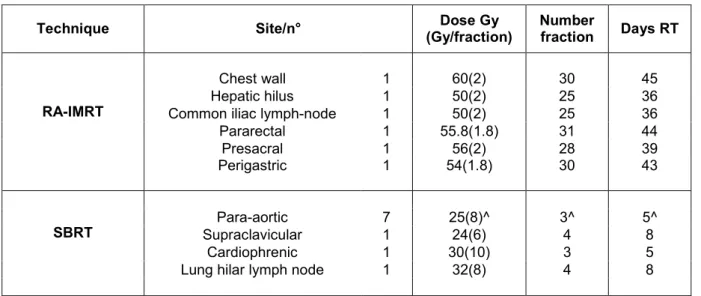
Table 2. Radiotherapy Techniques
Technique Site/n° (Gy/fraction)Dose Gy Numberfraction Days RT
RA-IMRT
Chest wall 1 60(2) 30 45 Hepatic hilus 1 50(2) 25 36 Common iliac lymph-node 1 50(2) 25 36 Pararectal 1 55.8(1.8) 31 44 Presacral 1 56(2) 28 39 Perigastric 1 54(1.8) 30 43
SBRT SupraclavicularPara-aortic 71 25(8)^24(6) 3^4 5^8 Cardiophrenic 1 30(10) 3 5 Lung hilar lymph node 1 32(8) 4 8 ^median dose Gy (Gy/fraction), median number fraction and median days RT
Legend: volumetric modulated Rapidarc radiotherapy (RA-IMRT), stereotactic body radiotherapy (SBRT)
Fig. 10 (A e B)
Fig. 11
Example of SBRT treatment planning
8.3 Results
Toxicity evaluation
Radiation Therapy Oncology Group/European Organisation for Research and Treatment of Cancer (RTOG/EORTC) criteria were used to evaluate treatment toxicity (ref). Acute toxicity was evaluated during radiotherapy treatment and during follow-up. In the RA-IMRT patients, gastrointestinal (GI) acute toxicity was: G0 (4 patients), G1 (2 patients); particularly 2 patients developed nausea and vomiting controlled with prophylactic
antiemetic treatment with ondansetron in oral administration. For SBRT patients: the acute toxicity was G0 (8 patients) and G1 (one patients). No haematological acute toxicity was observed in all patients (table 3).
Survival and local control
The patients were controlled with radiological imaging (CT scan after 40 days and CT or 18FDG-PET/CT ) after 3 months and during the follow-up), clinical evaluation and only in ovarian patients CA-125 serum level was controlled.
After a mean follow-up of 5 and 4.8 months (range 2-10 /1-13.3 months) for RA-IMRT and SBRT patients, 13/15 patients were evaluated (in 2 patients the response evaluation has not been yet performed).
Local response:
The local control rate ( the complete response of pathological disease after radiotherapy treatment) evaluated in 13/15 patients for a total number of 14 nodes (5/6 patients and nodes (RA-IMRT) and 8/9 patients, 9/10 nodes (SBRT) was 92.8% (5/5 nodes = 100% vs. 8/9 nodes = 88.9% for RA-IMRT and SBRT respectively).
Pattern of failure:
At the time of the analysis, October 2011, six patients are alive with no evidence of disease (2/5 = 40% and 4/8 = 50% patients RA-IMRT vs SBRT, respectively) , six patients are alive with clinically evident disease in other sites (2/5 = 40% and 4/8 = 50% patients RA-IMRT vs SBRT, respectively ), one patient died for systemic progression of disease, one patient was lost of follow-up and one patient was not evaluable at this time.
The overall survival at October 2011 was 92.3 % ( particularly was 80% for RA-IMRT and 100% for SBRT).
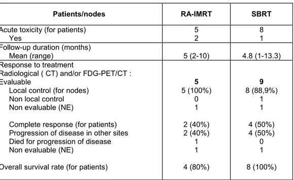
Table 3. Outcome
Patients/nodes RA-IMRT SBRT
Acute toxicity (for patients)
Yes 52 81
Follow-up duration (months)
Mean (range) 5 (2-10) 4.8 (1-13.3) Response to treatment
Radiological ( CT) and/or FDG-PET/CT : Evaluable
Local control (for nodes) Non local control Non evaluable (NE)
Complete response (for patients) Progression of disease in other sites Died for progression of disease Non evaluable (NE)
Overall survival rate (for patients)
5 5 (100%) 0 1 2 (40%) 2 (40%) 1 1 4 (80%) 9 8 (88,9%) 1 1 4 (50%) 4 (50%) 0 1 8 (100%)
Legend: computer tomography (CT) , 18-fluoro-deoxy-glucose positron emission tomography/CT (18FDG-PET/CT), volumetric modulated Rapidarc radiotherapy (RA-IMRT), stereotactic body radiotherapy (SBRT)