Alma Mater Studiorum – Università di Bologna
DOTTORATO DI RICERCA IN
Biologia Cellulare e Molecolare
Ciclo XXVII
Settore Concorsuale di afferenza: 05/E1 Settore Scientifico disciplinare: BIO/10
TITOLO TESI
OPA1 isoforms and protein domains in the rescue of mitochondrial
dysfunctions
Presentata da: Valentina Del Dotto
Coordinatore Dottorato Relatore
Prof. Davide Zannoni Prof.ssa Michela Rugolo
Correlatore
Dott.ssa Claudia Zanna
Esame finale: Bologna, Aprile 2015
ABSTRACT
Mutations in OPA1 gene have been identified in the majority of patients with Dominant Optic Atrophy (DOA), a blinding disease, and the syndromic form DOA-plus. OPA1 protein is a mitochondrial GTPase involved in various mitochondrial functions, present in humans in eight isoforms, resulting from alternative splicing and proteolytic processing. In this study we have investigated the specific role of each isoform through expression in OPA-/- MEFs, by evaluating their ability to improve the defective mitochondrial phenotypes. All isoforms were able to rescue the energetic efficiency, mitochondrial DNA (mtDNA) content and cristae integrity, but only the presence of both long and short forms could recover the mitochondrial morphology.
In order to identify the OPA1 protein domains crucial for its functions, we selected and modified the isoform 1, shown to be one of the most efficient in preserving mitochondrial phenotype, to express three specific OPA1 variants, namely: one with a different N-terminus portion, one unable to generate short form owing to deletion of S1 cleavage site and one with a defective GTPase domain. We demonstrated that the simultaneous presence of the N- and C-terminus of OPA1 was essential for the mtDNA maintenance; a cleavable isoform generating s-forms was necessary to completely rescue the energetic competence and the presence of the C-terminus was sufficient to partially recover the cristae ultrastructure.
Lastly, several pathogenic OPA1 mutations were inserted in MEF clones and the biochemical features investigated, to correlate the defective phenotypes with the clinical severity of patients. Our results clearly indicate that this cell model reflects very well the clinical characteristics of the patients, and therefore can be proposed as an useful tool to shed light on the pathomechanism underlying DOA.
Index
INTRODUCTION ... 1
1. MITOCHONDRIA ... 2
1.1 Structure and functions ... 2
1.2 OXPHOS system ... 4
1.3 Mitochondrial genome ... 8
1.4 Morphology ... 13
1.5 Fusion ... 15
2. OPA1 ... 17
2.1 OPA1 gene and protein ... 17
2.2 OPA1 and mitochondrial morphology ... 21
2.3 OPA1 and mitochondrial ultrastructure ... 23
2.4 OPA1 and apoptosis ... 24
2.5 OPA1 and energetic metabolism ... 26
2.6 OPA1 and mtDNA maintenance ... 27
3. Autosomal dominant optic atrophy ... 29
3.1 Genetics of DOA ... 29
3.2 Clinical features and histopathology ... 30
3.3 Pathophysiological mechanism in RGCs ... 31
AIMS ... 34
MATERIAL AND METHODS ... 36
1. Cells culture ... 37
2. Molecular cloning ... 37
3. Infection of OPA1-/- MEF ... 37
4. Mitochondrial morphology ... 38
5. Mitochondrial ultrastructure ... 38
6. Mitochondrial DNA measurement ... 39
7. SRB assay ... 39
8. Oxygen consumption rate (OCR) ... 39
9. Mitochondrial ATP Synthesis ... 40
10. Citrate synthase activity ... 41
11. Extraction of mitochondria from cultured cells ... 41
13. Total cellular lysates preparation ... 43
14. Mitochondria from mouse tissues ... 43
15. SDS-PAGE ... 43
16. OXPHOS complexes analysis by CN/BN-PAGE ... 43
17. Supercomplexes analysis by BN-PAGE ... 44
18. ImmunoBlotting ... 45
19. Primers list ... 45
20. Statistics ... 46
RESULTS ... 47
1. Characterization of OPA1 knock-out cells ... 48
2. Analysis of OPA1 isoforms ... 53
3. OPA1 domains ... 64
4. OPA1 mutations ... 71
DISCUSSION AND CONCLUSIONS ... 75
1. MITOCHONDRIA
1.1 Structure and functions
Mitochondria are cytoplasmatic organelles present in variable amount within eukariotic cells. Mitochondria are commonly referred to as the powerhouses of the cell because they are in charge of the ATP generation through the oxidative phosphorylation (OXPHOS). In addition, these organelles are involved in other important functions, like different types of cell death, regulation of calcium homeostasis, sugar and fatty acid catabolism, aminoacid metabolism, and reactive oxygen species (ROS) production.
According to the endosymbiotic theory, mitochondria origined from an aerobic bacterium entered into a symbiotic relationship with primitive host cells, giving energy to the eukaryotic cell in exchange of a stable environment and a continuum supply of nutrients (Gray et al, 1999). Mitochondria still retain some characteristics of bacterial cells as the presence of a double membrane, the ability to synthesize ATP, an independen protein synthesis apparatus and a circular DNA (mtDNA), which codes for tRNAs, rRNAs and proteins involved in OXPHOS (Friedman and Nunnari, 2014).
In contrast to the long-accepted static image of mitochondria, the introduction of tridimensional imaging technique with high resolution allowed to identify the so-called “dynamic mitochondrial network”. These are indeed dynamic organelles that frequently move within the cell toward regions of high-energy demand and form a network where morphological changes continuously occur during cellular cycle, development or tissue differentiation and apoptosis (Ishihara et al, 2013). These modifications can happen because they undergo membrane remodelling through cycles of fusion, in which two mitochondria join to form a single mitochondrion, and fission, in which a single mitochondrion divides into two mitochondria (Chan, 2012).
Mitochondria have a double membrane system: the outer mitochondrial membrane (OMM) bounds the organelle, while the inner mitochondrial membrane (IMM) separates the matrix space from the intermembrane space (IMS). OMM is characterized by a composition similar to the endoplasmatic reticulum and can also give rise to mitochondria-associated ER-membrane (MAM) in which lipids transfer between the two organelles takes place (Hayashi, 2009). OMM contains the voltage-dependent anion channel (VDAC), a multispanning β-barrel protein also called mitochondrial porin, which confers high permeability to small molecules and ions. In contrast, the IMM is impermeable to most small molecules and ions and only the species that have a specific transporter
insert inside the membrain can pass through (Nicholls, 2002). Many of these proteins within the IMM are components of the OXPHOS system. The space between the two membranes was termed inter-membrane space (IMS) and contains hydrosoluble proteins, such as the evolutionarily conserved cytochrome c. The IMM instead includes a gel-like substance, the matrix, which contains enzymes of the tricarboxylic acid cycle and β-oxidation, and multiple copy of mtDNA (Frey, 2000; Mao and Holt, 2009). Because IMM is the active site of OXPHOS, the selective permeability is a property required for maintenance of an electrochemical proton gradient between the matrix and the IMS.
The IMM is characterized by invaginations and organized in two contiguous but distinct membranes: the inner boundary membrane, which faces the OMM, and the cristae membranes, which extend into the mitochondrial interior as tubules or lamellae. This particular organization confers a large surface in a small volume. According to the classic Palade’s model, cristae were considered as simple invaginations of the IMM widely opened to the IMS (Palade, 1952), but now they are identified as a separated sub-compartment, resembling pleomorphic bags. Cristae can be tubular, branched or bag-shaped and are joined to the inner boundary membrane by narrow tubular structure called cristae junctions. These junctions are energetically favored structures, which typically have a diameter as small as 10-15 nm, may be altered by changes in matrix volume and respiratory activity. These structures are also important for mitochondrial processes involving internal diffusion of ions, metabolites and proteins (Mannella et al., 1997; Mannella et al., 2013). Also cristae shape is dynamic and the inner membrane topology is regulated by the cell to optimize mitochondrial function. In yeast mitochondria, cristae junctions are reversibly reformed within several minutes after extreme osmotic swelling and recontraction (Mannella et al, 2001), whereas in mammals, the number of cristae is increased upon starvation (Gomes et al, 2011; Pattern et al, 2014). Furthermore the cristae remodeling augment cytochrome c realease during apoptosis (Scorrano, 2002; Frezza, 2006).
1.2 OXPHOS system
OXPHOS is one of the metabolic processes that takes place inside the mitochondria and is the final step of cellular respiration. The OXPHOS provides the bulk of cellular energy in the form of ATP thanks to a flow of electrons that are transported through a series of protein complexes, which are embedded in the lipid bilayer of the mitochondrial IM (Balaban et al., 1990; Friedman and Nunnari, 2014). The OXPHOS system is composed of five enzymatic complexes (CI-V) and is constituted by 87 polypeptides. Only 13 are encoded by the mitochondrial DNA, while the remaining part is coded at the nuclear level and later imported into the mitochondria (Carelli and Chan, 2014). CI-IV, together with ubiquinone (CoQ) and cytochrome c, two mobile electron carriers, make up the electron transport chain (ETC) (Acin-Perez and Enriquez, 2014).
During the Krebs cycle or the β-oxidation of fatty acids, the electron carriers nicotinamide adenine dinucleotide (NADH) and flavin adenine dinucleotide (FADH2) are reduced. Subsequently, the
electrons resulting from NADH enter in the transport chain through the CI (NADH-ubiquinone oxidoreductase), while the FADH2 is oxidated at the level of CII (succinate-ubiquinone
oxidoreductase). CI is the largest of the respiratory complexes, comprising 45 subunits, of which seven are mitochondrially encoded (Carroll et al., 2006). CII, the only respiratory enzyme completely encoded by the nucleus, is composed of four subunits and catalyzes the oxidation of succinate to fumarate transfering the electrons to the covalently bound FAD. The electrons are further transported via a number of iron/sulphur clusters to CoQ, reducing it to CoQH2 (Lancaster
and Kroger, 2000). A third source transferring electrons to generate ubiquinol is the glycerol 3-phosphate dehydrogenase enzyme. CoQ after reduction can move and transport the electrons to CIII (ubiquinone-cytochrome c oxidoreductase). This complex, that couples electron transfer to the translocation of two protons across the IMM, has only one mtDNA-encoded subunit, cytochrome b; the other 10 subunits are nucleous-encoded (Berry et al., 2000, 2013). CIII is a functional dimer and transfers electrons from CoQH2 to cytochorme c, that donates electrons on the cytoplasmic
side of the IMM to complex IV (Darrouzet et al., 2001; Dibrova et al., 2013). CIV is the terminal enzyme of the respiratory chain, composed of 13 subunits, of which three are encoded by the mtDNA. This complex reduces dioxygen to water and harnesses the energy to translocate protons from matrix to the intermembrane space. In this step, four electrons have to be transfered from the
reduced cytochrome c pool to two molecules of oxygen, without generating any reactive oxygen species (Schultz and Chan, 2001; Ishigami et al., 2015).
During these processes, the energy released by the electrons flow through the respiratory chain allows the transport against gradient of protons from the matrix to the intermembrane space. The proton pumping occurs only at the level of CI, III and IV and creates an electrochemical gradient that is positive outside, and negative inside the IMM. The proton flux in the reverse direction provides the energy to sustain the activity of the complex V (ATP synthase) to synthesize ATP. This complex consists of two major functional domains, a large extra-membranous portion (F1) sector and a membrane-intrinsic portion (F0) sector joined together by central and peripheral stalks (Walker, 2013). CV has two subunits encoded by mtDNA (ATPase6 and ATPase8), that take part to the membrane-bound portion (F0) of the complex, and about 13 other subunits encoded by nDNA (Abrahams et al., 1994). Protons from the intermembrane space enter CV through the F0 complex leading to subunit rotation within the complex. The rotational energy of the motor is transmitted to the catalytic domain by the central stalk, which is attached directly to the rotary motor, and then used for ATP synthesis (Walker, 2012). ATP formation requires a sufficient supply of ADP and phosphate, and specific carriers transport both substrates across the mitochondrial membranes: the adenine nucleotide translocator (ANT) and the phosphate carrier (Seelert and Dencher, 2011).
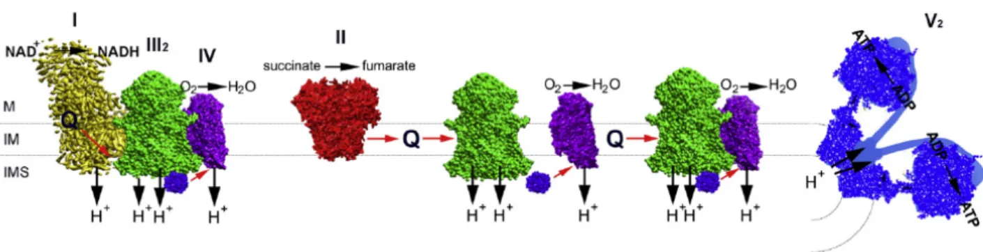
Although initially the respiratory components were proposed to be closely packed to guarantee a high efficient electron transport (‘solid’ model), according another model (‘fluid’ model) the OXPHOS complexes are independent entities embedded in the inner membrane, with CoQ and cytochrome c that freely diffuse among the complexes. However, thank to development of accurate techniques to isolate and identify stable associations of two or more respiratory complexes (respiratory supercomplexes, RSC) a reformulation of the solid model, the plasticity model, has been proposed. According to this model, the mitochondrial ETC can undergo to rearrangements within the IMM to adapt to fuel availability through a dynamic distribution between free respiratory complexes and SCs. In particular, data obtained by Blue-Native gel electrophoresis (BN-PAGE) unambiguously revealed that the respiratory complexes involved in this supramolecular association are the three proton-translocating enzymes, present in various configurations and stoichiometries. The I+III2+IV combination was denominated ‘respirasome’
Furthermore, there are suggestions that even higher levels of organisation may exist. RSC are proposed to increase the efficiency of electron flux through substrate channeling or enhanced catalysis (Genova and Lenaz, 2014; Acin-Perez and Enriquez, 2014; Chaban et al, 2014). Although the OXPHOS process is very efficient, a small percentage of electrons may be lost prematurely by redox centers present in the respiratory complexes, leading to the reduction of oxygen with the formation of ROS. Mitochondria therefore constitute the main source of ROS production within the cell. Therefore SCs allow acceleration of electron transport concomitant with sequestration of reactive intermediates to prevent generation of ROS. In this regard, independent approaches demonstrated that ROS generation by complex I was increased in the absence of SC formation (Acin-Perez and Enriquez, 2014). Furthermore, SCs are also linked with mitochondrial ultrastructure, in fact cristae shape may influence the assembly and stability of RCS and hence mitochondrial respiratory efficiency (Cogliati et al., 2013).
Figure 1. The plasticity model. Schematic representation of the OXPHOS system, where respiratory CI, III and IV are
partly organised into supercomplexes. CII was never found associated with other complexes. Red arrows show electron pathways. CI (yellow), CII (red), CIII2 (green), CIV (purple), CV (blue), cytochrome c (violet), Q, ubiquinol.
The positions of the matrix (M), the intermembrane space (IMS) and inner membrane (IM) are indicated (from Chaban et al., 2014).
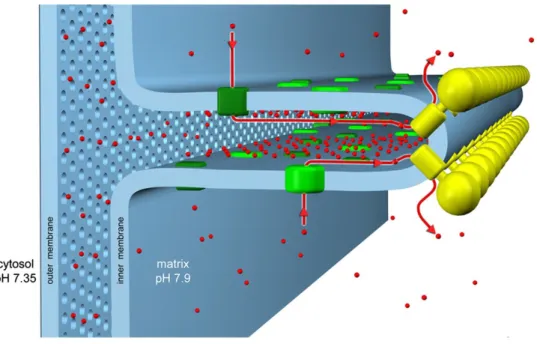
CV also can form various supramolecular structures, given that not only dimeric species but also higher molecular weight oligomers of the ATP synthase can be identified by BN-PAGE. Interestingly, ATP synthase dimers were found in strongly curved regions of the inner membrane (Davies et al., 2011) and they seem to be linked to cristae shape: in yeast mutants where the ATPase cannot dimerize, cristae are disorganized (Arselin et al., 2004), whereas in mammalian cells, increased cristae density favors ATPase dimerization during starvation (Gomes et al., 2011; Pattern et al., 2014).
Figure 2. Schematic organization of the cristae. CV and CI (as well as the other respiratory chain complexes) occupy
different regions of the crista membrane. CV forms dimer rows (yellow) in the strongly curved areas of the crista, ETC complexes (green), in particular CI, reside predominantly in the straight membrane regions. Protons (red), ejected into the intracristal space by the ETC, flow back into the matrix through the ATP synthase rotor, driving ATP production (from Davies et al., 2011).
1.3 Mitochondrial genome
1.3.1 Feature and structure of mtDNAThe mammalian mtDNA genome is a double-stranded, circular molecule of 16.659 base pair, localized in the mitochondrial matrix, where it is present in high copy number, ranging from tens to thousand copies per cell. The two strands of mtDNA can be distinguished into ‘heavy strand’ (H-strand) and ‘light strand’ (L- strand) strand, based on their nucleotide (GC) content (Fernandez-Silva et al., 2003). The mtDNA genome is highly compact compared to the organization of the nuclear genome, since there are no introns and intervening sequences among and between genes. Some genes overlap and certain stop codons necessary for proper mRNA translation are only generated after polyadenylation (Anderson et al., 1981).
Mitochondrial genome encodes 13 polypeptides, which are essential OXPHOS components. Of the other 24 genes, 22 are transfer RNAs and 2 ribosomal RNAs (12S e 16S), necessary for the mitochondrial synthesis of the 13 mtDNA gene products. As a result, all 37 mtDNA genes are essential for OXPHOS activity. The remaining around 1.500 genes encoding the mitochondrial proteome are scattered in the nuclear chromosomes. These proteins are translated by cytoplasmatic ribosomes and selectively imported into mitochondria through the specific translocases located in the inner and outer membrane (Harbauer et al., 2014). There are only two non-coding regions in the mtDNA. The first substantial noncoding region is the 1 kb D-loop (Displacement Loop), named because the triple-stranded structure formed by association of the nascent H-strand in this region during the replication. The D-loop contains the main regulatory elements for transcription and replication of mtDNA: the origin of H-strand DNA replication and the site of bidirectional transcription from opposing heavy and light strand promoters (Fernandez-Silva et al, 2003; Scarpulla, 2008). The second non-coding region contains the origin of L-strand replication (OL) and is located in a cluster of five tRNA genes around two thirds of the mtDNA length from the OH (Anderson et al., 1981; Fernandez-Silva et al., 2003).
Each mitochondrion contains multiple copies of mtDNA associated with specific proteins to form nucleoprotein complexes called nucleoids (Bogenhagen et al., 2008; 2012). Nucleoids are macromolecular structures, having an average mean size of about 100nm in mammals, regularly placed throughout the mitochondrial network, allowing an efficient maintenance of mtDNA in discrete segregating units (Gilkerson 2009, Kukat at al., 2011). Although is was previously reported that each nucleoid contained on average 5 to 10 copies of mtDNA, a recent study using
superesolution microscopy revealed that each nucleoid contains on average 1.4 mtDNA molecule only, meaning that the majority of the nucleoids just contain a single copy of mtDNA (Kukat at al, 2011).
A number of proteins have been identified to be part of the mitochondrial nucleoid. Mitochondrial transcription factor A (TFAM) is one of the major DNA-binding proteins associated in mammals, in particular about one thousand TFAM proteins were found for each mtDNA molecule. TFAM plays a crucial part in mtDNA transcription and its expression controls mtDNA copy number in cells, making it a central player in mtDNA maintenance and transmission (Kaufman et al. 2007, Kukat et al., 2011). Additional proteins crucial for mtDNA maintenance are the other components of nucleoids: the twinkle helicase, the mitochondrial polimerase gamma (Polγ), the single strand inding protein (mtSSB), mitochondrial transcription factors B1 and B2 (TFB1M and TFB2M) and a mitochondrial transcription termination factor (mTERF). A number of IMM proteins has been identified in purified nucleoid preparations, such as the adenine nucleotide translocase (ANT), the E2 subunit of α-ketoacid dehydrogenase, subunits of CI and CV, the SOD2 (Wang and Bogenhagen, 2006; Bogenhagen et al., 2008; Kienhöfer et al., 2009). Other types of nucleoid components identified are proteins with chaperone activity, including HSP70, HSP60, LRPPRC, prohibitin and the mAAA ATPase ATAD3A/B (Bogenhagen et al., 2008; Bogenhagen, 2012). In the model for the nucleoid structure proposed by these authors a central core, formed by proteins involved in mtDNA maintainance is surrounded by a peripheral region, composed by the metabolic proteins and chaperones (Bogenhan et al. 2008). This protein-enriched component not only protects the DNA from various insults, but also is likely to put constraints on any transactions involving the mtDNA, such as replication, repair, and transcription. In fact, mutations in genes encoding these and additional factors required for mtDNA maintenance are associated with a wide spectrum of human mitochondrial diseases (Spelbrink, 2010; Friedman and Nunnari, 2014). In post-mitotic tissues and cells, contrary to nuclear DNA, mtDNA is continuously replicated and the mitochondrial genome copy number is maintained at relatively constant level. The segregation of mtDNA partly depends on the unbrocken occurrence of mitochondrial network fission and fusionevents, therefore the balance between these processes is crucial for proper distribution and maintenance of mtDNA (Friedman and Nunnari, 2014). The link between mitochondrial dynamics and mtDNA transmission is consistent with the primary role of dynamics in the control of mitochondrial copy number. Complementation has thus been suggested to be the consequence of
mitochondrial network dynamics allowing for rapid and extensive exchange of genetic material (Schon and Gilkerson, 2009; Carelli and Chan, 2014). One of the peculariar features of mitochondrial genome is the uniparental inheritance of mtDNA in almost all the eukaryotes and, in the case of mammals, the maternal mtDNA is exclusively transmitted to the subsequent generations (Carelli and Chan, 2014). Because of the lack of protective histones, of an efficient DNA repair system, of intronic sequences and due to the fact that mitochondria are the main site of ROS production, mtDNA is extremely sensitive to oxidation or other genotoxic damage, and thus tends to accumulate mutations at a much higher rate than nuclear DNA (Liu and Demple, 2010). Four types of alterations of mitochondrial genome can be distinguished: partial deletions or duplications of mtDNA fragments, depletion of the mtDNA, accumulation of random mutations physiologically correlated to the ageing process and discrete point mutations (Vidoni et al., 2013). It has to ben noticed that in the same cell, tissue or individual, mutated mtDNA molecules can be present in a restricted proportion of the mtDNA population only, the condition being termed heteroplasmy. In contrast, when all copies of the mtDNA are identical (all wild-type or all mutant) the situation is referred to as homoplasmy. Therefore, the phenotype induced by a particular mutation depends not only on the type of mutation, but also on the ratio of mutated and wild-type copies, and become apparent when a percentage of mutated copies is reaches (threshold effect), which varies in different tissues (Rossignol et al., 2003). Since 1988, when a mtDNA mutation has been associated for the first time to a disease, hundreds of alteration of mtDNA causing mitochondrial pathology were discovered, showing a progressive, age-related clinical course and, because many of them involve central and peripheral nervous system and skeletal muscle, these are also known as mitochondrial encephalomyopathies (Carelli and Chan, 2014).
Figure 3. Schematic representation of the circular mtDNA genome, where the 13 genes encoding for the subunits of
CI, III, IV and V (blue-black), the 2 rRNAs (red), the 22 tRNAs (green) and the D-loop (yellow) are indicated (from Mishra et Chan, 2014).
1.3.2 Transcription, translation and replication
The transcription of mtDNA reflects its peculiar compact and functionally economic organization. Contrary to what occurs in the nucleus, mitochondrial genes are transcribed in a polycistronic manner, specifying more than one RNA gene or mRNA, and in human mitochondria this process starts at three different sites located in the D-loop region. The transcription of the light chain starts from a single promoter (LPS) and results in the production of a transcript that is processed originating 8 mRNAs and 1 tRNA. The heavy chain promoter (HSP) contains two start sites: H1 and H2. The transcription from H1 is responsible for the synthesis of 2 ribosomal RNAs , while from the site H2 produce a long transcript polygenic, that is processed to 14 tRNA, 12 mRNA and 2 rRNA (Scarpulla, 2008).
Factors encoded by nuclear genes are involved in the transcription process: the mitochondrial RNA polymerase (POLRMT), TFAM, mitochondrial transcription factor B (TFB1M and TFB2M), and the factor termination mitochondrial mTERF (Falkenberg et al, 2007). TFAM promotes bidirectional transcription after binding its site enhancer, which is located upstream the start site of the promoter, facilitating the interaction with the other components of the transcriptional apparatus. The sequence that determines the termination of the majority of transcripts, starting from the site H1, is recognized by MTER factor, which is able to bind this sequence and simultaneously the site H1, leading to the formation of a loop which includes the transcripts of the two 12S and 16S rRNAs.
Mitochondria contain an independent protein-synthesis machinery to produce polypeptides encoded by the mitochondrial genome. The two mitochondrial rRNAs and nuclear encoded proteins compose these ribosomes that translate the mitochondrial mRNAs in the matrix. The mitochondrial genetic system utilizes genetic codes that differ slightly from the universal nuclear code, with species-specific differences (Scarpulla, 2008).
The mtDNA replication occurs at the level of the matrix and is independent from that of nuclear DNA. The mtDNA replication and transcription are closely associated phenomena because, in addition to the spatial proximity between the start points of both processes, the heavy chain needs a RNA primer to start replication. The transcript from LSP provides RNA primers, then the POLRMT is replaced by the mitochondrial DNA polymerase γ (Pol γ) and the replication starts from the origin OH of the heavy chain (Fustè et al., 2010). The gene products of nuclear origin constitute the machinery necessary for mtDNA replication, in particular DNA synthesis takes place by means of Pol γ and other factors such as the helicase TWINKLE, TFAM and a ligase (Scarpulla, 2008). Two models have been proposed: the first, asymmetric, suggests that the process beginning at the level of the OH, localized in the D-loop, proceeds throughout the parent strand H to produce the L chain until it reaches the origin OL. At this point the synthesis of the L-strand begins and proceeds in the opposite direction respect to the replication of the H-chain. The second model, proposed by Holt and defined symmetrical, differs from the previous one and supposes that in reality the mtDNA replication occurs in a symmetrical and synchronous manner (Brown et al., 2005).
1.4 Morphology
In the cytosol, mitochondria form a highly dynamic reticulum whose morphology is primarily determined by processes of fusion and fission of both OMM and IMM (Liu et al., 2009; Chan, 2012). Fusion merges the membranes of two, separate mitochondria, and results in the unification and mixing of their compartments. Fission divides a single mitochondrion into two, new mitochondria. Inhibition of fusion causes fragmentation of the network, whereas inhibition of fission induces mitochondria to become extremely elongated and overly interconnected. Therefore, the balance of these opposing forces determines the shape and length of mitochondria (Chan, 2012; Scorrano, 2013). Over the last 20 years, fusion and fission have been studied in great detail and it has become clear the importance of these processes for cellular and organismal physiology. In fact mice lacking proteins involved in mitochondrial dynamics cannot survive past midgestation and show cell type–specific defects (Chen et al., 2003; Chen et al., 2007; Ishihara et al., 2009).
Two different typologies of fusion have been described: transient and complete fusion events. Mitochondrial fusion assays showed that transient fusion is a rapid and separable merge of the outer and inner membranes and allow a partial exchange only of soluble inter-membrane space and matrix proteins. This type of fusion does not change the morphology of mitochondria and has been termed “kiss-and-run.” Instead complete fusion events permit the exchange of all mitochondrial components and affect mitochondrial morphology (Liu et al., 2009).
Because mitochondrial proteoma is encoded by both mitochondrial and nuclear DNA, during mitochondrial biogenesis the levels of these two sets of proteins must be coordinately regulated. Without mitochondrial fusion, each mitochondria of the cell could have divergent biochemical e functional profiles. The tournover of fusion and fission makes the contents of the organelles homogeneous, allowing mitochondria to act as a coherent population (Chan, 2012). The morphology is important also for the respiratory capacity, changing on the basis of cellular energetic status. IMM fusion is tuned by the levels of OXPHOS and during starvation elongated mitochondria increase levels of dimerization and activity of ATP synthase to maintain ATP production (Mishra et al, 2014; Gomes et al., 2011). Metabolically active cells have a large mitochondrial network that acts as electrically united systems able to transmit mitochondrial membrane potential, allowing an efficient dissipation of the energy in the cell (Skulakev, 2001).
Dynamic control of mitochondrial structure is performed by a set of “mitochondria-shaping” proteins that include both pro-fusion and pro-fission members. All these proteins operate in healthy cells to maintain genetic and biochemical uniformity within the mitochondrial population.
During mitochondrial fission two classes of proteins are necessary to execute this process. The first includes the dynamin protein Drp1, which is recruited from cytosol to the mitochondrial surface and oligomerizes around the circumference of the mitochondrion, promoting membrane division (Chan, 2012). It has been shown that Drp1 is essential for mitochondrial fission provoked by events like mitosis or stress, and for development of the nervous system (Wakabayashi et al., 2009; Ishihara et al., 2009). The members of second class are integral, OM proteins that recruit Drp1 to the mitochondrial surface: Fis1, Mff, MiD49 and MiD51. Although in yeast the recruitment of the dynamin omologus requires Fis1 and the two WD40-containing adaptors, in mammals it has been reported that Fis1 can directly bind Drp1 (Griffin et al., 2005; Wells et al., 2007). Importantly, in mammals there are three other receptor proteins involved in fission events: Mff, MiD49 and MiD51. Knockdown of Mff caused reduced recruitment of Drp1 to mitochondria accompanied by robust elongation and recently it has been demostrated that Mff, MiD49 or MiD51 can function independently of one another to mediate mitochondrial fission. Particularly, it seems that Fis1 and Mff promote constitutive mitochondrial fission whereas the MiDs activate recruited Drp1 only during loss of respiration (Loson et al., 2013; Otera et al., 2010).
Interestingly, mitochondrial fission plays a key role in both cell life and death. The mitochondrial fission machinery actively participates in the programmed cell death pathway (apoptosis) by inducing fragmentation of the mitochondrial network before the release of the cytochrome c and caspase activation (Youle and Karbowski, 2005). Network fragmentation is important for the proper allocation of mitochondria to daughter cells during cellular division (Kashatus et al., 2011). In fact, changes in mitochondrial morphology during the cell cycle have been shown to influence the progression of the cell from G1 to S phase, suggesting that mitochondrial morphology can regulate the cell cycle (Mitra et al., 2009). Mitochondrial fission has also been linked to mitophagy, because interruption of fission reduces the efficiency of mitophagy, suggesting that fission segregates mitochondria with loss of membrane potential, allowing their removal from the rest of the population by autophagy (Frank et al., 2012; Stotland and Gottlieb, 2015).
The mitochondrial fusion is highly dependent on the mitochondrial membrane potential, and plays a central role also in the quality control of mitochondria. Mitochondria that are moderately
dysfunctional can be rescued by mitochondrial fusion, whereas those with severe dysfunction may be separated by fission and then degraded (Twig et al., 2008). Fusion is important also for mitochondrial calcium homeostasis (Szabadkaiet al., 2006), embryonic development (Chenet al., 2003) and spermatogenesis, and mtDNA maintenance (Chen et al., 2007). Moreover mitochondrial fusion plays a protective role in certain pathological conditions, because it allows complementation of mtDNA gene products in heteroplasmic cells that have accumulated different mutations (Sato et al., 2006).
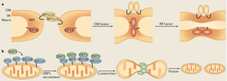
Figure 4. Schematic representation of the mitochondrial fusion and fission events. (a) OM fusion is mediated by
mitofusin 1 (MFN1) and 2 (MFN2) and subsequently followed by IM fusion, mediated by the optic atrophy 1 (OPA1) protein. (b) Mitochondrial fission requires the recruitment of dynamin-related protein 1 (DRP1) from the cytosol to OMM. Four DRP1 receptors located on the OMM exist in mammals: mitochondrial fission 1 (FIS1), mitochondrial fission factor (MFF), Mitochondrial dynamics protein of 2014).
1.5 Fusion
The fusion of two mitochondria requires that the inner and outer membranes of one mitochondrion be fused with the respective membranes of another. The first step of this process occurs with the fusion of the OMM, allowing the IMM to come into proximity for the second step, the IMM fusion. In mammals, three proteins carry out this event: the mitofusins MFN1 and MFN2 for the OMM fusion and OPA1 for the IMM fusion (Chan, 2012). These proteins belong to the dynamin-like protein superfamily and contain GTPase activity essential for their functions (Praefcke and McMahon, 2004).
The first mediator of mitochondrial fusion was identified in D. Melanogaster as the Fuzzy onion protein (Fzo1), which has two homologues in mammals, MFN1 and MFN2 (Santel and Fuller, 2001). These proteins include a GTPase domain, two hydrophobic heptad repeat motifs (HR1 and HR2), and are anchored at the OMM by two transmembrane domains, with both N- and C-terminal facing the cytosol (Chan, 2012). In vivo and in vitro studies have shown that the MFNs act in the initial step of fusion, are essential for OMM fusion and are required on adjacent mitochondria for fusion to occur (Koshiba et al., 2004; Song et al., 2009). However, they seem to play slightly different roles. MFN1 is more active in mitochondrial fusion exhibiting a higher capacity to induce this process, whereas the role of MFN2 is less defined, but it can forms hetero-oligomers with MFN1 and is suggested to participate in later steps of mitochondrial fusion (Koshiba et al., 2004; Ishihara et al., 2004). MFN2 has been shown to be involved also in endoplasmic reticulum interactions, bridging the two organelles close at the level of MAMs, and levels of MFN2 correlate with the oxidative metabolism of skeletal muscle (de Brito et al., 2008; Bach et al., 2003).
Fusion of both mitochondrial membranes appears coordinated because of the rapid nature of the two processes, yet mitochondria do not always undergo complete fusion and the OM can fuse without the subsequent fusion of the IM (Song et al., 2009; Mishra et al., 2014). Both the mitofusins and OPA1 are required to accomplish full fusion of mitochondria (Song et al., 2009). In yeast Mgm1p/Msp1p are conserved dynamin-related GTPases essential for fusion, morphology, inheritance, and genome maintenance of mitochondria (Wong et al., 2003; Jones and Fangman, 1992). The human orthologue is OPA1, which was identified in 2000 by two European groups (Delettre et al., 2000; Alexander et al., 2000). This protein is localized in the IMS, tightly anchored in the IM, and with its C-terminus facing the inter-membrane space (Wong et al., 2003). It has been shown as OMM fusion can be readily visualized in OPA1-null mouse cells in vivo without progression to IM fusion, suggesting that in mammals OPA1 is the main mediator of IM fusion (Song et al., 2009).
2. OPA1
2.1 OPA1 gene and protein
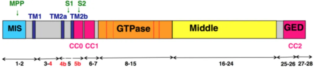
OPA1 gene is located on the long arm of chromosome 3q28–q29. Human OPA1 ORF is built from 31 exons and its mRNA transcripts differ by alternate splicing in the open reading frame, including or excluding exons 4, 4b and 5b. While exon 4 is evolutionarily conserved, both exons 4b and 5b are specific to vertebrates (Delettre et al., 2001; Guillery et al., 2008; Belenguer et al., 2013). The eight OPA1 mRNA variants encode proteins of 924–1014 aminoacids, whose N-terminal includes a mitochondrial targeting sequence (MTS), followed by a predicted transmembrane domain, anchoring the protein to the IMM, and the three peptides corresponding to the alternate spliced exons 4, 4b and 5b (Olichon et al., 2007). Exon 4 domain does not present any remarkable feature, whereas exon 4b and 5b corresponding domains encode two additional hydrophobic domains, TM2a and TM2b. The latter exon encodes also a coiled coil domain (CC0), predicted to oligo-polimerize. The following part of the protein includes the conserved dynamin regions: the GTPase domain bearing the dynamin signature, the middle domain with unknown function and the C-terminal GTPase effector domain (GED) containing a coiled coil domain (CC2) (Olichon et al., 2007; Belenguer et al., 2013).
Figure 5. Schematic representation of OPA1 protein structure. The features of dynamin family are: a GTPase domain
containing the three consensus GTP binding sequences (red bars) and the dynamin signature (red hatched bar), a middle domain and a GTPase effector domain (GED) containing a coiled-coil region (CCII). The mitochondrial import sequence (MIS) followed by a predicted transmembrane region (TM1), hydrophobic segments (TM2a and TM2b), and coiled-coil regions (CC0-1) are indicated. OPA1 exons numbers are schematized by double arrow and the alternative spliced exons 4, 4b and 5b are indicated in red. Matrix proteolytic cleavage sites for mitochondrial processing peptidase (MPP), paraplegin (S1) and YME1L (S2) are indicated (from Belenguer et al., 2013).
OPA1 expression is ubiquitous, but quantitatively variable according to the organ considered. High mRNA expression levels occur in retina, brain, liver, heart and pancreas. Also the relative abundance of the eight OPA1 splice forms shows great variability, although mRNAs containing ex4 are systematically abundant. In the brain, the presence of exons 4 and 4b, alone or combinated, predominate (Alexander et al., 2000; Misaka et al., 2002; Olichon, 2007).
Upon import of precursors, translated from the eight OPA1 mRNA, into the mitochondria, the MTS is cleaved by the mitochondrial processing peptidase to produce the long isoforms (l-isoforms) that are anchored to the IMM. Then the protein can also be subjected to a complex proteolytic processing that generates short isoforms (s-isoforms) lacking the transmembrane segment. These s-isoforms can be peripherally attached to the IMM, however, a fraction can diffuse in the IMS and associate to the OM (Satoh et al., 2003; Olichon et al., 2002; Song et al., 2007).
Figure 6. Schematic representation of the eight OPA1 isoforms. The mRNA splice forms differ in the presence or
absence of exons 4, 4b, and 5b. Cleavage of the mitochondrial targeting sequence (MTS) by MPP leads to the long isoforms. Additional cleavage at sites S1 (exon 5) or S2 (exon 5b) leads to the short isoforms. TM, transmembrane (from Song et al., 2007).
OPA1 processing can occur because the primary sequence of OPA1 presents two cleavage sites, named S1 and S2. All the isoforms present the S1 site, located at exon 5, whereas only isoforms
containing exon 5b can be processed at S2. Accordingly, teorically four isoforms may produce two short variants (Ishihara et al., 2006; Song et al., 2007). Actually, l-OPA1 isoforms containing exon 4b are totally processed into short forms (Song et al., 2007).
Numerous and discordant studies on the generation of s-OPA1 in human cells identified several types of proteases recognizing the two cleavage sites. The presilin-associated rhomboid-like protease (PARL) seems involved in the generation of a low abundance soluble OPA1 form at the IMS (Cipolat et al., 2006). Two m-AAA proteases, paraplegin and AFG3L2 form a high molecular weight complex within the IMM (Atorino et al, 2003). Overexpression of paraplegin induces a decrease of l-isoforms and accumulation of s-isoforms by cleavage at S1 (Ishihara et al., 2006). The contribution of two other subunits of the m-AAA complex, AFG3L1 and AFG3L2, in OPA1 processing was revealed by experiments conducted in yeast (Duvezin-Caubet et al., 2007). Also in mouse embryonic fibroblasts, down-regulation of AFG3L1 and 2 or the expression of a mutated AFG3L2 variant in human cells decreased the stability of OPA1 l-isoforms (Ehses et al., 2009). However neither PARL nor paraplegin are exclusive proteases involved in OPA1 processing, in fact their knock-down/out does not affect the ratio of l- to s-isoforms (Ishihara et al., 2006; Duvezin-Caubet et al., 2007). Down-regulation of the metallopeptidase OMA1 slightly reduced the level of OPA1 s-isoforms, which are generated by S1 cleavage and accumulate at low levels in MEFs (Ehses et al., 2009; Head et al., 2009). Furthermore, the i-AAA protease YME1L is responsible for cleavage at S2 (Griparic et al., 2007; Song et al., 2007). The knockdown of YME1L, indeed, has no effect on isoform 1 processing, which occurs exclusively at site S1, whereas for isoforms 4 and 7, which contain both sites S1 and S2, reduced level of the S2- and increased level of the S1-cleaved product were reported (Song et al., 2007). The OMA1 or YME1l knockout MEFs showed an OPA1 cleavage pattern containing only l-form (Anand et al., 2014). Furthermore also prohibitins can modify the processing of OPA1, in fact their alteration results in specific loss of l-OPA1 isoforms and accumulation of s-isoforms (Merkwirth et al., 2008). The knockout of another protease, Omi/HtrA2, also induces the selective increase of mild detergent extractable OPA1 levels (Kieper et al., 2010).
To make the picture even more complex, the OPA1cleavage can occur not only under physiological conditions but can be also induced by both apoptosis and dissipation of mitochondrial membrane potential. Collapse of membrane potential destabilizes l-OPA1 and enhances the cleavage at S1 and at other sites excluding S2 (Ishihara et al., 2006; Song et al., 2007; Griparic et al., 2007; Guillery et
al., 2008). The loss of mtDNA, dissipation of ∆Ψm, or decreased mitochondrial ATP levels induce OPA1 processing by OMA1 at the S1 site (Ehses et al., 2009). Recentily it has been reported that YME1L activity is regulated by mitochondrial respiration and/or ATP levels (Mishra et al., 2014). Altogether, these data reveal that OPA1 expression is highly regulated at both post-transcriptional and post-translational levels. This latter indeed comprises proteolytic processing, but probably also phosphorylation, ubiquitination, sumoylation and acetylation that have been shown to regulate other actors controlling mitochondrial dynamics. In this regard, the SIR3-dependent deacetylation of OPA1 lysines 926 and 931 has been reported, increasing OPA1 GTPase activity and recovering mitochondrial functions in OPA -/- null cells (Samant et al., 214). Further studies will certainly shed light on this complex processing mechanism.
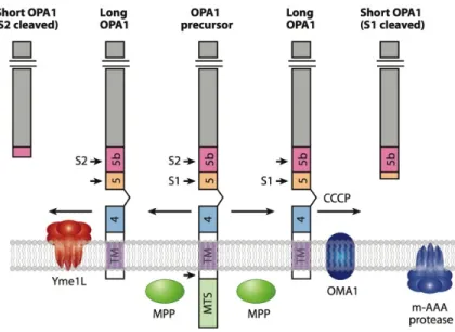
Figure 7. Processing of the precursor OPA1 splice form 7. After import of the N-terminus in the matrix, the
mitochondrial targeting sequence (MTS) is cleaved by the peptidase MPP to yield the membrane-anchored, l-isoform. Further processing at the S2 site by the YME1L protease yields a s-isoform (left side). Further cleavage at the S1 site also yields a s-isoform under physiological condition or after CCCP-induced mitochondrial depolarization, activating the OMA1 protease (right side). The m-AAA protease can also regulate OPA1 processing indirectly through OMA1. Abbreviations: HR, heptad repeat; TM, transmembrane segment; CCCP (carbonyl cyanide m-chlorophenylhydrazone) (from Chan, 2012).
2.2 OPA1 and mitochondrial morphology
OPA1 orthologs in yeast were initially identified as actors involved in maintaining the mitochondrial network fused. Subsequent data obtained in mammals indicated that, in accordance to its localization, this GTPase functions primarily in the fusion process at the IMM. The profusion activity of OPA1 was confirmed by different studies showing that loss of function of OPA1, by RNAi or gene knock-out, caused fragmentation of the mitochondrial reticulum (Misaka et al., 2006; Gripatic et a., 2004, Cipolat et al., 2004; Olichon et al., 2003; Song 2007). Interestingly, the
overexpression of wild-type or mutated OPA1 alleles also induces networkfragmentation, whereas in
cells where mitochondria are punctuated the overproduction of this GTPase promotes elongation of the reticulum (Cipolat et al, 2004; Olichon et al., 2002). All togheter these results suggest that OPA1 protein levels are finely regulated and that its depletion or excess counteracts its physiological pro-fusion activity.
Silencing of the three alternatively spliced exons ruled out the exon 4b and exon 5b involvement in the fusion process, suggesting that the exon4-containing variants are those required to maintain the network in a fusion state (Olichon et al., 2007).
It has been initially proposed that only OPA1 l-forms, without any processing, are necessary for fusion while s-forms are unable to fuse mitochondria (Ishihara et al., 2006). Additionally, a recent paper confirmed that normal fusion is mediated by the l-forms of OPA1 and that the s-forms are important for fission (Anand et al., 2014). However, this hypothesis not been then fully confirmed and other studies showed that both l- and s-forms are necessary for fusion (Herlan et al., 2003; Song et al., 2007). Splicing variants that produce both l- and s-OPA1 have indeed the capacity to restore defective mitochondrial fusion in OPA1−/− MEFs, in contrast with those that only produce s-OPA1, containing exon 4b. Interestingly, a non-cleavable form of OPA1 was not able to rescue morphology of OPA1−/− cells but co-expression with a s-form could restore the capacity of this non-cleavable l-isoform to mediate fusion, suggesting that both l- and s-forms of OPA1 participate in a complex that governs mitochondrial fusion (Song et al., 2007). In vitro experiments demonstrated that a recombinant s-form of human OPA1 can assemble on liposomes containing negatively-charged phospholipids also in the absence of a transmembrane domain and this interaction dramatically activates its GTP hydrolysis activity. This short polypeptide can also promote the protrusion of lipid tubules from the surface of cardiolipin-containing liposomes, furnishing the first evidence for a possible mechanism of the OPA1 fusogenic activity. Although able to tubulate membranes, this
peptide was unable to induce membrane fusion, indicating that in such an assay the presence of l-isoforms is also needed (Ban et al., 2010). Moreover, another study supports a model where l-forms of OPA1 form complexes and their processing to s-form stimulates GTPase activity and/or triggers structural rearraggements, promoting membrane fusion. Inhibition of the proteolityc OPA1 processing, indeed, blocked the OXPHOS-induced fusion and artificial cleavage of an engineered version of Opa1 was sufficient to promote IM fusion events (Mishra et al., 2014). These in vitro studies, together with those in cells expressing OPA1 isoforms, support a mechanism where both l- and s- forms are necessary for IMM fusion.
This scenario is even more complicated since under some specific cellular stress conditions, the OPA1 pattern can change. OPA1 l-form alone can promote mitochondrial elongation in the presence of cycloheximide, in contrast to the requirement during normal cell growth (Tondera et al., 2009). Collapse of membrane potential by addition of uncouplers, like CCCP, causes cleavage of the l-form and complete fragmentation of the mitochondrial network.
Mitochondrial fusion is a stepwise process and it has been demonstrated that OPA1 is needed only on one mitochondrion to facilitate fusion between pairs and deletion of OPA1 does not block OM fusion. Experiments using photo-activable GFP targeted to the OMM clearly demonstrated that OM fusion can continue in the absence of IM fusion, but merge of the IMMs cannot occur without OM fusion (Song et al., 2009). Moreover, OPA1 expression can induce mitochondrial tubulation in MFN2 knock-out but not in MFN1 knock-out cells, indicating that the fusogenic activity of OPA1 requires MFN1, while MFN2 is dispensable (Cipolat et al., 2004).
OPA1 integrity is also essential its fusogenic activity, as demonstrated by the fact that different mutations in the GTPase and GED domains severely impaired this activity (Ban et al., 2010).
Since mutations in the OPA1 gene have been associated with hereditary dominant optic atrophy (DOA), tissues and cells derived from patients have been investigated to shed light on the pathophysiology of this disease. Several studies described a fragmented mitochondrial network in fibroblasts, myotubes and skeletal muscle from DOA patients bearing different OPA1 mutations (Amati-Bonneau et al., 2005, 2008; Olichon et al., 2007; Zanna et al., 2008; Chevrollier et al., 2008; Spinazzi et al., 2008). Mitochondrial morphology was also affected in OPA1 knock-out retinal ganglionic cells (RGCs), in the rodent cortical primary neurons and in mouse models with OPA1 mutations (Kamei et al., 2005; Bertholet et al., 2013; Alavi et al., 2007; Davies et al., 2007).
2.3 OPA1 and mitochondrial ultrastructure
Human OPA1 is mainly associated within the IMM (Misaka et al., 2002; Olichon et al., 2002) although a small fraction consisting of s-isoforms was found associated with the OMM (Satoh et al., 2003). As the IMM is further sub-compartimentalized in the cristae membrane and in the inner boundary membrane, facing the OMM, it was postulated that OPA1 function may be to provide a dynamic intra-mitochondrial skeleton (Amutha et al., 2004). This hypothesis was tested by silencing OPA1 expression in HeLa cells, showing profoundly disorganised IMM structures with misshapen baggy cristae and major fission of the mitochondrial network, these alterations being apparent well before those typical of apoptosis (Griparic et al., 2004; Olichon et al., 2002). Strong support to OPA1 involvement on cristae stability was provided by EM analysis of OPA1-/- MEFs exhibiting severe ultrastructural defects, including swollen mitochondria and defective cristae organization, such as concentric inner membranes (Song et al., 2009). Another study unvealed the role of the protease PARL in the generation of a low abundance s-form of OPA1, forming a complex with l-form to maintain the integrity of the cristae junctions. Accordingly, loss of PARL was shown to generate s-OPA1, widen the cristae junction and augment the sensitivity of PARL−/− MEFs to pro-apoptotic stimuli (Cipolat et al., 2006). From these observations, it was suggested that OPA1 contributes to the IMM structures, cristae junctions and domains of interaction with the OMM (Lenaers, et al., 2009).
Moreover Cogliati et al., 2013, by using a conditional OPA1 ablation mouse model, reported ultrastructural defects with increased cristae width, whereas after overexpression of this GTPase
cristae were tighter. Interestingly, also under conditions of low substrate availability, OPA1 formed
oligomers and narrowed the cristae, event that promoted ATP synthase assembly, thus allowing maintainance of mitochondrial activity in a fusion-indipendent manner (Patten et al., 2014). A similar behavior was observed also during starvation, where the density of cristae increased in wild-type and MFN2-/- mitochondria that elongate, but not in OPA1-/- mitochondria (Gomes et al., 2011).
Several studies in fibroblast reported that often the increased mitochondrial network fragmentation was associated with cristae ultrastructure alterations (Amati-Bonneau et al., 2005, 2008; Olichon et al., 2007; Agier et al., 2012). Particularly, our group reported that under physiological conditions fibroblasts from patients bearing different mutations causing haploinsufficency exhibited a remarkable decrease in the number and organization of cristae, that were even more severely
destabilized after incubation in a glucose-free medium containing galactose (Zanna et al., 2008). In several mouse models, alteration of cristae shape was reported, confirming the importance of this protein in maintenance of mitochondrial ultrastracture (Alavi et al., 2007; Davies et al., 2007; Sarzi et al., 2012; Williams et al., 2012).
2.4 OPA1 and apoptosis
In addition to mitochondrial fragmentation, down-regulation of OPA1, or expression of pathogenic mutants, induced also increased sensitivity of cells to spontaneous and induced apoptosis (Olichon et al., 2003: Lee YJ, 2004; Olichon A, 2007). Since OPA1 is involved in cristae organization, its control on the triggering of apoptosis is likely to occur through the compartimentalization of the pro-apoptogenic factor cytochrome c (Olichon et al., 2003; Frezza et al., 2006). It has been proposed that some OPA1 isoforms could form the cristae junction bottleneck and act as a “cork” that holds cytochrome c within the cristae volume.
PARL is apparently upstream of OPA1 in a pathway where s-OPA1 may be important for regulating cytochrome c release during apoptosis (Cipolat et al., 2006). Expression of wild-type, but not mutated, OPA1 protected MEFs from death induced by intrinsic apoptotic stimuli, by controling the realease of cytochrome c release from mitochondrial cristae. Interestingly, this effect was also observed in MFN1−/− and double MFN1−/− and MFN2−/− cells, indicating that OPA1 can protect cells from apoptosis independently of its role in mitochondrial fusion (Frezza et al., 2006). Uncoupling of fusogenic and apoptotic functions of the dynamin was also observed upon knock-down of particular OPA1 splice variants, revealing that isoforms which contain either exon 4b or exon 5b regulated cytochrome c release without activation of fission or dissipation of the membrane potential. Furthermore, studies in cells overexpressing OPA1 variants including exon 5b suggested that this domain has a specific function or conformation that might positively affect the sequestration of cytochrome c in mammalian cells (Olichon et al., 2007). A similar effect was revealed in fibroblasts of a DOA patient bearing an OPA1 mutation in exon 5b destabilizing its coiled coil structure, which induced increased susceptibility to apoptosis, some respiration uncoupling, but no mitochondrial fission (Cornille et al., 2008). Together, these observations support the model where OPA1 exon 5b could specifically shape the cristae junction, a structure that when deranged would allow mobilisation of cytochrome c from the cristae volume to the IMS (Olichon et al., 2007).
Cristae junctions are the target of pro-apoptotic BH3-only pro-apoptotic proteins, like tBid
(Scorrano et al., 2002), which was shown to disassemble peculiar forms of OPA1, inducing subtle
cristae remodelling, and complete release of cytochrome c (Frezza et al., 2006; Yamaguchi et al.,
2008). Ultrastructural alterations and susceptibility to apoptosis have also been reported in fibroblasts bearing different OPA1 mutations (Zanna et al., 2008). Furthermore, the role of OPA1 disassembly in cytochrome c mobilization was evidenced in BNIP3-induced apoptosis. BNIP3 was shown to directly interact with OPA1, leading to mitochondrial fragmentation due to the inhibition of OPA1-mediated fusion. This interaction was necessary to trigger OPA1 oligomeric complex disruption leading to apoptosis, though a MFN1-dependent mechanism (see figure 8) (Landes et al., 2010; Alavi and Fuhrmann, 2013). An alternative hypothesis about the anti-apoptotic role of OPA1 derives from the evidence that OPA1 can bind more efficiently liposomes containing cardiolipin (Ban et al., 2010), a major phospholipid of the IMM, known to retain cytochrome c on the IMS face of the IM (Ott et al., 2007).
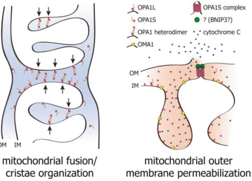
Figure 8. Model of the dual functions of OPA1 in IMM fusion, cytochrome c release and cell death. Membrane bound l-
and soluble s-OPA1 form a complex that is able to attract two mitochondrial membranes to enable IM fusion (left) and
cristae organization. Proteolytic cleavage of l-OPA1 to the soluble s-OPA1 allows formation of s-OPA1 complex, which
together with BNIP3 promotes OM permeabilization, cytochrome c release, and cell death (right) by bringing the IMM in proximity to OMM, thereby facilitating membrane hemi-fusion states that are energetically favorable for membrane permeabilization (from Alavi and Fuhrmann, 2013).
2.5 OPA1 and energetic metabolism
Considering the localization of OPA1 in the IMM, the major place of OXPHOS, it is not surprising that alterations in OPA1 expression also affect mitochondrial metabolism. Specific silencing of OPA1 alternative exons showed that the presence of exon 4 was associated with the ΔΨm maintenance (Olichon et al., 2007). Similarly, depletion of OPA1 by RNAi in MEFs led to loss of membrane potential and to severe reduction of basal respiration, unable to be stimulated by an uncoupler (Chen et al., 2005). In mitochondria ΔΨm values rapidly fluctuate and these electrical events can propagate along interconnected mitochondria. Recently it has been shown that in OPA1 knockout cells the flashes were completely lost, suggesting the importantce of OPA1 in these energy conservation events (Santo-Domingo et al., 2013). Defective OXPHOS with lowered mitochondrial ATP production on muscle from DOA patients was first documented for a common micro-deletion localized in the GED domain and further extended to several other OPA1 mutations (Lodi et al., 2004; Lodi et al., 2011). Coupling defect of OXPHOS, with reduced activity of CIV and increased activity of CV, has been observed in fibroblasts from DOA patients bearing various OPA1 mutations, associated or not with reduced mitochondrial ATP synthesis respectively (Chevrollier et al., 2008). Defective mitochondrial ATP synthesis was also documented in fibroblasts and in lymphoblasts from patients carrying different OPA1 mutations (Chevrollier et al., 2008; Amati-Bonneau et al., 2005). Our group also reported in fibroblasts from patients with different OPA1 mutations causing haploinsufficiency an impaired ATP synthesis driven by CI substrates and a decreased rate of mitochondrial fusion. Furthermore, OPA1 was shown to interact with respiratory CI, II and III, providing a potential link between OPA1 mutations and the respiratory chain defect (Zanna et al., 2008). Recently, the energetic impairment has been confirmed in studies on fibroblasts (Agier et al., 2012) and lymphoblasts (Van Bergen et al., 2011) from DOA patients carrying nonsense or missense OPA1 mutations. Interestingly, in the latter case, the OXPHOS dysfunction occurs in lymphoblasts from DOA patients with poor vision only. Patients with relative preserved vision could maintain normal mitochondrial ATP synthesis rate through increased respiration, suggesting a compensatory effect (Van Bergen et al., 2011). Moreover a study in OPA1 mouse model carrying the human c.2708delTTAG mutation, revealed that in different tissues there was a increased amount of cytochrome oxidase subunits, as a possible compensatory effect to contrast the premature age-related decrease in the stability of mitochondrial supercomplexes (Sarzi et al., 2012). These contrasting results may be explained by considering that in the studies were analysed different
mutations, that may cause different phenotypes, depending on the position of aminoacidic change also in the same protein domain.
2.6 OPA1 and mtDNA maintenance
Although Mgm1 in yeast has been identified thanks to its role in mtDNA maintenance, mtDNA deletion or depletion were never reported in skeletal muscles of DOA patients (Amati- Bonneau et al., 2008; Hudson et al., 2008; Yu-Wai-Man et al., 2010), which instead present a 2- to 4-fold increase in mtDNA copy number in COX-negative fibers (Yu-Wai-Man et al., 2010b). Furthermore only a slight reduction or no change in the mtDNA copy number was observed on blood lymphocytes or fibroblasts from DOA patients (Kim et al., 2005; Zanna et al., 2008). It was only in 2008 that OPA1 has been linked to mtDNA stability. Our and another group described the occurrence of a multisystemic disorder, named DOA-plus, characterized by severe optic atrophy and sensorineural deafness, associated with accumulation of multiple mtDNA deletions in skeletal muscles (Amati- Bonneau et al., 2008; Hudson et al., 2008).
As stated above, the nucleoids anchor the mtDNA to the matrix side of the IMM and several nucleoid proteins play important role in controlling mtDNA replication and transcription, but also in protecting this genome from damaging insults (Sperlink, 2010). Our group reported that down-regulation of OPA1 variants including exon 4b reduced the mtDNA content and replication, associated with altered nucleoid distribution throughout the mitochondrial network. In particular, the 11kDa N-terminus peptide of OPA1 containing exon 4b was shown to colocalize with mtDNA, physically interacting with nucleoids and rescuing the effect due to silencing. The hypothesis is that exon 4b-containing peptide might preserve mtDNA replication and proper distribution along the network by anchoring nucleoids to the IMM (Elachouri et al., 2011). In this regard, it has to be noticed that the N-terminus MGM1 domain was also found to be associated with nucleoids (Wong et al., 2000).
The effects of OPA1 in mtDNA stability could be also related to intactness of the IMM ultrastructure. Electron microscopy analyses indeed showed that mitochondria from OPA1-depleted or knock-out cells have an altered internal structure (Griparic et al., 2004; Song et al., 2009), similar to that reported in fibroblasts bearing OPA1 mutations causing aploinsufficieny (Zanna et al., 2008). OPA1 depletion or mutation, through changes in cristae morphology, could thus perturb mtDNA anchoring to the IM and thus influence its maintainance and replication.
Another possibility is that impairment of mitochondrial fusion, which is thought to be important for the maintenance of the mitochondrial functions by allowing the exchange of intra-mitochondrial content, might increase mtDNA instability, preventing damaged nucleoids from being repaired or complemented by fusion (Carelli and Chan, 2014).
These hypotheses, that need to be proven, are not mutually exclusive but, on the contrary, they take into consideration the multi-faceted functions of this protein, which might influence the stability of mitochondrial genome though different mechanisms.
Figure 9. Schematic representation of the different steps from the OPA1 gene to generation of N-terminal peptide
containing the exon 4b (NT-OPA1-Ex4b) and its interaction with the mitochondrial nucleoid. E1 to E29 are the OPA1 exons; (MTS) mitochondrial targeting sequence; (GED) GTPase effector domain; (TM) transmembrane domain; (MPP) mitochondrial processing peptidase; (YME1L) inner membrane protease cleaving OPA1-exon4b; (IMS) inner membrane space. (White spots) TFAM proteins; (black spot) POLG; (gray spot) unknown protein linking NT-OPA1-exon4b peptide to the mtDNA (from Elauchuri et al., 2010).
3. Autosomal dominant optic atrophy
3.1 Genetics of DOA
Autosomal dominant optic atrophy (ADOA, OMIM#165500), also known as Kjer’s optic atrophy, first described clinically by Batten (Batten, 1896) and Kjer (Kjer, 1959), is the most common of the hereditary optic neuropathies in the general population, with an estimated disease prevalence varing from 1:10,000 to 1:30,000. In 2000, two simultaneous studies identified OPA1 mutations in patients affected by ADOA (Alexander et al., 2000, Delettre et al., 2000). Two genes (OPA1, OPA3) encoding IMM proteins and three loci (OPA4, OPA5, OPA8) are currently responsible for this genetically heterogeneous disease. Additional loci and genes (OPA2, OPA6 and OPA7) are responsible for X-linked or recessive optic atrophy (Lenaers et al., 2012). About 70% of DOA has been linked to mutations in the OPA1 gene and more than 200 pathogenic mutations have been described so far, spread throughout the protein (OPA1 LSDB http://opa1.mitodyn.org; La Morgia et al., 2014). Two major mutations categories, detected throughout the entire gene, exist in patients with DOA. Stop-codon, frame-shift and deletion-insertion (about 50% of OPA1 mutations) leading to incomplete transcription and decreased protein content, with haploinsufficiency as the genetic mechanism. The remaining are missense mutations, which cause heterozygous amino acid substitutions and are thought to act through a dominant negative mechanism. In 2008, a subgroup of missense mutations affecting the GTPase domain have been linked to a multisystemic disorder form of DOA associated with sensorineural deafness, ataxia, late chronic progressive external ophthalmoplegia (CPEO) and mitochondrial myopathy with cytochrome c oxidase negative, defined as DOA “plus” syndrome (Amati-Bonneau et al., 2008; Hudson et al., 2008; Yu-Wai-Man et al., 2010). Importantly, these patients harboured multiple deletions of mtDNA in their skeletal muscles, confirming the OPA1 function in mtDNA stability, as reported for its orthologue in Saccharomyces cerevisiae (Jones and Fangman, 1992; Guan et al., 1993). Thus OPA1 has been added to the list of genes (ANT1, Twinkle, POLG, MFN2) associated with human disorders characterized by mtDNA deletions (CPEO syndromes) (Di Mauro and Schon, 2008; Rouzier et al., 2012). Recently, other phenotypes have been reported within the frame of DOA “plus” syndrome, including MS-like features, Behr-like spastic paraparesis and cases with absent or very mild ocular phenotype, thus expanding the spectrum of “OPAopathies” (Maresca et al., 2013).