Scuola Dottorale in Biologia
Sezione “Biologia Cellulare e Molecolare”
Ciclo di Dottorato XXIII
“Systems optimization for the selection of
phage display random peptide libraries”
“Ottimizzazione di sistemi per la selezione di
librerie peptidiche a sequenza casuale
espresse tramite Phage Display”
A.A. 2010/2011
Candidato:
Dott.ssa Anna Quintarelli
Docente Guida:
Prof. Pier Luigi Luisi
Coordinatore:
Prof. Paolo Mariottini
ABSTRACT
This doctoral work is part of the Never Born Protein project. It is based on idea that the proteins existing in our Earth are only an infinitesimal fraction of the all possible sequences. To use an analogy to explain the relationship between existing and possible proteins, we can say that the ratio between them corresponds roughly to the ratio between the Sahara desert and a single grain of sand.
Given the vast number of possible sequences, Nature could not have explored all possible amino acid combinations. Therefore, we are faced with the problem of how the “few” extant proteins were produced and/or selected: do extant proteins possess any special chemico-physical properties (such as solubility, fold, functionality, thermodynamic stability) that made their selection inevitable? Or rather are they the result of contingency, a frozen accident? If they are the result of contingency, there exists a universe of proteins which properties have never been sampled by nature: the Never Born Proteins – NBP.
To investigate the NBP world, random peptide 50 amino acid long libraries have been designed and selected for several characteristics by phage display technique.
To display the protein libraries we created 3 different phagemid systems, improving vectors used in previous experiments. The main improvements have been in the increase of efficiency in the library cloning and in the phage production steps. In addition, in the new vectors a His-tag replaced the old tag (a c-myc tag recognized by MAb 9E10 antibody). This involved a change of the purification system, in fact His-tagged proteins can be identified by Immobilized metal affinity chromatography (IMAC), whose efficiency is higher than antibody purification.
The complexity degree of phagemid libraries was estimated at 107 different sequences per ml. This complexity degree is a good compromise between a wide space of sequences and a good number of copies of each sequence. According to the production of phage libraries, by a 3+3 monovalent phage system we obtained phage particles with the phagemid incapsidated and a single NBP fused to the minor capsid protein (pIII protein).
The first selection aim has been to look for three-dimensional structure using the resistance to thrombin digestion as folding criterion.
All random sequences have in the middle a PRG (proline-arginine-glycine) site, corresponding to a thrombin cleavage site. Folded sequences preserve PRG during thrombin digestion, while un-folded sequences are cut since
PRG is exposed to enzyme cleavage. To recover His-tagged sequences un-digested by thrombin we chose the Ni-affinity purification, using ELISA Ni-coated plates. Results obtained after the second biopanning cycle showed a high proportion of resistant sequences (around 103 cfu/ml, the starting phage titre of 108 cfu/ml), indicating a good chance to select folded NBPs. Performed controls confirmed the results excluding un-specific bonds.
In parallel, a probable catalytic activity was tested, screening phage libraries against a TSA target, which mimics the transition state of an amide bond hydrolysis.
The aim of this selection is that a molecule which binds the transition state during a reaction can catalyze this reaction, lowering the activation energy. Therefore, selected NBPs could catalyze amide bond hydrolysis reaction. The procedure implied the selection of the phage library on a solid surface coated with the TSA. TSA was immobilized on the plate by a covalent bond between the acid group of TSA and secondary amine on the plate. In addition, we tested the influence of a metallic cofactor (Zinc) on affinity between NBPs and the target.
A good amount of phage library was recovered from wells coated with TSA (around105 cfu/ml, the starting phage titre of 109 cfu/ml). The metal cofactor did not seem to affect the affinity for the target, in fact the phage quantity recovered from the plate was similar both with and without Zn. Performed controls excluded un-specific bonds between capsid proteins and TSA and between NBPs and reagents used for TSA coupling. Un-specific bonds with plate were excluded too.
The last selection was to test possible interactions between NBPs and other proteins and investigate their possible use like inhibitors/activators of the target proteins.
The basic idea of this selection is that a ligand, to act as an inhibitor/ activator of a protein, first of all, has to bind such protein.
Chosen targets were 4 proteases: papain, pepsin, trypsin and α-chymotrypsin. They were immobilized on a polystyrene plate by adsorption. To avoid possible bounds between phage and plate surface, wells were incubated also with BSA.
Already after the second cycle, the distribution of selected phage is different for the 4 proteases: the number of phage from papain- and pepsin- wells is higher than those for the other proteases, in particular trypsin.
Performed controls excluded un-specific bonds between capsid proteins and each protease. Un-specific bond with BSA were excluded too.
The possibility of finding folded random proteins might suggest that folding is not a special property to be selected by Nature. This point could be taken as favouring the contingency hypothesis. Also the probability to find new sequences with a potential activity supports the idea that there may be an entire universe of possible proteins, with unknown properties.
RIASSUNTO
Il mio lavoro di dottorato fa parte del progetto Never Born Proteins. Questo progetto si basa sul concetto che le proteine esistenti sulla Terra sono solo una frazione infinitesimale di tutte le sequenze possibili. Per spiegare il rapporto tra le proteine esistenti e possibili, possiamo dire che il rapporto tra di esse corrisponde al rapporto tra il deserto del Sahara e un singolo granello di sabbia.
Dato il gran numero di possibili sequenze, la Natura non avrebbe potuto esplorare tutte le possibili combinazioni di amminoacidi. Pertanto, siamo di fronte al problema di come le "poche" proteine esistenti sono state prodotte e/o selezionate: le proteine esistenti hanno particolari proprietà chimico-fisiche (come solubilità, struttura terziaria, funzionalità, stabilità termodinamica) che hanno reso inevitabile la loro selezione? O piuttosto sono il frutto della contingenza? Se fossero il risultato della contingenza, esisterebbe allora un universo di proteine con proprietà mai testate dalla natura: le Never Born Proteins - NBP.
Per esplorare il mondo delle NBPs, librerie codificanti peptidi di 50 aminoacidi a sequenza casuale sono state progettate e poi selezionate per diverse caratteristiche con la tecnica del phage display.
Per esprimere le librerie abbiamo creato 3 diversi sistemi fagemidici, migliorando alcuni vettori utilizzati in precedenti esperimenti. I principali miglioramenti hanno riguardato un aumento dell’efficienza nel passaggio di clonazione della libreria e nella fase di produzione dei fagi. Inoltre, i nuovi vettori possiedono un His-tag al posto del vecchio tag (un c-myc tag riconosciuto dall’anticorpo MAb9E10). Ciò ha comportato un cambiamento del sistema di purificazione, infatti le proteine legate ad un His-tag possono essere identificate anche tramite cromatografia di affinità (Immobilized
metal affinity chromatography - IMAC) la cui efficienza è superiore alla
purificazione con anticorpi.
Il grado di complessità delle librerie fagemidiche è stato stimato 107 sequenze diverse per ml. Questo grado di complessità è un buon compromesso tra un ampio spazio delle sequenze e un buon numero di copie di ciascuna sequenza.
Per quanto riguarda la produzione delle librerie fagiche, tramite un sistema fagico monovalente 3+3 abbiamo ottenuto particelle fagiche con il fagemide incapsidato e una singola NBP fusa alla proteina minore del capside (la proteina pIII).
L'obiettivo della prima selezione è stato quello di cercare strutture tridimensionali utilizzando come criterio di folding la resistenza alla digestione da parte della trombina.
Tutte le sequenze casuali possiedono nella regione centrale un sito PRG (prolina-arginina-glicina), corrispondente ad un sito di taglio per trombina. Le sequenze strutturate proteggono il PRG dal taglio della trombina, mentre in quelle non strutturate il PRG è esposto alla digestione dell’enzima.
Per recuperare le sequenze legate all’His-tag e non tagliate dalla trombina abbiamo scelto la purificazione mediante affinità al Ni, utilizzando piastre ELISA ricoperte di Ni. I risultati ottenuti dopo il secondo ciclo di biopanning hanno mostrato un’elevata quantità di sequenze resistenti (circa 103 cfu/ml con un titolo di incubazione iniziale pari a 108 cfu/ml), ciò indica una buona possibilità di selezionare NBPs foldate. I controlli fatti hanno confermato i risultati escludendo legami aspecifici.
In parallelo le NBPs sono state testate anche per una possibile attività catalitica, selezionando le librerie fagiche contro un TSA, che mima lo stato di transizione della reazione di idrolisi del legame ammidico.
L’idea di base di questa selezione è che una molecola che lega lo stato di transizione durante una reazione può catalizzare questa reazione, abbassando l'energia di attivazione. Pertanto, le NBPs selezionate potrebbero catalizzare l’idrolisi del legame ammidico. La procedura implica la selezione della libreria dei fagi su una superficie solida rivestita con il TSA. Il TSA è immobilizzato su una piastra mediante un legame covalente tra il suo gruppo acido e l’ammina secondaria della piastra. Abbiamo anche verificato l'influenza di un cofattore metallico (zinco) sull’affinità di legame tra le NBPs e il TSA. Una quantità importante della libreria fagica è stata recuperata dai pozzi rivestiti con il TSA (circa 105 cfu/ml con un titolo di incubazione iniziale pari a 109 cfu/ml). Il cofattore metallico non sembra influenzare l'affinità per il target, infatti la quantità dei fagi recuperati dalla piastra è simile sia con che senza Zn. I controlli fatti hanno escluso legami aspecifici tra le proteine del capside e il TSA e tra le NBPs e i reagenti utilizzati per il coupling del TSA. Sono stati esclusi anche legami aspecifici con la piastra.
L'ultima selezione è stata effettuata per verificare le possibili interazioni tra le NBPs e altre proteine ed indagare il loro possibile uso come inibitori/attivatori delle proteine bersaglio.
L'idea di base di questa selezione è che un ligando per agire come inibitore/attivatore di una proteina, prima di tutto, deve legare tale proteina. I targets scelti sono stati 4 proteasi: papaina, pepsina, tripsina e α-chimotripsina. Esse sono stati immobilizzati su una piastra di polistirene
tramite adsorbimento. Per evitare possibili legami tra fagi e la superficie della piastra, i pozzetti sono stati incubati anche con BSA.
Già dopo il secondo ciclo, la distribuzione dei fagi selezionati è diversa per le 4 proteasi: il numero di fagi recuperati dai pozzetti con papaina e pepsina è superiore a quello per le altre proteasi, in particolare la tripsina. I controlli fatti hanno escluso legami aspecifici tra le proteine del capside e ogni proteasi. È stato escluso anche il legame aspecifico con la BSA.
La probabilità di trovare proteine a sequenza casuale con una struttura terziaria potrebbe suggerire che il folding non è una proprietà necessaria per essere selezionato dalla Natura. Questo punto potrebbe andare a favore della teoria della contingenza. Anche la probabilità di trovare nuove sequenze con una potenziale attività sostiene l'idea che ci possa essere un intero universo di possibili proteine, con proprietà sconosciute.
INDEX
1. INTRODUCTION 1
1.1 ABOUT ORIGIN OF LIFE 2
1.2 PEPTIDE FOLDING 6
1.3 PEPTIDES SYNTHESIS UNDER PREBIOTIC CONDITIONS 7
1.4 PHAGE DISPLAY TECHNOLOGY 10
1.4.1 Biology of filamentous phage 10
1.4.2 Phage display vector 15
1.5 PEPTIDE LIBRARIES BY PHAGE DISPLAY– THE AIM OF WORK
18
2. RESULTS 22
2.1 CONSTRUCTION OF PHAGEMID VECTORS 23
2.1.1 pIII-DUMMY-His-tag 23
2.1.2 pOCI1050 His-tag 25
2.1.3 pBluescript II KS His-tag 25
2.2 RANDOM LIBRARY 28
2.2.1 Construction of phagemid library 28 2.2.2 Computational analysis of the random sequences 30
2.3 PHAGE PRODUCTION 31
2.4 FOLDING SELECTION BY THROMBIN DIGESTION 32
2.4.1 Validation of selection method 33
2.4.2 Selection cycles 34
2.5 SELECTION AGAINST TSA 36
2.5.1 TSA stability check 36
2.5.3 Selection cycles 37
2.6 SELECTION AGAINST PROTEASES 39
2.6.1 Selection cycles 39
3. DISCUSSION 42
3.1 CONSTRUCTION OF PHAGEMID VECTORS 42
3.2 RANDOM LIBRARIES 43
3.3 PHAGE PRODUCTION 44
3.4 FOLDING SELECTION BY THROMBIN DIGESTION 45
3.5 SELECTION AGAINST TSA 46
3.6 SELECTION AGAINST PROTEASES 47
4. CONCLUSIONS AND OUTLOOK 48
1. INTRODUCTION
Science assumes that life on Earth originated from inanimate matter by a gradual and spontaneous increase of molecular complexity. Several different theoretical frameworks have been proposed to account for the spontaneous emergence of life. Wächtershäuser [Wächtershäuser, 1988] identifies enzyme-free metabolic cycles as the pivotal system underpinning life’s origin; Kauffmann proposes auto-catalytic peptide cycles as the primary motor [Kauffmann, 1996], whereas Cech fostered the idea that RNA was the scaffold of the first living system [Cech, 1993]. Conversely, Luisi emphasises the autopoietic nature of life [Luisi, 2003]; whereas Lancet proposes composition inheritance as the foundation of life [Segre, 2000]. Despite the differences, all theories must confront the same fundamental question: is life an obligatory pathway given certain initial conditions (i.e. determinism)? Or it is rather the result of the simultaneous interplay of different contingent factors (i.e. contingency)?
Within this debate, one of the most interesting questions in modern life science is how prebiotic evolution of the first biopolymers occurred and so how functional known biopolymers were selected.
It is common knowledge that for specific functionality, such as binding or catalysis, specific sequences which fold specifically are necessary. In fact, protein activity is the result of a specific three-dimensional structure, which, in turn, is determined by the amino acid primary sequence. Therefore, based on this premise, the idea of the present work is to investigate the folding frequency of random polypeptide sequences, their potential catalytic activity and finally their possible use like inhibitors/activators for several proteases.
The random polypeptide library has been designed with no sequence or structural constrains so it can be reasonably considered as a mirror image of a peptide population produced under plausible prebiotic conditions. The selection for folding is based on the concept that folded polypeptides are more protected against digestion by a protease than unfolded ones, while the ability to bind a transition state analogue of the ester and amide bond hydrolysis is at the bottom of catalytic selection.
1.1 ABOUT ORIGIN OF LIFE
The Oparin-Haldane theory [Oparin, 1954 (I - II), 1924; Haldane, 1954, 1929] about the origin of life on Earth, stating that life is the result of a series of spontaneous events which produced the first self-reproducing protocells starting from inanimate matter, is one of the most recognized. By definition, this transition to life via prebiotic molecular evolution excludes panspermia, (the idea that life on earth comes from space), and divine intervention.
Life is based on the action of proteins and nucleic acids whose functions are the result of a specific sequence that produces their structural fold and so the activity. These amino acid long sequences are co-polymeric structures, formed by macromolecules having chemically different monomer units in the same chain.
The assumption that life derives from inanimate matter bears the simple consequence that it could be reproduced in laboratory, a basic concept that is the linchpin of origin of life experimental research. Even assuming that this is possible, a big question remains unanswered as clearly formulated by Eschenmoser and Kisakürek [Eschenmoser, 1996]: “Is the pathway that goes from inanimate to animate matter determined by the laws of physics and chemistry? Or is it due to a unique event due to the contingent parameters operating in a particular time/space situation – something that in the old nomenclature would be called chance?”.
From a deterministic point of view, life arose from inanimate matter through a series of causally linked events ruled by the laws of physics and chemistry. Conversely, contingency may be defined as the outcome of a particular set of concomitant effects that apply in a particular space-time situation and thus determine the outcome of a given event. In addition, in a strictly deterministic situation, the state of a system at any point in time determines the future behaviour of the system – with no random influences. By contrast, in a non-deterministic (i.e. stochastic system) it is not generally possible to predict the time evolution of the system.
The deterministic view gained a broad support by de Duve and Morowitz [de Duve, 2002; Morowitz, 1993]. De Duve [de Duve, 1995] wrote: “[…] given the suitable initial conditions, the emergence of life is highly probable and governed by the laws of chemistry and physics [...]”, suggesting the idea that life on Earth was inevitable.
To support the contingency theory one may cite Jacques Monod’s Chance
and Necessity [Monod, 1971], his colleague François Jacob [Jacob, 1982]
and Stephen Jay Gould [Gould, 1989]. Contingency, in this particular context, can be defined as the simultaneous interplay of several concomitant effects to shape an event in a given space/time situation. In this scenario any change in the starting contingent conditions would dramatically affect the final result. The implications of contingency are dramatic, as Stephen Jay Gould illustrates when he says: “[...] run the tape again, and the first step from prokaryotic to eukaryotic cell may take twelve billions years instead of two” [Gould, 1989].
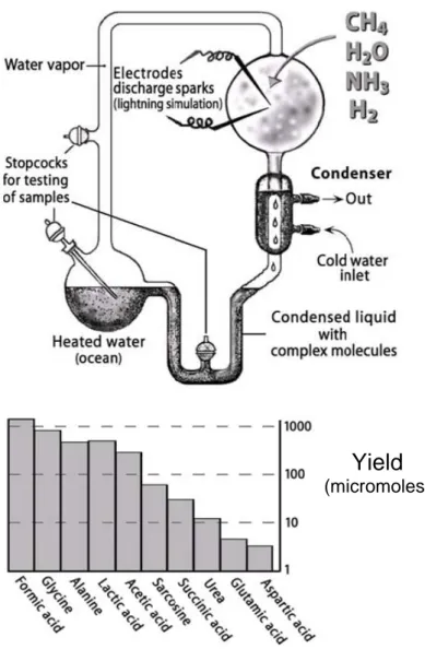
The first scientist who consciously did chemistry in pursuit of the origin of life was the graduate student Stanley Miller, fascinated by Oparin’s idea [Oparin, 1961]. Miller obtained the formation of several α-amino acids and other relatively complex substances of biological importance by passing electrical discharges through a flask filled with hydrogen, ammonia, methane, and water vapour (assumed by Oparin to be the constituents of prebiotic reductive atmosphere) (Figure 1.1) [Miller, 1953]. Although the nature of the primitive atmosphere is strongly controversial, the key point of Miller’s experiments is that relatively complex biochemicals can be formed from a mixture of very simple gaseous components in a plausible prebiotic way.
Following 1953 Miller’s experiment, many chemists successfully attempted the synthesis of other compounds of biochemical relevance under prebiotic conditions. For instance Shen and Orò reported the prebiotic synthesis of histidyl-histidine [Shen, 1990 (I – II)] and Plankensteiner and co-workers investigated the possible catalytic activity of short oligopeptides synthesized under prebiotic evolution [Plankensteiner, 2002].
Figure 1.1 Stanley Miller’s experiment. The assumption was a strongly
reduced atmosphere consisting of the four gaseous components assumed by Oparin to be the constituents of a prebiotic atmosphere (hydrogen, ammonia, methane and water vapour), and electric discharges as energy source. The graph represents the products and the relative yields obtained (Adapted from Luisi 2006).
Yield
The sequences, or primary structures, of existing biopolymers are believed to be a product of evolution. From the molecular point of view, biological evolution can be viewed as a random walk and optimisation through the sequence space. This space is astronomically big because the number of all possible sequences is exponentially proportional to the length of the polymer. Indeed, it is commonly accepted that the proteins existing in our Earth are only an infinitesimal fraction of the possible sequences [de Duve, 2002; Levinthal, 1968].
To use an analogy to explain the relationship between existing and possible proteins, we can say that the ratio between them corresponds roughly to the ratio between the Sahara desert and a single grain of sand [Luisi, 2006]. Given the vast number of possible sequences, Nature could not have explored all possible amino acid combinations and, therefore, many proteins with interesting new properties may have never been sampled by Nature - this is true even for relatively short peptides. For example, to summarize some well known calculations, taking a polypeptide of 50 residues there are 2050 (~1065) possible 50mer products. If only one molecule of each of these peptides were to be synthesized, approximately 1.8•1041 moles of material with a total mass of ~1042 kg would be produced. This quantity corresponds to ~1018 times the weight of the earth. Moreover, if this set of peptides could be synthesized at a rate of 106 molecules per second, it would take ~3•1051 years to complete their synthesis.
In light of these figures, we are faced with the problem of how the “few” extant proteins were produced and/or selected during prebiotic molecular evolution and subsequently, through a series of spontaneous steps of increasing molecular complexity, they could produce life in terms of self-reproducing protocells.
In other words, do extant proteins possess any special chemico-physical properties (such as solubility, fold, functionality, thermodynamic stability) that made their selection inevitable? Or rather are they the result of contingency, a frozen accident? If they are the result of contingency, there exists a universe of proteins which properties have never been sampled by nature: the Never Born Proteins - NBP [Luisi, 2006].
1.2 PEPTIDE FOLDING
It is common knowledge that protein activity is the result of a specific three-dimensional structure, specific to its function, such as binding or catalysis, and that this structure by the primary amino acid sequence.
Although some polypeptide chains fold spontaneously into the native state, others require the assistance of enzymes. For example, some polypeptide chains require enzymes to catalyse the formation and exchange of disulfide bonds and many others require the assistance of chaperones. These last are proteins that promote the folding of polypeptide chains and at the same time prevent the formation of illicit associations between such chains and other proteins.
A protein in its native state is not static. The secondary structural elements of the domains, as well as the entire domains, continually undergo small movements in space. The energy difference between the native state and the denatured one, in physiological conditions, is quite small, about 5-15 kcal/mol, not much more than the energy contribution of a single hydrogen bond, which is of the order of 2-5 kcal/mol [Branden, 1998]. From a biological point of view, it is important that this free energy difference is small because cells must be able to degrade proteins as well as synthesize them, and the functions of many proteins require structural flexibility.
When a fully extended unfolded polypeptide chain begins to fold, hydrophobic residues tend to be buried in the interior, greatly restricting the number of possible conformations the chain can assume, and therefore allowing proteins to fold in seconds rather then years.
The first observable event in the folding pathway of at least some proteins is a collapse of the flexible disordered unfolded polypeptide chain into a partly organized globular state, the molten globule. The molten globule has most of the secondary structure of the native state and in some cases even native-like positions of the α helices and β strands. However, it is less compact than the native structure and the proper packing interactions in the interior of the protein have not been formed. Instead, the interior side chains may be mobile, more closely resembling a liquid than the solid-like interior of the native state. In addition, loops and other elements of surface structure remain largely unfolded, with different conformations. For the single native form to be reached in the final stage of folding, the formation of native interactions throughout the protein, including hydrophobic packing in the interior as well as the fixation of surface loops must take place.
Since the fundamental prerequisite for a polypeptide to serve as a protein is a three-dimensional structure, the question whether a random sequence is likely to fold into a native state is basic to understand the possibility of the emergence of functional proteins in the “primordial soup”.
To tackle the highly challenging problem of protein folding, a number of models have been developed; one of these is the lattice Monte Carlo simulation [Hilhorst, 1975; Verdier, 1973]. This simulation showed that the necessary condition for a polypeptide to have a kinetically reachable stable conformation is that it has a pronounced global energy minimum [Shakhnovich, 1993; Gutin, 1993; Goldstein, 1992]. It was estimated [Gutin, 1995] that only a small fraction of random sequences satisfies this requirement for chains of realistic lengths. The implication of this finding is that it is unlikely that folded protein will be found as a result of random exploration of the sequence space.
Although stable proteins might be only a tiny fraction of all possible sequences, stable polypeptides were not hydrolyzed and accumulated over time, whereas more frequently occurring unstable sequences underwent hydrolysis.
To experimentally address these hypothesises it is necessary to produce large random libraries of polypeptides. The construction and study of such libraries is discussed in the following chapters.
1.3 PEPTIDES SYNTHESIS UNDER PREBIOTIC CONDITIONS Unfortunately, we do not know how the first proteins were formed in the prebiotic Earth. The characteristics of such polymers are so unique that it is only possible to speculate about their formation and, of course, try different solutions to reproduce them under prebiotic conditions; but, even knowing a useful method to produce such polymers, the problem would not be solved. In fact, as discussed previously, by synthesizing a random 50-mer chain using all 20 different amino acids it is theoretically possible to produce about 1065 different sequences and the probability to sample two identical chains is approximately equal to zero.
Furthermore, it is very difficult to predict the formation of a specific sequence due to the different elements playing a role in structure’s formation. It is useful, to consider the dynamics behind the synthesis of a copolymer formed by two monomers, A and B (Luisi 2006) (Figure 1.2).
Figure 1.2 Growing of copolymers constituted by only two monomers A and B (Adapted from Luisi 2006)
The growing chain can terminate with A or B and in the next step for each of the two sequences there is the same theoretical possibility to acquire either A or B (Figure 1.2). Four kinetic constants (kAA, kBB, kAB, and kBA) define the probability that a monomer is incorporated into the chain in a classic linear polymerization, while rA=kAA/kABand rB=kBB/kBAthe tendency of the chain to assume a certain composition and sequence. When rAand rB are both significantly larger than one, any polymer chain will tend to grow by incorporating a monomer equal to the last added, which will result in long stretches of homo-polymers. Conversely, when rA and rB are both significantly smaller than one the two monomers will be alternated in the sequence. In both cases the composition of the sequences could be roughly predicted knowing the monomers initial concentration. However, when rA and rB are both close to one the polymerization will proceed in a pure random fashion with an unpredictable final result. In the case of proteins or nucleic acids the situation is even more complicated because they are formed by several different monomers.
Furthermore, since the final result could be influenced by kinetic and thermodynamic parameters, the composition of prebiotic biopolymers could not be directly deduced even if we had known the amino acid composition of the “prebiotic soup”. Indeed, neither the high or low abundance in the starting solution of a specific amino acid expresses its relative frequency in a poly-condensed product. Taking all these factors into account, it is reasonable to assume that the prebiotic synthesis of biopolymers occurred in an unpredictable random way.
ABAABABBAAA KAB KAA ABAABABBAAAB ABAABABBAAAA ABAABABBAAB KBB KBA ABAABABBAABB ABAABABBAABA
The creation of polypeptides has also been explored under prebiotic conditions. For example, Fox and Dose [Fox, 1977] show that proteinoids (bodies containing polymerized amino acids) can be formed by heating mixtures of amino acids (containing a 10 fold excess of residues with reactive side chains, such as glutamic acid, aspartic acid or lysine) at 180° for a few hours. However, this procedure results in a high abundance of branched products and therefore is not considered a reliable method to produce biopolymers. Nevertheless, it was however reported that when using amides, the presence of clay increases the yields during repeated drying and heating, and Ito and co-workers [Yanagawa, 1990] reported a substantial arrays of polypeptides prepared in this way. But generally, one can accept the statement that no reliable method is known to produce high molecular weight of linear co-polypeptides under prebiotic conditions. It is also interesting to mention the condensation of N-carboxyanhydrides (known as Leuch’s anhydrides). The relevance of this reaction lies in the fact that NCA-amino acid derivatives are supposed to be prebiotic compounds [Taillades, 1999]. As noticed by Ferris [Ferris, 2002], this synthetic route has proved advantageous with regard to other synthetic paths, in fact, the synthesis can occur in water solution. Since the polymerization is faster than the hydrolysis rate, there is no racemization and therefore the synthesis is specific for α-amino acids. Oligomers up to 10-mers can be obtained in one single step, but this is the limit of the method, as it appears impossible to reach significantly higher polymerization degrees. However, decamers can be used to synthesise longer polypeptides by means of fragments condensation.
Finally, a new interesting development is offered by Orgel and Ghadiri’s groups [Leman, 2004]; they have shown that carbonyl sulfide (COS), a simple volcanic gas, brings about the formation of peptides from amino acids under mild conditions in aqueous solution, reaching high yields (around 80%) at room temperature. Following this procedure, dipeptides and tripeptides have been successfully synthesised.
Alongside the chemical approach producing long peptides, there is the biological approach. In this case, it is necessary to put aside any focus on the mechanism and assume that the polymeric sequences were formed in a wide distribution of randomly produced polypeptide chains in which there is a specific percentage of folded chains which may have catalytic activity. The aim of my work is to use the phage display method for obtaining this broad distribution of chains and investigate the existence of a stable fold and then their potential catalytic properties.
1.4 PHAGE DISPLAY TECHNOLOGY
Phage Display technology, introduced by G. Smith [Smith, 1985], is a good method for selecting specific molecules from large peptide or proteins libraries [Siegel, 2001; Ladner, 2000; Hoogenboom, 1998; Burton, 1995; Ladner, 1995; Neri, 1995 (II); Winter, 1994; Griffiths, 1993; Barbas, 1993]. Phage display has been successfully applied to a wide range of different purposes such studying:
− protein-protein interactions [Hertveldt, 2009; Sidhu, 2007; Cesareni, 1992];
− receptor and antibody-binding sites [Winter, 1994; Griffiths, 1993; Better, 1988; Skerra, 1988];
− protein stability [Kotz, 2004; Hoess, 2001; Forrer, 1999],
− new enzyme substrates and inhibitors [Hawinkels, 2007; Sedlacek, 2005; Deperthes, 2002; Kay, 2001; Hyde-DeRuyscher, 2000];
− the design of catalytic antibodies and enzymes with novel specificities [Fernandez-Gacio, 2003].
This technology utilizes the ability to express foreign proteins on the surface of phage particles which are fused to the coat proteins. The DNA encoding the displayed protein of interest is inserted into the single-stranded genome of filamentous phage, providing a physical link between genotype and phenotype. Instead of having to genetically engineer proteins or peptide variants one-by-one and subsequently express, purify, and analyze them, phage display enables the construction of large libraries of protein. Specific clones can then be selected and their sequence easily determined by sequencing the DNA contained in the phage particle. The selection format is usually based on biopanning of the library on solid phase surfaces carrying immobilized ligands. In addition, the high in vitro stability of the phage particle permits the use of a wide range of selection conditions, such as high temperature, denaturants, pH and ion concentration.
1.4.1 Biology of filamentous phage
Filamentous phage have a single-stranded DNA genome which is encased in a long cylinder approximately 6 nm wide by 900 to 2000 nm in length (Figure 1.3) and they are able to infect a variety of gram negative bacteria. Three bacteriophage, M13, fl, and fd, are the most able to infect E.Coli cells, containing the F conjugative plasmid. Their genome has been
completely sequenced resulting 98% homologous DNA sequences [Hill, 1982; Beck, 1981;Van Wezenbeek, 1980].
Figure 1.3 Schematic drawing of phage particles. pIII is the minor coat
protein three, while pVIII represents the major coat protein eight. The phage genome is constituted by a single stranded DNA (ss DNA) encased into a cylindrical phage particle.
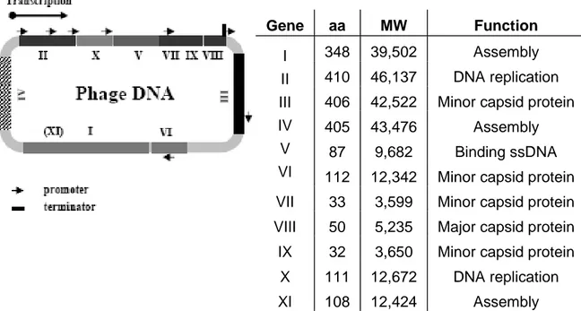
The entire genome of these phage consists of 11 genes (Figure 1.4). Two of these genes, X and XI, overlap and are in-frame with the larger genes II and I [Rapoza, 1995; Model, 1988]. The arrangement of the genes on DNA is based on their functions in the life cycle of the bacteriophage. Two genes (gII and gX) encode proteins required for DNA replication while a third one (gV) encodes for a protein necessary both at the assembly and DNA level; a group of three genes (gI, gIV and gXI) is involved in the phage assembly process at membrane level, while a last group encodes the capsid proteins. In addition to the regions which encode proteins, is the “Intergenic Region” which contains the sites of origin for the synthesis of the (+) strand (phage DNA) or (-) strand as well as a hairpin region which is the site of initiation for the assembly of the phage particles (packaging signal) (Figure 1.4). A phage expresses about 2700 copies of the major coat protein (pVIII, 50 aa long), and 3 to 5 copies of the minor coat protein (pIII, a 406 aa long) [Russel, 1991].
Figure 1.4 Genome and gene products of the fl bacteriophage. The
single-stranded DNA contains 11 genes, listed in the table. It has 6407 nucleotides which are numbered from the unique HindII site located in gene II.
Infection of E.Coli by the bacteriophage is a two-step process. In the initial phase the pIII end of the phage particle interacts with the tip of the F conjugative pilus (Figure 1.5). The pilus is retracted so the tip of the phage is moved to the membrane surface. The retraction of the pilus presumably occurs the normal polymerization-depolymerization cycles inherent to the pili or may be the attachment of the phage can trigger pilus retraction [Frost, 1993].
The integration of the pVIII major capsid proteins and perhaps the other capsid proteins into the inner membrane together with the translocation of the DNA into the cytoplasm is the last step of infection. After that, the host DNA replication machinery converts the single-stranded phage DNA into the double-stranded plasmid like replicative form (RF). The RF serves as template for expression of the phage proteins and to produce new ssDNA. Phage progeny are assembled by packaging of ssDNA into protein coats and extruded through the bacterial membrane into the medium (Figure 1.5). The assembly of filamentous phage begins where the inner and outer membranes of the E.Coli cells are in close contact [Lopez, 1985]. These assembly sites may be the result of specific interactions between pI, pIV
Gene aa MW Function
348 39,502 Assembly
410 46,137 DNA replication 406 42,522 Minor capsid protein
405 43,476 Assembly 87 9,682 Binding ssDNA I II III IV V
VI 112 12,342 Minor capsid protein VII 33 3,599 Minor capsid protein VIII 50 5,235 Major capsid protein
IX 32 3,650 Minor capsid protein X 111 12,672 DNA replication
and pXI (Figure 1.5). The event that initiates the assembly of the particle is probably the interaction of the pV-phage DNA complex with proteins in the assembly site. The single-stranded DNA binding protein pV is 87 amino acids long and its biological functional entity is a homodimer; it is a multifunctional protein that not only is implicated as a scaffolding and/or chaperone during the phage assembly process, but regulates viral DNA replication and gene expression at the level of mRNA translation.
Subsequently the pV dimers are displaced from, and the capsid proteins added to the DNA during the extrusion through the bacterial envelope into the media.
The assembly process is conveniently divided into three parts, initiation, elongation and termination, reflecting the events required for packaging the different ends and the long cylinder of the phage [Endeman, 1995; Russel, 1991; Russel, 1989; Lim, 1985] (Figure 1.5).
Prior to assembly the coat proteins are imbedded in the inner membrane with the C termini in the cytoplasm. pVII and pIX are firstly incorporated at one end of the particle and then pVIII molecules are added along the length of the particle in thousands of copies.
The conclusion of the phage assembly process occurs when the end of the DNA is reached, and protein pVI and pIII added. In the absence of either of these proteins, assembly goes on with pVIII continuing to encapsulate another DNA producing polyphage containing multiple copies of the genome. The assembled phage particle is then released from the bacterial envelope into the extracellular environment.
Since the capsids are assembled around the DNA, there are no constraints on the length of DNA packaged. This property led to its use as a cloning vehicle. On the other hand, the membrane-associated assembly properties of the capsid proteins allow the packaging of chimeric proteins into the phage particle. The flexibility of the assembly process has led to an impressive array of applications for the use of phage display.
Filamentous phage does not produce a lytic infection in E.Coli, but rather induces a state in which the infected bacteria produce and secrete phage particles without undergoing lysis and the bacteria continue to grow and divide.
Figure 1.5 Schematic representation of the bacteriophage life cycle. The
phage starts the infection of E.Coli by the specific interaction with the tip of the F pilus. (+), the bacteriophage single-stranded DNA; (-), the complementary DNA strand; pV, the product of the bacteriophage gene V essential for the assembly process; pIII, pVI, pVII and pIX, the minor coat proteins; pVIII, the major coat protein; pI, pIV and pXI, the assembly proteins, the phage is extruded through an assembly site formed by these proteins.
1.4.2 Phage display vector
According to Smith’s classification [Smith, 1997, 1993], there are different formats for Phage Display (Figure 1.6):
− type 3 / type 8:
In these formats, the exogenous peptide or protein is expressed on the virion capsid as fusion protein to either the pIII or the pVIII protein. In particular, the corresponding encoding gene is cloned upstream of the gIII or gVIII into a wild-type viral vector. The result is that every copy of the capsid protein displays the fusion producing a multivalent display.
− type 33 / type 88:
In these systems, the vectors are phage that carry two copies of gene gIII or gVIII, one of which is fused with exogenous gene; bacterial cells infected with these phage incorporate both wild-type and fusion copies of pIII or pVIII into the same viral particles.
− type 3+3 / type 8+8:
In these formats, the protein fusion gene can be placed in a phagemid vector. Phagemids are hybrids of phage and plasmid vectors, usually containing an M13 origin of replication, the packaging signal site, multiple cloning sites and an antibiotic-resistance gene in addition to the elements required for plasmid propagation in E.Coli cells [Mead, 1988]. The phagemid replicates in E.Coli as a double-stranded plasmid and co-infection occurs with a helper phage, resulting in the production of single-stranded phagemid DNA, which is packaged into phage particles. The helper phage provides all the proteins necessary for phage assembly, including wild-type copies of all the coat proteins. In general, for these display formats are mainly used pIII as well as pVIII. In the case of using pIII, the resulting phage particles may incorporate either pIII derived from the helper phage or the pIII fusion protein, encoded by the phagemid, producing a monovalent display.
The level of display for different polypeptides varies greatly. The ratio between fusion proteins and wild type pIII may range between 1:9 and 1:1000, depending on both the length and the sequence of the displayed peptide and the growth conditions.
Using a 3+3 system the infectivity is not compromised because wild-type copies of pIII are also provided. Fused protein domains are more accessible when linked to domain 2 of pIII. Most protein display experiments have
utilized this truncated form of pIII for efficient display. However, replacement of domain I (aa 1-198) of pIII with an exogenous sequence leads to non-infectious phage when in multivalent phage display (Figure 1.7).
Figure 1.6 Classification of phage display vectors. The black boxes and
spheres correspond to the foreign genetic elements and their encoded peptides, respectively. Wild-type phage type 3 type 8 type 3+3 type 8+8 type 33 type 88
Figure 1.7 Phage system and Phagemid system in comparison.
3 MULTIVALENT PHAGE DISPLAY
Helper phage
In designing a vector for phage display, a series of consideration has to be taken into account, such as the insertion of antibiotic resistance genes, linkers, tags and stop codons. For example, a displayed element can be separated from pIII by short linkers. While there have been no formal experiments on the best linker sequences to use, many vectors use some variation of the sequence GGGGS. It is also possible to include a proteolytic cleavage site between the displayed peptide/protein and the capsid protein. While to reduce the level of non-recombinants in phage display libraries, a stop codon is inserted in the fragments of gene III. The stop codon, TAG, can be suppressed efficiently in bacteria containing supE or supF; when these vectors are propagated in such bacterial strains either a glutamine (Q) or tyrosine (Y) is inserted at the TAG codon, respectively. However, when TAG containing gene III is in bacterial strains that lack either supE or supF, no full-length pIII accumulates and therefore no virus particles are generated.
Furthermore, the vectors can be engineered to express short peptide “tags”, recognized by specific antibodies, in the N terminal region of fused protein to immunologically discriminate between parental and recombinant phage particles. One such sequence is the c-myc epitope [Evan, 1985], which is recognized by MAb 9E10, or a hexa-histidine tag [Hochuli, 1987], which is useful for a recovery of a specific protein using by Ni-column affinity chromatography.
1.5 PEPTIDE LIBRARIES BY PHAGE DISPLAY – THE AIM OF WORK
It is noteworthy that the synthesis of peptide libraries with random amino acid sequences, followed by a procedure that selects new peptides with desirable properties, has become a relatively widely exploited area of research. Many investigators, however, have a biotechnological or pharmaceutical background and focus on the isolation of functional proteins [Siegel, 2001; Ladner, 2000, 1995; Neri, 1995 (I)]. Their main interest is the isolation of proteins with improved stability, new or optimized catalytic properties [Dumon, 2008; Seelig, 2007; Lingen, 2002; Meyer, 2002] or proteins that bind target molecules with enhanced affinity [Ho, 2006; Doorbar, 1994]. Generally, however, such investigations have been done starting from selected extant protein scaffolds and randomizing either restricted regions or the entire gene [Cirino, 2003; Murakami, 2003; Murakami, 2002; Shao, 1998; Cadwell, 1994]. Alternatively, using
recombination techniques, DNA fragments have been mixed to obtain novel combinations [Aguinaldo, 2003; Coco, 2003; Joern, 2003; Lutz, 2003; O’Maille, 2002]. These approaches can be defined as “directed randomizations” [Neylon, 2004], in the sense that randomization is performed in order to achieve certain desired properties.
Others investigations have been made in de novo sequence libraries with different levels in terms of randomization. For example, using a binary pattern of polar and non-polar amino acids [Bradley, 2006; Wei, 2003 (I-II); Moffet, 2001; West, 1999; Cordes, 1996; Kamtekar, 1993]. Nevertheless, using this method to construct the library, proteins were forced to assume a specific secondary structure [Hecht, 2004; Moffet, 2001]. An even more reductively methodology, in terms of sequence, has been used in Sauer’s group [Davidson, 1995, 1994]. Here the idea has been to investigate the possibility to obtain stable three-dimensional structures into random sequence libraries prepared using 3 amino acids only: glutamine, leucine and arginine.
In contrast to these approaches the rationale of this work is completely different. It is aimed at investigating the frequency of folding and of catalytic activity in a totally random library where is no bias towards any given sequence or structural feature. This approach to create de novo libraries can be defined as a “total randomization”.
We created 50 amino acid long random peptide libraries encoding proteins non extant in nature. As discussed previously, by synthesizing a random 50-mer chain using all 20 different amino acids it is theoretically possible to produce about 1065 different sequences and the probability to sample two identical chains is approximately equal to zero.
We chose the 50 amino acid length because it was quite short to resemble simple prebiotic oligopeptides but sufficient long to be folded. Indeed, in nature there are small proteins (around 30 amino acids long) with a stable three-dimensional structure, as the APP (Avian Pancreatic Peptide). On the contrary, smaller peptides are unstable since their mobility is increased by thermic agitation. Therefore, the 50 amino acid length seemed to be a good compromise. In addition, the structure of 50 amino acid long peptide can be investigated by NMR spectroscopy easily.
The random sequences were first cloned in phagemid vectors and then the resulted phagemid libraries were converted in phage libraries by the phage display technique.
The phage libraries were selected to look for NBPs with a folded structure and, parallel to this screening, NBPs were investigated for potential catalytic activities and possible interaction with other proteins.
According to the investigation of folded proteins, we used the resistance to proteolytic digestion as folding criterion. We chose thrombin as digestion enzyme because it is more selective than other proteases and not affected by flanking residues. On the contrary, other enzymes are inhibited when specific residues are near the cleavage site. As target site of thrombin we chose the PRG (proline-arginine-glycine) site which was placed in the middle of random sequence.
The base of this selection is that in a folded sequence the PRG is preserved during thrombin digestion while in un-folded sequence the PRG is exposed to enzyme cleavage.
The next step of this work concerns the selection of NBPs for their potential catalytic activity. Our screening strategy is based on the transition state theory [Jencks, 1969; Glasstone, 1941]. According to this theory, a catalyst enhances the reaction rate stabilizing high-energy transition state structures that are formed during the reaction. Therefore, any protein that stabilizes the transition state is a potential candidate for catalyzing that reaction.
In regard to the catalytic reaction, we focused on amide bond hydrolysis. This reaction spontaneously occurs in aqueous environment. As reported by Luisi and co-workers [Gorlero, 2008], under anhydrous conditions, or following the precipitation of the product, the entire equilibrium of the reaction can be shifted towards the synthesis. Within this framework, peptides capable of hydrolysing the amide bond may catalyse the reverse reaction, synthesizing short peptides. Accordingly, amide-hydrolytic peptides might have triggered off the emergence of primordial metabolism [Nakashima, 1980] or auto-catalytic peptides cycles [Kaufamann, 1996]. In addition, amide hydrolysis involves the formation of a high-energy tetrahedral intermediate [Tanaka, 2002] which can be mimicked by a phosphonates or phosphomamidates TSA (Figure 1.8).
Figure 1.8 Molecular structure of TSA. The upper part of figure represents
the mechanism of amide hydrolysis, in the lower there is the structure of transition state analog (TSA), that can be a phosphonate (X=O) or a phosphonamidate (X=NH). (Adapted from Tanaka, 2002).
The last part of this work explores the possible interactions between NBPs and other proteins, in particular proteases. In theory, peptides are able to bind to a protein anywhere on its solvent-exposed surface. However, most peptides bind at sites that coincide with natural ligand-binding sites. In fact, this site seems to have features that predispose it for ligand binding [Sidhu, 2000]. Consequently, peptides bound to the natural ligand-binding site could act as inhibitors/activators like the natural ligand, even with better results. Therefore, the aim of our work is to select NBPs against four proteases immobilized by adsorption on a plate to have potential new inhibitors/ activators of target-proteases. Positive selected sequences, with a demonstrated activity, could be used in pharmaceutical and biotechnological field.
2. RESULTS
The first part of this work describes the development of 3 different phagemid vectors employed to display a totally de novo random library encoding totally new proteins (the Never Born Proteins).
The 150 bp long DNA library is totally random except for tripeptide proline-arginine-glycine (PRG) which is the substrate for thrombin used for folding selection. Another shared characteristic is the hexa-histidine tag (His-tag) at the N-terminal of all sequences, useful for the recovery of NBPs. These sequences were inserted into each phagemid vector described hereafter, obtaining 3 different phagemid libraries. Each library was expressed by the phage display technique. Using the 3+3 monovalent phage system (described in section 1.4.2), a single NBP-pIII fusion protein was displayed on each phage (Figure 2.1). The other pIII proteins were wild-type and thus phage infectivity was not compromised.
Figure 2.1 Schematic drawing of random peptide fused with pIII. The
random peptide is exposed as fusion protein to pIII (minor coat protein) on the top of the phage while pVIII represents the major coat protein. The PRG (proline-arginine-glycine) site is in the middle of random peptide, while the His-tag is exposed at the N-terminal of the random sequence.
Phage libraries were selected for several characteristics using the ELISA panning technique. The first selection was to look for three-dimensional structures using the resistance to thrombin digestion as folding criterion. In parallel, a probable catalytic activity was tested, screening phage libraries against a TSA target, which mimics the transition state of an amide bond hydrolysis. Finally, NBPs were selected to analyse the possible interaction with other proteins, in particular 4 different proteases: papain, pepsin, trypsin and α-chymotrypsin.
2.1 CONSTRUCTION OF PHAGEMID VECTORS 2.1.1 pIII-DUMMY-His-tag
The pIII-DUMMY-His-tag comes from pOCI1050 c-myc (courtesy of Dr. Chiarabelli, University of Roma Tre) that allows in-frame expression of: - the pelB signal sequence, to ensure a reliable membrane translocation; - the c-myc tag sequence (16 residues long), to recognize by MAb 9E10
antibody; - a linker;
- a gene of interest;
- the C-terminal part of gene III (sequence D197-S406).
The signal sequence is cleaved when the protein is exported to the periplasm. A gene of interest can be cloned into the unique NotI, XbaI sites and an amber codon is located between the gene of interest and the C-terminally truncated gene III (Figure 2.2).
One of the main differences between pIII-DUMMY-His-tag and pOCI1050 c-myc is the His-tag replaces the c-myc tag. This involves a change of the purification system: His-tag fusion proteins can be identified by Immobilized metal affinity chromatography (IMAC), whose efficiency is higher than antibody purification.
In addition, the new vector has the “Dummy” sequence and the Xa site. The first is an insertion sequence (738 bp long) harbouring 3 stop codons in the 3 different reading frames in order to allow visual control during the cloning step and minimise the production of non-recombinant fusion proteins in case of phagemid self-ligation. The Xa site is the cleavage site of a protease,
the activated coagulation Factor X, to allow the purification of exogenous proteins separately from the tag.
Figure 2.2 Maps of phagemid vectors pOCI1050 and pIII-DUMMY-His-tag . Both vectors have a ribosome binding sequence (RBS) upstream pelB
and a p-Lac promoter (pLacZ gene) to control the expression of foreign proteins. These last can be inserted using NotI and XbaI cloning sites.
pelB c-myc Linker Gene III
NcoI NotI XbaI
rbs ATG pLacZ HindIII EcoRI pOCI1050 c-myc Or i M13 Ampr ColE 1 Tag pIII
pelB c-myc Linker Gene III
NcoI NotI XbaI
rbs ATG
pLacZ
HindIII EcoRI
pelB c-myc Linker Gene III
NcoI NotI XbaI
rbs ATG
pLacZ
HindIII EcoRI
pelB c-myc Linker Gene III NcoI
NcoI NotINotI XbaIXbaI
rbs ATG
pLacZ HindIII
HindIII EcoRIEcoRI
pOCI1050 c-myc Or i M13 Ampr ColE 1 Tag pIII pOCI1050 c-myc Or i M13 Ampr ColE 1 Tag pIII
pIII Dummy His-tag
Ori M13
Ampr
Col
E
1
Tag Dummy pIII
pelB His-tag Linker Gene III
rbs ATG
pLacZ Xa Dummy
NcoI NotI XbaI
HindIII EcoRI
pIII Dummy His-tag
Ori M13
Ampr
Col
E
1
Tag Dummy pIII
pIII Dummy His-tag
Ori M13
Ampr
Col
E
1
Tag Dummy pIII
pelB His-tag Linker Gene III
rbs ATG
pLacZ Xa Dummy
NcoI NotI XbaI
HindIII EcoRI
pelB His-tag Linker Gene III
rbs ATG
pLacZ rbs ATG pelB His-tag Xa Linker Dummy Gene III
pLacZ Xa Dummy
NcoI
NcoI NotINotI XbaIXbaI
HindIII
2.1.2 pOCI1050 His-tag
This vector derives from pOCI1050 c-myc described before. The c-myc tag is replaced with a His-tag from pET14b (Novagen) cloning into the pOCI1050 c-myc between NcoI and NotI sites (upstream and downstream of c-myc sequence respectively) (Figure 2.3).
Figure 2.3 Map of pOCI1050 His-tag. The vector has a ribosome binding
sequence (RBS) upstream pelB and a p-Lac promoter (pLacZ gene) to control the expression of foreign proteins. These last can be inserted using NotI and XbaI cloning sites.
2.1.3 pBluescript II KS His-tag
The pBluescript II KS His-tag derives from a commercial vector, the pBluescript II KS.
The pBluescript II phagemids have an extensive polylinker with 21 unique restriction enzyme recognition sites. The polylinker and T7 and T3 RNA polymerase promoter sequences are present in the N-terminal portion of a lacZ gene fragment. A total of 131 amino acids of β-galactosidase coding sequence are present in the pBluescript II phagemid, but the coding sequence is interrupted by the large polylinker (Figure 2.4).
pelB His-tag Linker Gene III
NcoI NotI XbaI
rbs ATG pLacZ HindIII EcoRI pOCI1050 His-tag Or i M1 3 Ampr ColE1 Tag pIII
pelB His-tag Linker Gene III
NcoI NotI XbaI
rbs ATG
pLacZ
HindIII EcoRI
pelB His-tag Linker Gene III
NcoI NotI XbaI
rbs ATG
pLacZ
HindIII EcoRI
pelB His-tag Linker Gene III
NcoI
NcoI NotINotI XbaIXbaI
rbs ATG
pLacZ HindIII
HindIII EcoRIEcoRI
pOCI1050 His-tag Or i M1 3 Ampr ColE1 Tag pIII pOCI1050 His-tag Or i M1 3 Ampr ColE1 Tag pIII
Figure 2.4 Circular map and list of features for the pBluescript II KS (wild-type). Red arrows indicate plasmid restriction sites eliminated.
The pBluescript II KS wild-type was modified to insert a His-tag and change its restriction system. The first modification consisted of the inclusion of a new fragment, corresponding to the region in pOCI 1050 His-tag between the rbs and gIII (~790 bp long) (Figure 2.3), which was cloned into the pBluescript II KS between HindIII and EcoRI sites (Figure 2.5). After insertion of the His-tag, the new plasmid was modified in its restriction system to allow cloning of the library. The XhoI site in the pBluescript II KS (Figure 2.4) was removed to avoid problems during the last step of the library cloning. In fact, to obtain the DNA sequence coding for the PRG site it is necessary to digest the DNA annealing region with the
XhoI enzyme, as described in the next section. To modify this plasmid
region, pBluescript His-tag was digested with SalI and XhoI. The small fragment between the two enzyme sites was then eliminated by purification and plasmid reclosed.
The XbaI site in the pBluescript II KS (Figure 2.4) was also removed because it is present a second XbaI site in the new fragment inserted into the pBluescript II KS during the first modification. To do this, the plasmid was digested with SmaI and SacI and the small fragment between the two enzyme sites was eliminated by purification. After incubation with Klenow polymerase to remove the 5’ protruding, the pBluescript His-tag blunt ends were ligated.
Figure 2.5 pBluescript II KS His-tag map. The vector has a ribosome
binding sequence (RBS) upstream pelB and a p-Lac promoter (pLacZ gene) to control the expression of foreign proteins. These last can be inserted using NotI and XbaI cloning sites.
pelB His-tag Linker Gene III
NcoI NotI XbaI
rbs ATG pLacZ HindIII EcoRI pBluescript II KS His-tag Ampr Tag pIII
pelB His-tag Linker Gene III
NcoI NotI XbaI
rbs ATG
pLacZ
HindIII EcoRI
pelB His-tag Linker Gene III
NcoI NotI XbaI
rbs ATG
pLacZ
HindIII EcoRI
pelB His-tag Linker Gene III NcoI
NcoI NotINotI XbaIXbaI
rbs ATG
pLacZ HindIII
HindIII EcoRIEcoRI
pBluescript II KS His-tag Ampr Tag pIII pBluescript II KS His-tag Ampr Tag pIII
2.2 RANDOM LIBRARY
2.2.1 Construction of phagemid library
The DNA library cloned in the phagemid systems was synthesized using two groups of oligonucleotides (forward and reverse) with codon schemes NNK and NNM, where N is an equimolar mixture of all four bases and where K is either G or T and M either C or A. These schemes use 32 codons to encode all 20 amino acids and 1 stop codon (TAG), yielding an acceptably low frequency of stop codons when used to encode short polypeptides.
The oligonucleotides consist of random nucleotides, encoding 23 amino acids in the forward group and 24 in the reverse, flanked by 11-18 fixed residues that are necessary for annealing and cloning (Figure 2.6). After annealing and incubation with RedTaq polimerase, the DNA library was cloned in all described vectors using the unique NotI, XbaI sites. The bases in excess in the annealing region were subsequently removed by cleavage with XhoI restriction enzyme and in this way PRG site was formed.
All the vectors were transformed into E.Coli cells (XL1Blue MRF’ strain) by electroporation. This technique was chosen because produces higher efficiency than the best chemical methods and this is crucial in order to achieve large, representative primary libraries.
The final number of different random sequences present in each library in the bacteria was estimated to be about 107 per ml.
Figure 2.6 Library construction.
ForwardOligonucleotides ReverseOligonucleotides
NNK XhoI NotI XbaI XhoI NNM Ampr Phagemid vector Gene III His-tag XbaI XhoI NotI XhoI
NotI XhoI XhoI XbaI
His-tag Gene III
Digestion and ligation
NotI XhoI XbaI
His-tag P-R-G Gene III
ForwardOligonucleotides ReverseOligonucleotides
NNK XhoI NotI XbaI XhoI NNM Ampr Phagemid vector Gene III His-tag XbaI XhoI NotI XhoI
NotI XhoI XhoI XbaI
His-tag Gene III
Digestion and ligation
NotI XhoI XbaI
His-tag Gene III
ForwardOligonucleotides ReverseOligonucleotides
NNK
XhoI NotI
XbaI
XhoI NNM
ForwardOligonucleotides ReverseOligonucleotides
NNK XhoI NotI NNK XhoI NotI XhoI NotI XbaI XhoI NNM XbaI XhoI NNM XbaI XhoI NNM Ampr Phagemid vector Gene III His-tag XbaI XhoI NotI XhoI Ampr Phagemid vector Gene III His-tag Ampr Phagemid vector Gene III His-tag XbaI XhoI NotI XhoI XbaI XhoI NotI XhoI
NotI XhoI XhoI XbaI
His-tag Gene III
NotI XhoI XhoI XbaI
His-tag Gene III
Digestion and ligation
NotI XhoI XbaI
His-tag Gene III
Digestion and ligation
NotI XhoI XbaI
His-tag Gene III
NotI XhoI XbaI
2.2.2 Computational analysis of the random sequences
From each phagemid library, we analyzed DNA sequence of 10 samples at random to verify their correctness and randomness. The majority of sequences, about 80%, were found to be correct with random sequence in-frame in the vector. The remaining part of the sequences consisted of either not in-frame sequences or vectors lacking random sequences.
Of all the correct sequences, randomness at the amino acid level was analysed comparing experimental and expected data (Figure 2.7). The graph shows that the random sequences have an amino acid frequency in line with the expected data. The experimental value of glutamine residues (Q) is the result of both, the codons for Q in the random sequences, and the translation of TAG codons into Q residues due to the E.Coli Amber mutation strain used.
Figure 2.7 Randomness of phage peptide library at amino acid level
Randomness at a nucleic acid level of the correct clones was also investigated (Figure 2.8) taking into account the NNK scheme used in the design of the library. Also in this analysis experimental values are in line with theoretical ones.
0,0 1,0 2,0 3,0 4,0 5,0 6,0 7,0 8,0 9,0 10,0 A R N D C Q E G H I L K M F P S T W Y V % a a THEORETICAL EXPERIMENTAL % of a a in ph ag e l ibr ar y 0,0 1,0 2,0 3,0 4,0 5,0 6,0 7,0 8,0 9,0 10,0 A R N D C Q E G H I L K M F P S T W Y V % a a THEORETICAL EXPERIMENTAL % of a a in ph ag e l ibr ar y
1st position 2nd position 3rd position %Theor. %Exp. %Theor. %Exp. %Theor. %Exp.
T 25 24.7 25 25.5 50 49.8
C 25 23.3 25 24.4 0 2.2
A 25 25.7 25 24.3 0 0
G 25 26.3 25 25.8 50 47.9
Figure 2.8 Randomness of phage peptide library at nucleic acid level.
2.3 PHAGE PRODUCTION
E.Coli cells, electroporated with all phagemid libraries, are used to produce
3 different phage libraries.
As mentioned in the introduction, a co-infection with a helper phage is necessary for the production of phage particles, with the phagemid incapsidated and a single NBP-pIII fusion protein on the surface.
Helper phage M13K07 used in this step is an M13 derivative. It is able to replicate itself in the absence of phagemid, but, in the presence of a phagemid bearing a wild-type M13 origin, single-stranded phagemid is packaged preferentially.
Electroporated cells need also of IPTG, since fusion gene (random sequence-gIII) is controlled by a pLac promoter.
Four different types of phage come from co-infected cells (Figure 2.9), but only one is the recombinant phage with exogenous peptide on the capsid and the corresponding exogenous gene in its genome. Only the recombinant phage is suitable for biopanning selections.
The phage libraries were recovered by precipitation with PEG 6000 + NaCl and solubilized in PBS buffer. The phage titre was estimated to be about 1011 cfu/ml (Colony Forming Unit per millilitre) for all phage libraries.
Figure 2.9 Overview of the possible phage types produced during E. Coli infection. (a) Recombinant phage with exogenous peptide (phenotype) and
the corresponding encoding gene (genotype); (b) non-recombinant phage lacking the phenotype; (c) non-recombinant phage lacking the genotype; (d) non-recombinant phage lacking both phenotype and genotype, i.e. helper phage.
2.4 FOLDING SELECTION BY THROMBIN DIGESTION
Resistance to thrombin digestion was used as a first, rudimentary folding criterion.
Formed from prothrombin, thrombin is a serine protease: a protein-cutting enzyme that uses a serine amino acid to perform the cleavage (Figure 2.10). Other examples of serine proteases are trypsin and chymotrypsin, enzymes involved in digestion. Thrombin, however, is more specific than these gastrointestinal cleavage machines. It selectively cleaves Arg-Gly bonds in fibrinogen to form fibrin and release fibrinopeptides A and B.
Figure 2.10 Thrombin structure. Thrombin is a serine protease, the active
site is localized in the structure of activated thrombin at the base of a deep groove. The oxygen atom of the key serine amino acid is shown in bright red, and the two bright blue nitrogen atoms are part of a histidine that activates the serine.
All random sequences have in the middle a PRG (proline-arginine-glycine) site, corresponding to a thrombin cleavage site. Folded sequences preserve PRG during thrombin digestion, while un-folded sequences are cut since PRG is exposed to enzyme cleavage.
2.4.1 Validation of selection method
Immobilized metal affinity chromatography (IMAC) is widely used for the purification and identification of recombinant fusion proteins with histidine tags. The affinity of the His-tag for the nickel chelate is sequence dependent but generally very high. This allows the histidine-containing protein to be captured on a solid support (agarose, multiwell plate, magnetic beads, etc.) that contains a chelated nickel ion.
In this work, the HIS-Select® High Capacity (HC) Nickel Coated Plates were chosen for two main reasons: they can recover little amount of proteins with His-tag and their 96 wells allow to screen several samples and/or several conditions at once. A preliminary incubation of phage library resulted in a capacity affinity binding of 7•105 phage per μl.