ABSTRACT
The objective of this article is to detail and present our experience on the incidence and management of parathyroid dysfunction after thyroid surgery. Selective evaluation of original articles and reviews that were retrieved by a PubMed search over the years 1990 to 2018, as well as of the recommendations of medical societies including the American, European and Asian Thyroid/Endocrine Associations. The literature presents several contributions, with controversial results. The recommended management for the diagnosis and treatment of parathyroid dysfunction after bilateral thyroid surgery or recurrent surgery consists of an intact parathyroid hormone (iPTH) determination 12–24 hours after surgery and calcium substitution in iPTH <15 pg/mL, no substitution with iPTH ≥15 pg/mL. This procedure is safe for the patient and is accepted by patients and social insurances (for short hospital stay).
Keywords: Thyroid gland; Thyroid disease; Parathyroid glands; Parathyroid hormone
PREFACE
Postoperative hypoparathyroidism is a frequent, if not diagnosed or late recognized clinically serious complication after bilateral thyroid surgery or recurrent surgery in addition to the paresis of the recurrent laryngeal nerve (RLN).
The definition of hypoparathyroidism varies widely in literature (1-5).
Based on current investigations, postoperative hypoparathyroidism after thyroid surgery is defined as an intact parathyroid hormone (iPTH) below 15 pg/mL with normal, low average or decreased serum calcium (Ca, protein corrected) (1).
It is a transient phenomenon when normalization of normal level of Ca level without continuing the Ca substitution happens within the first 6 months (2). Permanent parathyroid
Review Article
Received: Feb 25, 2018 Revised: Apr 1, 2018 Accepted: Apr 14, 2018 Correspondence to Gianlorenzo DionigiDivision for Endocrine and Minimally Invasive Surgery, Department of Human Pathology in Adulthood and Childhood “G. Barresi”, University Hospital G. Martino, University of Messina, Via C. Valeria 1, Messina 98125, Italy. E-mail: [email protected]
Copyright © 2018. Korean Association of Thyroid and Endocrine Surgeons; KATES This is an Open Access article distributed under the terms of the Creative Commons Attribution Non-Commercial License (https:// creativecommons.org/licenses/by-nc/4.0/). ORCID iDs
Gianlorenzo Dionigi
https://orcid.org/0000-0003-0864-6087 Author Contributions
Conceptualization: Gianlorenzo Dionigi; Data curation: Salvatore Lazzara, Alberto Barbera, Guido Nicola Zanghì, Francesco Freni, Grazia Pagano, Andrea Cogliandolo, Ozer Makay, Gianlorenzo Dionigi; Formal analysis: Salvatore Lazzara, Alberto Barbera, Guido Nicola Zanghì, Francesco Freni, Grazia Pagano, Andrea Cogliandolo, Ozer Makay, Gianlorenzo Dionigi; Investigation: Salvatore Lazzara, Alberto Barbera, Guido Nicola Zanghì, Francesco Freni, Grazia Pagano, Andrea Cogliandolo,
Salvatore Lazzara1, Alberto Barbera1, Guido Nicola Zanghì2, Francesco Freni3,
Grazia Pagano1, Andrea Cogliandolo1, Ozer Makay4, Gianlorenzo Dionigi 5 1 Surgical Oncology Division, Department of Human Pathology in Adulthood and Childhood “G. Barresi”,
University Hospital G. Martino, University of Messina, Messina, Italy
2 Department of Surgery, Policlinico Vittorio Emanuele University Hospital - General Surgery and Oncology
Unit, University of Catania, Catania, Italy
3 Division of ENT Surgery, Department of Human Pathology in Adulthood and Childhood “G. Barresi”,
University Hospital G. Martino, University of Messina, Messina, Italy
4Division of Endocrine Surgery, Department of General Surgery, Ege University Hospital, Izmir, Turkey 5 Division for Endocrine and Minimally Invasive Surgery, Department of Human Pathology in Adulthood and
Childhood “G. Barresi”, University Hospital G. Martino, University of Messina, Messina, Italy
Prevention, Identification and
Management of Postoperative
Hypoparathyroidism
Ozer Makay, Gianlorenzo Dionigi; Project administration: Salvatore Lazzara, Alberto Barbera, Guido Nicola Zanghì, Francesco Freni, Grazia Pagano, Andrea Cogliandolo, Ozer Makay, Gianlorenzo Dionigi; Writing - original draft: Gianlorenzo Dionigi. Conflict of Interest
No potential conflict of interest relevant to this article was reported.
gland dysfunction occurs when no iPTH normalization, continuation of Ca substitution proceeds 6 months postoperatively (2).
If this strict definition is used, it is found to have a transient hypoparathyroidism in 21%–50% after thyroidectomy and 1% to 10% permanent prevalence.
Most patients with hypocalcemia after total thyroidectomy will recover the parathyroid function in a few weeks, but some 20%–30% of them will still be in the need for replacement therapy one month after surgery and about 5%–10% of those will develop permanent hypoparathyroidism (3). Many efforts are required to address the problem of a consensus for best define this
complication. But there are inconsistent definition, different parameters and parameter combinations currently used for hypoparathyroidism (3-5). As a obvious consequence, a wide range of reported incidences of transient and permanent postoperative hypoparathyroidism are presented in literature (1-5).
Effective treatment of hypoparathyroidism requires consistent serial determinations. The information about low iPTH concentration allows to start the proper and timely pharmacotherapy and avoid clinical manifestation of hypocalcaemia.
Acute hypocalcemia may lead to syncope, congestive heart failure, and angina due to the multiple cardiovascular effects (5). Neuromuscular and neurologic symptoms may also occur as numbness and tingling sensations in the perioral area or in the fingers and toes, muscle cramps, particularly in the back and lower extremities; may progress to carpopedal spasm (i.e., tetany), wheezing; may develop from dysphagia, bronchospasm. Laryngospasm is feared complication in a patient who already has an associated RLN paralysis (5).
GENESIS
Although postoperative hypocalcemia has been related to several demographic and metabolic causes, iPTH decline, resulting from inadvertent excision or devascularization of the
parathyroid glands, are the most common causes (6).
There are surgical techniques described to help preserve the inferior and superior parathyroid gland in situ during thyroidectomy (Fig. 1). Applying the proper surgical concepts improvs the rate of inferior and superior parathyroid gland preservation in situ and decreases the incidence of transient postoperative hypoparathyroidism (6).
A sound knowledge of regional anatomy combined with a meticulous operative technique are essential to avoid injury to these structures (Fig. 2).
Intraoperative causes of postoperative hypoparathyroidism include manipulation, misdirected blood flow, unrecognized parathyroid devascularization as part of thyroid dissection and unrecognized (accidental) removal of one or more parathyroid glands (7-10). Not insignificantly, the microvasculature of the parathyroid glands is burdened by the long-term/difficult identification of the RLNs (6). Early and definitive identification of the RLN with intraoperative monitoring (IONM) may minimize parathyroid glands trauma.
Postoperatively, compression by a local hematoma may be another cause (7-10).
Postoperative hypocalcaemia due to dysfunction of the parathyroid glands (inferior) is the most common complication after total thyroidectomy plus central neck dissection (CND) (10). This is a very important issue due to the increased frequency of central compartment dissections in last years (10-20).
The number of parathyroid glands remaining in situ is a key variable to understand the pathogenesis of protracted hypoparathyroidism and the chances for restoration of the parathyroid function. Thus, targeted identification (if possible of all parathyroid glands) and atraumatic, capsule-dissection of the glands can reduce postoperative dysfunctions. It is important to respect the venous drainage.
The exact knowledge of anatomy and its variation in position and number is indispensable (11,12). An indispensable technical aid for better identification, more accurate preparation and better assessment of the perfusion of the parathyroid glands are the magnifying glasses (2.5 to 3.5-fold magnification) in open surgery and the high definition (HD) cameras in endoscopic/ robotic procedures (Fig. 3) (13).
.
.
.
.
A B
Fig. 1. The terms “superior” and “inferior” refer to a gland's embryologic origin. (A) The superior parathyroid glands are usually one to 2 centimeters cranial to the junction of the RLN with the inferior thyroid artery and within 1 cm of the entry point for the RLN into the ligament of Berry and the cricoid cartilage. Superior parathyroid glands can be undescended, or can be parapharyngeal, retropharyngeal, or retrotracheal within the middle cervical/mediastinal compartment. (B) The 2 inferior parathyroid glands reside in the anterior mediastinal compartment, anterior to the RLN. They are most often found in the thyrothymic tract, or just inside the thyroid capsule on the inferior portion of the thyroid lobes. The parathyroid glands are variable in number: 3 or more small glands. Occasionally, some individuals may have 6, 8, or even more parathyroid glands.
The critical use of currently used vessel sealing devices, bipolar coagulation and/or ultrasonic accessories, seems to reduce peri- and postoperative circulatory disturbances (14,15). However, there are no prospective, randomized studies that support this subjective impression.
If the perfusion of a parathyroid gland cannot be reliably preserved during the preparation, it should be autografted into a muscle pouch of the sternocleidomastoid muscle (according to previous histological confirmation of the gland) (16). The “prophylactic en-principe autotransplantation” of a parathyroid gland during thyroidectomy did not yield any advantages (17).
Fig. 2. The superior parathyroid glands receive their blood and drainage from the inferior thyroid vessels in more than 90% of cases, rarely from the superior ones. The inferior parathyroid glands receive a variable blood supply, from either the ascending branch of the inferior thyroid arteries or the thyroid ima artery. The inferior thyroid artery arises from the subclavian arteries. Each parathyroid vein drains into the superior, middle and inferior thyroid veins. The superior and middle thyroid veins drain into the internal jugular vein, and the inferior thyroid vein drains into the brachiocephalic vein.
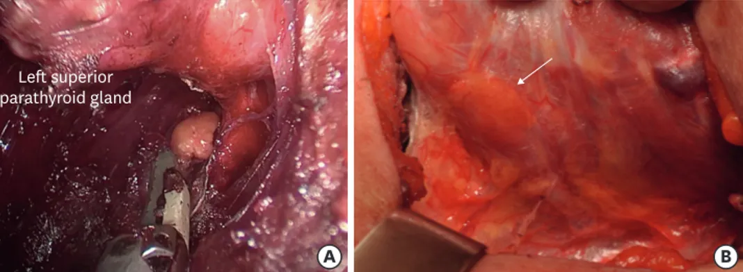
Left superior parathyroid gland
A B
Fig. 3. Identification and preservation of parathyroid gland. (A) Transoral endoscopic thyroidectomy vestibular approach. (B) Open conventional thyroidectomy.
It is the important principle that every parathyroid glands should be treated as if it were the last remaining one.
A recent study introduce the ‘TBP layer’ (thymus, blood vessel, inferior parathyroid gland layer)
concept for preserving the inferior parathyroid gland in situ during CND, and to evaluate its effectiveness (21). The study group included patients with primary papillary thyroid cancer who underwent total thyroidectomy with CND using the new surgical concept (21). The control group included sex- and age-matched patients. The proportion of inferior parathyroid glands preserved in situ and postoperative hypoparathyroidism rates in the 2 groups were compared. There were 181 patients in the study group and 306 in the control group (21). There were no significant differences between the groups in tumor size, multifocality, extrathyroidal extension, and number of harvested and metastatic central lymph nodes. The rate of inferior parathyroid gland preservation in situ was significantly improved from 37.9 to 76.3 per cent on the left side (P<0.001), and from 52.0 to 77.9 per cent on the right side (P<0.001), in the study group compared with the control group. The incidence of transient hypoparathyroidism decreased significantly from 35.0 to 7.2 per cent (P<0.001) (21).
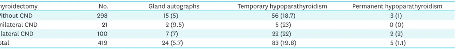
The view that gland autograph, parathyroid dysfunction significantly increases with the extent of the surgical intervention (subtotal resection vs. thyroidectomy, CND yes vs. no) is not confirmed from a review from our experience, summarized in Table 1.
INDOCYANINE GREEN (ICG)
It is worth nothing that identification of parathyroid glands does not equal safe preservation, as some studies demonstrated that it is not the number of parathyroid glands identified, but the number of parathyroid glands preserved in situ that matters. Therefore, a non-invasive objective and reliable way to localize parathyroid glands and assess their viability intra-operatively is warranted. In this aspect, modern technology such as the ICG as near-infrared fluorescent dye for real-time in situ parathyroid glands perfusion monitoring may have a potential role (Fig. 4). ICG angiography reliably predicts the vascularization of the parathyroid glands and obviates the need for postoperative measurement of Ca and PTH, and supplementation with Ca in patients with at least one well perfused parathyroid gland (22).
A randomized controlled trial was undertaken to determine whether intraoperative parathyroid gland angiography with ICG could predict postoperative hypoparathyroidism, and obviate the need for systematic blood tests and oral Ca supplementation.
Between September 2014 and February 2016, patients who had at least one well perfused parathyroid gland on ICG angiography were randomized to receive standard follow-up
Table 1. Frequency of parathyroid gland autographs, temporary and permanent hypothyroidism after thyroidectomy with and without CND, level 6 and 7 Thyroidectomy No. Gland autographs Temporary hypoparathyroidism Permanent hypoparathyroidism
Without CND 298 15 (5) 56 (18.7) 3 (1)
Unilateral CND 21 2 (9.5) 5 (23) 0 (0)
Bilateral CND 100 7 (7) 22 (22) 2 (2)
Total 419 24 (5.7) 83 (19.8) 5 (1.1)
Values are presented as number (%). No statistical significance differences are found when comparing between groups (P>0.05). CND = central neck dissection.
(measurement of Ca and PTH on postoperative day (POD) 1 and systematic supplementation with Ca and vitamin D; control group) or no supplementation and no blood test on POD 1 (intervention group) (22). In all patients, Ca and PTH levels were measured 10–15 days after thyroidectomy. The primary endpoint was hypocalcaemia on POD 10–15. A total of 196 patients underwent ICG angiography during thyroid surgery, of whom 146 had at least one well perfused parathyroid gland on ICG angiography and were randomized. None of these patients presented with hypoparathyroidism, including those who did not receive Ca supplementation (22). The intervention group was statistically non-inferior to the control group (exact 95% confidence interval [CI] of the difference in proportion of patients with hypocalcaemia −0.053 to 0.053; P=0.012). Eleven of the 50 excluded patients, in whom no well perfused parathyroid gland could be identified by angiography, presented with hypoparathyroidism on POD 1, and six on POD 10–15, which was significantly different from the findings in randomized patients (P=0.007) (22).
PARATHYROID HORMONE (PTH) AND CA LEVELS
Traditionally, sequential measurement of Ca levels is recommended after bilateral thyroid interventions, at least on the first 2 to 3 PODs (1). In contrast to the very good prediction of the parathyroid function on the basis of the iPTH level, the Ca as “success parameter” alone on the first POD hardly correlates with the prognosis regarding the development of postoperative hypoparathyroidism (1,2).
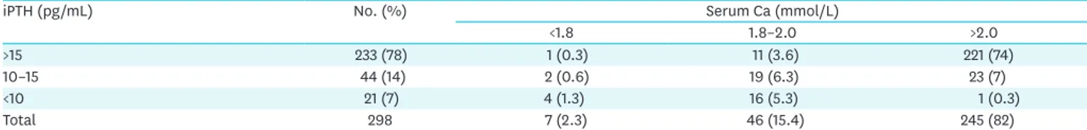
Table 2 summarize our experience on PTH and Ca levels determination on the 1st POD after 298 bilateral thyroidectomy. Ca levels in the first POD is not predictive, iPTH instead significantly anticipates parathyroid function (Table 2). Only the course of the Ca level on other days allows the reliable detection of parathyroid dysfunction, but is neither
Fig. 4. ICG as near-infrared fluorescent dye for real-time in situ parathyroid glands perfusion monitoring. ICG = indocyanine green.
Table 2. PTH and Ca levels on the 1st postoperative day after bilateral thyroidectomy
iPTH (pg/mL) No. (%) Serum Ca (mmol/L)
<1.8 1.8–2.0 >2.0
>15 233 (78) 1 (0.3) 11 (3.6) 221 (74)
10–15 44 (14) 2 (0.6) 19 (6.3) 23 (7)
<10 21 (7) 4 (1.3) 16 (5.3) 1 (0.3)
Total 298 7 (2.3) 46 (15.4) 245 (82)
Values are presented as number (%). Normal range: serum Ca: 2.0–2.6 mmol/L, iPTH: 15–65 pg/mL. Central lymph node dissection cases are excluded from the analysis.
comfortable for the patient (multiple blood samples) nor for the social cost (prolonged hospitalization) (1).
Intraoperative iPTH monitoring (IOPTH) with documentation of adequate decrease of elevated iPTH levels to predict (targeted) normocalcemia is established in primary hyperparathyroid surgery (23). IOPTH was also recommended as part of radical thyroid surgery to assess the function of the normal in situ remaining parathyroid glands (23). This method is costly because at least 3 iPTH determinations are required to document intraoperative iPTH history and additional medical technical assistance must be provided. Also, iPTH accessory meters are not always available, especially in smaller surgical departments hospitals (23).
Prospective randomized trial of prophylactic parathyroid autografting after thyroidectomy vs. iPTH levels have shown that patients with an iPTH level <10 pg/mL (and only on this condition) are subject to parathyroid autografting “by principle” compared to patients with “on-demand” parathyroid autotransplantation (subjective criteria) will benefit (but no for permanent hyperparathyreoidismus in this patient group) (24). However, an iPTH level cannot be used until 10–20 minutes after the end of the operation to decide whether parathyroid autotransplantation is necessary (24).
The iPTH level 4 hours postoperatively is the most meaningful (24-26). More detailed studies on the validity of iPTH measurements at the end of the operation compared to later measurements showed that iPTH levels are best predictable at 4 hours postoperatively and later in actual parathyroid function (25,26). Thus, IOPTH and the measurement immediately after the end of the operation have less practical significance than originally hoped (24-26).
Although rare, postoperative life-threatening rebleeding occurs within the first 24 hours postoperatively (27-29). Concepts with inpatient short-term stays take into account these experiences and recommend at least 24 hours stationary observation (28). Therefore, the best time for the postoperative examination of the parathyroid metabolism is the one-time measurement of the iPTH level on the morning of the first POD (2).
In a prospective “standard protocol” it could be shown that a once-only measured iPTH level 12–24 hours postoperatively, independent of the serum Ca level, can predict the postoperative parathyroid function with a sensitivity and specificity of 99% (2).
Short-term and long-term observations have shown that patients with an iPTH level ≥15 pg/ mL (normal value 15–65 pg/mL) have normal parathyroid metabolism, no Ca and/or vitamin D substitution need to be discharged and without further checks in home care (2).
CA AND VITAMIN D SUBSTITUTION
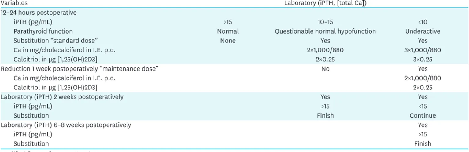
Table 3 describes Selberherr et al. (2,30) protocol for Ca/vitamin D substitution as a function of iPTH levels 12–24 hours postoperatively after bilateral thyroidectomy (with/without CND). Patients with an iPTH level <10 pg/mL can be discharged without symptoms with an oral “standard substitution” consisting of Ca carbonate and cholecalciferol, standard dose (Table 3).
The addition of calcitriol [1α, 25 (OH) 2-cholecalciferol = 1α, 25 (OH) 2 vitamin D3 or 1,25 (OH) 2D] improves intestinal Ca uptake (2).
According to the protocol, after 1 week the dosage is reduced by one third (“maintenance dose”) in order to prevent iatrogenic hypercalcaemia (2,30).
Ca or vitamin D doses must be closely monitored.
In a few patients, iPTH levels between 10 and 15 pg/mL are documented. The development of a normal or subfunction is not predictable. These patients may either be discharged with a “standard dosage” on the 1st POD or remain stationary for further laboratory control on the 2nd postoperative day. Parathyroid function has already been normalized in at least one third of patients (iPTH ≥15 pg/mL) (2,30). Discharge is possible without substitution, while patients with an iPTH in the “gray area” must be substituted according to the standard protocol (Table 3).
Depending on the 14-day postoperatively measured iPTH level, substitution is maintained at the maintenance dose (iPTH <15 pg/mL) or when normalized (iPTH ≥15 pg/mL) (Table 3).
CONCLUSION
Many thyroidectomies are performed by unexperienced surgeons. We believe that incidence of permanent hypoparathyroidism is underestimated. Endocrine surgeons are to perform functionally safe thyroidectomy and reduce the surgical complications related to parathyroid injury (30). And to do so, the best surgical tips for parathyroid preservation is to take time, keep trying, never give-up, and do the surgeon's best, despite the low success rate of parathyroid preservation in the beginning of thyroid surgery. Surgeons should try to identify and preserve both the inferior and superior parathyroid glands, preserve the vascular supply and venous dranage of the glands (31).
There is no formulaic technique for parathyroid preservation. However, to improve such surgical skill, the surgeons must learn about the actual anatomy of the parathyroid glands,
Table 3. Ca/vitamin D substitution as a function of iPTH levels 12–24 hours postoperatively and in the course of (almost total) thyroidectomy (with/without CND, level 6–7)
Variables Laboratory (iPTH, [total Ca])
12–24 hours postoperative
iPTH (pg/mL) >15 10–15 <10
Parathyroid function Normal Questionable normal hypofunction Underactive
Substitution “standard dose” None Yes Yes
Ca in mg/cholecalciferol in I.E. p.o. 2×1,000/880 3×1,000/880
Calcitriol in µg [1,25(OH)2D3] 2×0.25 3×0.25
Reduction 1 week postoperatively “maintenance dose” No Yes
Ca in mg/cholecalciferol in I.E. p.o. 2×1,000/880
Calcitriol in µg [1,25(OH)2D3] 2×0.25
Laboratory (iPTH) 2 weeks postoperatively Yes Yes
iPTH (pg/mL) >15 <15
Substitution Finish Continue
Laboratory (iPTH) 6–8 weeks postoperatively Yes
iPTH (pg/mL) >15
Substitution Finish
Modified from references 2 and 30.
focus on the micro-looking branches of parathyroid supplying vessels that originate from the main vessel trunk, and explore every possible location that might bear the parathyroid glands (31). To increase the chance of preserving the final vessel branches that supply the parathyroid, one must not ligate the vessels at their origin. Instead, approach from the distal end of the branches and forward proximally. The surgeon must clearly see the pulse of the peripheral vessel branches originating from either superior thyroidal artery or inferior thyroidal artery that supply the preserved parathyroid gland with no color change at the end of the procedure (31).
The presented management records the parathyroid gland function safely by an iPTH measurement 12–24 hours postoperatively. In contrast to IOPTH, the recommended procedure immediately and postoperatively considers unpredictable functional impairments of the parathyroid glands by (clinically irrelevant) hematomas and/or swellings. The one-off evaluation of postoperative parathyroid metabolism with an iPTH measurement is cost-effective at 17.60 EUR per determination (2). Inpatient short-term stay further reduces inpatient treatment costs (2,30).
REFERENCES
1. Asari R, Passler C, Kaczirek K, Scheuba C, Niederle B. Hypoparathyroidism after total thyroidectomy: a prospective study. Arch Surg 2008;143:132-7.
PUBMED | CROSSREF
2. Selberherr A, Scheuba C, Riss P, Niederle B. Postoperative hypoparathyroidism after thyroidectomy: efficient and cost-effective diagnosis and treatment. Surgery 2015;157:349-53.
PUBMED | CROSSREF
3. Pattou F, Combemale F, Fabre S, Carnaille B, Decoulx M, Wemeau JL, et al. Hypocalcemia following thyroid surgery: incidence and prediction of outcome. World J Surg 1998;22:718-24.
PUBMED | CROSSREF
4. Sitges-Serra A, Ruiz S, Girvent M, Manjón H, Dueñas JP, Sancho JJ. Outcome of protracted hypoparathyroidism after total thyroidectomy. Br J Surg 2010;97:1687-95.
PUBMED | CROSSREF
5. Bergenfelz A, Jansson S, Kristoffersson A, Mårtensson H, Reihnér E, Wallin G, et al. Complications to thyroid surgery: results as reported in a database from a multicenter audit comprising 3,660 patients. Langenbecks Arch Surg 2008;393:667-73.
PUBMED | CROSSREF
6. Veyseller B, Aksoy F, Yildirim YS, Karatas A, Ozturan O. Effect of recurrent laryngeal nerve identification technique in thyroidectomy on recurrent laryngeal nerve paralysis and hypoparathyroidism. Arch Otolaryngol Head Neck Surg 2011;137:897-900.
PUBMED | CROSSREF
7. Kara M, Tellioglu G, Krand O, Fersahoglu T, Berber I, Erdogdu E, et al. Predictors of hypocalcemia occurring after a total/near total thyroidectomy. Surg Today 2009;39:752-7.
PUBMED | CROSSREF
8. Shaha AR, Burnett C, Jaffe BM. Parathyroid autotransplantation during thyroid surgery. J Surg Oncol 1991;46:21-4.
PUBMED | CROSSREF
9. Reeve T, Thompson NW. Complications of thyroid surgery: how to avoid them, how to manage them, and observations on their possible effect on the whole patient. World J Surg 2000;24:971-5.
PUBMED | CROSSREF
10. Hallgrimsson P, Nordenström E, Bergenfelz A, Almquist M. Hypocalcaemia after total thyroidectomy for Graves' disease and for benign atoxic multinodular goitre. Langenbecks Arch Surg 2012;397:1133-7.
PUBMED | CROSSREF
11. Mohebati A, Shaha AR. Anatomy of thyroid and parathyroid glands and neurovascular relations. Clin Anat 2012;25:19-31.
12. Policeni BA, Smoker WR, Reede DL. Anatomy and embryology of the thyroid and parathyroid glands. Semin Ultrasound CT MR 2012;33:104-14.
PUBMED | CROSSREF
13. Testini M, Nacchiero M, Piccinni G, Portincasa P, Di Venere B, Lissidini G, et al. Total thyroidectomy is improved by loupe magnification. Microsurgery 2004;24:39-42.
PUBMED | CROSSREF
14. Dionigi G, Van Slycke S, Rausei S, Boni L, Dionigi R. Parathyroid function after open thyroidectomy: a prospective randomized study for ligasure precise versus harmonic FOCUS. Head Neck 2013;35:562-7.
PUBMED | CROSSREF
15. Saint Marc O, Cogliandolo A, Piquard A, Famà F, Pidoto RR. LigaSure vs clamp-and-tie technique to achieve hemostasis in total thyroidectomy for benign multinodular goiter: a prospective randomized study. Arch Surg 2007;142:150-6.
PUBMED | CROSSREF
16. Niederle B, Roka R, Brennan MF. The transplantation of parathyroid tissue in man: development, indications, technique, and results. Endocr Rev 1982;3:245-79.
PUBMED | CROSSREF
17. Zedenius J, Wadstrom C, Delbridge L. Routine autotransplantation of at least one parathyroid gland during total thyroidectomy may reduce permanent hypoparathyroidism to zero. Aust N Z J Surg 1999;69:794-7.
PUBMED | CROSSREF
18. Barczyński M, Konturek A, Stopa M, Cichoń S, Richter P, Nowak W. Total thyroidectomy for benign thyroid disease: is it really worthwhile? Ann Surg 2011;254:724-9.
PUBMED | CROSSREF
19. Erbil Y, Barbaros U, Salmaslioğlu A, Yanik BT, Bozbora A, Ozarmağan S. The advantage of near-total thyroidectomy to avoid postoperative hypoparathyroidism in benign multinodular goiter. Langenbecks Arch Surg 2006;391:567-73.
PUBMED | CROSSREF
20. Dralle H. Postoperative hypoparathyroidism: central neck dissection is a significant risk factor. Chirurg 2012;83:1082.
PUBMED | CROSSREF
21. Wang JB, Wu K, Shi LH, Sun YY, Li FB, Xie L. In situ preservation of the inferior parathyroid gland during central neck dissection for papillary thyroid carcinoma. Br J Surg 2017;104:1514-22.
PUBMED | CROSSREF
22. Vidal Fortuny J, Sadowski SM, Belfontali V, Guigard S, Poncet A, Ris F, et al. Randomized clinical trial of intraoperative parathyroid gland angiography with indocyanine green fluorescence predicting parathyroid function after thyroid surgery. Br J Surg 2018;105:350-57.
PUBMED | CROSSREF
23. Riss P, Kaczirek K, Heinz G, Bieglmayer C, Niederle B. A “defined baseline” in PTH monitoring increases surgical success in patients with multiple gland disease. Surgery 2007;142:398-404.
PUBMED | CROSSREF
24. Grodski S, Serpell J. Evidence for the role of perioperative PTH measurement after total thyroidectomy as a predictor of hypocalcemia. World J Surg 2008;32:1367-73.
PUBMED | CROSSREF
25. Barczyński M, Cichoń S, Konturek A, Cichoń W. Applicability of intraoperative parathyroid hormone assay during total thyroidectomy as a guide for the surgeon to selective parathyroid tissue autotransplantation. World J Surg 2008;32:822-8.
PUBMED | CROSSREF
26. Barczyński M, Cichoń S, Konturek A. Which criterion of intraoperative iPTH assay is the most accurate in prediction of true serum calcium levels after thyroid surgery? Langenbecks Arch Surg 2007;392:693-8.
PUBMED | CROSSREF
27. Raffaelli M, De Crea C, Carrozza C, D'Amato G, Zuppi C, Bellantone R, et al. Combining early postoperative parathyroid hormone and serum calcium levels allows for an efficacious selective post-thyroidectomy supplementation treatment. World J Surg 2012;36:1307-13.
PUBMED | CROSSREF
28. Doran HE, England J, Palazzo FBritish Association of Endocrine and Thyroid Surgeons. Questionable safety of thyroid surgery with same day discharge. Ann R Coll Surg Engl 2012;94:543-7.
PUBMED | CROSSREF
29. Doran HE, Palazzo F. Day-case thyroid surgery. Br J Surg 2012;99:741-3.
30. Selberherr A, Niederle B. Avoidance and management of hypoparathyroidism after thyroid gland surgery. Chirurg 2015;86:13-6.
PUBMED | CROSSREF
31. Sung TY, Lee Y, Yoon JH, Chung KW, Hong SJ. The best surgical tips for parathyroid preservation in thyroid surgery. VideoEndocrinology 2015;2.