Inpatient vs outpatient management and timing of delivery
of uncomplicated monochorionic monoamniotic twin
pregnancy: the MONOMONO study
The MONOMONO Working Group
#K E Y W O R D S: Cesarean delivery; chorionicity; cord accident; cord entanglement; healthcare; monochorionic; multiple
gestation; perinatal death; respiratory distress syndrome; twin pregnancy
ABSTRACT
Objectives Monoamniotic twin pregnancies are at increased risk of perinatal complications, primarily owing to the risk of cord entanglement. There is no recommen-dation on whether such pregnancies should be managed in hospital or can be safely managed in an outpatient setting, and the timing of planned delivery is also a sub-ject of debate. The aim of this study was to compare the perinatal outcomes of inpatient vs outpatient fetal surveil-lance approaches employed among 22 participating study centers, and to calculate the fetal and neonatal death rates according to gestational age, in non-anomalous monoam-niotic twins from 26 weeks’ gestation.
Methods The MONOMONO study was a multinational
cohort study of consecutive women with monochorionic monoamniotic twin pregnancies, who were referred to 22 university hospitals in Italy, the USA, the UK and Spain, from January 2010 to January 2017. Only non-anomalous uncomplicated monoamniotic twin pregnancies with two live fetuses at 26+ 0 weeks’
gestation were included in the study. In 10 of the centers, monoamniotic twins were managed routinely as inpatients, whereas in the other 12 centers they were managed routinely as outpatients. The primary outcome was intrauterine fetal death. We also planned to assess fetal and neonatal death rates according to gestational age per 1-week interval. Outcomes are presented as odds ratio (OR) with 95% CIs. The main outcome was analyzed using both standard logistic regression analysis, in which each fetus was treated as an independent unit, and a generalized mixed-model approach, with each twin pair treated as a cluster unit, considering that the outcome for a twin is not independent of that of its cotwin.
Correspondence to: Dr G. Saccone, Department of Neuroscience, Reproductive Sciences and Dentistry, School of Medicine, University of Naples Federico II, Naples, Italy (e-mail: [email protected])
#Participants of The MONOMONO Working Group are listed at end of article. Accepted: 3 July 2018
Results 195 consecutive pregnant women with a
non-anomalous uncomplicated monoamniotic twin gestation (390 fetuses) were included. Of these, 75 (38.5%) were managed as inpatients and 120 (61.5%) as outpatients. The overall perinatal loss rate was 10.8% (42/390) with a peak fetal death rate of 4.3% (15/348) occurring at 29 weeks’ gestation. There was no significant difference in mean gestational age at delivery (31 weeks), birth weight (∼1.6 kg), or emergency delivery rate between the inpatient and outpatient surveillance groups. Based on generalized mixed-model analysis, there was no statistically significant difference in fetal death rates between inpatient management commencing from around 26 weeks compared with outpatient surveillance protocols from 30 weeks (3.3% vs 10.8%; adjusted OR 0.21 (95% CI, 0.04–1.17)). Maternal length of stay in the hospital was 42.1 days in the inpatient group, and 7.4 days in the outpatient group (mean difference 34.70 days (95% CI, 31.36–38.04 days). From 32+ 0 to 36 + 6 weeks, no fetal or neonatal death in either group was recorded. 46 fetuses were delivered after 34+ 0 weeks, and none of them died
in utero or within the first 28 days postpartum.
Conclusion In uncomplicated monoamniotic twins,
inpa-tient surveillance is associated with similar fetal mortality as outpatient management. After 31+ 6 weeks, and up to 36+ 6 weeks, there were no intrauterine fetal deaths or neonatal deaths. Copyright© 2018 ISUOG. Published by John Wiley & Sons Ltd.
INTRODUCTION
Monochorionic monoamniotic twinning accounts for about 1–2% of monozygotic twin pregnancies worldwide1–6. Monoamniotic twins are at increased risk
diamniotic or dichorionic twin pregnancies3–5. Perinatal
mortality is reported to be high in monoamniotic twins, primarily owing to cord entanglement5. Early- and
mid-pregnancy loss before 26 weeks’ gestation seems to be correlated mainly with the incidence of twin reversed arterial perfusion (TRAP) sequence, conjoined twins and major congenital anomalies, the rate of these complica-tions being as high as 60% in studies evaluating high-risk referred populations5,7. Regarding pregnancy loss after
26 weeks, the largest review of monoamniotic twins, including 60 studies and 133 non-conjoined monoam-niotic twin pregnancies, reported a non-anomalous perinatal mortality rate of about 20%, with a sig-nificant rise in mortality after 32 weeks’ gestation4. These data are often used to justify planned preterm delivery from 32 weeks in otherwise uncomplicated monoamniotic twins5,8; however, robust data on which
a decision about the timing of delivery can be based are missing.
Inpatient management of monoamniotic twin pregnan-cies from viability until delivery has been reported5. No
recommendation has been made on whether these women should be managed in hospital or whether they can be managed safely in an outpatient setting, and The Ameri-can College of Obstetricians and Gynecologists (ACOG) concluded that ‘the optimal management of these patients remains uncertain’8.
The aim of this study was to compare the perina-tal outcomes of inpatient vs outpatient feperina-tal surveillance approaches employed in 22 participating study centers, and to calculate the fetal and neonatal death rate accord-ing to gestational age, in non-anomalous monoamniotic twins at≥ 26 weeks’ gestation.
SUBJECTS AND METHODS Study design and participants
This was a multinational, retrospective, cohort study of pregnant women with a monochorionic monoamniotic twin pregnancy who were referred to 22 university hospitals in Italy, the USA, the UK and Spain (Table 1) between January 2010 and January 2017. Clinical records were collected in a dedicated merged database. Only women with a confirmed diagnosis of monoamnionicity after delivery were included.
All reported variables were collected for all the subjects included in the study. Inclusion criteria were gestational age of at least 26+ 0 weeks with both fetuses alive, and confirmation of monoamnionicity at delivery and/or by pathologic examination of the placenta. Only uncom-plicated monoamniotic twin pregnancies were included. Exclusion criteria were pseudomonoamnionicity (iatro-genic creation of a single amniotic space because of an invasive procedure); conjoined twins; major fetal abnor-mality; intrauterine growth restriction (IUGR) or selective IUGR (i.e. one or both fetuses with ultrasound-estimated fetal weight < 10th centile); twin-to-twin transfusion
syndrome; TRAP sequence; acardiac twins; spontaneous
miscarriage before 26 weeks; and higher-order multiple pregnancies. Women who underwent selective reduc-tion were also excluded. Therefore, all women included in the study had a non-anomalous uncomplicated monoamniotic twin pregnancy with both fetuses alive at 26 weeks.
In 10 of the centers, all monoamniotic twins were managed routinely as inpatients, while in the other 12 centers all monoamniotic twins were managed routinely as outpatients (Table 1). In outpatient care, frequent follow-up was employed, with regular evaluation of fetal wellbeing by ultrasound assessment of fetal growth (fetal biometry of both twins and amniotic fluid volume assessment using deepest vertical pocket) every 3 weeks, ultrasound Doppler (umbilical artery Doppler and middle cerebral artery peak systolic velocity for both twins) every 2 weeks9, and non-stress tests (NST) usually once a week with either standard NST or computerized cardiotocography.
Women managed as inpatients were admitted from 24+ 0 to 29 + 0 weeks until delivery. Patient management in this group included NST two or three times a day, ultrasound assessment of fetal growth every 3 weeks and Doppler ultrasound every 2 weeks (Table 1)9. Continuous fetal heart rate monitoring was not performed in any of the participating centers.
All women in the inpatient group (study group) were delivered following one admission. For women included in the outpatient group (comparison group), who had one or more admissions, the total length of stay was calculated.
In both groups, planned Cesarean delivery was sched-uled usually at 32+ 0 to 34 + 6 weeks, according to local protocols and at the provider’s discretion (Table 1)8,10. Any indication for earlier delivery was recorded. Antena-tal corticosteroids for feAntena-tal lung maturation were offered before planned Cesarean delivery. Assessment of cervi-cal length by transvaginal ultrasound for the prevention of preterm birth was not performed routinely in either group, given the lack of treatment for twin pregnancies with a short cervix11–16.
Outcomes
Primary and secondary outcomes were compared between the inpatient and outpatient groups. The primary outcome was intrauterine fetal death (i.e. stillbirth) after 26 weeks’ gestation. Secondary outcomes were gestational age at delivery, total antenatal maternal length of stay (LOS) in the hospital, indication for delivery, birth weight, LOS in the neonatal intensive care unit (NICU) (days from admission to the NICU until discharge), neonatal death (i.e. death of a liveborn baby within the first 28 days postpartum) and perinatal death (i.e. either fetal or neonatal death).
We also planned to assess fetal and neonatal death rates according to gestational age at 1-week intervals; this secondary analysis was performed separately for the inpatient and outpatient groups.
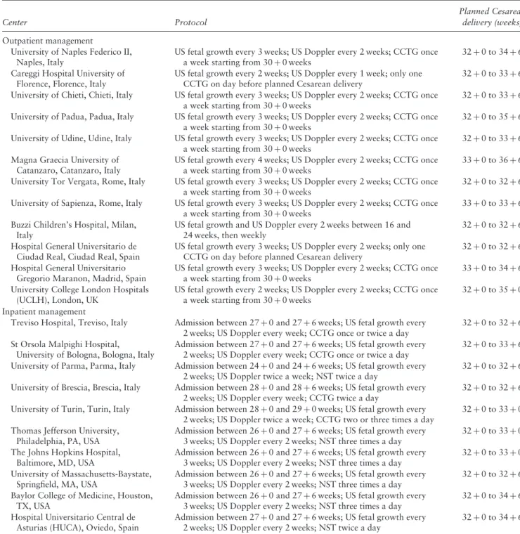
Table 1 Protocol followed in participating centers for inpatient or outpatient management of uncomplicated monochorionic monoamniotic
twin pregnancy with two live fetuses at 26 weeks’ gestation
Center Protocol
Planned Cesarean delivery (weeks)
Outpatient management
University of Naples Federico II, Naples, Italy
US fetal growth every 3 weeks; US Doppler every 2 weeks; CCTG once a week starting from 30+ 0 weeks
32+ 0 to 34 + 6 Careggi Hospital University of
Florence, Florence, Italy
US fetal growth every 2 weeks; US Doppler every 1 week; only one CCTG on day before planned Cesarean delivery
32+ 0 to 33 + 6 University of Chieti, Chieti, Italy US fetal growth every 3 weeks; US Doppler every 2 weeks; CCTG once
a week starting from 30+ 0 weeks
32+ 0 to 33 + 6 University of Padua, Padua, Italy US fetal growth every 3 weeks; US Doppler every 2 weeks; CCTG once
a week starting from 30+ 0 weeks
32+ 0 to 35 + 6 University of Udine, Udine, Italy US fetal growth every 3 weeks; US Doppler every 2 weeks; CCTG once
a week starting from 30+ 0 weeks
32+ 0 to 33 + 6 Magna Graecia University of
Catanzaro, Catanzaro, Italy
US fetal growth every 4 weeks; US Doppler every 2 weeks; CCTG once a week starting from 30+ 0 weeks
33+ 0 to 36 + 6 University Tor Vergata, Rome, Italy US fetal growth every 3 weeks; US Doppler every 2 weeks; CCTG once
a week starting from 30+ 0 weeks
32+ 0 to 32 + 6 University of Sapienza, Rome, Italy US fetal growth every 3 weeks; US Doppler every 2 weeks; CCTG once
a week starting from 30+ 0 weeks 33+ 0 to 33 + 6
Buzzi Children’s Hospital, Milan, Italy
US fetal growth and US Doppler every 2 weeks between 16 and 24 weeks, then weekly
32+ 0 to 32 + 6 Hospital General Universitario de
Ciudad Real, Ciudad Real, Spain
US fetal growth every 3 weeks; US Doppler every 2 weeks; only one CCTG on day before planned Cesarean delivery
32+ 0 to 32 + 6 Hospital General Universitario
Gregorio Maranon, Madrid, Spain
US fetal growth every 3 weeks; US Doppler every 2 weeks; CCTG once a week starting from 30+ 0 weeks
33+ 0 to 34 + 6 University College London Hospitals
(UCLH), London, UK
US fetal growth every 2 weeks; US Doppler every 2 weeks; CCTG once a week starting from 30+ 0 weeks
32+ 0 to 35 + 0 Inpatient management
Treviso Hospital, Treviso, Italy Admission between 27+ 0 and 27 + 6 weeks; US fetal growth every 2 weeks; US Doppler every week; CCTG once or twice a day
32+ 0 to 32 + 6 St Orsola Malpighi Hospital,
University of Bologna, Bologna, Italy
Admission between 27+ 0 and 27 + 6 weeks; US fetal growth every 2 weeks; US Doppler every week; CCTG once or twice a day
32+ 0 to 33 + 6 University of Parma, Parma, Italy Admission between 24+ 0 and 24 + 6 weeks; US fetal growth every
2 weeks; US Doppler twice a week; NST twice a day
32+ 0 to 32 + 6 University of Brescia, Brescia, Italy Admission between 28+ 0 and 28 + 6 weeks; US fetal growth every
2 weeks; US Doppler every week; CCTG twice a day
32+ 0 to 32 + 6 University of Turin, Turin, Italy Admission between 28+ 0 and 29 + 0 weeks; US fetal growth every
2 weeks; US Doppler twice a week; CCTG two or three times a day
32+ 0 to 33 + 0 Thomas Jefferson University,
Philadelphia, PA, USA
Admission between 26+ 0 and 27 + 6 weeks; US fetal growth every 3 weeks; US Doppler every 2 weeks; NST three times a day
32+ 0 to 33 + 0 The Johns Hopkins Hospital,
Baltimore, MD, USA
Admission between 26+ 0 and 27 + 6 weeks; US fetal growth every 3 weeks; US Doppler every 2 weeks; NST three times a day
32+ 0 to 33 + 0 University of Massachusetts-Baystate,
Springfield, MA, USA
Admission between 26+ 0 and 27 + 6 weeks; US fetal growth every 3 weeks; US Doppler every 2 weeks; NST three times a day
32+ 0 to 32 + 6 Baylor College of Medicine, Houston,
TX, USA
Admission between 26+ 0 and 27 + 6 weeks; US fetal growth every 3 weeks; US Doppler every 2 weeks; NST three times a day
32+ 0 to 34 + 6 Hospital Universitario Central de
Asturias (HUCA), Oviedo, Spain
Admission between 27+ 0 and 27 + 6 weeks; US fetal growth every 2 weeks; US Doppler every 2 weeks; NST twice a day
32+ 0 to 34 + 6
Ultrasound (US) assessment of fetal growth included fetal biometry of both twins and amniotic fluid volume assessment using deepest vertical pocket method. US Doppler assessment included umbilical artery Doppler and middle cerebral artery peak systolic velocity assessment, in both twins. CCTG, computerized cardiotocography; NST, non-stress test.
Statistical analysis
Statistical analysis was performed using the Statistical Package for Social Sciences (SPSS) v. 19.0 (IBM Inc., Armonk, NY, USA). Data are shown as mean± SD, median (range) or n (%). Univariate comparisons of dichotomous data were performed using the chi-square test with continuity correction. Comparisons between groups were performed using the t-test to test means
with SD by assuming equal within-group variance, and the Mann–Whitney U-test to test group medians with range. Primary and secondary outcomes are presented as odds ratios (ORs) with 95% CIs17. In addition to
standard logistic regression analysis, in which each fetus was treated as an independent unit, we used a gener-alized mixed-model approach in which each twin pair was a cluster unit. This model was used because the outcome of a twin is not independent of that of its
cotwin. Two-sided P-values were calculated, and P < 0.05 was considered to indicate statistical significance18. The study was reported following the STROBE guidelines19.
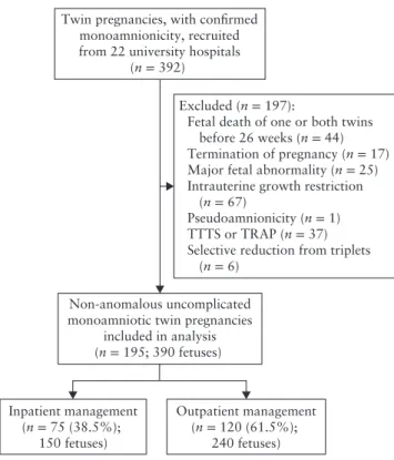
RESULTS
In total, 195 consecutive women with a non-anomalous uncomplicated monoamniotic twin pregnancy (390 fetuses) and both fetuses alive at 26 weeks, were included in the study (Figure 1). Of these, 75 (38.5%) women were managed as inpatients and 120 (61.5%) as outpatients. Inpatient and outpatient management policies were highly variable between the included centers. Inpatient monitor-ing usually started at about 26 weeks’ gestation, whereas in the outpatient group monitoring was usually instituted after 30 weeks (Table 1). Demographic characteristics were similar between the two groups (Table 2). Mean maternal age was about 30 years in both groups. One woman in the inpatient group and one in the outpatient group had a history of stillbirth in a prior pregnancy.
Primary analysis
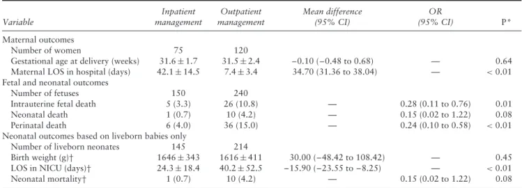
Based on standard logistic regression analysis, non-anomalous uncomplicated monoamniotic twin pregnan-cies managed as inpatients from 26 weeks to delivery had a significantly lower rate of intrauterine fetal death (3.3%
vs 10.8%; OR, 0.28 (95% CI, 0.11–0.76)) and perinatal
death (4.0% vs 15.0%; OR, 0.24 (95% CI, 0.10–0.58)), and shorter length of NICU stay by approximately 16 days (mean difference (MD) –15.90 days (95% CI, –23.6 to –8.25 days)), compared with pregnancies managed as out-patient. Mean maternal LOS in the hospital was 42.1 days in the inpatient group and 7.4 days in the outpatient group (MD, 34.70 days (95% CI, 31.36–38.04 days)) (Table 3).
However, based on generalized mixed-model ana-lysis considering each twin pair as a cluster unit, non-anomalous uncomplicated monoamniotic twin preg-nancies managed as inpatient had a similar rate of intrauterine fetal death as did those managed as out-patient (raw rates: 3.3% vs 10.8%; adjusted OR, 0.21 (95% CI, 0.04–1.17); Table 4).
Indications for delivery are shown in Table 5. 70.7% of the women in the inpatient group and 68.3% of the women in the outpatient group delivered via scheduled Cesarean section on the planned date.
Secondary analysis
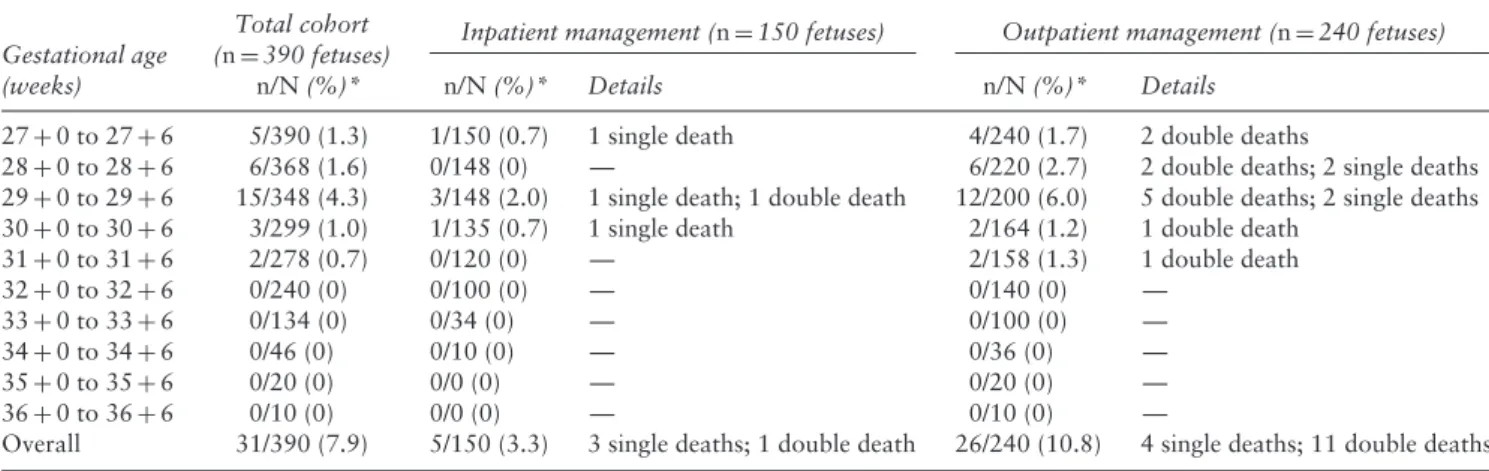
The overall fetal, neonatal and perinatal death rates in our cohort were 7.9% (31/390), 2.8% (11/390) and 10.8% (42/390), respectively. Four (5.3%) women in the inpatient group and 15 (12.5%) in the outpatient group experienced intrauterine fetal death. Details of fetal, neonatal and perinatal deaths according to gestational age at delivery are shown in Tables 6, S1 and S2, respectively. The intrauterine fetal death rate per 1-week gestational-age interval ranged from 0% to 4.3%. The highest
Twin pregnancies, with confirmed monoamnionicity, recruited from 22 university hospitals
(n = 392)
Non-anomalous uncomplicated monoamniotic twin pregnancies
included in analysis (n = 195; 390 fetuses) Inpatient management (n = 75 (38.5%); 150 fetuses) Outpatient management (n = 120 (61.5%); 240 fetuses) Excluded (n = 197):
Fetal death of one or both twins before 26 weeks (n = 44) Termination of pregnancy (n = 17) Major fetal abnormality (n = 25) Intrauterine growth restriction
(n = 67)
Pseudoamnionicity (n = 1) TTTS or TRAP (n = 37) Selective reduction from triplets
(n = 6)
Figure 1 Flowchart showing inclusion in study of uncomplicated
monochorionic monoamniotic twin pregnancies with two live fetuses at 26 weeks’ gestation. TRAP, twin reversed arterial perfusion; TTTS, twin–twin transfusion syndrome.
Table 2 Baseline characteristics of 195 women with uncomplicated
monochorionic monoamniotic twin pregnancy with two live fetuses at 26 weeks’ gestation, according to whether they were managed as inpatients or outpatients Characteristic Inpatient management (n= 75) Outpatient management (n= 120) P Age (years) 28.8± 5.5 30.0± 4.3 0.12
Assisted reproductive technology 5 (6.7) 10 (8.3) 0.88 Body mass index (kg/m2) 25.5± 5.3 26.2± 5.5 0.40 Smoker during pregnancy 9 (12.0) 11 (9.2) 0.71
Ethnicity 0.12 White 59 (78.7) 107 (89.2) African-American 6 (8.0) 6 (5.0) Other* 10 (13.3) 7 (5.8) Gravidity 2 (1–8) 2 (1–7) 0.54 Parity 1 (0–4) 1.5 (0–4) 0.77 Prior stillbirth 1 (1.3) 1 (0.8) 0.99
Data are presented as mean± SD, n (%) or median (range). *Includes Asian and Hispanic.
weekly intrauterine fetal death rate was observed between 29+ 0 and 29 + 6 weeks, in both the inpa-tient group (rate, 2.0%) and the outpainpa-tient group (rate, 6.0%).
From 32+ 0 to 36 + 6 weeks, no fetal or neonatal death occurred in either group (Tables 6 and S1), with 0/46 perinatal deaths between 34+ 0 and 34 + 6 weeks, 0/20 between 35+ 0 and 35 + 6 weeks and 0/10 between 36 + 0 and 36+ 6 weeks (Table S2).
Table 3 Maternal, fetal and neonatal outcomes of 195 uncomplicated monochorionic monoamniotic twin pregnancies with two live fetuses
at 26 weeks’ gestation, according to whether they were managed as inpatients or outpatients
Variable Inpatient management Outpatient management Mean difference (95% CI) OR (95% CI) P* Maternal outcomes Number of women 75 120
Gestational age at delivery (weeks) 31.6± 1.7 31.5± 2.4 –0.10 (–0.48 to 0.68) — 0.64 Maternal LOS in hospital (days) 42.1± 14.5 7.4± 3.4 34.70 (31.36 to 38.04) — <0.01 Fetal and neonatal outcomes
Number of fetuses 150 240
Intrauterine fetal death 5 (3.3) 26 (10.8) — 0.28 (0.11 to 0.76) 0.01
Neonatal death 1 (0.7) 10 (4.2) — 0.15 (0.02 to 1.22) 0.08
Perinatal death 6 (4.0) 36 (15.0) — 0.24 (0.10 to 0.58) <0.01
Neonatal outcomes based on liveborn babies only
Number of liveborn neonates 145 214
Birth weight (g)† 1646± 343 1616± 411 30.00 (–48.42 to 108.42) — 0.45
LOS in NICU (days)† 24.3± 18.4 40.2± 52.5 –15.90 (–23.55 to –8.25) — <0.01
Neonatal mortality† 1 (0.7) 10 (4.2) — 0.15 (0.02 to 1.22) 0.08
Data are presented as n, mean± SD or n (%). *Standard logistic regression analysis, in which each fetus was treated as an independent unit. †Outcomes based on liveborn babies only. LOS, length of stay; NICU, neonatal intensive care unit; OR, odds ratio.
Table 4 Potential predictors of intrauterine fetal death in
uncomplicated monochorionic monoamniotic twin pregnancies with two live fetuses at 26 weeks’ gestation
Predictor OR (95% CI) P*
Inpatient vs outpatient management 0.21 (0.04–1.17) 0.07 Gestational age, 1-week increase 0.55 (0.36–0.89) 0.006 Birth-weight discordance, 1% increase 1.03 (0.93–1.14) 0.6 *Generalized mixed-model analysis, treating twin pair as cluster unit. OR, odds ratio.
DISCUSSION Main findings
In addition to the general risks of monochorionicity and twin pregnancy, monoamniotic twins are at greatly increased risk of neonatal death owing to umbilical cord entanglement. In the past, these risks have been associated with a loss rate as high as 70%5,13–20. In this cohort of
390 uncomplicated monoamniotic fetuses, the overall rate of perinatal death was 10.8%.
Based on standard logistic regression analysis, in which each fetus was treated as an independent unit, the rate of intrauterine fetal death was significantly lower in the women managed as inpatients than in those managed as outpatients. We also observed an improvement in neonatal outcomes, with inpatient management associated with a shorter LOS in the NICU. However, using generalized mixed-model analysis, with each twin pair treated as a cluster unit, the rate of intrauterine fetal death was similar between inpatient- and outpatient-management groups. Therefore, our study shows that when treating each fetal death as an independent event, there appears to be a statistically significant difference between inpatient and outpatient groups. However, because second twins have an increased risk of fetal death after the death of a first twin, this finding
did not reach statistical difference when twin pairs were analyzed as a cluster unit.
Inpatient monitoring usually started at about 26 weeks’ gestation, whereas monitoring was delayed until 30 weeks in the outpatient group. Our study showed that the difference in fetal mortality between the inpatient and the outpatient groups mainly occurred in the 26–30-week window, and that once monitoring has been instituted (be it in the inpatient or in the outpatient group) survival of monoamniotic twins is excellent, and outcomes of inpatient and outpatient groups are similar. Therefore, this study shows clearly that close monitoring is needed to achieve good outcomes in monoamniotic pregnancies, regardless of the surveillance setting.
An important finding of this study is the markedly improved perinatal survival compared with that reported in older literature. This could be explained by improve-ments in the diagnosis and treatment of monoamniotic twin pregnancies but also by the fact that most losses in these pregnancies are attributable to fetal abnormal-ities and spontaneous early miscarriage5, which were
excluded from our study. Therefore, these data may truly represent the natural history of non-anomalous uncom-plicated monoamniotic twins once viability has been reached.
Our secondary analysis also showed that fetal death in non-anomalous uncomplicated monoamniotic twins occurred up to 31+ 6 weeks. Indeed, the major important and novel finding of the MONOMONO study was the lack of ‘late-gestational-age’ deaths, suggesting that putting back the planned timing of delivery from 32 to 34+ 6 weeks may be a safe approach for pregnant women with an uncomplicated monoamniotic twin pregnancy. However, while no deaths occurred after 34 weeks, only 23 pregnancies continued beyond this gestation, hence the study was underpowered to draw conclusions on the optimal timing of delivery to avoid stillbirth.
Table 5 Indication for delivery by Cesarean section in 195 uncomplicated monochorionic monoamniotic twin pregnancies with two live
fetuses at 26 weeks’ gestation, according to whether they were managed as inpatients or outpatients
Indication for delivery
Inpatient management (n= 75) Outpatient management (n= 120) OR (95% CI) P Maternal 1 (1.3)* 6 (5.0)† 0.26 (0.03–2.18) 0.21 Fetal 11 (14.7) 8 (6.7) 2.41 (0.92–6.29) 0.07 Planned delivery 53 (70.7) 82 (68.3) 1.12 (0.60–2.09) 0.73
Spontaneous onset of labor before planned delivery 10 (13.3) 24 (20.0) 0.62 (0.28–1.37) 0.24 Data are presented as n (%). *One case of pre-eclampsia. †Four cases of pre-eclampsia; two cases of placental abruption. OR, odds ratio.
Table 6 Incidence of intrauterine fetal death in 195 uncomplicated monochorionic monoamniotic twin pregnancies (n= 390 fetuses) with
two live fetuses at 26 weeks’ gestation, according to whether they were managed as inpatients or outpatients, by 1-week gestational-age intervals
Inpatient management (n= 150 fetuses) Outpatient management (n= 240 fetuses) Gestational age
(weeks)
Total cohort (n= 390 fetuses)
n/N (%)* n/N (%)* Details n/N (%)* Details
27+ 0 to 27 + 6 5/390 (1.3) 1/150 (0.7) 1 single death 4/240 (1.7) 2 double deaths
28+ 0 to 28 + 6 6/368 (1.6) 0/148 (0) — 6/220 (2.7) 2 double deaths; 2 single deaths
29+ 0 to 29 + 6 15/348 (4.3) 3/148 (2.0) 1 single death; 1 double death 12/200 (6.0) 5 double deaths; 2 single deaths 30+ 0 to 30 + 6 3/299 (1.0) 1/135 (0.7) 1 single death 2/164 (1.2) 1 double death
31+ 0 to 31 + 6 2/278 (0.7) 0/120 (0) — 2/158 (1.3) 1 double death 32+ 0 to 32 + 6 0/240 (0) 0/100 (0) — 0/140 (0) — 33+ 0 to 33 + 6 0/134 (0) 0/34 (0) — 0/100 (0) — 34+ 0 to 34 + 6 0/46 (0) 0/10 (0) — 0/36 (0) — 35+ 0 to 35 + 6 0/20 (0) 0/0 (0) — 0/20 (0) — 36+ 0 to 36 + 6 0/10 (0) 0/0 (0) — 0/10 (0) —
Overall 31/390 (7.9) 5/150 (3.3) 3 single deaths; 1 double death 26/240 (10.8) 4 single deaths; 11 double deaths *Denominator is total number of live fetuses at that gestational age, i.e. excluding fetal deaths and delivered babies. OR, odds ratio.
The most important limitation of our study was the retrospective non-randomized approach. Owing to the retrospective nature of the study, it was not possible to separate the importance of hospitalization vs the increased frequency of testing per se. Because this was not a randomized comparison, the findings were subject to bias. Moreover, since continuous fetal heart rate monitoring was not performed in any of the institutions following inpatient management, it was not possible to assess whether such a monitoring approach could further decrease the rate of fetal death. Inpatient and outpatient management were highly dissimilar between the included centers (Table 1), and therefore variations in management among the different institutions could have influenced our findings. Data on patient satisfaction, neonatal outcomes and economic implications were not available.
Comparison with the literature and implications
Several small studies evaluating perinatal outcomes in monoamniotic twins have been published (Table S3)7,20–26. In a retrospective study, Heyborne et al.20
assessed the effectiveness of inpatient monitoring of monoamniotic twins, and observed improved neonatal survival among women who were admitted electively for inpatient monitoring. On the other hand, Van Mieghem et al.22concluded that if close fetal surveillance
is instituted after 26–28 weeks and delivery takes place
at approximately 32–34 weeks, the risk of perinatal complications is low, regardless of the surveillance setting. Fetal demise is a major concern as a monoamniotic twin pregnancy approaches term, and early delivery would prevent this occurrence. It is indeed common for monoamniotic twins to be delivered preterm with planned Cesarean section at about 32 weeks’ gestation5,8. In 2016,
the Royal College of Obstetricians and Gynaecologists, ACOG and the Society for Maternal Fetal Medicine recommended that monoamniotic twin pregnancies should be delivered by Cesarean section between 32 and 33 weeks because of the high risk of intrauterine fetal death8,27. These recommendations are based on
studies demonstrating that the perinatal mortality rate roughly doubles beyond 34 weeks (7%) compared with that at 33 weeks (4%)4. However, the justification for
preterm delivery should be balanced against the likelihood of respiratory distress syndrome (5%) at 32 weeks1,7,
despite the use of antenatal steroids28–30 in otherwise
uncomplicated pregnancies1,3,4,8. This balance may not
be achieved if fetal loss in uncomplicated monoamniotic twins is low1,4. Our study showed no fetal or neonatal
death between 31+ 6 weeks and 36 + 6 weeks.
Conclusions
In uncomplicated monoamniotic twins, inpatient surveil-lance is associated with similar fetal mortality to that
of outpatient management. As the raw rates of fetal mor-tality were 3.3% in the inpatient group and 10.8% in the outpatient group, further research is necessary.
Our data also suggest that, in non-anomalous uncom-plicated monoamniotic twins, the fetal and neonatal death rates do not increase after 32+ 0 weeks, therefore planned Cesarean delivery at 33+ 0 to 34 + 6 weeks is a reasonable strategy to discuss with the patient. Data beyond 34 weeks are too limited to make a recommendation. Owing to the retrospective nature of this study, caution should be exercised before changes in practice are employed. A ran-domized controlled trial would provide the best evidence on the preferred method of monitoring for monoamniotic twins, however, this would be logistically difficult given the rarity of such pregnancies.
The MONOMONO Working Group
Gabriele Saccone*, Department of Neuroscience, Repro-ductive Sciences and Dentistry, School of Medicine, University of Naples Federico II, Naples, Italy
Vincenzo Berghella, Division of Maternal-Fetal Medicine, Department of Obstetrics and Gynecology, Sidney Kimmel Medical College of Thomas Jefferson Univer-sity, Philadelphia, PA, USA
Mariavittoria Locci, Department of Neuroscience, Repro-ductive Sciences and Dentistry, School of Medicine, University of Naples Federico II, Naples, Italy
Tullio Ghi, Department of Obstetrics and Gynecology, University of Parma, Parma, Italy
Tiziana Frusca, Department of Obstetrics and Gynecol-ogy, University of Parma, Parma, Italy
Mariano Lanna, Fetal therapy Unit ‘U Nicolini’, Buzzi Children’s Hospital University of Milan, Milan, Italy Stefano Faiola, Fetal therapy Unit ‘U Nicolini’, Buzzi
Children’s Hospital University of Milan, Milan, Italy Anna Fichera, Department of Obstetrics and Gynecology,
University of Brescia, Brescia, Italy
Federico Prefumo, Department of Obstetrics and Gyne-cology, University of Brescia, Brescia, Italy
Giuseppe Rizzo, University of Roma Tor Vergata, Division of Maternal Fetal Medicine, Ospedale Cristo Re Roma, Rome, Italy
Costanza Bosi, University of Roma Tor Vergata, Division of Maternal Fetal Medicine, Ospedale Cristo Re Roma, Rome, Italy
Bruno Arduino, Department of Neuroscience, Reproduc-tive Sciences and Dentistry, School of Medicine, University of Naples Federico II, Naples, Italy
Pietro D’Alessandro, Department of Neuroscience, Reproductive Sciences and Dentistry, School of Medicine, University of Naples Federico II, Naples, Italy
Maria Borgo, Department of Neuroscience, Reproductive Sciences and Dentistry, School of Medicine, University of Naples Federico II, Naples, Italy
Silvana Arduino, Department of Obstetrics and Gynecol-ogy, 2nd University of Turin, AO Town of Health and
Science, Turin, Italy
Elisabetta Cantanna, Department of Obstetrics and Gynecology, 2nd University of Turin, AO Town of Health and Science, Turin, Italy
Giuliana Simonazzi, Department of Medical Surgical Sciences, Division of Obstetrics and Prenatal Medicine, St Orsola Malpighi Hospital, University of Bologna, Bologna, Italy
Nicola Rizzo, Department of Medical Surgical Sciences, Division of Obstetrics and Prenatal Medicine, St Orsola Malpighi Hospital, University of Bologna, Bologna, Italy
Giorgetta Francesca, Department of Medical Surgical Sciences, Division of Obstetrics and Prenatal Medicine, St Orsola Malpighi Hospital, University of Bologna, Bologna, Italy
Viola Seravalli, Department of Health Science, Division of Pediatrics, Obstetrics and Gynecology Careggi Hospital University of Florence, Florence, Italy; Johns Hopkins Center for Fetal Therapy, Department of Gynecology & Obstetrics, Johns Hopkins University School of Medicine, Baltimore, MD, USA
Jena L. Miller, Johns Hopkins Center for Fetal Therapy, Department of Gynecology & Obstetrics, Johns Hopkins University School of Medicine, Baltimore, MD, USA
Elena Rita Magro-Malosso, Department of Health Science, Division of Pediatrics, Obstetrics and Gyne-cology Careggi Hospital University of Florence, Flo-rence, Italy
Mariarosaria Di Tommaso, Department of Health Science, Division of Pediatrics, Obstetrics and Gynecology Careggi Hospital University of Florence, Florence, Italy
Andrea Dall’Asta, Department of Obstetrics and Gyne-cology, University of Parma, Parma, Italy
Letizia Galli, Department of Obstetrics and Gynecology, University of Parma, Parma, Italy
Nicola Volpe, Department of Obstetrics and Gynecology, University of Parma, Parma, Italy
Silvia Visentin, Department of Woman’s and Child’s Health, University of Padua, Padua, Italy
Erich Cosmi, Department of Woman’s and Child’s Health, University of Padua, Padua, Italy
Laura Sarno, Department of Neuroscience, Reproductive Sciences and Dentistry, School of Medicine, University of Naples Federico II, Naples, Italy
Claudia Caissutti, Department of Experimental Clinical and Medical Science, DISM, Clinic of Obstetrics and Gynecology, University of Udine, Udine, Italy
Lorenza Driul, Department of Experimental Clinical and Medical Science, DISM, Clinic of Obstetrics and Gynecology, University of Udine, Udine, Italy
Hannah Anastasio, Division of Maternal-Fetal Medicine, Department of Obstetrics and Gynecology, Sidney Kimmel Medical College of Thomas Jefferson Univer-sity, Philadelphia, PA, USA
Daniele Di Mascio, Department of Gynecological, Obstetrical and Urological Sciences, Sapienza Univer-sity of Rome, Rome, Italy
Pierluigi Benedetti Panici, Department of Gynecological, Obstetrical and Urological Sciences, Sapienza University of Rome, Rome, Italy
Flaminia Vena, Department of Gynecological, Obstetrical and Urological Sciences, Sapienza University of Rome, Rome, Italy
Roberto Brunelli, Department of Gynecological, Obstetri-cal and UrologiObstetri-cal Sciences, Sapienza University of Rome, Rome, Italy
Andrea Ciardulli, Department of Obstetrics and Gynecol-ogy, Catholic University of Sacred Heart, Rome, Italy Francesco D’Antonio, Department of Obstetrics and
Gynecology, University Hospital of Northern Norway, Tromsø, Norway; Department of Obstetrics and Gynecology, University of Chieti, Chieti, Italy
Corina Schoen, Division of Maternal-Fetal Medicine, Department of Obstetrics and Gynecology, University of Massachusetts-Baystate, Springfield, MA, USA Anju Suhag, Division of Maternal-Fetal Medicine,
Department of Obstetrics and Gynecology, Baylor College of Medicine, Houston, TX, USA
Zita Maria Gambacorti-Passerini, Department of Obstet-rics and Gynecology, Hospital General Universitario de Ciudad Real, Ciudad Real, Spain
Maria Angeles Anaya Baz, Department of Obstetrics and Gynecology, Hospital General Universitario de Ciudad Real, Ciudad Real, Spain
Giulia Magoga, Department of Obstetrics and Gynecol-ogy, Treviso, Italy
Enrico Busato, Department of Obstetrics and Gynecology, Santa Maria di Ca’ Foncello Hospital, Treviso, Italy Elisa Filippi, Department of Obstetrics and Gynecology,
Santa Maria di Ca’ Foncello Hospital, Treviso, Italy Mar´ıa Jos´e Rodriguez Su ´arez, Department of Obstetrics
and Gynecology, Hospital Universitario Central de Asturias (HUCA), Oviedo, Spain
Francisco Gamez Alderete, Department of Obstetrics and Gynecology, Hospital General Universitario Gre-gorio Maranon, Madrid, Spain
Paula Alonso Ortuno, Department of Obstetrics and Gynecology, Hospital General Universitario Gregorio Maranon, Madrid, Spain
Amerigo Vitagliano, Department of Woman’s and Child’s Health, University of Padua, Padua, Italy
Antonio Mollo, Department of Neuroscience, Reproduc-tive Sciences and Dentistry, School of Medicine, University of Naples Federico II, Naples, Italy
Antonio Raffone, Department of Neuroscience, Repro-ductive Sciences and Dentistry, School of Medicine, University of Naples Federico II, Naples, Italy
Marianne Vendola, University College of London (UCLH), London, UK
Preethi Navaneethan, University College of London (UCLH), London, UK
Ruwan Wimalasundera, University College of London (UCLH), London, UK
Raffaele Napolitano, University College of London (UCLH), London, UK
Carmen Imma Aquino, School of Medicine, University of Salerno, Salerno, Italy
Serena D’Agostino, Department of Obstetrics and Gynaecology, School of Medicine, Magna Graecia University of Catanzaro, Catanzaro, Italy
Cinzia Gallo, Department of Obstetrics and Gynaecology, School of Medicine, Magna Graecia University of Catanzaro, Catanzaro, Italy
Giuseppe Maria Maruotti, Department of Neuroscience, Reproductive Sciences and Dentistry, School of Medicine, University of Naples Federico II, Naples,
Italy
Maria Elena Flacco, University of Ferrara, Ferrara, Italy Ahmet A. Baschat, Johns Hopkins Center for Fetal
Therapy, Department of Gynecology & Obstetrics, Johns Hopkins University School of Medicine, Balti-more, MD, USA
Roberta Venturella, Department of Obstetrics and Gynaecology, School of Medicine, Magna Graecia University of Catanzaro, Catanzaro, Italy
Maurizio Guida, School of Medicine, University of Salerno, Salerno, Italy
Pasquale Martinelli, Department of Obstetrics and Gynaecology, School of Medicine, Magna Graecia University of Catanzaro, Catanzaro, Italy
Fulvio Zullo, Department of Neuroscience, Reproductive Sciences and Dentistry, School of Medicine, University of Naples Federico II, Naples, Italy
*First author of this work.
ACKNOWLEDGMENT
We thank Lamberto Manzoli, Full Professor at Local Health Unit, University of Pescara, Pescara, Italy, for providing assistance with the statistical analysis.
REFERENCES
1. Cordero L, Franco A, Joy SD. Monochorionic monoamniotic twins: neonatal outcome. J Perinatol, 2006; 26: 170–175.
2. Hall JG. Twinning. Lancet 2003; 362: 735–743.
3. Ishii K. Prenatal diagnosis and management of monoamniotic twins. Curr Opin
Obstet Gynecol 2015; 27: 159–164.
4. Roqu´e H, Gillen-Goldstein J, Funai E, Young BK, Lockwood CJ. Perinatal outcomes in monoamniotic gestations. J Matern Fetal Neonatal Med 2003; 13: 414–421. 5. Dias T, Thilaganathan B, Bhide A. Monoamniotic twin pregnancy. Obstet Gynaecol
2012; 14: 71–78.
6. Maruotti GM, Saccone G, Morlando M, Martinelli P. First-trimester ultrasound determination of chorionicity in twin gestations using the lambda sign: a systematic review and meta-analysis. Eur J Obstet Gynecol Reprod Biol 2016; 202: 66–70. 7. Ezra Y, Shveik D, Ophir E, Nadjari M, Eisenberg VH, Samueloff A,
Rojansky N. Intensive management and early delivery reduce antenatal mortality in mono-amniotic twin pregnancy. Acta Obstet Gynecol Scand 2005; 84: 432–435. 8. The American College of Obstetricians and Gynecologists and Society for Maternal
Fetal Medicine. Practice Bulletin Number 169: Multifetal Gestations: Twin, Triplet, and Higher-Order Multifetal Pregnancies. Obstet Gynecol 2016; 128: e131–146. 9. Khalil A, Rodgers M, Baschat A, Bhide A, Gratacos E, Hecher K, Kilby MD, Lewi L,
Nicolaides KH, Oepkes D, Raine-Fenning N, Reed K, Salomon LJ, Sotiriadis A, Thilaganathan B, Ville Y. ISUOG Practice Guidelines: role of ultrasound in twin pregnancy. Ultrasound Obstet Gynecol 2016; 47: 247–263.
10. Saccone G, Berghella V. Planned delivery at 37 weeks in twins: a systematic review and meta-analysis of randomized controlled trials. J Matern Fetal Neonatal Med 2016; 29: 685–689.
11. Berghella V, Saccone G. Twins with short cervix: hope ahead. BJOG 2017; 124: 1174.
12. Saccone G, Rust O, Althuisius S, Roman A, Berghella V. Cerclage for short cervix in twin pregnancies: systematic review and meta-analysis of randomized trials using individual patient-level data. Acta Obstet Gynecol Scand 2015; 94: 352–358.
13. Roman A, Rochelson B, Martinelli P, Saccone G, Harris K, Zork N, Spiel M, O’Brien K, Calluzzo I, Palomares K, Rosen T, Berghella V, Fleischer A. Cerclage in twin pregnancy with dilated cervix between 16 to 24 weeks of gestation: retrospective cohort study. Am J Obstet Gynecol 2016; 215: 98.e1–11.
14. Roman A, Rochelson B, Fox NS, Hoffman M, Berghella V, Patel V, Calluzzo I, Saccone G, Fleischer A. Efficacy of ultrasound-indicated cerclage in twin pregnancies.
Am J Obstet Gynecol 2015; 212: 788.e1–6.
15. Society for Maternal–Fetal Medicine (SMFM). Electronic address: [email protected], McIntosh J, Feltovich H, Berghella V, Manuck T. The role of routine cervical length screening in selected high- and low-risk women for preterm birth prevention. Am
J Obstet Gynecol 2016; 215: B2–B7.
16. Gordon MC, McKenna DS, Stewart TL, Howard BC, Foster KF, Higby K, Cypher RL, Barth WH. Transvaginal cervical length scans to prevent prematurity in twins: a randomized controlled trial. Am J Obstet Gynecol 2016; 214: 277.e1–7. 17. Smith AH, Bates MN. Confidence limit analyses should replace power
calcu-lation in the interpretation of epidemiologic studies. Epidemiology 1992; 3: 449–452.
18. McNamee R. Regression modelling and other methods to control confounding.
Occup Environ Med 2005; 62: 500–506.
19. Von Elm E, Altman DG, Egger M, Pocock SJ, Gøtzsche PC, Vandenbroucke JP; STROBE Initiative. The Strengthening the Reporting of Observational Studies in Epidemiology (STROBE) statement: guidelines for reporting observational studies.
Lancet 2007; 370: 1453–1457.
20. Heyborne KD, Porreco RP, Garite TJ, Phair K, Abril D; Obstetrix/Pediatrix Research Study Group. Improved perinatal survival of monoamniotic twins with intensive inpatient monitoring. Am J Obstet Gynecol 2005; 192: 96–101.
21. DeFalco LM, Sciscione AC, Megerian G, Tolosa J, Macones G, O’Shea A, Pollock MA. Inpatient versus outpatient management of monoamniotic twins and outcomes.
Am J Perinatol 2006; 23: 205–211.
22. Van Mieghem T, De Heus R, Lewi L, Klaritsch P, Kollmann M, Baud D, Vial Y, Shah PS, Ranzini AC, Mason L, Raio L, Lachat R, Barrett J, Khorsand V, Windrim R, Ryan G. Prenatal management of monoamniotic twin pregnancies. Obstet Gynecol 2014; 124: 498–506.
23. Murata M, Ishii K, Kamitomo M, Murakoshi T, Takahashi Y, Sekino M, Kiyoshi K, Sago H, Yamamoto R, Kawaguchi H, Mitsuda N. Perinatal outcome and clinical features of monochorionic monoamniotic twin gestation. J Obstet Gynaecol Res 2013: 39: 922–925.
24. Quinn KH, Cao CT, Lacoursiere DY, Schrimmer D. Monoamniotic twin pregnancy: continuous inpatient electronic fetal monitoring – an impossible goal? Am J Obstet
Gynecol 2011; 204: 161.e1–6.
25. Pasquini L, Wimalasudera RC, Fichera A, Barigye O, Chappell L, Fisk NM. High perinatal survival in monoamniotic twins managed by prohylactic sulindac, intensive ultrasound surveillance, and Cesarean delivery at 32 weeks’ gestation. Ultrasound
Obstet Gynecol 2006; 28: 681–687.
26. Prefumo F, Fichera A, Pagani G, Marella D, Valcamonico A, Frusca T. The natural history of monoamniotic twin pregnancies: a case series and systematic review of the literature. Prenat Diagn 2015; 35: 274–280.
27. Kilby MD, Bricker L on behalf of the Royal College of Obstetricians and Gynaecologists. Management of monochorionic twin pregnancy. Green Top Guideline No. 51. BJOG 2016; 124: e1–45.
28. Saccone G, Berghella V. Antenatal corticosteroids for maturity of term or near term fetuses: systematic review and meta-analysis of randomized controlled trials. BMJ 2016; 355: i5044.
29. Royal College of Obstetricians and Gynaecologists (RCOG). Preterm Labour and
Birth. Royal College of Obstetricians and Gynaecologists: London, UK, 2015.
30. Roberts D, Brown J, Medley N, Dalziel SR. Antenatal corticosteroids for accelerating fetal lung maturation for women at risk of preterm birth. Cochrane Database Syst
Rev 2017; 3: CD004454.
SUPPORTING INFORMATION ON THE INTERNET
The following supporting information may be found in the online version of this article:
Table S1 Details of neonatal deaths according to gestational age at delivery Table S2 Details of perinatal deaths according to gestational age at delivery