1
UNIVERSITÀ DEGLI STUDI DELLA TUSCIA DI VITERBO DIPARTIMENTO DI SCIENZE BIOLOGICHE E ECOLOGICHE
Corso di Dottorato di Ricerca in
Ecologia e Gestione Sostenibile delle Risorse Ambientali XXIX Ciclo
Life beyond the borders: the resistance of the black fungus
Cryomyces antarcticus to radiation and space environment
(s.s.d. BIO/03)
Tesi di dottorato di:
Dott. Claudia Pacelli
Coordinatore del corso Tutors
Prof. Daniele Canestrelli Prof. Silvano Onofri
Prof. Laura Selbmann
2 “Science is much more than a body of knowledge. It is a way of thinking. A way of skeptically interrogating the universe with a fine understanding of human fallibility.” CARL SAGAN (1934-1996)
3
Abstract
Our understanding of potential life in extraterrestrial environment and its biosignature has mainly focused on studying extreme environments on Earth. Terrestrial extreme environments, indeed, offer a rich source of information allowing us to determine how extreme conditions affect life and molecules associated.
Extremophilic organisms, inhabiting these environments, have adapted to the most stunning conditions on Earth environments; among these, the Antarctic cryptoendolitic black fungus Cryomyces antarcticus CCFEE 515, isolated from the McMurdo Dry Valleys in Antarctica, is one of the most resistant eukaryotic microorganism known to date, survived over 1.5 years real space during the LIFE experiment.
In the frame of different astrobiological projects (BIOMEX and STARLIFE), the fungus was exposed to different kind of stressors, including vacuum, temperature ranges, Martian atmosphere, simulated space conditions and even real space exposure, which included various types of radiations as UVC, whole UV spectrum, ionizing radiation (γ- and x-rays), deuterons and heavy ions (helium); these rays, being part of the cosmic radiation environment, are space-relevant radiations.
The role of the melanin, a pigment composing the fungal cell wall, in the photo-protection was investigated and demonstrated, by comparing melanized and non-melanized strain of the fungus; melanin as biosignature molecule was analyzed too.
In conclusion, this research demonstrated the strong endurance of the Antarctic cryptoendolitic black fungus Cryomyces antarcticus in simulated and real space conditions contributing to our knowledge on the limits of life on Earth, on the potential survival of this eukaryotic microorganism on other planets and to the identification of biosignatures for searching life beyond Earth.
4
Contents
1 Introduction ... 7
1.1 Astrobiology ... 7
1.2 Dry Valleys in Antarctica are considered one of the extraterrestrial analogue environments ... 7
1.3 The black fungi of the Antarctic cryptoendolitic communities ... 10
1.4 The black fungus C. antarcticus, an eukaryotic model for astrobiology ... 13
1.5 Astrobiological Projects ... 15
1.5.1 BIOlogy and Mars EXperiment Project ...15
1.5.2 STARLIFE Project ...18
1.6 Aim of the thesis and chapters description ... 20
2 BIOMEX experiment: Ultrastructural alterations, molecular damage and survival of the fungus Cryomyces antarcticus after the Experiment Verification Tests 22 2.1 Introduction ... 23
2.2 Material and methods ... 25
2.2.1 Fungal strain preparation ...25
2.2.2 Tests facilities and exposure conditions ...26
2.2.3 Survival Tests ...27
2.2.4 DNA extraction and PCR analyses ...28
2.2.5 Random amplification of polymorphic DNA assay ...28
2.2.6 Transmission Electron Microscopy ...28
2.3 Results... 29 2.3.1 Cultivation test ...29 2.3.2 DNA damage ...32 2.3.3 Ultrastructural Damage ...33 2.4 Discussion ... 35 2.5 Conclusions ... 37
3 BIOlogy and Mars Experiment: responses of the black fungus Cryomyces antarcticus to the EXPOSE-R2 Science Verification Test ... 39
3.1 Introduction ... 40
3.2 Materials and Methods ... 42
3.2.1 Organisms and culture conditions ...42
3.2.2 Ground-based simulations ...43
3.2.3 Survival assessment ...45
3.2.4 DNA damage revealed by PCR-based assays ...46
3.2.5 Evaluation of cell integrity through Transmission Electron Microscopy ...46
3.2.6 Raman Confocal Spectroscopy ...46
3.3 Results... 47
5
3.3.2 Metabolic activity analyses ...49
3.3.3 DNA damage ...50
3.3.4 Ultrastructural Damage ...51
3.3.5 Raman Spectroscopy ...53
3.4 Discussion ... 54
3.5 Conclusions ... 57
4 STARLIFE V: Survival, DNA integrity and ultrastructural damage in Antarctic cryptoendolithic eukaryotic microorganisms exposed to ionizing radiation 58 4.1 Introduction ... 59
4.2 Materials and Methods ... 60
4.2.1 Samples preparation ...60
4.2.2 Tests facilities and exposure conditions ...61
4.2.3 DNA extraction and PCR reactions ...62
4.2.4 Random amplification of polymorphic DNA (RAPD) assay ...63
4.2.5 Clonogenic assay ...63 4.2.6 Ultrastructural damage ...64 4.3 Results... 64 4.3.1 DNA integrity ...64 4.3.2 Clonogenic survival ...67 4.3.3 Ultrastructural damage ...68 4.4 Discussion ... 70
5 Melanin is effective in protecting fast and slow growing fungi from various types of ionizing radiation ... 74
5.1 Introduction ... 75
5.2 Material and Methods ... 76
5.2.1 Microorganisms ...76
5.2.2 Irradiation conditions ...77
5.2.3 Mechanistic modeling of clonogenic survival data. ...78
5.2.4 Determination of metabolic activity of melanized and non-melanized cells subjected to deuteron and x-rays radiation by XTT and MTT assays. ...80
5.2.5 ATP assay ...81
5.3 Results... 82
5.3.1 Protective effects of melanin on both fungal species ...82
5.3.2 Comparison of the IR and LQ models ...84
5.3.3 Metabolic activity assays ...86
5.4 Discussion ... 90
6 Synthesis and Conclusions ... 93
7 Appendixes ... 97
A.A Preparation of samples ... 97
6 A.C Preliminary results on samples exposed to actual space and Martian simulated condition in space. ... 99
7 1 Introduction
1.1 Astrobiology
Astrobiology is a rather young field of research gathering scientists from different backgrounds around the questions of the origin, evolution, distribution and future of life on Earth and in the Universe. This interdisciplinary field encompasses researches on the origin and evolution of planetary systems, origins of organic compounds in space, rock-water-carbon interactions, abiogenesis on Earth, planetary habitability, search of biosignatures for life detection, and studies on the potential for life to adapt to challenges on Earth and in outer space (Sullivan and Baross, 2007). Astrobiology is not only diverse in terms of disciplines. It also traverses a very wide spectrum of spatial and temporal scales: from the molecular level to ecosystems and planetary systems, at scales ranging from Earth’s (sub)surface to planetary objects detected thousands of lightyears away, and from understanding the origins of life to its future evolution and destiny (Horneck et al., 2016). Platforms placed in Low Earth Orbit (LEO) and ground based simulators have been developed to expose organisms and molecules to the space environment. New generation astrobiological experiments have been carried out by using nanosatellites, e.g. cubesats (Shiroma et al., 2011); that orbit where the radiation doses are significantly higher (at least one order of magnitude) than on the ISS (Woellert et al., 2011). All the efforts should answer to the astrobiological questions: What is life? How did it start? Are we alone in the Universe? Did living organisms arise on the planet Mars and do they survive today? Does life exist in the subterranean oceans of Europa, one of the Moons of Jupiter? What is the future of life on Earth and beyond?
1.2 Dry Valleys in Antarctica are considered one of the extraterrestrial analogue environments
As reviewed by Cockell et al. (2016) we have to define all the conditions that life require in order to investigate deeply the origin of life on Earth, its persistence on the planet since its emergence, and the search for evidence of life on other planets. To answer these points many astrobiologists have attempted to further explain habitability and the requirements for presence of life. They focused mainly on defining the basic requirements for life to be
8 metabolically active or to reproduce in planetary environments and the processes to be sustained over geological periods within the lifetimes of planetary bodies.
Our knowledge of the boundaries for life on Earth defines the standard parameters that define these boundaries include, for example, extremes of temperature, pH, salinity, pressure, redox states, radiation, gravitation, the availability of electron donors and acceptors, and thermodynamic laws (Stevenson et al., 2015; Horneck et al., 2016). Some extreme environments on Earth are characterized by only one or two of these parameters. However, several environments are characterized by multiple extremes (Harrison et al., 2013).
Recent findings (Lin et al., 2006; Pointing et al., 2009; Shtarkman et al., 2013) reveal that life may thrive in environments we thought previously uninhabitable, suggesting we have not yet encountered the limits of life on our own planet. Extreme environments on Earth, previously thought to be incompatible with active life, are perfect models for studying the limits of habitability on Earth. A large number of analogue sites have been identified similar to the environmental conditions we would expect elsewhere in our Solar System: hot deserts, cold and dry polar regions, permafrost soils, deep seas, alkaline and hypersaline habitats (González-Toril et al., 2003; McKay et al., 2003; Gunde-Cimerman et al., 2005; Gilichinsky et al., 2007; Onofri et al., 2007a; Stevenson et al., 2015). Examples from hot deserts such as the Atacama and the Tunisian Sahara desert Gypsum crusts (Dong et al., 2007; Stivaletta et al., 2010) suggest that life is possible in deserts localizes and specializes towards areas that offer protection from the harsh UV radiation, and dry conditions. The super-arid and cold location of the McMurdo Dry Valleys in Antarctica are analogue of Mars environment. Subglacial Antarctic lakes as Vostok or Vida, ice-bound systems presumably isolated with solar-derived organic carbon and coincident microbial life which has survived for millennia since isolation, are analogue to the icy moons around Jupiter and Saturn, Europa and Enceladus. Such aphotic and anoxic ecosystems provide potential analog for habitats on other icy worlds where water-rock reactions may co-occur with saline deposits and subsurface oceans (Murray et al., 2012).
After the discovery of life in evaporates, halite is now the new frontiers for astrobiologists: from the drought of the Atacama Desert to salt deposits up to Permian in age and 2000 meters in burial depth, living microbes have been found. Because halite is geologically
9 stable and impermeable to ground water, the microbes allegedly are the oldest organisms known to live on Earth (Jaakkola et al., 2016). The now-frozen water on Mars may have been liquid in the past, offering a prerequisite for life; sometimes microbes become trapped inside these inclusions, where liquid water allows them to remain viable and wait for the environmental conditions to become more hospitable (Oren et al., 1995). There is evidence of evaporitic minerals on the planet (Osterloo et al., 2008), and if there ever was life on Mars, remnants of it could now be in slumber under the surface. Other potential targets for searching for halophilic life in the Solar System are Jupiter's moon Europa and Saturn's moon Enceladus (Jaakkola et al., 2016).
Among the environments described above, the McMurdo Dry Valleys, located in Southern Victoria Land in Antarctica, being the coldest hyper-arid desert environment on Earth (Cowan et al., 2014), are considered to be, among the terrestrial ‘extreme’ environments, the closest analogue of Mars (Fig 1.1). A combination of very cold and very dry conditions, very poor nutrient availability, and large fluxes of UV-light characterizes the environment (Horneck, 2000; Finster et al., 2007; Onofri et al., 2007a, b).
They have a total ice-free area of 4500 km2, making them the largest (15%) ice-free land portion of the continent (Cary et al., 2010; Levy, 2013). This region has a mean annual surface temperature of near -20°C (Doran et al., 2002) with temperatures dropping down to -60°C in the winter (Horowitz et al., 1972; Cary et al., 2010). Frequent daily temperature fluctuations of >20°C often result in multiple freeze-thaw cycles (Aislabie et al., 2006; Barrett et al., 2008).
Further environmental factors that pose extreme stresses on microbial life are the low bioavailability of water [<10 cm yr-1 water equivalent precipitation (Witherow et al.,
2006)], high salt concentrations, low nutrient availability (<1% by weight) (Vishniac, 1993; Burkins et al., 2000) and high radiation, including UV (Onofri et al., 2007b). Microbial biomass is low in these regions but just like in other extreme environments, life tends to seek for shelter in areas that provide protection against the most destructive effects of desiccation, freeing and radiation.
All the environments characterized by these kind of stressors are hostile for organisms; however, a small group of microbes, the so called “extremophiles” are adapted to live even in the Martian-like region of the McMurdo Dry Valleys and to cope with extreme
10 desiccation. For this they are supposed to have the potential to survive some extra-terrestrial conditions.
Obviously, our knowledge of life and resistance of living microorganism on Earth will be essential for the new missions searching for past or present life on Mars, Europa, Enceladus and other planetary bodies.
1.3 The black fungi of the Antarctic cryptoendolitic communities
As explained above, Earth is our only reference for studying the possibility of life on other planetary bodies: extreme terrestrial environments host specifically adapted organisms, among which anhydrobionts are generally considered the best models for exobiological studies (Finster et al., 2007; Stevenson et al., 2015). The extremely arid McMurdo Dry Valleys, therefore, represent a natural source of stress resistant microorganisms. In this hostile environment, fungi and cyanobacteria have adopted a strategy to escape most of the stress parameters by colonizing the inside of rocks (Friedmann, 1982). When conditions become too harsh and epilitic life is not possible on the surface, cracks, fissures and porosity within the rocks represent the main sites for colonization. Cryptoendolithism is one of the most spectacular adaptation of microbes to the environmental pressure and the predominant life-form in the inner part of continental Antarctica (Friedmann et al., 1993). The lichen dominated community is one of the most complex among the endolithic communities (Friedmann, 1982): these communities, showed in Figure 1.2, represent a borderline adaptation,but actually the last chance to survive in that area.
11 The cryptoendolithic lichen dominated communities include several prokaryotic and eukaryotic organisms that live at the limit of their biological potential (Onofri et al., 2004; Ruisi et al., 2007): black meristematic fungi are invariably present in these communities (Selbmann et al., 2005, 2008) and are one of the most impressive examples of adaptation (Gunde-Cimerman et al., 2005; Sterflinger, 2005).
For long time, Exobiology has focused on prokaryotic models (Horneck et al., 1994; Nicholson et al., 2000) because of their less complex organization, their earlier emergence, and putative higher resistance to stresses compare to eukaryotes. Nowadays, eukaryotes demostrated to be able to survive or even thrive in different extreme environments (Zettler et al., 2002), and are increasingly attracting interest for astrobiological studies. In particular, the black meristematic fungi have been suggested as the best eukaryotic model for exobiological speculations (Onofri et al., 2007b). Nevertheless, data on their tolerance to different stresses and about actual limits of surviving are scant or even missing.
Rock Inhabiting Fungi (RIF) are extremophilic or extremotollerant organisms sharing peculiar features allowing them to survive in oligotrophic environments characterized by
Figure 1.2 Cryptoendolithic lichen dominated community (Battleship Promontory, Convoy Range,
12 extremely high or low temperatures, high UV radiation and osmotic stress, combined together. They are usually melanized, often able to reproduce by unicellular growth, at least for a part of their life cycle and organized in microscopic colonies on the rocky substrate, so they are also called black yeasts or microcolonial fungi (MCF) (Staley et al., 1982; Sterflinger, 2005; Selbmann et al., 2014b); because of their ability to form cells’ clumps with a peculiar isodiametric expansion, they are also called meristematic fungi (Sterflinger et al., 1999).
They are characterized by thick and melanized cells wall, which protect cells against extreme temperatures and desiccation, as well as UV irradiation. Melanins are high molecular weight pigments responsible of the characteristic dark, brown or black color of RIF, largely contributing to their resistance to chemical and physical stresses. Melanins are negatively charged idrophobic molecules, often aggregated to proteins and carbohydrates, formed by phenols and indolic compounds polymerization (Butler and Day, 1998). They can be DOPA-melanin (3,4-dihydroxyphenylalanine) or DHN-melanin (1,8-dihydroxynaphtalene) (Butler and Day, 1998). They are synthesized in the cell wall (Butler and Day, 1998) or released as extracellular polymers (Kogej et al., 2004). Some melanized fungal species have been found in nuclear reactors and their cooling water systems, suggesting that melanins could confer a remarkable tolerance to ionizing radiation (Zhdanova et al., 2000), even being responsible of ionizing gamma radiation’s conversion into chemical energy by still unknown mechanisms (Dadachova et al., 2007).
Furthermore solutes such as trehalose and sucrose, which possess water-retention properties, have been widely detected in endolithic microorganisms, including RIF (Friedmann et al., 1993). In particular trehalose is very efficient for its cryoprotective effects during freezing or desiccation (Weinstein et al., 2000), acting as stabilizer of enzyme conformation and phospholipid bi-layers of membranes allowing these surprising organisms to survive complete dehydration (Onofri et al., 2012).
RIF have also very slow growth rate; meristematic growth (i.e. isodiametric cellular expansion) represents an additional advantage resulting in a minimal surface/volume ratio, which allows survival in dry conditions (Wollenzien et al., 1995). The ability to modify cellular polarity (Yoshida et al., 1996), a scarce morphological differentiation and the capacity to rely on air-borne sparse nutrients exclusively (oligotrophism) are crucial
13 features for extreme environments inhabitants (Gunde-Cimmerman et al., 2005; Zettler et al., 2002).
Regulation of metabolic activities is a strategy to balance energy expense according to changes in the composition of the atmosphere and climate. Sterflinger et al. (2012) suggests that in polar environments for large part of the year fungi incur dormancy, a reversible state that stops only when temperature rises and melting water is available. Rock inhabiting fungi are invariably asexual since the genetic machinery for recombination active implies too high energetic expense. Moreover, in order to decrease energy expense, the life-cycle in these fungi is extremely simplified, usually limited to just a few cells that subdivide and fall apart for passive dispersal.
A paradigmatic example of rock inhabiting fungi tolerance to environmental stresses is represented by the genus Cryomyces, isolated from cold Antarctic and Alpine rocks, among which the Antarctic endemic C. antarcticus Selbmann et al. (2005) is one of the most‐ stress-tolerant organisms known to date.
1.4 The black fungus C. antarcticus, an eukaryotic model for astrobiology
As reviewed by Selbmann et al. (2013, 2015), during the last years strains of Cryomyces spp. have been subjected to a number of experiments in order to test stress tolerance.
14 Antarctic strains, which have typical psychrophilic profiles (Van Uden, 1984) with optimal growth temperatures around 10 °C or at least 15 °C, are unable to grow above 20 °C. In the Antarctic deserts, during summer temperature fluctuactions on rock surfaces can be wide and sudden and cause a repeated freeze-thawing stress to lithobionts; during Austral winter organisms live in permanently frozen conditions. Antarctic black fungi may actually easily tolerate this stress: repeated treatment to – 20 + 20 °C did not affect growth ability (Onofri et al., 2007b; Onofri et al., 2008). Moreover, strains were proved to tolerate even very high temperatures, since germination ability of Cryomyces spp. is not affected after exposition at 90 °C for 1 hour (Onofri et al., 2008). In cold environments, resistance to osmotic stress represents an additional challenge, since water availability decreases during ice crystals formation. Moreover, rock fungi evolved specific adaptation to tolerate considerable high salt concentration. For instance, strains of Cryomyces spp. are still able to grow at NaCl concentration of 25% (Onofri et al., 2007b), demonstrating a remarkable tolerance to osmotic stresses. Moreover, using a proteomic approach, it was demonstrated that C. antarcticus did not not actively respond to stress temperature or even Mars simulated conditions; yet it just to down-regulates its metabolism, suggesting that both trehalose and mannitol might play a cell protective role in those fungi (Tesei et al., 2012; Zakharova et al., 2012). The ability to survive long-term desiccation makes these isolates pre-adapted to the extreme conditions of space, since high-vacuum conditions produce an extreme dehydrating effect.
Resistance to radiation has been largely reported in Cryomyces spp. C. antarcticus maintains its ability to germinate after high UV exposition (Onofri et al., 2007b) and even after space radiation (Onofri et al., 2012) by resisting, rather than repairing potential DNA damages (Selbmann et al., 2011). Therefore, C. antarcticus is able to withstand short-term, Mars-simulated ground-based exposition when actively growing (Zakharova et al., 2014) and long-term exposition (up to 1.5 years) when dehydrated (Onofri et al., 2015).
To summarize C. antarcticus (Figure 1.3) is able to resist extremes of temperatures, high salt concentration, UV radiations and even real space exposure and simulated Martian conditions (Onofri et al., 2007a, 2007b, 2008, 2012, 2015; Selbmann et al., 2011) and for these reasons it has been chosen as best eukaryotic model for the astrobiological experiments, including this study.
15
1.5 Astrobiological Projects
With the development of space technology, many experiments were performed to simulate the harsh conditions expected in space. The impossibility to completely reproduce on the ground the full-spectrum of solar irradiation or the combined effects of all the space constraints, namely vacuum, radiations and temperature cycles, necessarily implies to perform experiments in real space conditions.
As reviewed by Horneck et al., (2010) outer space is a harsh and inhospitable environment for terrestrial organisms due to lethal effects of vacuum, solar and galactic cosmic radiations and temperature extremes. The record of survival in space remains that of 6 years of Bacillus subtilis spores (Horneck et al., 2010). Ultimately, organisms from the three domains of life (Bacteria, Archaea and Eukaryota) have survived space exposure in either the BIOPAN or the more recent EXPOSE missions (Horneck et al., 2012; Onofri et al., 2012, 2015; Tepfer et al., 2012; Brandt et al., 2014).
Among the last astrobiological projects, BIOMEX and STARLIFE, two experiments of real space exposure and simulated environments experiments, have been performed.
1.5.1 BIOlogy and Mars EXperiment Project
BIOlogy and Mars Experiment (BIOMEX) is one of the four space experiments on the exposure facility EXPOSE-R2 onboard the ISS, using ground based facilities for reference studies. The experiment focuses on desiccation-tolerant organisms, including halophyles, bacteria and cyanobacteria, fungi, and their cellular components, such as pigments, membranes and proteins. The aim of the project is to get new insights about stability and degradation of the exposed extremophiles and their constituents, grown on terrestrial, lunar and Martian analogue mineral substrates. Investigating the degradation of microorganisms and their metabolites, which may be induced by the space environment (radiation, vacuum and Mars atmosphere), BIOMEX will provide an efficient characterization and list of organic compounds (e.g., amino acids, nucleobases, lipids) for searching of extant or extinct life. These molecules are essential for life on Earth and are also prime targets in the search for life beyond Earth, with a special focus on Mars (de Vera et al., 2012). Biosignatures degrade over time; in situ environmental conditions influence the preservation of those molecules. Nonetheless, upon shielding (e.g., by mineral surfaces),
16 particular biosignatures can persist for billions of years, making them of vital importance in answering questions about the origins and limits of life on early Earth, Mars or other habitable worlds.
Hence, the choice of targets to detect life (namely biosignatures) and the understanding of their degradation under different extraterrestrial conditions is a key feature for future missions to recognize life when encountered (Gómez and Parro, 2012). Methods and analytical tools in the field of life science are continuously improving; in particular, amplification methods are very useful for the detection of low concentrations of genomic material but most other organic molecules are not prone to amplification methods (Aerts et al., 2014). Putative biosignatures have been identified by Raman spectroscopy in photoprotective and antioxidant molecules, UV screening compounds in rock-inhabiting communities from hot and cold deserts which are perceived as critically important for the forthcoming ExoMars mission for the robotic search of life on Mars (Jorge-Villar and Edwards, 2013; Vítek et al., 2010).
Furthermore, BIOMEX samples, selected microbes intermixed with Martian and lunar mineral analogues, will be investigated to both have insights of potential effects of substrates on microbes and to consider life endurance in the contest of the Litho-Panspermia theory (Arrhenius, 1908; Martins et al., 2008). This theory based on the idea of the possibility of an interplanetary travel of microbe inside meteorites.
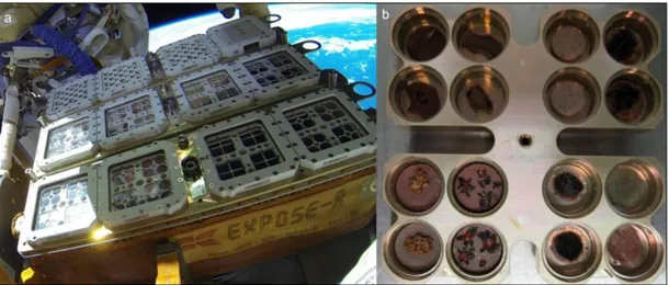
Taking advantage of the combined harsh conditions present in LEO (UV and ionizing radiations, temperature extremes, space vacuum and microgravity), in the EXPOSE-R2 space mission selected organisms have been exposed for 1.5 years not only to space conditions, but also to a simulated Mars-like environment (CO2 atmosphere and UV > 200
nm). The space mission was successfully launched to the ISS on July 24th, 2014 on board the space cargo Progress 56P, whereas on August 18th, was installed outside the ISS on the
Russian Svezda module (Figure 1.4). The experiment ended in June 2016.
A set of microorganisms, representing species of all three main branches of the tree of life were exposed, including microorganisms which are known to be relevant for Mars (e.g. methane producing archaea, cyanobacteria and iron bacteria) and to corroborate the Litho-Panspermia theory (Figure 1.4, de Vera et al., 2012).
17 Among the microorganisms, BIOMEX included the Antarctic cryptoendolithic black fungus C. antarcticus, as eukaryotic model organism for investigating astrobiological topics. In the preparation of EXPOSE-R2 space mission, selected space and Martian simulations (EVTs for Experiment Verification Tests and SVT for Science Verification Tests) were scheduled to test whether the selected samples could withstand real exposure. Colonies of C. antarcticus were grown on substrates including Lunar, P-MRS, S-MRS analogues, or sandstone, which is the Original Substrate (OS). The results on EVTs and SVT tests were analyzed in this thesis (Chapters 2 and 3).
Figure 1.4 Top: EXPOSE-R2 launch onboard the space cargo Progress 56P
from the Baikonur Cosmodrome, Kazakhstan (credit Roscosmos). Bottom: Retrieve EXPOSE-R experiment from the Universal Work Platform on the Russian Zvezda module (credit NASA).
18
1.5.2 STARLIFE Project
Among other space parameters as extreme vacuum, desiccation and strong thermal contrasts (Nicholson et al., 2000), exposure to space environment includes numerous types of ionizing radiation as high-energy photons and particles of different masses, charges, and energies (Moeller et al., 2010; Dartnell, 2011).
Figure 1.5 The rDNA analysis-based phylogenetic tree of terrestrial life including the selected organisms
(highlighted in the tree) for the BIOMEX space experiments onboard the ISS. Bottom left: mineral pellets selected for BIOMEX (de Vera et al., 2012).
19 The complex space radiation has been considered one of the main hazardous components of space environment for any biological system to survive long periods in space (Ferrari and Szuszkiewicz, 2009); yet, a deeper understanding of the biological effects of this parameter is required for assessing the radiation risks in space. This pertains to astronauts, the cabin microflora and accompanying bioregenerative life support systems during long-term exploratory missions (Badhwar and O'Neill, 1994; Cucinotta, 2015) as well as to any microorganism accidentally traveling through space after being ejected from its planet’s surface by a meteorite impact, as described in the scenario of Litho-panspermia.
To address these questions, a set of different astrobiological model systems have been studied within the STARLIFE radiation campaigns (Moeller et al., in press). The STARLIFE group aims to investigate the responses of different astrobiological model systems to the different types of ionizing radiation (x-rays, gamma-rays, heavy ions), which represent major parts of galactic cosmic radiation spectrum. Low and high-energy charged particles radiation experiments have been performed at the HIMAC facility (Heavy Ion Medical Accelerator) at the National Institute of Radiological Sciences (NIRS) in Chiba, Japan. X- or γ-rays were used as reference radiation at DLR (German Aerospace Center, Cologne, Germany).
Exposure to this particulate radiation in space causes a wide range of different types of genomic lesions, e.g., single and double strand breaks, abasic sites, modified (mainly oxidized) bases or interstrand crosslinks (Asaithamby and Chen, 2011; Friedberg, 2003; Yokoya et al., 2008) with consequences of gene mutations, chromosome exchanges, cancer induction and cell death (Cacao et al., 2016).
Various biological endpoints have been investigated with a combination of different biochemical and molecular biological methods, e.g., colony formation assays, most probable number, vitality staining, different microscopic analysis, RAMAN spectroscopy, PCR/RT-PCR, metabolism, and key biosignatures stability. They allowed gaining a clearer understanding and broader spectrum of the effects of galactic cosmic rays on astrobiological model systems.
In the frame of STARLIFE, three Antarctic endolithic microorganisms, namely C. antarcticus, Umbilicaria sp. mycelium and Stichococcus sp., were exposed to high doses of γ-rays; findings will give insights to define the limit of life under radiation environment.
20
1.6 Aim of the thesis and chapters description
The present thesis aims to describe the responses of the Antarctic meristematic black fungus Cryomyces antarcticus CCFEE 515 to stresses, in the context of astrobiological studies; focus was on resistance to the ground-based simulations, both in the preparation of the BIOMEX LEO experiments, STARLIFE project and to 1.5 years of real space exposure outside the ISS, under anydrobiotic condition. The role of melanin pigments in the protection of the fungus, comparing melanized and non-melanized strain of C. antarcticus under physiological condition, were investigated too.
The first chapter is an introduction to the astrobiology research, focusing on the importance of extreme environments on Earth as references for the space environment, especially the McMurdo Dry Valleys, in Antarctica, which is the closest terrestrial analogue to Mars. The introduction continues with the description of the cryptoendolitic communities, which live inside the rocks, and of C. antarcticus, one of the most resistant eukaryote know to date. The fungus, being perfect eukaryotic astrobiological model, after the high survival reported after the LIFE project, was investigated in BIOMEX and STARLIFE projects, which are deeply described at the end of the introduction.
The following chapters of this thesis describe our understanding on the survival of the fungus C. antarcticus after the ground-based simulations performed in the preparation for the ISS exposure of BIOMEX experiment. The ground-based simulations (second and third chapters) were carried out in the framework of Experiment Verification Tests (EVTs) and Science Verification Test (SVT), respectively, and performed using the Planetary and Space Simulation facilities of the Institute of Aerospace Medicine (German Aerospace Center, DLR, Köln, Germany).
In chapter 2, the BIOMEX experiment is described with the first results obtained during the EVTs on survival, DNA and ultrastructural damage of C. antarcticus grown of Martian and Lunar regulites, to selected space and Martian simulated conditions. The main techniques used in the following chapters are also presented as a proof of concept in this chapter. Chapter 3 deals with the second ground test, the SVT, looking at the survivability of the fungus for the real space mission. XTT assay has been optimized for the black fungus, to evaluate the fungal metabolic activity too. This chapter focuses also on the suitability of melanin as fungal biosignature, using the RAMAN spectroscopy; limits and
21 limitation of this technique on our model is put forward. Results shown in chapter 4 are achieved in the contest of STARLIFE project, a simulation experiment which includes numerous types of ionizing radiation as high-energy photons and γ-radiation. In this contest we have exposed three different Antarctic microorganisms to very high level of space relevant ionizing radiation (60Co) to evaluate the microbes survival and the persistence of
DNA, as biosignature. Again, C. antarcticus has been showed as the most resistant among our test microorganisms, both in term of survival and DNA resistance. In the fifth chapter, the protective role of fungal melanin is investigated after exposure to space-relevant densely and sparsely ionizing radiation, by comparing melanized and non-melanized strains of C. antarcticus, under physiological condition. Survival was analyzed by plating CFU’s, the metabolic activity both by XTT and MTT assays, while the ATP content in the cells was analyzed by the ATP assay.
General conclusions and synthesis are presented in chapter 6, supplementary data and preliminary results on samples exposed to real space and simulated Martian atmosphere in space for 1.5 years are reported in the Appendixes.
22
Chapter 2
2 BIOMEX experiment: Ultrastructural alterations, molecular damage and survival of the fungus Cryomyces antarcticus after the Experiment Verification Tests
Abstract
The search for traces of extinct or extant life in extraterrestrial environments is one of the main goals for astrobiologists; due to their ability to withstand stress producing conditions, extremophiles are perfect candidates for astrobiological studies. The BIOMEX project aims to test the ability of biomolecules and cell components to preserve their stability under space and Mars-like conditions, while at the same time investigating the survival capability of microorganisms. The experiment has been launched into space and is being exposed on the EXPOSE-R2 payload, outside of the International Space Station (ISS) over a time-span of 1.5 years. Along with a number of other extremophilic microorganisms, the Antarctic cryptoendolithic black fungus Cryomyces antarcticus CCFEE 515 has been included in the experiment. Before launch, dried colonies grown on Lunar and Martian regolith analogues were exposed to vacuum, irradiation and temperature cycles in ground based experiments (EVT1 and EVT2). Cultural and molecular tests revealed that the fungus survived on rock analogues under space and simulated Martian conditions, showing only slight ultra-structural and molecular damage.
Keywords: BIOMEX, cryptoendolithic black fungus, DNA damage, Mars, space
simulations, survival.
Pacelli, C., Selbmann, L., Zucconi, L., De Vera, J. P., Rabbow, E., Horneck, G., de la Torre, R., and Onofri, S. (2016) BIOMEX Experiment: Ultrastructural Alterations, Molecular
Damage and Survival of the Fungus Cryomyces antarcticus after the Experiment
23
2.1 Introduction
The question whether extraterrestrial life exists has always intrigued scientists. Extremophilic and extreme-tolerant microorganisms have, as their natural niches, environments previously thought to be incompatible with active life; for this reason they are perfect models for studying the limits of habitability on Earth. The McMurdo Dry Valleys of Antarctica, Arctic regions, permafrost soils and cold deserts, for instance, are considered to be good analogues of Mars environments due to their permanently cold and dry conditions (Hansen et al., 2007). Microbes living there are pushed to the absolute limits of adaptability and represent a perfect tool for astrobiological research (Finster et al., 2007). The resistance of these terrestrial extremophiles under both space simulation and LEO (Low Earth Orbit) has been documented (Horneck et al., 2010). Both ground based and space experiments on the International Space Station (ISS), i.e. the LIFE experiment on Expose-E (Rabbow et al., 2012, 2014), showed that some organisms, such as spores of bacteria, meristematic black fungi, and lichens, are able to survive and reactivate their metabolism after space simulations or direct exposure to space (Demets et al., 2005; Horneck et al., 1994; Olsson-Francis et al., 2009; Onofri et al., 2008, 2012; Raggio et al., 2011; Sancho et al., 2007, 2008) and even simulated Martian conditions (Baqué et al., 2013; Meeßen et al., 2013a; Moeller et al., 2012a; Sánchez et al., 2012).
This work is a part of BIOMEX, whose main goal is to detect signatures of extinct or extant life on Mars, investigating the fate of selected extreme-tolerant organisms and the stability of associated biomolecules, after exposure to actual space and simulated Mars conditions. Investigations will be based on sensitive and non-destructive approaches such as Infrared (Igisu et al., 2006, 2009) and Raman spectroscopies (Böttger et al., 2012, 2013; de Vera et al., 2012), using for comparison an international Raman library whose construction is in progress.
Another main objective of BIOMEX is to test survival in extra-terrestrial conditions of selected extreme-tolerant/extremophilic lithobionts, such as bacteria, meristematic black fungi and lichens grown on Mars and Lunar regolith analogues.
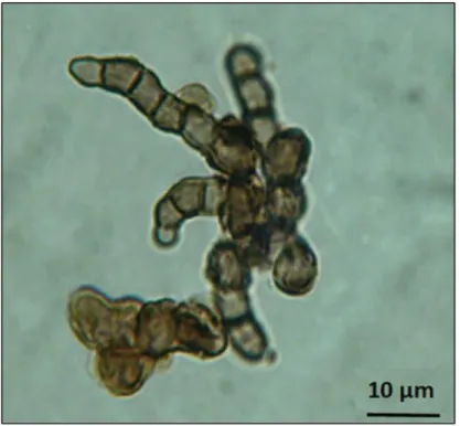
Among the selected organisms, the cryptoendolithic black fungus Cryomyces antarcticus CCFEE 515, from the McMurdo Dry Valleys in Antarctica, is an excellent eukaryotic model due to its exceptional stress resistance and ability to grow inside the rock. Its survival
24 in dried conditions after 18 months of exposure to actual space outside of the ISS, as well as to simulated Mars conditions in space, which was recently demonstrated, gave new insights to the Litho-Panspermia theory (transfer of life between neighbor planets within a meteorite) (Onofri et al., 2012).
On July 24th 2014 the EXPOSE-R2 facility (Fig. 2.1): was launched onboard a Russian
Progress cargo spacecraft (Fig. 2.2) from the Baikonur Cosmodrome, Kazakhstan to the ISS and mounted outside the ISS Zvezda module.
Fig. 2.1 BIOMEX launch on July 24th 2014.
The EXPOSE-R2 facility carried BIOMEX, along with the Biofilm Organisms Surfing Space (BOSS), Photochemistry on the Space Station (PSS) and an experiment from the Russian Institute of Biomedical Problems (IBMP).
This work focuses on the preparatory ground-based EVTs (Experiment Verification Tests) which include space and Martian simulations, performed on C. antarcticus in support of the actual space exposure.
25 Results give clues in searching for life in future Mars exploration missions (de Vera et al., 2012) in detecting putative biosignatures.
2.2 Material and methods 2.2.1 Fungal strain preparation
Cryomyces antarcticus CCFEE 515 was isolated by R. Ocampo-Friedmann from sandstone collected at Linnaeus Terrace (Southern Victoria Land) by H. Vishniac, in the Antarctic expedition 1980-81.
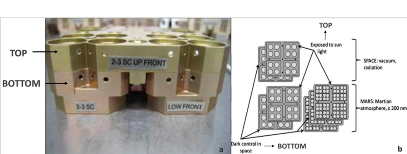
For the EVT tests, cell suspensions were spread on MEA (malt extract agar: malt extract, powdered 30 g/L; peptone 5 g/L; agar 15 g/L; Applichem, GmbH) in Petri dishes, mixed with Antarctic sandstone (15 g/L), Lunar and Martian analogues (1 g/L), prepared to optimize mineral/microorganisms interactions. Sandstone was the original substrate (OS) for the test fungus, Lunar analogue (L) was constituted mainly of anorthosite (Mytrokhyn et al., 2003) and two specific Martian analogues were composed of sulfatic Mars regolith (S-MRS) and phyllosilicatic Mars Regolith (P-MRS), simulating late basic Mars and early acidic Mars surface lithosphere composition, respectively (Böttger et al., 2012). Mars analogue composition was developed and produced by the Naturkundemuseum Berlin, according to the data of Mars research missions (Bibring et al., 2005; Chevrier and Mathé et al., 2007; Poulet et al., 2005). Colonies were grown at 15 °C for 3 months. Disks, cut to fit within the wells of exposure carrier (12 mm diameter) (Fig. 2.2b), were drilled under sterile conditions.
Untreated samples, prepared as above and stored in the dark at room temperature, were used as controls in all the tests performed.
Fig. 2.2 a) EXPOSE R2 Facility mounted outside the ISS from October 22th 2014; b)
26
Table 2.1 Exposure conditions during the Experiment Verification Tests (EVTs). 2.2.2 Tests facilities and exposure conditions
Ground-based simulations (EVTs) were performed using the Planetary and Space Simulation facilities (PSI) at the Institute of Aerospace Medicine (German Aerospace Center, DLR, Köln, Germany). Tests were performed in triplicate and exposure conditions were as reported in Table 2.1.
EXPOSE-R2 EVT part 1 exposure experiments
Test Parameters Performed
Vacuum Vacuum
Mars atmosphere Mars atmosphere
Temperature min and max: 10 °C to +45 °C
Temperature min and max: 25 °C / +60 °C Irradiation
UVC (254 nm) irradiation with Hg low pressure lamp at 80 mW/cm2 1 h, pressure 3.86 x 10⁻³ ± 0.12 Pa 7 h, pressure 8.50 x 10⁻⁵ ± 0.12 Pa 1 h, pressure 6.08 x 10² ± 0.12 Pa 7d, pressure 6.00 x 10² ± 0.12 Pa 66 cycles 2 h 10 °C ± 1 °C; 2 h +45 °C 1 h 25 °C ± 0.5 °C; 1 h +60 °C ± 0.5°C 0 J/m2 12 s, 9.6 J/m2 2 min, 5 s, 96 J/m2 20 min, 50 s, 1000 J/m2 208 min, 20 s 10,000 J/m2
EXPOSE-R2 EVT part 2 exposure experiments
Test Parameters Performed
Irradiation
Polychromatic UV irradiation (200-400 nm) with SOL2000 at 1271 Wm-2 28 d dark 0 kJ/m 5,5 x 10² kJ/m (7min 12 sec) (0,1%ND) 5,5 x 10³ kJ/m (1 h 12 min) (1%ND) (1 wd) 1,4 x 10⁵ kJ/m (1 h 12 min) (1%ND) (1 wd) 2.7 x 10⁵ kJ/m (30 h)(4 wd)(60 h)(9 wd) 5,5 x 10⁵ kJ/m (120 h) (18 wd @ 7 h /wd)
27
2.2.3 Survival Tests
Cultivation test
Survival of C. antarcticus was determined by its colony forming ability as percentages of CFU (Colony Forming Units). For the test, three of the treated colonies was suspended in 1 mL of physiological solution (NaCl 0.9%), and diluted to a final concentration of 3,000 cells/mL, 0.1 mL of the suspension was spread on Petri dishes supplemented with MEA (5 replicates), incubated at 15 °C for 3 months and counted.
PMA assay
The test was performed by adding the Propidium MonoAzide (PMA, Biotium, Hayward, CA) at a final concentration of 200 μM to 1-2 re-hydrated fungal colonies. PMA penetrates only damaged membrane cells, crosslinks to DNA after light exposure and thereby prevents Polymerase Chain Reaction (PCR).
Following DNA extraction and purification, quantitative PCR (Biorad CFX96 real time PCR detection system) was used to quantify the number of fungal Internal Transcribed Spacer (ITS) ribosomal DNA fragments present in both PMA treated and non-treated samples. Five μL of purified genomic DNA (0.1 ng/ml) was added to 12 μL of PCR cocktail containing 1X Power Sybr-Green PCR Master Mix (Applied Bios, Foster City, CA), as well as NS91 forward (5‘-gtc cct gcc ctt tgt aca cac-3‘) and ITS51 reverse (5‘-acc ttg tta cga ctt tta ctt cct c-3‘) primers, each at 5 pmol final concentration. Sterile water was added to reach the final volume of 25 μL. These primers amplify a 203 bp product spanning the 18S/ITS1 region of rRNA encoding genes.
A standard Q-PCR cycling protocol, consisting of a denaturation steps at 95°C for 2 min, followed by 35 cycles of denaturing at 95 °C for 30 s, annealing at 55 °C for 30 s, and elongation at 72 °C for 30 s, was performed. Fluorescence measurements were recorded at the end of each annealing step. After forty cycles, a melt curve analysis was performed by recording changes in fluorescence as a function of raising the temperature from 60-90 °C in 0.5 °C per increments. All tests were performed in triplicate.
Statistical analyses
For multiple data points, the calculation of the mean and standard deviations were performed. Statistical analyses were performed by one-way analysis of variance (Anova)
28 and pair wise multiple comparison procedure (Tukey test), carried out using the statistical software SigmaStat 2.0 (Jandel, USA).
2.2.4 DNA extraction and PCR analyses
DNA was extracted from rehydrated colonies, using Nucleospin Plant kit (Macherey-Nagel, Düren, Germany) following the protocol optimized for fungi.
ITS and LSU amplification were performed using BioMix (BioLine GmbH, Luckenwalde, Germany) adding 5 pmol of each primer and 20 ng of template DNA at final volume of 25 μL. The amplification was carried out using MyCycler Thermal Cycler (Bio-Rad Laboratories GmbH, Munich, Germany) equipped with a heated lid.
The rDNA regions were amplified as follows: for the ITS region the first denaturation step at 95 °C for 2 min was followed by denaturation at 95 °C for 30 s, annealing at 55 °C for 30 s, extension at 72 °C for 30 s and for the LSU region the first denaturation step at 95 °C for 3 min was followed by denaturation at 95 °C for 45 s, annealing at 52 °C for 30 s, extension at 72 °C for 3 min. The last three steps were repeated 35 times, with a last extension 72 °C for 5 min for ITS and 7 min for LSU. Primers ITS5, ITS4 (White et al. 1990), LR5 and LR7 (Vilgalys and Hester 1990) were employed to amplify ITS and LSU rDNA portions, respectively.
2.2.5 Random amplification of polymorphic DNA assay
RAPD was performed using BioMix (BioLine GmbH, Luckenwalde, Germany) adding 5 pmol of the primer and 1 ng of template DNA at final volume of 25 μL. The primer used for RAPD was GGA7 (GGA GGA GGA GGA GGA GGA GGA) (Kong et al., 2000). The
conditions for amplification were: first denaturation step at 94 °C for 2 min followed by denaturation at 94 °C for 20 s, annealing at 49 °C for 60 s, extension at 72 °C for 20 s. The last three steps were repeated 40 times, with a last extension 72 °C for 6 min.
2.2.6 Transmission Electron Microscopy
Controls and UV-irradiated colonies were treated with 5% glutaraldehyde/cacodylate sucrose buffer 0.1 M (pH 7.2) for 12 h at 4 °C, washed three times in the same buffer for 1 h each at 4 °C and fixed with 1% OsO4 + 0.15% ruthenium red in 0.1 M cacodylate buffer
29 (pH 7.2) for 3 h at 4 °C. Samples were washed in distilled water (2 times for 30 min at 4 °C), treated with 1% uranyl acetate in distilled water for 1 h at 4 °C and washed in distilled water (2 times, 30 min at 4 °C). Samples were then dehydrated in ethanol solutions: 30%, 50%, 70% (15 min each, at room temperature) and 100% EtOH (1 h at room temperature), critical point dried and infiltrated in ethanol 100%: LR White series with accelerator, in rotator, at 4 °C (2:1 for 3 h; 1:1 for 3 h, 1:2 overnight) and embedded in pure resin for 1 day and overnight; as final step, samples were included in pure resin in gelatinous capsule for 2 days at 48–52 °C.
2.3 Results
2.3.1 Cultivation test
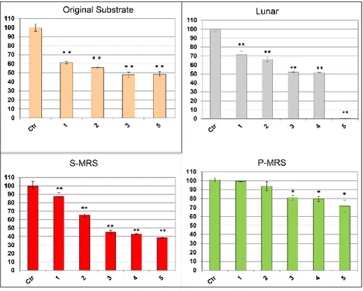
C. antarcticus retained the colony-forming ability after both EVT1 and EVT2 treatments. In EVT1 (Fig. 2.3) the fungus showed a similar trend of survival on all the substrates tested: in general, survival decreased with increased UV-irradiation doses. Yet, colonies formed even at the highest dose, 10.000 J/m², where percentage of survival was 5%, 18%, 38% and 46% in Lunar, Original Substrate, P-MRS and S-MRS analogues, respectively. The fungus, grown on Martian analogues, was able to survive exposure to the Martian atmosphere with no significant decrement on S-MRS with respect the control and almost 60 % of survival on P-MRS. Surprisingly, lower vitality was observed, for all substrates tested, at -25 °C than at 60 °C.
In the EVT2 treatments (Fig. 2.4), a progressive increase of mortality was observed with increasing of UV-irradiation doses, but 48%, 38% and 72% of germination was recorded even at highest doses for colonies grown on Original substrate, S-MRS and P-MRS, respectively. Colony-forming ability was maintained in most cases after EVT2 treatments; the only exceptions was the highest irradiation in Lunar sample.
30 Vitality, specifically, the integrity of the plasma membrane, was also tested through PMA assay on control and EVT2 samples treated with medium and maximum irradiation doses for each substrate (Fig. 2.5). This test gave a higher percentage of possible survival (measured as cells with intact membrane) compared to the results from the cultural test, even at the highest dose; for instance, no colonies were recorded under Lunar conditions but 35% was observed with PMA assay. No significant differences between treated samples and control were obtained on P-MRS.
Fig. 2.3 Cultural test after EVT1 treatments: Percentages of CFU’s of C. antarcticus on different substrates, relative to controls on the same substrate.Control (Ctr), vacuum 7h (Vac), Martian atmosphere 7g (M atm), temperature cycles (TC), minimum temperature (-25 °C), maximum temperature (+60 °C), irradiation at different intensities (0, 10, 100, 1000 and 10,000 J). Significant differences were calculated by Tukey test with * = p > 0.05. and ** = p > 0.001.
31 Fig. 2.4 Cultural test after EVT2 treatments of C. antarcticus grown on
different substrates: CFU’s following exposure to UV light relative to controls on the same substrate. Control (Ctr), increasing polychromatic UV irradiation doses 1: 1.5x10³, 2: 1.5x10⁴, 3: 1.5x10⁵, 4: 5.0x10⁵, 5: 8.0x10⁵kJ/m2. The statistical analyses were performed as Figure 2.3.
Fig. 2.5 Results of PMA assay coupled with qPCR after EVT2 treatments in a selection of samples (Samples correspond with those in Fig. 4): A: Percentages of C. antarcticus cells with damaged membrane B: Percentages of C. antarcticus cells with intact membrane. The statistical analyses were performed as in Figure 2.3.
32
2.3.2 DNA damage
The integrity of genomic DNA in treated samples was tested by assessing its ability to serve as a PCR template both after EVT1 and EVT2 treatments.
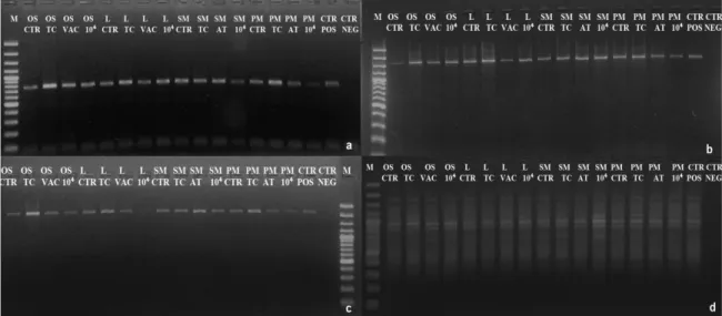
All the EVT2 samples were analyzed, while for EVT1, on the basis of colony forming results, only samples exposed at maximum treatments were chosen: Temperature Cycles, Vacuum 7h, Martian atmosphere 7d, maximum dose of UV (254 nm) irradiation at 104J/m². Amplifications worked out for all the gene-lengths in EVT1 (Fig. 2.6a, b, c) and EVT2 (Fig. 2.7a, b, c) exposed samples. A reduced intensity of PCR bands was evident in panel c, with the largest gene length, in Lunar EVT1 samples exposed to 10,000 J/m2 (Fig. 2.6c, Lane L 104) and for colonies grown on S-MRS at the highest dose of UV (>200 nm) irradiation in EVT2. Although there was an overall decrease in band intensity, mainly for the highest molecular weight (MW) bands (about 2200 bp) the RAPD profiles were well preserved in all samples both after EVT1 and EVT2 treatments (Fig. 2.6d, 2.7d), demonstrating a good preservation of the whole genomic DNA.
Fig. 2.6 Increasing detrimental effect of EVT1 treatments on the DNA integrity of genes of different lengths a) ITS rRNA gene (ITS5-ITS4 primers)(700bp) b) LSU rRNA gene (ITS5-LR5 primers) (1600bp). c) LSU rRNA gene (ITS5-LR7 primers) (2000 bp). d) RAPD profile of C.
antarcticus. Treatments were as follows: Control, Thermal Cycles (TC), Vacuum (VAC) and 104 J/m², Positive PCR Control (CTRL POS), Negative PCR Control (CTRL NEG), DNA ladder (M). Substrates: OS, Original Substrate; L, Lunar; SM, S-MRS; PM, P-MRS.
33
2.3.3 Ultrastructural Damage
Ultrastructural damage in C. antarcticus cells was observed by TEM. The samples treated at the highest irradiation dose of EVT2, for which the effect on vitality and DNA damage were evident (Fig. 2.8) were compared with laboratory controls
Control cells maintained mostly an intact cell membrane and a well-organized and defined cytoplasm after dehydration (Figs. 2.8a, c, e, g, i). By contrast, the majority of cells in the irradiated samples showed extended damage with irregular shapes, damaged cell walls, discontinuous cell membranes and a compromised organization of the cytoplasm (fig. 2.8b, d, f, h, j).
Fig. 2.7 Increasing detrimental effect of EVT2 treatments on the DNA integrity on genes of different
lengths a) ITS rRNA gene (ITS5-ITS4 primers) (700bp). b) LSU rRNA gene (ITS5-LR5 primers) (1600bp). c) LSU rRNA gene (ITS5-LR7 primers) (2000 bp). d) RAPD profile of C. antarcticus after EVT2 treatments. Order as follows: DNA ladder (M), irradiation (sample correspondence as for Fig. 2.3), Control (CTR). Substrates: OS, Original Substrate; L, Lunar; SM, S-MRS; PM, P-MRS.
34
Fig. 2.8 TEM micrographs. Untreated (CTR) and treated
(maximum irradiation of EVT2, 8.0x10⁵ kJ/m2) microcolonies of C. antarcticus on sandstone (OS) (a, b); Lunar (L) (c, d); S-MRS (e, f); P-MRS CTR (g, i) Unirradiated (a,c,e,g,i) and irradiated (b,d,f,h,j) respectively. The structure of the cytoplasm was not conserved in many cases in the irradiated samples, the organelles are not visible (b, d, f, white arrows) and the continuity of the cell membrane (f, black arrow) and of the cell wall (b, d, black arrow) interrupted. The irradiated cells were better preserved in colonies cultivated on P-MRS where the structure of mithocondria were still well discernable (j, white arrow).
35
2.4 Discussion
Ground-based simulations, EVT1 and EVT2, performed in the frame of the BIOMEX experiment currently on-board the EXPOSE-R2 platform fixed outside the ISS, were preliminary experiments to validate samples for the space mission. One of the aims of the project was to study dried cells of C. antarcticus, grown on lunar regolith analogue rocks and two Mars regolith analogue mixtures, to test survival as well as DNA and ultra-structural damage.
Although a reduced survival was observed after exposure to the most stressing parameters, C. antarcticus retained some colony forming ability after UV irradiation as well as after simulated treatment with other outer space stressors. These results were confirmed by TEM observations showing that, in addition to cells showing ultrastructural damage after the highest irradiation dose, a number of cells were still in good condition (not shown), which agrees with the survival rate recorded in the colony forming assays. The main damaging factors of the EVTs were UV irradiation and temperature cycles. C. antarcticus survival was higher after +60 ºC than after -25 ºC treatment. The surprising ability of this psychrophilic fungus to tolerate very high temperatures was previously observed, when it was found to retain 100% survival after exposure to 1 h at 90 °C (Onofri et al. 2008). Survival was high even under simulated Mars atmosphere and vacuum since more than 50% of colonies developed in all cases. If survival was comparable in the irradiated samples on Martian analogues, values were different on the two substrata when the fungus was exposed to Martian atmosphere: no significant decrement was recorded on S-MRS with respect the control whereas here was less than 60 % of survival for P-MRS (Fig. 2.3). This apparently incongruent result is difficult to explain and requires additional investigations.
Most samples showed a higher percentage of apparent survival in PMA assays as compared to colony forming tests. This apparent inconsistency may be due to the coincidental preservation of cell membrane integrity in cells that have lost the ability to multiply that have, preventing PMA to penetrate and react with DNA; this could have led to an overestimation of vitality in some cases, i.e. Lunar sample. The membrane may be less susceptible to damage than DNA or the DNA replication enzymes under some of these conditions. Similar results were reported by Bryan et al. (2015) who also obtained higher
36 values with the indirect XTT assay method with respect the clonogenic approach for testing survival for irradiated fungal cells of Cryptococcus neoformans. Data obtained in Lunar sample (8x10⁵ kJ/m2), where survival was 35% in PMA assay compared to zero in cultural
test, may be due to the loss of the ability to multiply in some cells where UV treatment caused extensive molecular damage, since UV targets more specifically the DNA rather than the membrane.
It is worth noting that the highest survival, both from cultural analysis and PMA assay, was obtained for C. antarcticus grown on P-MRS; the same fate was recently reported for the mycobiont of Buellia frigida suggesting a protective role of the substratum (Meeßen et al., 2015). The same authors observed that the highest viability was obtained when the lichen was exposed on the original rock substratum. Differently, in the frame of BIOMEX experiment, Baquè et al. (2014) reported a higher survival of the cyanobacterium Chroococcidiopsis when mixed with S-MRS regolith. These contrasting results led us to conclude that a possible protective role of the analogues is difficult to be sustained for now and needs to be further studied. Our results clearly demonstrate the high resistance of C. antarcticus to all EVT treatments, including exposure to vacuum, simulated Mars atmosphere, and different doses of monochromatic (254 nm) and polychromatic (>200 nm) UV radiation.
Further studies on C. antarcticus in the last ground tests (Science Verification Test, SVT) are still in progress and will clarify the real role of substrates in the protection; it was suggested that survival in space could benefit from the shielding provided by melanin. Melanin is a biological macromolecule mainly known for its protective role against UV, extreme temperatures, desiccation and osmotic stress (Sterflinger 2006; Plemenitaš et al. 2008). The high tolerance to UV-B exposure of single cells of black fungi has been reported by Onofri et al. (2007b). It is known that melanin strongly absorbs UVB, UVA, and PAR, thereby protecting fungi and lichens against those stressors (Nybakken et al., 2004, Meeßen et al., 2013b). Moreover, it was observed that melanized fungal spores, in addition to resistance to UVR, even resist γ-ray and X-ray treatment better than melanin-deficient ones (Bell and Wheeler, 1986) suggesting a role for this pigment in radioprotection of fungi (Henson et al., 1999; Dadachova et al., 2007). In this study, the presence of either Martian and Lunar analogues did not affect molecular analyses since genomic DNA was
37 successfully extracted and amplified even from samples that had lost the ability to form colonies. In agreement with what is reported in the literature, PCR band intensity decreased mainly in the highest molecular weight fragments in single-gene PCR (Atienzar et al., 2002). However, most of the amplicons were still obtained even at highest doses of UV-irradiation and RAPD profiles were well preserved in all samples. This surprisingly high DNA resistance to UV-irradiation, if protected by screening pigments, the outer cell envelop or a dust layer, suggests DNA as a possible biosignature candidate in future exploration missions (Lyon et al., 2010). Of course its resistance to UV-radiation needs to be proved over much longer timescales.
It is worth noting that ancient DNA was actually recovered on Earth from samples between 400 thousand and 1.5 million years old (Sankaranarayanan et al., 2014); it was also postulated that present Mars conditions (in terms of dryness and low temperatures) may even preserve ancient DNA much better than Earth conditions (Sephton, 2010) with a theoretical preservation of a 100 bp fragment of DNA along a timescale of 3.4 x 10⁹ years at -50 °C and 3 x 10²¹ years at -110 °C at the Martian polar ice caps (Willerslev et al., 2004). Moreover, some specific conditions could improve the long-term preservation of ancient DNA in halite crystals, permafrost, amber depositions and marine sediments (Panieri et al., 2010). Of course, some other damaging factors, such as ionizing radiation (Hassler et al., 2014) and oxidative environments (Yen et al., 2000; Hecht et al., 2009) on Mars, must be taken into consideration; but our simulation experiments suggest that we may have a good chance to reveal the presence of DNA, in present, in putative extraterrestrial samples, by using some low-specificity based approach such as random primers (as for RAPD) or non-specific staining such as orange acridine or non-toxic ones such as gel red and sytox green.
2.5 Conclusions
C. antarcticus is an astonishingly resistant fungus, able to withstand even long term exposure to actual Space conditions (Onofri et al., 2012); the present study proved that the fungus survives space simulated stressors even when grown on extraterrestrial rock analogues. The awareness that a terrestrial microbe may survive extraterrestrial conditions is an important clue in searching for life on other planets, above all on Mars. Many efforts
38 are now devoted to the definition of proper biosignatures to detect whether life was ever present in an extraterrestrial sample from a putatively habitable region. Our results show that genomic DNA can be successfully extracted even in the presence of Martian or Lunar analogues; the ease of isolation and detection are key characteristics for a suitable biosignature, and optimizing the extraction is an important challenge in detecting biomarkers (Aerts et al., 2014). PCR was also successful and amplifications were obtained for most of the genes even at a length of up to 2000 bp; this was regardless of the treatments, revealing a high DNA persistence. Further analyses on samples treated in more stressing ground based experiments (SVT) or exposed to actual space conditions in the frame of the BIOMEX experiment, will provide further information on the detectability of this molecule and its suitability as biomarker in future exploration missions. Studies are in progress to define additional biomolecules to be used as good biomarkers using non-destructive approaches as Raman and Infrared spectroscopies, according to the instruments available on ExoMars.
Acknowledgments. This research was funded by the ASI and was supported by the
German Helmholtz Association through the Helmholtz-Alliance “Planetary Evolution and Life”. The authors thank DLR for space simulation tests. The PNRA (Italian National Program for Antarctic Research) for supporting Antarctic campaigns for sample collection and research laboratory studies in Italy. The Italian National Antarctic Museum ‘‘Felice Ippolito’’ is kindly acknowledged for funding CCFEE. The authors thank PhD. Ruth A. Bryan for English revision.
39
Chapter 3
3 BIOlogy and Mars Experiment: responses of the black fungus
Cryomyces antarcticus to the EXPOSE-R2 Science Verification Test
Abstract
The BIOlogy and Mars Experiment (BIOMEX) is part of the ESA space mission EXPOSE-R2 in Low Earth Orbit (LEO). In the frame of this international and interdisciplinary experiment, dried colonies of the Antarctic cryptoendolithic black fungus Cryomyces antarcticus CCFEE 515, grown on Martian and Lunar analog regolith pellets, were exposed for 1.5 years to LEO space and simulated Mars conditions on the International Space station (ISS). In preparation for this mission, several preflight tests, Experiment Verification Test (EVT) and Science Verification Test (SVT), were performed to investigate the space mission candidate organisms’ resistance to space stressors, the potential interference of extraterrestrial rock analogues on fungal survival and the detection of biomolecules as potential biomarkers. The present results demonstrate that C. antarcticus was able to tolerate the conditions of the SVT experiment, regardless of the substratum where it was grown. DNA demonstrated high stability after treatments and it was confirmed as a possible biosignature molecule, while melanin, a molecule chosen as a target for biosignature detection, was impossible to unambiguously detect by Raman spectroscopy.
Keywords: Astrobiology, Radiation resistance, Biosignatures, Extremophilic microorganisms, Raman spectroscopy.
Pacelli, C., Selbmann L., Zucconi L., Coleine C., de Vera J.P., Rabbow E., Dadachova E., Böttger U., and Onofri S. BIOlogy and Mars Experiment: responses of the black fungus Cryomyces antarcticus to the EXPOSE-R2 Science Verification Test. (under Revision in Astrobiology).