University of Pisa
BIOS Research Doctorate School in BIOmolecular Sciences
Course in Experimental
and Molecular Oncology
XXII Cycle (2008-2010)
THESIS
Evolution of HPV lesions in HIV-infected and HIV-uninfected women
in Mozambique: HPV genotype characterization
Tutor: Prof. Generoso Bevilacqua
Supervisor: Prof. Generoso Bevilacqua
Index
Page
Abbreviations 4
Abstract 5
1. Introduction 6
1.1 Cervical cancer – epidemiology 1.2 Classification of HPVs
1.3 Geographic prevalence of HPV types
1.4 Status of the global HIV epidemic in sub-Saharan Africa 1.5 Overview of the AIDS epidemic in Mozambique
1.6 Epidemiology of HPV infections and cervical cancer in the era of HIV
2 Aim of the Research 21
3. Methods 22
3.1 The Drug Resource Enhancement against AIDS and Malnutrition (DREAM) Programme
3.2 Study population 3.3 Study design
3.4 Selection of participants
3.5 Collection of specimens, procedures and testing for HPV genotypes 3.6 Outcomes in HIV patients
4. Results 26
5. Discussion 30
6. Conclusions 35
7. Acknowledgments 37
8. References 38
Figures and Tables
Figure 2 - The organisation of episomal and integrated HPV DNA 8 Figure 3 – HIV regional prevalence, 2004- 2009 (Mozambique) 13 Figure 4 – Age-standardised incidente rate of cervical cancer in Africa 16 Figure 5 – Exstimated age-standardised incidence rate per 100.000 cervix uteri 17 Figure 6 –. Mozambique: estimated number of cancer cases, all ages 17 Figure 7 – Type specific prevalence of HPV in HIV positive women in the World 18
Figure 8 – Screening protocol 29
Table 1: comparative table of Pap test vs HPV genotypes (HR and probable HR) 32
Attachment 1- Favourable opinion of Mozambican
Ministry of Health’s Bioethics Committee (Portoguese) 49
Attachment 2 – Authorization of Local Authority of Health
Abbreviations
ADC adeno- and adenosquamous carcinoma
ASCUS atypical squamous cells of undetermined significance
BMI body mass index
CI confidence interval
CIN cervical intraepithelial neoplasia
HIV-1 Human immunodeficiency virus type 1 HPV human papillomavirus
HR hazard ratio
HSIL high-grade squamous intraepithelial lesion IARC International Agency for Research on Cancer ICC invasive cervical cancer
LSIL low-grade squamous intraepithelial lesion LOH loss of heterozygosity
MA microsatellite alteration OR odds ratio
PCR polymerase chain reaction SCC squamous cell carcinoma SIL squamous intraepithelial lesion
SIR standardized incidence ratio TASO The AIDS Support Organization
TMB tetramethylbenzidine VL viral load
ABSTRACT
Background: Women infected by human immunodeficiency virus (HIV) display an higher rates of human papillomavirus (HPV) infection and cervical dysplasia than HIV seronegative women. Our study explore the relationship between HPV and HIV-1 in Mozambique to determine the prevalence of different HPV genotypes in a group of 1-infected women and in a control group of HIV-uninfected women and to correlate these findings with cervical cytological results. Moreover the persistence of the HPV infections and the evolution of the lesions has been investigated.
Methods: A prospective, two arms, observational study involving infected and HIV-uninfected women has been performed. The enrolment period lasted from August 2007 to May 2010, whereas the observation period ended on February 2011.
Results: The study involved 331 participants: 247 HIV-infected and 84 HIV-uninfected women. HPV was found in 179/247 (72,5%) HIV-infected and in 45/84 (53,6%) HIV-uninfected subjects (p <0.001). The most frequent HPV genotypes identified in HIV-infected subjects were HPV: 58 (24,9 %, p<001), 16 (19,1 %) 61 (14,2 %,), 53 (13,7 %), 6 (11,3%) ; the HPV types identified in HIV-uninfected subjects were: 16 (17,7%) , 6 (16,1%), 66 (14,5%), 53 (11,3%), 18 (9,7%)
Conclusions: the HPV types identified are partially different from those more commonly identified in Western countries. It is necessary to improve screening for HPV and monitoring and treating programmes for SIL in HIV patients in Mozambique and in other sub-Saharan African countries.
1. INTRODUCTION
1.1 Cervical cancer – epidemiology
Cervical cancer is the second most frequent female neoplasia worldwide and the most common female cancer in large areas of the developing world where an estimated 80% of new cases arise every year (Parkin et al, 1997; Ferlay J et al., 2004, Cronje HS, 2004, Odendal L., 2011).
Approximately half million new cases of cervical cancer are diagnosed per year worldwide, with a mortality of about 30% of the cases: 85% of these deaths will occur in women in the developing world. Cervical cancer remains the second most common cause of cancer-related death in women and accounts for 13% of all female cancers. The occurrence rate in developed countries has shown a tendency to decrease year by year, mainly due to effects of early diagnosis using the Papanicolaou (Pap) smear method. On the contrary cervical cancer is increasing in developing countries where screening programmes are not extensively setted up.
The International Agency for Research on Cancer (IARC) predict that the number of cervical cancer cases anticipated by 2020, all other things being equal, will increase of 40% globally (Fig. 1).
This event is dramatically dependent on the socioeconomic status. The countries in Africa, Latin America and Asia are predicted to have a 50–55% increase in the number of cases. Europe
and North America will also experience a modest increase in the number of cases, in the order of 6% in Europe and 23% in Northern America (Ferlay et al., 2004).
1.2 HPVs and Cervical cancer
Infection with genital HPVs has been established as the primary cause of cervical squamous intraepithelial lesion (SIL) and invasive cervical cancer (ICC) (zur Hausen, 1987). zur Hausen noted that twelve clades of human papillomaviruses (HPV) had been isolated from papillomatous and Bowenoid lesions of the human genital tract. HPV 6, 11, 16 and 18 were most frequently found, respectively. Recently Odendal (Odendal L. 2011) reported that between the more than 100 human papillomaviruses isolated, 13 are considered “high risk” of oncogenic behaviour, and from them 70% of the world’s cervical cancer are caused by HPV types 16 and 18. HPV types 6 and 11, on the contrary, caused typical genital warts (condylomata acuminata) and mild dysplastic lesions of the cervix characterized by a high degree of koilocytotic atypia. The majority of the remaining tumours revealed evidence for infections with additional types of HPV, also because co-infection with different types of HPV is normal.
Studies in 22 countries, coordinated by the IARC, identified HPV DNA in almost all (99.7%) of about 1,000 cases of cervical cancer (Bosch FX et al., 1995; IARC, 1995; Walboomers et al., 1999). The presence of HPV in virtually all cervical cancers implies the highest worldwide attributable fraction so far reported for a specific cause of any major human cancer. The extreme rarity of HPV-negative cancers reinforces the rationale for HPV testing in addition to cervical cytology in routine cervical screening.
Epidemiological studies, performed mainly on human immunodeficiency virus (HIV) uninfected women, have shown that, despite the high prevalence and strong association with cervical neoplasia, the majority of HPV infections with high- and low-risk types are transient and only a fraction of persistent infections progress on to high-grade SIL and invasive cancer, underscoring the interplay of a number of environmental, viral and host factors in HPV-related tumour progression (Ho GY et al., 1998; Massad et al., 1999; Castellsagué X & Muñoz N, 2003; Wang SS & Hildesheim A, 2003). Thus, infection alone is not sufficient to cause cervical cancer.
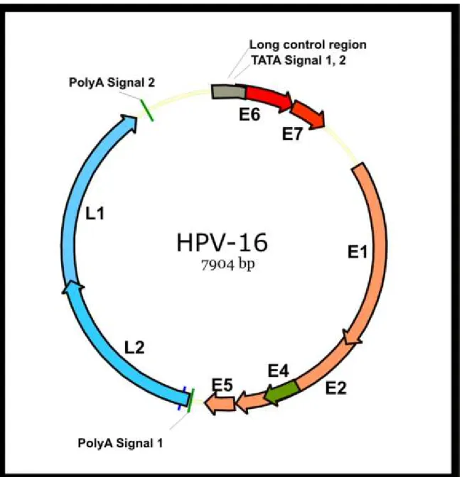
HPVs are small, non-enveloped viruses that contain circular double-stranded DNA genomes of approximately 8,000 base pairs (Fig. 2) (Hebner CM et al., 2006; Narisawa-Saito M et al., 2007). HPV genomes replicate episomally in host cells, but HPV DNA is frequently found to be integrated into chromosomes in cervical cancer cells. The timing of viral integration appears to correspond to the development of high-grade CIN as a consequence of a high-level expression of the viral proteins E6 and E7 (von Knebel Doeberitz M, 2002). Those proteins have various
biological activities in addition to inactivation of the major tumour suppressors, p53 and pRB, respectively. Lines of investigation indicate that these two viral proteins do not merely immortalise normal human epithelial cells but that they confer some tumourigenic properties of transformed cells by multiple mechanisms.
Source: Yugawa and Kiyono, 2009
Fig. 2 The organisation of episomal HPV DNA. The HPV genome is a circular double-stranded DNA of approximately 8,000 base pairs. The genetic map of HPV16 is illustrated. The genome contains an upstream regulatory region (URR) and eight open reading frames (ORFs) of six early genes (E1, E2, E4, E5, E6, E7) and two late genes (L1, L2). Early genes have roles in viral replication and late genes encode viral capsid proteins. E1 is a viral helicase and is recruited to the viral replication origin located in URR by E2, where it forms the DNA replication machinery with cellular proteins. HPV genomes replicate episomally in their life cycle. HPV DNA is frequently found to be integrated into host chromosomes in cervical cancer cells and displays consistent features, with disruption of E2 ORF and retention of E6/E7 ORFs and URR containing the early promoter for E6/E7 transcription. Within a cell containing a mixture of episomal and integrated HPV DNA, the viral replication proteins E1 and E2 expressed from HPV episomes may cause chromosomal abnormalities such as rearrangements and/or translocations.
1.3 Classification of HPVs
Around 40 HPV genotypes of more than 100 identified, infect the genital mucosa. Those have been classified with reference on the probability to induce a malignancy, as follow:
(i) ‘high-risk’ viruses, associated with an high risk of cervical cancer (HPV types 16, 18, 31, 33, 35, 39, 45, 51, 52, 56, 58 and 59). Probable carcinogenic viruses, associated with cervical cancer in a few case-control studies (HPV types 26, 53, 66, 68, 73 and 82).
(ii) ‘low-risk’ viruses, associated with benign epithelial proliferations (HPV types 6, 11, 40, 42, 43, 44, 54, 61, 70, 72, 81 and 89).
(iii) ‘undetermined risk’ viruses whose oncogenicity has not yet been determined ((HPV types 2a, 3, 7, 10, 27, 28, 29, 30, 32, 34, 55, 57, 62, 67, 69, 71, 74, 77, 83, 84, 85, 86, 87, 90 and 91), Muñoz et al., 2003, Muñoz et al., 2006; Smith et al., 2007).
High-risk HPV types are found in virtually all ICC (Bosch et al., 2002; IARC, 2007, Odendal, 2011) and they are significantly associated with progression to ICC. High-risk HPV such as type 16, 18 and 31, are associated with more than 90% of cervical cancers (Walboomers et al., 1999), where up to 70% of ICC are associated with types 16 and 18 (Odendal, 2011). High-risk HPV infections are also implicated in the development of other anogenital malignancies, including vulvovaginal, penile and anal cancers (Steenbergen RD et al., 2005; Kayes O et al., 2007) as well as a subset of head and neck cancers. Most of these high-risk types are phylogenetically related to either HPV16 (31, 33, 35, 52 and 58) or HPV18 (39, 45, 59 and 68) (Chan et al, 1995). Limited evidence suggests that their distribution may vary by region (Bosch et al, 1995).
Infection by multiple HPV types in some studies was not associated with a greater risk of cervical cancer than infection by a single HPV type (Herrero R, 2000; Kleter B, 1999; Muñoz, N, 2003).
1.4 Geographic prevalence of HPV types
There are regional variations in the contribution made by different human HPV types to ICC, to squamous cell carcinoma (SCC) and to adeno- and adenosquamous carcinoma (henceforth collectively termed ADC).
In a meta-analysis of HPV, a total of 10,058 ICC cases from 85 studies were included prevalence (Clifford MG, 2003). The majority of cases came from studies performed in Asia (31%) and Europe (33%), with African studies representing the smallest proportion of cases (6%). HPV
prevalence was reported stratified by histological type for 73% of the cases: 5825 cases of SCC and 1,508 cases of ADC. In total, 12 studies included only SCC and seven studies included only ADC. Owing to their similar overall and type-specific HPV prevalence, ICC of unspecified histology were combined with SCC for comparison of HPV type-specific prevalence by histological type.
The most common HPV types identified were, in order of decreasing prevalence, HPV16, 18, 45, 31, 33, 58, 52, 35, 59, 56, 6, 51, 68, 39, 82, 73, 66 and 70. Other HPV types were detected in no more than 0.2% of cases of ICC.
In SCC, HPV16 was the predominant type in all regions studied, varying from 45.9% in Asia to 62.6% in North America and Australia. HPV18 was found consistently in 10–14% of cases of SCC. In most regions, HPV45 (2–8%), 31 (2–7%) and 33 (3–5%) were the most prevalent types in SCC after types 16 and 18.
In cases from Africa, the prevalence of HPV45 (8.0%) was more than twice that of either 31 (2.7%) or 33 (3.2%). In cases from Asia, HPV58 (5.8%) and 52 (4.4%) were found more commonly than HPV45, 31 and 33. Other HPV types varied considerably in their prevalence from region to region, but accounted for no more than 2% of ICC cases from any region. Sufficient ADC-specific data existed for the comparison of HPV specific prevalence across Asia, Europe and North America and Australia (Figure 3). HPV18 was the predominant type (37.7%), found consistently in 37–41% of cases of ADC in these regions, with HPV16 accounting for a smaller proportion (26–36%). HPV45 was the third most prevalent type in each region, present in 5–7% of cases of ADC and only in 2–4% of cases of SCC from these regions. The HPV16 phylogenetically related types 31, 33, 52 and 58 (but not 35) were all less prevalent in cases of ADC than in cases of SCC from each region.
In a recent meta analysis (Tiggelaar SM. Et al 2012) including one hundred seventeen study has been reported that HPV 16 has a seroprevalence ranged from 0%-31% in North America, 21%-30% in Africa, 0%-23% in Asia/Australia, 0%-33% in Europe, and 13%-43% in Central and South America. Moreover the prevalence of HIV 16 is higher with respect of HIV 18 and also the age peak of HIV 16 infection is located within 25 to 40 years whereas HIV 18 shows a wider range of age. In this study the serology of the patients has been compared with the molecular analysis of nucleic acid of HPV in order to find an affordable marker suitable for a vaccine intervention.
1.6 Status of the global HIV epidemic in sub-Saharan Africa
Sub-Saharan Africa remains the region most heavily affected by HIV. In 2010, about 68% (22,4 million) of all people living with HIV resided in sub-Saharan Africa, a region with only 12% of the global population. Sub-Saharan Africa also accounted for 70% of new HIV infections in 2010, although there was a notable decline in the regional rate of new infections. The epidemic
continues to be most severe in southern Africa, with South Africa having more people living with HIV (an estimated 5.6 million) than any other country in the world. Almost half of the deaths from AIDS-related illnesses in 2010 occurred in southern Africa. AIDS has claimed at least one million lives annually in sub-Saharan Africa since 1998. Since then, however, AIDS-related deaths have steadily decreased, as free antiretroviral therapy has become more widely available in the region. The total number of new HIV infections in sub-Saharan Africa has dropped by more than 26%, down to 1.9 million [1.7 million–2.1 million] from the estimated 2.6 million [2.4 million–2.8 million] at the height of the epidemic in 1997. In 22 sub-Saharan countries, research shows HIV incidence declined by more than 25% between 2001 and 2009. This includes some of the world’s largest epidemics in Ethiopia, Nigeria, South Africa, Zambia and Zimbabwe. The annual HIV incidence in South Africa, though still high, dropped by a third between 2001 and 2009 from 2.4% [2.1%–2.6%] to 1.5% [1.3%–1.8%]. Similarly, the epidemics in Botswana, Namibia and Zambia appear to be declining. The epidemics in Lesotho, Mozambique and Swaziland seem to be levelling off, albeit at unacceptably high level.(UNAIDS world AIDS DAY REPORT 2011)
For the region as a whole, women are disproportionately affected in comparison with men, with stark differences between the sexes in HIV prevalence among young people. In southern Africa, reductions in HIV prevalence are particularly striking in Zimbabwe, where HIV prevalence in pregnant women attending antenatal clinics fell from 26% in 2002 to 18% in 2006 (Ministry of Health and Child Welfare [Zimbabwe], 2007). In Botswana, a drop in HIV prevalence among pregnant 15–19-year-olds (from 25% in 2001 to 18% in 2006) suggests that the rate of new infections could be slowing (Ministry of Health [Botswana], 2006). The epidemics in Malawi and Zambia also appear to have stabilized, amid some evidence of favourable behaviour changes (Heaton, Fowler & Palamuleni, 2006; Sandoy et al., 2007) and signs of declining HIV prevalence among women using antenatal services in some urban areas (Ministry of Health and Population [Malawi], 2005; Ministry of Health [Zambia], 2005; Michelo et al., 2006; National AIDS Commission [Malawi], 2007). Meanwhile, the 26% HIV prevalence found in adults in Swaziland in 2006 is the highest prevalence ever documented in a national population based survey anywhere in the world (Central Statistical Office [Swaziland] & Macro International Inc., 2007).
In Lesotho and parts of Mozambique, HIV prevalence among pregnant women is increasing. In some of the provinces in the central and southern zones of the country, adult HIV prevalence has reached or exceeded 20%, while infections continue to increase among young people (ages 15–24) (Conselho Nacional de Combate ao HIV/SIDA, 2006). The HIV prevalence in the comparatively smaller epidemics in East Africa has either reached a plateau or is receding. After dropping dramatically in the 1990s (Asamoah-Odei, Garcia-Celleja & Boerma, 2004; Kirungi et al., 2006),
adult national HIV prevalence in Uganda has stabilized at 5.4% [5.0%–6.1%]. However, there are signs of a possible resurgence in sexual risk-taking that could cause the epidemic to grow again. For example, the proportion of adult men and women who say they had sex with a person who was not a spouse and did not live with the respondent has grown since 1995 (from 12% to 16% for women and 29% to 36% for men) (Kirungi et al., 2006; Ministry of Health [Uganda] & ORC Macro, 2006; Uganda Bureau of Statistics & Macro International Inc, 2007). Most of the comparatively smaller HIV epidemics in West Africa are stable or are declining — as is the case for Burkina Faso,
Ivory Coast, and Mali. In Côte d’Ivoire, and the HIV prevalence among pregnant women in urban areas fell from 10% in 2001 to 6.9% in 2005 (Ministère de la Santé et de l’Hygiene Publique
de la Côte d’Ivoire & CDC/RETRO-CI/ MEASURE Evaluation, 2007). The largest epidemic in West Africa — in Nigeria, the continent’s most populous country — appears to have stabilized at 3.1% [2.3%–3.8%], according to HIV infection trends among women attending antenatal clinics (Federal Ministry of Health [Nigeria], 2006).
1.7 Overview of the AIDS epidemic in Mozambique
GENERAL STATISTICS, Mozambique, 2011
Total population: 22,948,000
Gross national income per capita (PPP international $): 770 Life expectancy at birth m/f (years): 47/51
Healthy life expectancy at birth m/f (years, 2003): 36/38 Probability of dying under five (per 1 000 live births):142
Probability of dying between 15 and 60 years m/f (per 1 000 population): 557/434 Total expenditure on health per capita (Intl $, 2005): 50
Total expenditure on health as % of GDP (2005): 5.7
H I V a n d A I D S E S T I M A T E S , M o z a m b i q u e , 2 0 0 9
Number of people living with HIV: 1 400 000 [1 200 000 - 1 500 000] Adults aged 15 to 49 prevalence rate: 11.5% [10.6% - 12.2%]
Adults aged 15 and up living with HIV: 1 200 000 [1 100 000 - 1 400 000] Women aged 15 and up living with HIV: 760 000 [680 000 - 840 000] Children aged 0 to 14 living with HIV: 130 000 (69 000 - 180 000] Deaths due to AIDS: 74 000 [57 000 - 92 000]
Fonte: MISAU/INS, 2009]
Figure 3: HIV Regional Prevalence, 2004-2009.
1.8 Epidemiology of HPV infections and cervical cancer in the era of HIV
Infection with HIV is an important risk factor for the HPV infection and the development of HPV-associated lesions in the female genital tract. HPV DNA is 2 to 3 times as frequent in cervicovaginal-lavage specimens and almost 15 times as common in anal-swab specimens from HIV-infected women as in those from HIV-uninfected women (Vermund SH et al., 1991; Laga M et al., 1992; Kreiss JK et al., 1992; Sun XV et al., 1995; Hillemanns P et al., 1996; Chiasson MA et al., 1997). In addition, HIV-infected women are about five times as likely as HIV-uninfected women to have squamous intraepithelial lesions, vulvovaginal condyloma acuminata, or anal intraepithelial neoplasia (Laga M et al., 1992; Kreiss JK et al., 1992; Sun XV et al., 1995; Hillemanns P et al., 1996; Chiasson MA et al., 1997; Wright TC Jr et al., 1994; Williams AB et al., 1994).
HIV-infected women are at a significantly increased risk for ICC (Frisch M et al., 2000; Dal Maso L et al., 2003; Clifford GM et al., 2005; Mbulaiteye SM et al., 2006), which cannot be explained purely by a higher incidence of HPV infection among these women. Indeed, HPV infections are more likely to persist in HIV-positive women than in HIV-negative women (Minkoff
H et al., 1998; Moscicki AB et al., 2004; Ahdieh L et al., 2001), and this persistence contributes to a higher prevalence of HPV infection among HIV-positive women (Palefsky JM eal., 1999; de Vuyst H et al., 2003; Chaturvedi AK et al., 2005; ], and a higher-risk for low-grade SIL (LSIL) (La Ruche G et al., 1998; Ellerbrock TV et al., 2000; Hawes SE et al., 2003).
There is evidence suggesting that HIV-positive women without cytological abnormalities may be infected with a broader range of HPV types than HIV-negative women (Cappiello G et al., 1997;Goncalves MA et al., 1999 ; Levi JE et al., 2002 ; Palefsky JM et al., 2003; Baay MF et al., 2004; Chaturvedi AK et al., 2005). Furthermore, HPV prevalence among HIV-positive women increases with lowering immune status (Strickler HD et al., 2003), with HPV16 being notably more weakly associated with immune status than other HPV types (Cappiello G et al., 1997; La Ruche G
et al., 1998; Goncalves MA et al., 1999; Palefsky JM et al., 1999; Ellerbrock TV et al, 2000; Levi
JE et al., 2002;de Vuyst H et al., 2003; Strickler HD et al., 2003; Hawes SE et al., 2003; Baay MF et al., 2004; Chaturvedi AK et al., 2005).
Several mechanisms may explain the increased prevalence and more aggressive course of HPV-associated disease in HIV-infected individuals. These include direct interactions between
the two viruses and, attenuated immune response and chromosomal instability (Sun XW, et al., 1997; Ahdieh et al., 2001; Rowhani-Rahbar et al., 2007). HIV may interact with HPV at the molecular level through the action of the HIV-1 tat protein, which has been shown to transactivate the HPV long control region in vitro, leading to increased expression of the HPV E6 and E7 oncogenes (Vernon et al.,1993).
Local immune response at the tissue level may be especially important, Levi et al. (2005) showed that the number of Langerhans cells is decrease at the increase of HIV viral load in CIN lesions of HIV positive women, compared with HIV-positive women. Kobayashi et al. (2004) have shown that, in CIN in HIV-positive women, immune cell densities (CD4+ T-cells, macrophages, neutrophils, and natural killer cells) and expression of interferon-gamma were significantly decreased compared with CIN in HIV-negative women. Regulatory cytokines were also down-regulated in HIV-positive women. Analysis of these data indicates that both pro- and anti-inflammatory responses present in high-grade CIN lesions are suppressed in HIV-infected women (Kobayashi et al., 2004). HIV infection may also be associated with perturbations in circulating cytokines, which in turn may modulate HPV infection at the tissue level. (Takeshita et al., 1995; Woodworth et al., 1990, 1992, 1995; Woodworthand Simpson 1993; Iglesias et al., 1995). Immune response appears to have a limited role in protection against the progression of high-grade lesions to invasive cancer. If so, then other non-immune factors must be operative. There is now increasing evidence that progression to cancer may reflect genetic damage in the lesions. Several studies have
shown that HPV DNA is integrated into the host cell genome at increasing frequency with the progression of CIN to cervical cancer (von Knebel Doeberitz,
2002).
An increasing number of technologies has recently been developed to allow for the large-scale analysis of genomic alterations in cancer tissues. Solid tumors usually present with
complex chromosomal re-arrangements, leading to multiple DNA copy-number imbalances (Albertson and Pinkel, 2003). Copy-number abnormalities (CNAs) affect host gene expression when there is over-expression of a potential oncogene in amplified regions, or loss of a potential tumor suppressor in regions that have been deleted. CNAs in CIN lesions from HIV-positive individuals have been described by comparative genomic hybridization (CGH) (Haga et al., 2001).
The most common regional DNA copy-number change was gain mapped to chromosome arm 3q. This finding was of interest, since this alteration was previously reported to be among the most common alterations in cervical cancer (Umayahara et al., 2002), suggesting a common molecular pathway for HPV-associated neoplasies. In one study chromosome 3p deletions were frequently detected in CIN precursor lesions and there were no differences in the 3p loss of heterozygosity (LOH) frequencies between HIV-associated and sporadic CIN lesions. Microsatellite alterations (MAs), which reflect widespread genomic instability, occurred with a greatly increased frequency in HIV-associated CIN. Although the mechanism underlying the development of increased MAs is unknown, it may play a crucial role in the development of many HIV-associated neoplasias (Wistuba II et al., 1999).
In addition to CNA, epigenetic mechanisms may affect gene expression. This includes methylation of gene promoter regions, leading to down-regulation of expression of those genes. IGSF4 is a tumor suppressor gene whose promoter has been shown to be methylated in nearly two-thirds of cervical cancers. In another recent study, Gustafson et al. (2004) reported that there was increased methylation with the increased severity of CIN, and the mean number of methylated genes was significantly higher in high-grade lesions compared with low-grade lesions or normal cervical tissues. Other genes methylated with greater frequency included DAPK1 and HIC1.
Similar findings of increased methylation in association with increased grade of CIN were reported by Widschwendter et al. (2004). It has not yet been determined if methylation occurs more frequently in lesions of a given grade in HIV-positive individuals, compared with HIV-negative individuals, as a mechanism to explain the higher incidence of high-grade disease among the former.
The type-specific prevalence among HIV-infected women, stratified by geographical region and by cervical cytology was evaluated in a recent meta-analysis of HPV (Clifford, 2006). A total
of 5,578 HIV-infected women from 20 studies were included in the analysis. HIV-positive women came predominantly from North America (58.2%) but also from countries in Europe (15.2%), Africa (13.9%), South/Central America (7.8%) and Asia (4.8%). The overall prevalence of HPV infection among HIV-infected women was 36.3% for those without cytological abnormalities, and increased to 69.4% for those with atypical squamous cells of undetermined significance (ASCUS)/LSIL and 84.1% for those with high-grade SIL (HSIL). The prevalence of infection in HIV-infected women with multiple HPV types was 11.9% for those without cytological abnormalities (32.8% HPV-positive); 34.7% for those with ASCUS/LSIL (50.0% HPV-positive) and 41.1% for those with HSIL (48.9% HPV-positive). HPV prevalence was 56.6% in Africa, 31.1% in Asia, 32.4% in Europe, 31.4% in North America and 57.3% in South/Central America. HPV16 was the most commonly identified type, present in 4.5% of all HIV-infected women without cytological abnormalities (12.4% HPV positive).
Fig 5
The next most common high-risk types among women without cytological abnormalities were, in decreasing order of prevalence, types 58 (3.6%), 18 (3.1%), 52 (2.8%), 31 (2.0%) and 33 (2.0%). The most common low-risk type was HPV 53 (4.4%). A total of 26 individual types were each found in more than 1.0% of all HIV-positive women without cytological abnormalities. The relative distribution of HPV types appeared to vary by geographical region. The strongest differences by region were seen for HPV 31 (p<0.001) and HPV 35 (P<0.001), which were particularly high in Africa; for HPV 39 (P<0.001), particularly high in Asia and for HPV 68 (P = 0.004), which was particularly high in South/Central America. The type-specific HPV prevalence among 2,053 HIV-positive women with ASCUS/LSIL and 295 with HSIL highlights that HPV 16 was nearly three times more prevalent in those with HSIL (31.9%) than in those with ASCUS/ LSIL (12.0%) (P<0.001). HPV types 18, 31 and 33 were also significantly more prevalent in those with HSIL than in those with ASCUS/LSIL (p = 0.012, p = 0.032 and p<0.001, respectively). HPV6 was significantly less prevalent in those with HSIL than in those with ASCUS/ LSIL (p = 0.050). For all other HPV types, prevalence in HSIL was not significantly different to that in ASCUS/ LSIL.
Prevalence of any HPV was similar for HSIL in HIV-positive women (84.1%) and in the general female population (84.2%), but HIV-positive women with HSIL were much more likely to be infected with multiple HPV types (41.4%) than their counterparts from the general female population (6.7%) (odds ratio [OR], 9.3; 95% confidence interval (CI), 6.9–12.4). HSIL among HIV-positive women was significantly less likely to harbour HPV16 than HSIL in the general female population (OR, 0.6; 95% CI, 0.4– 0.7). HPV35 also appeared slightly under-represented in HSIL in positive women, but the difference was not significant. In contrast, HSIL in
HIV-positive women was approximately 50% more likely to harbour HPV types 18 and 33, approximately twice more likely to harbour HPV types 51, 52 and 58, and over three times more likely to harbour HPV types 11, 53 and 61, which were rarely detected (<2.5%) in HSIL from the general female population.
At least one of the types that made up the HPV 16-related phylogenetic group (including HPV 31, 33, 35, 52, 58 and 67 was detected in 19.8% of the cases compared to 3.1% of the controls (P< 0.001), and the HPV 18-related group (HPV 39, 45, 59, 68 and 70) was detected in 13.0% of the cases compared to 4.6% of the controls ( P = 0.016). HPV 16, HPV 33, HPV 16-related and HPV 18-16-related types were significantly more frequent in HIV-1- infected women than uninfected women . Women infected with both HIV-1 and HPV 16, HPVs 16/18/33 or HPV 16-related types were at much higher risk for LSILs than women infected with HIV-1 or these HPV types separately. Among HPV-infected women, more than a single type of HPV (multiple infections) was found in 42.2% of LSILs compared to 19.2% of the controls ( P = 0.034). Multiple HPV infections were also more frequent in HIV-1-infected women (49.1%) than among uninfected women (21.6%, P = 0.012). When multiple infections were excluded from the analysis, the associations between LSILs and HPVs 16 or 18, HPV 33, and HPV 16-related group were the only ones to persist. In HIV-1- infected women, SILs occurred at an early stage of HIV disease. Invasive cancer was linked to HIV-2 infection in univariate analysis only. The results, according with the researches, suggest that the relation of SILs with HIV-1 infection is mainly explained by HPV infection and that HIV-1-infected African women may not often reach the invasive stage of cervical cancer.
In a study in Mozambique (Castellsagué X et al., 2001), the genotype distribution of HPV infections in an age-stratified sample of 262 women in Mozambique was studied. The researchers found that HPV-16 was not the dominant type. Instead, HPV 35 was the most commonly identified genotype among HPV-positive women (16/96 [17%]) and women with cervical neoplasia (7/23 [30%]).
The same group (Castelsangue, 2008) collected cervical samples from 262 women from the general population and 241 tumour samples from women with invasive cervical cancer and tested them for HPV genotyping with the SPF10-LiPA25 PCR system. Among the 195 women without cervical abnormalities by cytology the HPV prevalence was 75.9%. In this group of women, the most frequently identified HPV types among HPV-positive women were, in descending order of frequency: HPV51 (23.6%), HPV35 (19.6%), HPV18 (14.2%), HPV31 (13.5%) and HPV52 (12.8%). In women with cervical cancer detection of HPV DNA was 100%. The type-specific distribution of the most frequent types in descending order of frequency was: HPV16 (47.0%),
HPV18 (31.3%), HPV51 (14.8%), HPV52 (14.3%), HPV45 (12.6%), HPV35 (10.4%), HPV33 (4.8%) and HPV31 (2.6%). HPVs 16/18 and HPVs 16/18/31/45 were detected in 71.7% and 80.9% of cervical cancer tissue, respectively. While HPVs 51 and 35 were the two most common types in cytologically normal women in Mozambique, HPVs 16 and 18 remained the two most frequently identified types in cervical cancer.
In another study in Mozambique (Carrilho, 2005) the aim was to evaluate HPV infection in whole cervical cone specimens with CIN. An additional aim was to evaluate the relation between the presence of CIN lesions and HPV infection and the expression of Ki-67, p53, cytokeratins, Gp230 glycoprotein, and simple mucin-type carbohydrates In this study all cases showed high risk HPV types, namely types 16, 33, 35, and 58. Four of the five patients were infected by multiple viral types. HPV-58 was always seen in CIN III, whereas HPV-35 was more frequent in CIN I. The expression of Ki-67 and p53 was higher in CIN III lesions. The expression of cytokeratins 8 and 17 showed complete or almost complete overlap with CIN III. An altered expression of Gp230, Tn, and sialyl-T was often seen in all grades of CIN.
2. AIM OF THE RESEARCH
The aim of this work was to study the relationship between HIV and HPV infections in Mozambique, particularly regarding the female genital tract. Special attention has been focused on the differences in the evolution of the HPV related lesions in HIV positive/negative women in Mozambique.
Specific Objectives:
a. to evaluate the prevalence and persistence of HPV infection in a cohort of HIV-infected/uninfected women in Mozambique.
b. to evaluate the prevalence of SIL in the two groups of HIV-infected/uninfected women. c. to determine the prevalence and persistence of different HPV genotypes among
HIV-infected/uninfected women
d. to correlate the findings regarding HPV genotypes with cervical cytological results: normal, ASCUS, LSIL and HSIL.
e. to plan a cervical cancer screening programme for HIV-infected women through the collaboration between the University Hospital in Pisa, the Central Hospital in Maputo and the Mozambican Health Ministry.
3. METHODS
3.1The Drug Resource Enhancement against AIDS and Malnutrition (DREAM) Programme
DREAM (Drug Resource Enhancement against AIDS and Malnutrition) is an holistic program of care and monitoring of HIV living people in Africa. This program, started by the Community of Sant’Egidio on 2002 in Mozambique, at this time operates in 33 health centres and 20 advanced diagnostic laboratories within 10 countries in sub-saharan Africa.
The DREAM’s main aim is to make the pharmacological treatment of HIV/AIDS and food supplementation available free of charge to the populations which are the most affected by HIV pandemia and malnourishment. DREAM offers an holistic approach that includes HAART (high active anti retroviral therapy), nutritional supplementation, home care and diagnostic tests. The programme includes infected children, adults and pregnant women in order to prevent the diffusion of the infection to the newborn. At the same time the programme encourages the patients’ participation and adherence to the treatment being free of charge and through many other factors. DREAM is still the first and only African programme that includes the HIV virological monitoring of its patients.
The project is intended to serve as a model for a wide-ranging scale-up of the response to the epidemic, and Community Care and Home Care services (CCHC) and Mother and Child Prevention and Care (MCPC), respectively, are key components to reach that goal.
Elements of the DREAM programme 1. Predisposing-Cultural factors
a. Health information groups b. Health education groups c. Peer support groups d. Counselling:
i. By the physician ii. By the pharmacist iii. By the coordinator
iv. HIV pre-test counselling v. HIV post-test counselling
vi. Nutritional counsellingStaff training specifically about adherence
2. Enabling-Organizational factors
a. Free access to HAART and opportunistic infections treatment b. Free access to laboratory diagnostics
c. Computerization of DREAM centres and laboratories d. Home Care
e. Coordinator's responsibility for the team and the patients’ adherence f. Free access to nutritional support for the patient and his family g. Integration and collaboration with the National Health System h. Staff incentives according to the results achieved
i. Different coloured cards for the patients according to appointment type j. Health education leaflets and illustrations showing how to take the medicine
3. Reinforcing-Participative factors
a. Employment of local staff at all levelsEmployment of community health workers in the programmeInvolvement of the patient’s family in the therapeutic programme d. Involvement of Health Ministries and national and local health
authoritiesInvolvement of local political and religious leaders
3.2 Study population
The participants were enrolled in the Health Centre of Benfica, northern surrounding of Maputo, Mozambique. Benfica has about 300,000 residents and the health centre covers a radius of about 15 km. The Benfica Health Centre is a private health centre, integrated into the network of the National Health System. The Health Centre is divided in two branch: the DREAM Centre, specialised in the care of HIV-infected patients, and the first-level Health Centre where people refers for prevention and treatment for malaria, infectious diseases and paediatrics.
A local gynaecologist comes once a week and, with a medicine technician, is responsible of the HPV study.
3.3 Study design
This is a prospective, two-arm, observational study. The first arm includes HIV- infected women in care for HIV/AIDS at the Benfica DREAM Centre and the second arm includes the control group of HIV-uninfected women who attended the Benfica first level Health Centre. The enrolment period lasted from August 2007 to May 2010, whereas the observation period ended on February 2011, the follow up will be continue for other 15 years. The protocol was approved by the Minister of Health in Maputo in May 2007.
3.4 Selection of participants
Starting from August 2007, HIV- infected women and a control group of HIV-infected women, between the ages of 15 and 50, were invited to take part in the research. Health education meetings were organised in order to promote the women’s participation. Participation was on a voluntary basis. All the HIV-uninfected women were invited to take the rapid HIV test before inclusion in the study. Written informed consent was requested. A questionnaire was administered to all study subjects including questions regarding age at first coitus, lifetime number of sex partners, number of sex partners in the past year, condom use, oral contraceptive use, alcohol and smoking habits.
3.5 Collection of specimens, procedures and testing for HPV genotypes
For each woman enrolled, the gynaecologist collected two cervical samples using an Ayre spatula and cytobrush. The samples were collected by scraping each woman’s uterine cervix, both the ecto- and the endocervix, and the cells were then spread onto slides (Pap smears). The slides were sent to the Central Hospital of Maputo where they were read by staff pathologists at the Department of Pathology. The results were reported according to the CIN classification and then according to the Bethesda System: Pap smears were considered to be abnormal if they contained ASCUS or LSILs or HSILs.
During the patient’s first visit, two specimens were collected. The second specimen, collected at the same time as the first one, remained on the spatula, which together with cytobrush, was placed in a tube with fixative alcohol at 70°. The tubes were sent to the Department of Pathology of the Hospital in Pisa where the HPV DNA isolation, amplification and hybridisation procedures were performed, using the Clinical Arrays (GENOMICA) methodology (Lillo F, 2006; Verdasca N et al., 2007). The DNA was extracted with a column system, and 5 µl of the extracted DNA was used for PCR (all the reagents were supplied by the manufacturer). For genotyping, 5 µl of the PCR product were applied to the array tube. The methodology is based on a simple principle - fixing a low-density micro-array at the bottom of a microtube. This microarray (3x3 mm) included 120 cDNA spots immobilised on a polymer-coated slide which hybridised with specific DNA sequences from the sample. Amplified DNA was marked with biotin and added to the array tube. Amplified products hybridised with the specific probes. These labelled products recognised the specific probes on the microarray during hybridization, and were immobilised. The microarray was incubated with a streptavidin-peroxidase conjugate which binds to the amplified products via a reaction between the streptavidin and the biotin label. In the presence of tetramethylbenzidine
(TMB), the peroxidase activity of the conjugate induces the appearance of an insoluble product, which precipitates at the hybridisation sites on the microarray.
3.6 Outcomes in HIV women
HIV rapid tests. Two rapid test methodologies were used: Determine and Unigold (Rouet et al., 2004; Tegbaru B et al., 2004). Peripheral blood CD4+ and CD8+ cell counts were determined by flow cytometry - Becton Dickinson (Glencross DK et al., 2008) . Accurate CD4 T-cell enumeration is pivotal for correct clinical management of HIVþ/AIDS patients including treatment for opportunistic infection (OI) and initiation of anti-retroviral therapies. Flow cytometry is the recently established biomedical platform typically used for such CD4 enumeration, which has acquired its universal diagnostic significance with the arrival of HIV disease and AIDS. In Africa where programs to expand ART are being widely implemented, a strong need therefore exists to create awareness of thenecessity for Good Clinical Laboratory Practice incorporating both internal quality control (IQC) and external quality assessment (EQA) for flow cytometric CD4 T cell enumeration. These quality concepts, an essential measure in all areas of laboratory medicine, have been exemplified for routine cholesterol and serum creatinine tests. Such an integrated approach is vital to ensure that CD4 laboratory testing is of sufficient quality to ensure that treatment is appropriately and uniformly implemented in HIV/AIDS patients and that patients are accurately monitored on therapy, irrespective of methodology or geographic location. the ease of which the African laboratories handle simpler CD4 T cell counting systems: this includes the FACSCountTM with automated pipetting and analysis.
For this study we used the most recent viral load measured (within 90 days) before the date of sample collection.
Viral load was measured using the branched DNA (bDNA) assays - System 340, version 3.0; SIEMENS Diagnostic (Tsongalis GJ, 2006). Viral quantification or viral load testing has become part of the routine management of patients infected with HIV-1 or hepatitis C virus (HCV). There are currently several molecular technologies that are available for use in the clinical laboratory setting. Of these, only the bDNA assays are FDA-approved for HIV-1 and HCV viral load testing. This signal amplification technology is built on a series of hybridization reactions that are highly amenable to full automation and thus lessen the amount of labor required to perform this type of analysis.
HIV plasma viral loads, CD4+ T-cell counts and antiretroviral therapy regimens were obtained from the patients’ records available from the DREAM Programme in Mozambique.
4. RESULTS
Based on the above mentioned criteria, 331 subjects were selected for the study, which included 247 HIV-infected and 84 HIV-uninfected women..
4.1 Demographic characteristics
The mean age (± SD) of the 247 HIV-infected women was 35.6 ±. 7.8 years. Mean age (±. SD) of the 84 HIV-uninfected women was 36.1 ±. 9.5 years (p = 0.693).
35 (14.2%) of the 247 HIV–infected women, had a very low level of instruction, while 8 (10%) of the 84 HIV-uninfected women had a very low level of instruction (P = 0.144).
28 (11,4%) of the 247 HIV-infected women consume alcohol regularly (one non responder) while 24 (28%) of the 84 HIV-uninfected women consume alcohol and 36 do not (P <0.05).
The mean of number of current partners in the HIV-infected group was 3.52 while it was 2.44 in the HIV-uninfected group (P <0.05).
4.2 Pap smears results
Among the 331 subjects selected for the study, 242 (73.1 %) were negative at the Pap1 (first Pap-test) while 89 (26.8%) had abnormal Pap1 smears; within them we found 22 (6.6 %) ASCUS, 55 (16.6 %) LSIL, and 12 (3.6 %) HSIL respectively.
In the 247 HIV-infected women 179 (72.5%) were negative at the Pap1 while 68 (27.5%) were positive, from them we observed 17 (6,9%) ASCUS, 40 (16,2%) LSIL, and 11 (4,5%) HSIL. Moreover 63 (75,0 %) of the 84 HIV-uninfected women were negative at the Pap1, and 21 (25%) were positive with 5 (6,0 %) ASCUS, 15 (17,8 %) LSIL and 1 (1,2 %) HSIL.
Following the screening program 90 patients underwent to a Pap2 (second Pap test). From them 70 (77,8%) were negative whereas from the positive Pap2 we had 6 (12,3%) ASCUS, 12 (20,8%) LSIL and 2 (2,8%) HSIL.
A further analysis display that 71 (78,9 %) patients were HIV positive with 53 (74,6%) negative at Pap2, 3 (7,0 %) ASCUS, 11 (15,5 %) LSIL and 2 (2,8%) HSIL. Among the 19 (21,1%) HIV negative we observed 17 (89,5%) were also negative at Pap2, 1 (5,3 %) ASCUS and 1 (5,3 %) LSIL : no HSIL has been observed in this group.
4.3 HPV infection
HPV was found in 224 (67.8%) subjects, 179 (73.5%) of them being HIV positive and in 45 (51.8 %) HIV negative (P <0.001), OR :2,58 IC 95% (1,53-4,34).
In infected women there were 133 (74.3%) HPV-multiple-infections while in HIV-uninfected women there were 30 (66.7%) HPV-multiple-infections (P = 0.375).
4.4 HPV genotypes
Twenty-nine distinct HPV types were identified among the 247 HIV-infected subjects: Sixteen viral types (16, 18, 31, 33, 35, 39, 45, 51, 52, 53, 56, 58, 59, 66, 68 and 82) classified as “high-risk” and “probable high-risk”, eight types (6, 11, 40, 54, 61, 70, 72, 81) as “low risk” and five types (62, 63, 71, 83 and 84) as “undetermined risk”.
Nineteen distinct HPV genotypes were identified among the 84 HIV-uninfected subjects. Ten types (16, 18, 31, 33, 45, 51, 53, 58, 66 and 82) classified as “risk” and “probable high-risk”, three viral types as ‘probable high-risk’ viruses, six types (6, 11, 44, 61, 70 and 81) as” low-risk” and three types (62, 83 and 84) as “undetermined low-risk”.
The most common types identified in HIV-infected subjects were HPV types 58 (24,9 %) (P <001), 16 (19,1 %) (P = 0.837), 61 (14,2 %) (P =0.155), 53 (13,7 %) (P = 0.5), 6 (11,33 %) (P = 0.39), 33 (11,0 %) (P=0,118), 18 (9,7 %) (P=0,8885), 66 (8,0 %) (P=0,163), 83 (7,3 %) (P=0,734), 51 (6,5 %) (P=0,563), 45 (6,0 %) (P=0,651), 82 (4,0 %) (P=0,852).
The most common types identified in HIV-uninfected subjects were HPV types 16 (17,7%), 6 (16,1%), 66 (14,5%), 53 (11,3 %), 18 (9.7%), 61 (8,1 %), 45 (8,1 %), 83 (6,5%), 82 (4,9%), 33 (4,8 %), 58 (4,8 %), 51 (4,6 %).
Distribution of HPV types (High risk and probable High risk) in 331 subjects, according to the Pap-test results, is shown in Table IV
In HIV-infected women, the HPV types 16 (12.7%), 6 (7.9 %), 61 and 70 (7.1%), as single or multiple infections, were the most prevalent in ASCUS/ LSIL ; HPV 58 (28.6 %) was most associated to HSIL . Conversely, HPV type-specific prevalence among the 21 ASCUS/LSIL were HPV types 51, 61 and 70 (14.3%).
4.5 HPV Screening Programme
The screening programme in order to monitor the HPV lesions evolution, particularly in HIV-infected women is presented as a result because in Mozambique the Minister of Public Health did not plan any screening in public health programme. This research has been the opportunity to
study it with the Department of Gynaecology of the Central Hospital, the minister of Health and according to their resources. All the women who accepted to take part in the operational research were aware that they had to attend a follow-up over the following 15 years. The screening protocol is different for HIV-infected women and HIV-uninfected women. The frequency of gynaecological visits and pap-test depends on previous pap test results, HIV status, HPV status and the HPV genotype.
4.6 Screening protocol
1) HIV neg/ HPV neg: Pap test every two years. 2) HIV neg/ HPV pos:
a. LR HPV subtype: yearly Pap test b. HR HPV subtype: six monthly Pap test 3) HIV pos/ HPV neg: yearly Pap test
4) HIV pos/ HIV pos:
a. LR HPV subtype: six monthly Pap test b. HR HPV subtype: quarterly Pap test 5) HPV genotype is checked every three years
FIG 8: screening protocol
HIV
HPV subtypeHPV
HPV
POS POS NEG NEG NEG POS LR HRpap-test
each two
years
yearly
pap-test
HPV subtype LR HRyearly
pap-test
six monthly
pap-test
six monthly
pap-test
quarterly
pap test
5. DISCUSSION
Up to now we don’t have a big amount of data on HPV infection among women HIV positive in Mozambique, and our aim is to give a picture to the relationship between HPV infection and cervix lesions in HIV positive/negative women.
Moreover we have not carried out research with a target population like other researches (Didelot-Rousseau M-N et al., 2000) who decided to study HIV and HPV in a population of highly sexually exposed women but we started to study two general cohorts, whose only difference is whether they are HIV-infected or HIV-uninfected. Both cohorts are attending a public health programme in Maputo. The control group of HIV-uninfected women do not go to the health centre for specific and gynaecological problems but for common health problems, so they are similar to the general population.
The group of 331 women selected in this study is not quite large but will become more numerous because enrolment is continuing. While the epidemiology of genital HPV types has been studied relatively better in Southern and Eastern African countries such as Kenya (Temmerman et al, 1999; De Vuyst et al, 2003), Malawi (Miotti et al, 1996), Mozambique (Castellsaguè et al, 2001; Castellsaguè et al, 2008), Tanzania (Mayaud et al, 2003), Uganda (Serwadda et al, 1999), Zimbabwe (Gravitt et al, 2002; Baay et al, 2004), there are not as much data related with HPV in HIV positive women. There have also been studies in some West African countries like Ivory Coast (La Ruche et al, 1998) and Mali (Bayo et al, 2002), but detailed HPV types were only reported from Senegal (Xi et al, 2003), Nigeria (Thomas et al, 2004), Gambia (Wall et al, 2005), and Burkina Faso (Didelot-Rousseau M-N et al., 2006).
The results of our study are interesting because they show many differences between the epidemiology of HPV in the group of HIV-infected women and in the group of HIV-uninfected women.
The HIV-infected women were similar to those who were HIV-uninfected in terms of age, race/ethnic group and education, which makes the results obtained in the two groups more comparable.
Both groups, HIV-infected and HIV-uninfected, were highly significantly different regarding their consumption of alcohol (more in the HIV-uninfected women than HIV-infected ones) and number of sexual partners (more in HIV-infected women than in HIV-uninfected ones). These findings encourage some reflection regarding how to improve the health education strategies for people living with HIV/AIDS who are in care.
It is worth pointing out that the percentage of abnormal Pap smears (ASCUS, LSIL and HSIL) in the 331 women involved in the study, is quite high: 89/331 (26,8%), from them 77
(23,2%) were ASCUS/LSIL and 12 (3,6%) were HSIL. The percentage of abnormal Pap smears increases among the HIV-infected women: 68/247 (27,5%) from them 57 (23%) were ASCUS/LSIL and 11 (4,5%) HSIL, while in the HIV-uninfected women: 21/84 (25%) from them 20 (23,8%) LSIL and 1 (1,2%) HSIL: the analysis of the lesions clearly underline that in the HIV positive women there are roughly four time the presence of HSIL and so the need to be treated. This is in agreement with the higher risk due to the concurrent infection of HIV and HPV.
PAP2 results display that during the observation the capacity of self repair of the lesions decrease in presence of HIV. Infact if we have in the PAP1 75% and 72,5% of negative smears in HIV negative and HIV positive patients respectively, we observe 89,5 % and 74,6 % of negative smears in HIV negative and HIV positive patients. So the amount of negative smears increase during the time in HIV negative women (from 75% to 89,5%) whereas it remains quite invariant in the HIV positive group (from 72,5% to 74,6%). HIV positive patients show lower capacity of regression from LSIL with respect to HIV negative patients. This suggestion is supported also by the fact that in the PAP1 the LSIL is 23,8% in HIV negative patients and 23% in HIV positive patients whereas in the PAP2 LSIL is 10,6% in HIV negative and 22,5% in HIV positive. The decrease of the amount of LSIL and the increase of the negative smears between the two pap test in HIV negative patients is not observed in HIV positive patients where the percentage of the negative smears and LSIL remain the same between the two pap test. On the other hands there are no HSIL in the HIV negative patients at the second pap test whereas there are 2,8% of them in the HIV positive group. The decrease of the HSIL is not meaningful because should be corrected by the number of patients that underwent to LEEP or surgical intervention, but the number of cases doesn’t allow us such kind of analysis.
Two hundred twenty-four of the 331 subjects selected in the study (67,8%) were HPV positive. There is a great difference between the two groups: HIV-infected and HIV-uninfected. In fact a far larger percentage of HIV-infected women are also HPV positive 179 (73,5%) compared to the group of HIV-uninfected women 45 (51,8%) and the HPV is associated to HIV in a highly significant way (P<0.001). There is a higher percentage of multiple HPV infections in the group of HIV-infected women 133 (74,3%) vs HIV-uninfected women 30 (66.7%) but there is no statistical association between multiple infections and the HIV status (P=0,375)
One very important aspect of our study concerns the range of high risk and probable high risk HPV genotypes (16 HPV types in the HIV-infected women: 16, 18, 31, 33, 35, 39, 45, 51, 52, 53, 56, 58, 59, 66, 68 and 82 vs. 10 types in HIV-uninfected women) Taking all the viral types into consideration, there are 29 HPV genotypes present in the HIV-infected women compared to 19 in the group of HIV-uninfected women.
Table 1: comparative table of Pap test vs HPV genotypes (HR and probable HR) Pap Test HPV genotypes* Normal n (%) LSIL n (%) HSIL n (%) Total n (%) 35 18 2 55 16 63,64% 32,73% 3,64% 16,22% 18 11 1 30 18 60,00% 36,67% 3,33% 8,85% 11 5 0 16 31 68,75% 31,25% 0,00% 4,72% 22 7 1 30 33 73,33% 23,33% 3,33% 8,85% 1 4 1 6 35 16,67% 66,67% 16,67% 1,77% 1 4 0 5 39 20,00% 80,00% 0,00% 1,47% 14 6 0 20 45 70,00% 30,00% 0,00% 5,90% 7 12 0 19 51 36,84% 63,16% 0,00% 5,60% 7 3 0 10 52 70,00% 30,00% 0,00% 2,95% 27 14 0 41 53 65,85% 34,15% 0,00% 12,09% 3 5 0 8 56 37,50% 62,50% 0,00% 2,36% 38 11 3 52 58 73,08% 21,15% 5,77% 15,34% 2 0 0 2 59 100,00% 0,00% 0,00% 0,59% 21 8 0 29 66 72,41% 27,59% 0,00% 8,55% 1 2 1 4 68 25,00% 50,00% 25,00% 1,18% 7 5 0 12 82 58,33% 41,67% 0,00% 3,54% 215 115 9 339 Total 63,42% 33,92% 2,65% 100,00%
As reported in the results the most common types identified in HIV-infected subjects were HPV types 58 (24.9 %) (P<001), 16 (19.8 %) (P = 0.837), 61 (14.2 %) (P<0.155), 53 (13.7 %) (P = 0.55) and 6 (11.33 %) (P = 0.390). The most common types identified in HIV-uninfected subjects were HPV types 16 (17.7%), 6 (16.1%), 66 (14.5%), 53 (11.3%) and 18 (9.7%).
It is interesting underline that genotype 58 is highly associated with the HIV-positive status (P<0.001). This genotype has also been identified as an important pathogen in previous studies in
Mozambique, where it was identified in 100% of the cases of CIN 3 (Carrilho C, 2005). Moreover genotype 58 was also identified as an important pathogen in the epidemiology of cancer of the uterus in previous studies in Africa also associated with HIV (Clifford GM et al., 2003; Carrilho C et al. 2005; Clifford GM et al. 2006; Didelot-Rousseau M-N et al., 2006). Genotype 58 also seems to be associated with HSIL but we do not have enough data to confirm it.
Genotype 16 is the most frequent of the HPV clades in HIV-uninfected women, whereas the genotype 18 shows the same rate (9,7%) in both HIV infected than uninfected women. Genotype 53 is also found with a relevant rate in both the groups, i.e. 13,7% in HIV infected and 11,3% in HIV uninfected women. Two more HR genotypes have been found with a relevant rate, the genotype 51 (6,5% in HIV positive and 4,6% in HIV negative women) and genotype 45 (6% in HIV positive and 8,1% in HIV negative patients).
In this study therefore we found HR HPV genotypes, correlated with various SIL, that differ from what is normally found in western countries, even if the increasing amount of data on HPV spread in those settings displays an evolving pattern.
This evidence enhance the need of prevention and screening protocols with particular attention to HIV positive women.
In fact the research has also given me the opportunity to study the follow up protocol for HIV-infected women and for HIV-uninfected ones. This protocol, shown in the results, regards the different phases of the screening, diagnostics and treatment.
The results of this research convinced us that prevention of cancer of the uterus by monitoring the lesions with the Pap test, their association with HPV genotypes, colposcopy, biopsy and treatment (medical and surgical).
The treatment consists on LEEP (Loop Electrosurgical excision procedure) session or, if ICC is evidenced, on hysterectomy. During the follow up 18 LEEP and 2 hysterectomies has been performed. All of them were in HIV positive women.
After 50 years of Pap smears and close to a decade of evaluation of HPV-based screening, a number of considerations regarding its value and sustainability have been reached by several major international review parties (International Agency for Research on Cancer, 2005; Arbyn et al, 2006; Cuzick et al, 2006; Kitchener et al, 2006).
Cervical cancer is highly preventable through cytological screening programs that facilitate the detection and treatment of precancerous lesions. Such screening, however, requires an established laboratory, highly trained cytotechnologists, and up to three visits for screening, evaluation of cytological abnormalities, and treatment and is therefore difficult to implement and sustain in settings with limited resources. Alternative methods, such as DNA testing for HPV and
simple visual screening, may prove more practical when incorporated into new strategies that are less dependent on existing laboratory infrastructure and require fewer visits.
In Zambia a program for cervix cancer prevention has been setted up on the basis of a “see and treat” strategy (Mulindi H. et al, Aids 2009). Cervical cancer screening using visual acid (VIA) has proven to be a realistic and simple intervention that gives results on-the-spot and is suitable for large-scale implementation at a population level. VIA inspection with possibly cryo-therapy can be implemented in rural settings by training paramedical health workers, as nurses and midwives. Studies, including community randomized trials, have documented the efficacy, safety, acceptability, and cost-effectiveness of a single-visit see-and-treat methodology based on VIA and same-visit cryo-therapy of eligible lesions inspection.
Moreover the patients not eligible for cryo-therapy should be referred to an histopathological evaluation and possibly LEEP treatment.
A different study compared the HPV-DNA test, after a self-made home sampling, with regular pap smear in Mexico. Despite the much lower positive predictive value for HPV testing of self-collected vaginal specimens compared with cytology, such testing might be preferred for detecting CIN 2 or worse in low-resource settings where restricted infrastructure reduces the eff ectiveness of cytology screening programmes. Because women at these sites will be screened only a few times in their lives, the high sensitivity of a HPV screen is of paramount importance.
A special point is the usage of vaccine.
It is known that both divalent than trivalent vaccines display a cross protection for genotypes 31, 33 and 52, the divalent also for 45 and 58 (Xavier Castellsaguè, Int.J.Cancer:122, 1901-1904 2008), (Zidda G. Phd Issue 2010).
There is an urgent need for a cost-effective, broad-spectrum HPV prophylactic vaccine in developing countries, which necessitates substantial cost subsidization of the virus-like particle (VLP) based vaccines licensed in industrialized countries or an alternative approach with second-generation vaccines that are specifically designed for delivery to women in resource-poor communities.
For the future of my research it is important to continue studying the HPV genotypes in the two cohorts of women, HIV-infected and HIV-uninfected, in order to evaluate the persistence of the genotypes and their association with the lesions at the Pap test. Moreover, it will be important to evaluate the impact of HAART on the persistence of the genotypes and the evolution of the lesions.
6. CONCLUSIONS
This study starts to monitor the HPV infection within a public health programme, the DREAM Programme, which aims to treat HIV-positive women with HAART. Our study may therefore have different premises from other studies in this field because aims to deal with a serious clinical problem, the risk of cervical cancer in HIV-positive patients, with the prospect of prevention and cure. A control group of HIV-negative women was found in order to achieve the aims of the research. For ethical reasons, the control group was also included in a public health programme of prevention, monitoring and treatment, which is free of charge. An important factor is that our cohort of HIV-positive and HIV-negative women will become increasingly numerous and the follow-up will continue for the next 15 years in a prospective, observational study.
With these premises, this operative research has been approved by the Mozambican Ministry of Health’s ethical committee. The research has also received great collaboration from the Ministry’s executives, particularly in the field of gynaecology and pathology. The other important subject in this collaboration is the University Hospital of Pisa which, through the Department of Pathology directed by Prof. Generoso Bevilacqua and the Department of Gynaecology directed by Prof. Andrea Genazzani, has started an official collaboration with the Mozambican Ministry of Health in the field of training for African personnel, the offer of technologies in medicine and biology and scientific collaboration at many levels. Synergies and original contributions from various Institutions like the Mozambican Ministry of Health, the University Hospital of Pisa and the Community of Sant’Egidio have created an original programme in Mozambique, which can not only obtain important research results in the present and in the future but will also have an impact on the health of Mozambican people. In fact, through this approved operational research in Mozambique, there is the opportunity to create a model of public health with the purpose of monitoring, preventing and treating SIL and cervical cancer disease in Mozambican women, both in HIV-infected and HIV-uninfected women. The collaboration between the University Hospital of Pisa and the Ministry of Health do not only concern the phases of screening and characterisation of the HPV genotypes but also the phase of treatment of HSIL. In fact the Department of Pathology directed by Professor Bevilacqua has donated the LEEP 1000 System technology (Loop Electrosurgical Excision Procedures), which is very useful for surgically treating HSIL.
It is important to underline that this study is in developing phase because the role of the HAART (highly active antiretroviral therapy) in preventing HPV infection and restoring SIL, has not been clarified yet.