R E S E A R C H
Open Access
A KHDC3L mutation resulting in recurrent
hydatidiform mole causes genome-wide
DNA methylation loss in oocytes and
persistent imprinting defects
post-fertilisation
Hannah Demond
1, Zahra Anvar
2,3,4*, Bahia Namavar Jahromi
2,3, Angela Sparago
5, Ankit Verma
4,5, Maryam Davari
3,6,
Luciano Calzari
7, Silvia Russo
7, Mojgan Akbarzadeh Jahromi
8, David Monk
9, Simon Andrews
10,
Andrea Riccio
4,5*and Gavin Kelsey
1,11*Abstract
Background: Maternal effect mutations in the components of the subcortical maternal complex (SCMC) of the human oocyte can cause early embryonic failure, gestational abnormalities and recurrent pregnancy loss.
Enigmatically, they are also associated with DNA methylation abnormalities at imprinted genes in conceptuses: in the devastating gestational abnormality biparental complete hydatidiform mole (BiCHM) or in multi-locus imprinting disease (MLID). However, the developmental timing, genomic extent and mechanistic basis of these imprinting defects are unknown. The rarity of these disorders and the possibility that methylation defects originate in oocytes have made these questions very challenging to address.
Methods: Single-cell bisulphite sequencing (scBS-seq) was used to assess methylation in oocytes from a patient with BiCHM identified to be homozygous for an inactivating mutation in the human SCMC component KHDC3L. Genome-wide methylation analysis of a preimplantation embryo and molar tissue from the same patient was also performed. Results: High-coverage scBS-seq libraries were obtained from fiveKHDC3Lc.1A>Goocytes, which revealed a genome-wide deficit of DNA methylation compared with normal human oocytes. Importantly, germline differentially methylated regions (gDMRs) of imprinted genes were affected similarly to other sequence features that normally become methylated in oocytes, indicating no selectivity towards imprinted genes. A range of methylation losses was observed across genomic features, including gDMRs, indicating variable sensitivity to defects in the SCMC. Genome-wide analysis of a pre-implantation embryo and molar tissue from the same patient showed that following fertilisation methylation defects at imprinted genes persist, while most non-imprinted regions of the genome recover near-normal methylation post-implantation.
Conclusions: We show for the first time that the integrity of the SCMC is essential for de novo methylation in the female germline. These findings have important implications for understanding the role of the SCMC in DNA methylation and for the origin of imprinting defects, for counselling affected families, and will help inform future therapeutic approaches. Keywords: Epigenetics, DNA methylation, Genomic imprinting, Single-cell analysis, Oocytes, Embryos
© The Author(s). 2019 Open Access This article is distributed under the terms of the Creative Commons Attribution 4.0 International License (http://creativecommons.org/licenses/by/4.0/), which permits unrestricted use, distribution, and reproduction in any medium, provided you give appropriate credit to the original author(s) and the source, provide a link to the Creative Commons license, and indicate if changes were made. The Creative Commons Public Domain Dedication waiver (http://creativecommons.org/publicdomain/zero/1.0/) applies to the data made available in this article, unless otherwise stated.
* Correspondence:[email protected];
[email protected];[email protected]
2
Infertility Research Center, Shiraz University of Medical Sciences, Shiraz, Iran
4Institute of Genetics and Biophysics‘Adriano Buzzati-Traverso’, Consiglio
Nazionale delle Ricerche (CNR), Naples, Italy
1Epigenetics Programme, Babraham Institute, Cambridge, UK
Background
The subcortical maternal complex (SCMC), a multi-protein structure of the mammalian oocyte, orchestrates a number of essential cellular processes during the oocyte-to-embryo transition, such as spindle assembly, chromosome alignment and symmetric cell division in cleavage-stage embryos [1, 2]. In humans, mutations in SCMC proteins cause various developmental abnormal-ities, including early embryonic arrest and reproductive failure [3–9]. A recurrent, but unexplained, finding is ab-normalities in genomic imprinting. Maternal recessive mutations in NLRP7 and KHDC3L, both indicated to en-code SCMC components [3, 4, 10, 11], are the predom-inant cause of biparental, complete hydatidiform mole (BiCHM; also referred to as recurrent, familial hydatidi-form mole; OMIM 231090 and 614293), a rare gesta-tional abnormality characterised by trophoblast overgrowth and absence of embryo development. NLRP7 or KHDC3L mutations are found in the majority of BiCHM cases (NLRP7 ~ 75%, KHDC3L 5–10%) and are associated with widespread loss of methylation (LoM) of germline differentially methylated regions (gDMRs) of imprinted genes in molar tissue [3, 4, 12, 13]. In addition, mutations in other SCMC components, includ-ing PADI6, OOEP, NLRP5 and NLRP2, have been de-scribed in single imprinting syndromes or multi-locus imprinting disturbance (MLID), and PADI6 has been as-sociated with molar pregnancies [5,8,14]. However, the molecular aetiology of BiCHM and MLID is obscure, as connections between the SCMC under the oocyte plasma membrane and the nuclear DNA methylation machinery have not been defined. This lack of molecular understanding prevents a meaningful development of therapeutic approaches or satisfactory counselling of af-fected families. Mouse models have thus far not been in-formative, because there are no direct homologues of KHDC3L or NLRP7, and because maternal effect muta-tions in Nlrp5/Mater, Ooep/Floped and Tle6 lead to very early developmental arrest [1, 15, 16]. Analysis of a mouse Nlrp2 knockout, which is compatible with devel-opment to term but with reduced fertility, has implicated a defect in methylation maintenance of imprinted genes post-fertilisation. It was shown that localisation of the maintenance DNA methyltransferase DNMT1 with the SCMC is disrupted in Nlrp2 knockout oocytes while DNMT3A, the predominant methyltransferase respon-sible for de novo methylation in the oocyte, retains nor-mal chromosome association [17]. However, while mid-gestation embryos and neonates from Nlrp2-deficient oocytes exhibit limited methylation alterations of some imprinted genes, there is no generalised LoM of imprints typical of molar tissue [17]. Furthermore, with the rarity of BiCHM, especially with a KHDC3L mutation, and be-cause patients do not benefit from assisted reproduction
techniques other than egg donation, human oocyte or early embryo material to study the origins of BiCHM and MLID has been even more difficult to obtain. Therefore, until now, there has been no assessment of whether DNA methylation defects occur in oocytes ei-ther in mouse models or human cases. Amongst the key outstanding questions are (1) whether there is a defect-ive establishment of methylation in oocytes or a failure to maintain methylation specifically at imprinted loci in pre-implantation embryos during genome-wide erasure of gametic methylation and (2) whether LoM is limited to imprinted loci or is more widespread. All these possi-bilities could pertain because, in addition to undergoing genome-wide de novo methylation [18], the oocyte pro-vides the key factors required for the complex methyla-tion reprogramming events in the cleavage-stage embryo, including gDMR methylation maintenance [19,
20]. Genome-wide methylation analysis of BiCHM cases with NLRP7 mutations using methylation arrays reveals a widespread and apparently selective effect on imprinted gDMRs [13], but this may not reflect the methylation defect as it originates in the oocyte or pre-implantation embryo, because molar tissues were exam-ined after remethylation of the genome at implantation. Until the development of single-cell genome-wide methylation profiling (scBS-seq) [21], it has been impos-sible to answer these long-standing questions. Here, we had the unique opportunity to examine methylation both in oocytes and molar tissue from the same patient homozygous for a loss-of-function mutation in KHDC3L. We found that DNA methylation establish-ment in the oocyte was globally impaired and that this LoM persisted at imprinted loci until post-implantation. This is the first study to assess the origin of an imprint-ing disorder in the human germline, demonstratimprint-ing the importance of the integrity of the SCMC for DNA methylation establishment in the oocyte.
Methods
Patient details
Patient D was registered at the Mole Clinic of the Shiraz University of Medical Sciences with a history of two suc-cessive complete hydatidiform molar pregnancies and identified by the Infertility Research Centre for subse-quent mutation testing. A KHDC3L mutation was con-firmed using Sanger sequencing.
Tissue sample preparation
A tissue sample from one of the molar conceptuses and an endometrium tissue sample from patient D were col-lected and formalin-fixed paraffin-embedded (FFPE) for further analysis. As controls, FFPE tissue samples were obtained from the placenta from two control patients, as well as maternal endometrium and molar tissue from a
patient with a sporadic case of androgenic complete hydatidiform mole (AnCHM). DNA was extracted using the QIAamp DNA FFPE Tissue Kit (Qiagen), and con-tamination of molar tissue was tested by Chromoquant QF-PCR kit (CyberGene AB).
Oocyte collection and ICSI procedure
Oocytes were obtained voluntarily from patient D at the IVF centre of the Ghadir Mother and Child Hospital affili-ated to Shiraz University of Medical Sciences with signed informed consent of the patient and her husband and the approval of the Ethics Committee of Shiraz University of Medical Sciences (ethics codes: IR.sums.rec.1395.S718 for oocyte retrieval and IR.sums.rec.1396.S779 for embryo production). Mature oocytes were obtained after ovar-ian stimulation using a standard gonadotropin-releasing hormone (GnRH) antagonist protocol. Oo-cytes were collected in G-IVF plus (Vitrolife) and cleaned in G-MOPS (Vitrolife) supplemented with 80 IU/ml hyaluronidase (HYASE-10X, Vitrolife). Out of nine oocytes, seven were collected for subsequent scBS-seq analysis. Intracytoplasmic sperm injection (ICSI) was performed followed by 6 days embryo cul-ture with the two remaining oocytes, resulting in one embryo, which was collected in < 5μl RLT buffer for whole-embryo BS-seq analysis.
DNA methylation array
DNA methylation of FFPE tissue samples was analysed by chip array. DNA was sodium bisulphite treated using the EZ DNA Methylation Kit (D5001, Zymo Research). Single-strand bisulphite-converted DNA was quantified with the NanoPhotometer Pearl (Implen GmbH), and DNA length was restored using the Infinium HD FFPE Restore Kit (WG-321-1002, Illumina). Genome-wide methylation was performed on the Infinium Methylatio-nEPIC Bead Chip (WG-317-1001, Illumina) following Infinium HD FFPE Methylation Assay instructions and using Illumina-supplied reagents and conditions. Fluor-escence intensities were captured using Illumina HiScan SQ (Illumina). The methylation profiles of 2 of the 11 control placentas were processed together with the sam-ples of the KHDC3Lc.1A>G and AnCHM patient. The other 9 control placentas were processed separately.
DNA methylation array analysis
The chip array data were analysed using R (v. 3.5.3). Beta values were extracted from‘idat’ files by using the ‘Load’ module of the ‘Champ’ R package (v. 2.12.0) [22], with quality control options set as default. After this quality control step, 745,259 probes were retained and used for further analysis. SWAN normalisation [23] was applied, with the ‘method’ option set to ‘minfi’. The
SWAN-normalised samples were assigned with respective gen-ome coordinates based on probe name and manifest file (Illumina). The coordinates for the array data were then converted to human genome version hg38 (GRCh38) using CrossMap (version 0.2.5) [24].
A genome-wide analysis was performed with a tile-based approach using the ‘makewindows’ parameter of bedtools (v2.25.0) [25] to generate fixed-sized 20-kb con-secutive genome windows to compare across samples (n = 160,724) of which 101,127 were retained after the intersection with the array data. Further filtering was ap-plied to ensure a minimum of 3 informative CpGs cov-ered by the array per 20-kb window. Only windows with the required minimal coverage in all samples were taken into account, resulting in the assessment of 65,339 win-dows (64.6%).
To analyse DNA methylation of imprinted DMRs, annotations of gDMRs were taken from Sanchez-Delgado et al. [13] and Hanna et al. [26] and cate-gorised as described in Additional file 1: Table S1. The coordinates of the array data were intersected with DMRs viz. classic maternal and paternal gDMRs, placenta-specific gDMRs and secondary DMRs using bedtools. Probes over CpG islands (CGIs) were defined using CpG island features from EnsEMBL v90. Placenta methylated (> 70%) and unmethylated (< 20%) CGIs were filtered using publically available whole-genome bisulphite sequencing (WGBS) DNA methylation data of placenta [27] as a reference. Non-CGI regions were called by filtering 20-kb windows that were not overlapping CGIs. To analyse the DNA methylation loss in different genomic regions, genome features from EnsEMBL v90 were used. Plots were generated using the R packages ggplot2 for the scatter plot and PCA, gplots (heatmap.2 function) for heatmaps and ggpubr R package for boxplots. The methylation pro-file in array data was visualised using the UCSC genome browser (GRCh38). Hypomethylated regions in the KHDC3Lc.1A>Gmole were defined as 20-kb windows with a mole/control placenta methylation ratio of < 0.65 and at least ten covered CpGs per window.
Pyrosequencing
DNA from FFPE tissues was bisulphite converted using the EpiTect Bisulfite Kit (Qiagen). Methylation analysis (CpG) pyrosequencing assays were designed with PyroMark Assay Design SW 2.0. Primers sequences are listed in Add-itional file2: Table S2. The PyroMark PCR kit (Qiagen) was used to amplify 200 ng of converted DNA. Quantitative DNA methylation analysis was performed using the Pyro-Mark Q48 Autoprep, according to the manufacturer’s in-structions (Qiagen). The results were analysed with the Q-CpG software (V.1.0.9Pyrosequencing).
Bisulphite sequencing
DNA methylation of single oocytes was assessed using WGBS according to the single-cell adaptation (scBS-seq) of the post-bisulphite adaptor tagging (PBAT) method as previously described [21,28]. The PBAT protocol was also employed to analyse DNA methylation of the embryo, using a slightly adapted method for bulk samples as de-scribed elsewhere [29]. Single-cell libraries were amplified for 14 cycles, and the embryo library was amplified for 12 cycles. Libraries were sequenced on the Illumina MiSeq to assess library quality and to screen for somatic cell con-tamination. Good-quality libraries were then sequenced deeper on the Illumina NextSeq platform.
Public datasets
As controls for our DNA methylation data, we used publi-cally available datasets. Raw sequencing reads were ob-tained from DDBJ and GEO databases (https://www.ddbj. nig.ac.jp/index-e.html and https://www.ncbi.nlm.nih.gov/ geo/) for the following datasets and processed with the pa-rameters detailed below: bulk DNA methylation from oo-cytes, sperm and blastocysts (accession DRP002710) [30]; single-cell DNA methylation from oocytes and embryos (accession GSE81233) [31]; bulk DNA methylation from the placenta (accession GSM1186665) [27]; and single-cell RNA sequencing from oocytes (accession GSE44183) [32].
Library mapping and trimming
Raw fastq sequence files were initially quality trimmed and adaptor trimmed with Trim Galore v0.4.2. Single-cell data were trimmed with the ‘--clip_r1 9’ parameter added in a single-end mode. Bulk PBAT data was trimmed with the ‘--clip_r1 9’. RNA-seq data were clipped with default parameters. Mapping and methyla-tion calling of bisulphite sequencing data were per-formed with Bismark v0.19.1 against the human GRCh38 genome assembly. PBAT data were called using the --pbat mode. Single-cell bisulphite data were called using the --non_directional mode. RNA-seq data were mapped against the human GRCh38 genome assembly using Hisat2 v2.1.0 using --no-softclipping and --dta and guided by splice junctions extracted from EnsEMBL v94.
DNA methylation sequencing analysis
DNA methylation was quantified in the KHDC3Lc.1A>G oocytes and control oocytes in SeqMonk. Individual KHDC3Lc.1A>G oocyte libraries were compared to pub-lished single-cell libraries [31]. Since DNA methylation in the KHDC3Lc.1A>Goocytes appeared similar, datasets were grouped for part of the analysis to increase genome-wide coverage of CpGs and compared to grouped single-cell datasets of MII oocytes [31] and a bulk dataset of GV/MI oocytes [30]. Using Seqmonk, a tile-based method was ap-plied to bin consecutive genomic windows with a fixed
length (50 kb for single-cell and 20 kb for grouped ana-lysis) to facilitate comparison across individual samples. Methylation values were quantified with bisulphite se-quencing pipeline quantification, which calculates per-base methylation percentages and then averages these within each window. Filters were applied to ensure a mini-mum coverage of five or ten observed cytosines per probe window for single-cell and grouped data analysis, respect-ively. Only windows with the required minimal coverage in all samples were taken into account, allowing for the as-sessment of 26.6% of probes (n = 16,448) for single-cell analysis and 92.1% of probes (n = 142,204) for grouped data analysis. The initial quality of single-cell oocyte li-braries was analysed by MiSeq using 100-kb overlapping windows with a 10-kb step size. Methylated and unmethy-lated domains were called using publically available DNA methylation data in oocytes and sperm [30] as a reference. Adjacent 10-kb windows with > 70% or < 30% DNA methylation, respectively, were merged and filtered for a size of≥ 50 kb.
DNA methylation maintenance was analysed by calcu-lating the embryo to oocyte methylation ratio, under the assumption that perfect maintenance would be reflected as a ratio of 0.5. Only the regions that had ≥ 20% re-sidual methylation in KHDC3Lc.1A>G oocytes were in-cluded in the maintenance analysis. Methylated domains were filtered for those that were uniquely methylated in one parental germline but not the other. Probes over CGIs were defined using CpG island features from EnsEMBL v90. Again, DNA methylation of control oo-cytes was used to filter for methylated CGIs (> 70%) and unmethylated CGIs (< 20%). Annotations of gDMRs are listed in Additional file 1: Table S1. Genome features used to assess DNA methylation loss were from EnsEMBL v90. For analysis of repetitive elements, se-quencing libraries were mapped against of prototypic consensus sequences of repetitive DNA elements, ex-tracted from Repbase in 2015 (https://www.girinst.org/
repbase/). Average DNA methylation for each element
was quantitated. Repetitive elements were grouped into classes (ERV, L1, LTR, SINE, microsatellite repeats). Microsatellite repeats were excluded from further ana-lysis because of insufficient coverage. CpGs in ZFP57-binding sites were defined as CpGs within the hexanu-cleotide TGCCGC described by Quenneville et al. [33] and alternative binding site GGCCGC from Anvar et al. [34] that were contained within maternal gDMRs (390 binding sites). The percentage of methylated binding sites was calculated for each single cell (control and KHDC3Lc.1A>G).
Statistical analysis
Statistical analysis was conducted using GrapPad Prism 7 and R. Normal distribution of datasets was analysed
using a Shapiro-Wilk normality test. DNA methylation differences between KHDC3Lc.1A>G mole and control placenta were analysed with a Wilcoxon signed-rank test. Differences between KHDC3Lc.1A>Gand control oo-cytes in global CpG methylation and methylation of ZFP57-binding sites were determined using an unpaired two-tailed t test. The Wilcoxon signed-rank test was employed to assess DNA methylation differences be-tween KHDC3Lc.1A>Gand control oocytes of methylated and unmethylated domains, methylated and unmethy-lated CGIs, and maternal gDMRs, as well as the DNA methylation maintenance ratio. To determine the effect size of DNA methylation changes in gDMRs, methylated CGIs, unmethylated CGIs and non-CGI regions in KHDC3Lc.1A>G mole and control placenta, a Brown-Forsythe and Welsh ANOVA followed by Dunnett’s T3 multiple comparisons test were carried out.
Results
Methylation defects in biparental complete hydatidiform mole caused byKHDC3L mutation
Patient D, a 27-year-old woman homozygous for a confirmed loss-of-function mutation (A to G at the + 1 pos-ition in start codon) in KHDC3L with multiple consanguin-eous marriages in her extended family (Additional file 3: Figure S1A,B), had a history of 2 BiCHMs. A sample of 1 of these molar conceptuses with no evidence of contamination from maternal tissue (Additional file3: Figure S2A) was in-vestigated. The presence of 2 alleles with balanced peaks for each informative microsatellite marker analysed confirmed that the molar tissue was derived from a biparental concep-tus (Additional file3: Figure S2A). Similar to patient D, the husband and the KHDC3Lc.1A>G mole were found to be homozygous for the KHDC3L mutation (Additional file 3: Figure S1A and S2B); this result, which could be explained by the parents’ consanguinity (Additional file3: Figure S1B), excludes the presence of normal KHDC3L in the conceptus as well as in oocytes. We used the Illumina Human Methyla-tionEPIC BeadChip array to assess the genome-wide DNA methylation profile of the KHDC3Lc.1A>Gmole as well as the endometrium of patient D, an unrelated androgenetic mole (AnCHM) and corresponding maternal endometrium, and 2 control placentas. After quality control filtering, methylation data for ~ 745,000 CpG sites for each sample were obtained. We also included methylation data from a further 9 control placentas obtained independently on the same platform to generate a robust control placenta dataset. The KHDC3Lc.1A>Gmole clustered with the AnCHM but separ-ately from the control placentas and endometrium when analysed by principal component analysis (PCA; Add-itional file 3: Figure S3A). Analysis of 20-kb genomic win-dows showed a reduction of CpG methylation levels in the KHDC3Lc.1A>G mole compared to the control placenta (Fig.1a). However, the general pattern of DNA methylation
of the KHDC3Lc.1A>G mole was reminiscent of that of the control placentas (Fig. 1b). Analysis of genomic features showed that the majority of features, including genes, pro-moters, exons, introns and intergenic regions, had a reduc-tion in mean methylareduc-tion of between 6.3 and 9.6% in the KHDC3Lc.1A>G mole compared with the control placenta, whereas no reduction was observed in CpG islands (CGIs; Additional file3: Figure S3B). We then specifically evaluated methylated at imprinted gDMRs as listed in Additional file1: Table S1. For the purpose of this and subsequent analyses, maternal gDMRs are defined as DMRs methylated in oocytes but not in sperm and associated with known imprinted genes (32 features); paternal gDMRs as DMRs methylated in sperm but not oocytes and associated with known imprinted genes (1 feature); placenta-specific gDMRs as CGIs methyl-ated in oocytes but not sperm and retaining allelic differen-tial methylation selectively in placenta (15 features [13];); and secondary gDMRs as elements in known imprinted loci that acquire allelic differential methylation after fertilisation (14 features). In contrast to the small decrease in methylation throughout the genome, there was a substantial reduction in methylation of many maternal gDMRs (23% on average), for example, at the KCNQ1OT1 locus, whereas paternal gDMRs retained normal methylation (e.g. H19) or increased methyla-tion in the case of the secondary DMR at GNAS-NESP (Fig.1b–d, Additional file3: Figure S3B,C). These methyla-tion abnormalities were confirmed by pyrosequencing at three maternal gDMRs, as well as the normal methylation at the H19 paternal gDMR (Additional file3: Figure S3D). The reductions in maternal gDMR methylation in the KHDC3Lc.1A>G mole were very similar to the AnCHM (Fig.1c, Additional file3: Figure S3C) and to those described previously for molar tissue arising from a mutation in NLRP7 [13]. The placenta methylome is characterised by intermedi-ate methylation levels throughout the genome, with the ma-jority of CGIs being unmethylated (12,889 CGIs < 10%) and 4062 CGIs being methylated (> 70%) [27]. In the KHDC3Lc.1A>G mole, these characteristics are still evident, and although methylated CGIs and non-CGI regions show decreased DNA methylation, the reduction is significantly less than at maternal gDMRs (Fig.1e, Additional file3: Fig-ure S3E, Additional file 4: Table S3). It is possible that the slight hypomethylation of these other genomic features re-flects differences in cellular composition in the mole com-pared with the control placenta.
These findings suggest that the KHDC3Lc.1A>G con-ceptus is competent to methylate the genome as a whole during the post-implantation remethylation phase, but that any LoM in gDMRs that might have occurred in the oocyte or preimplantation embryo cannot be fully re-stored. To evaluate this further, we used the methylation EPIC array data to identify regions with similar hypome-thylation as observed for gDMRs in the KHDC3Lc.1A>G mole compared to the control placentas. To do this, we
filtered for 20-kb windows with a mole to control placenta methylation ratio of < 0.65. This analysis identified 479 hypomethylated regions (Additional file5: Table S4), cor-responding to 1.9% of all 20-kb windows analysed (25, 030). Of the hypomethylated windows, 17 overlapped with
1 of the 47 primary maternal gDMRs that we included in the study (Additional file 5: Table S4) and a further 27 overlapped with placenta-specific gDMRs identified by Hanna et al. [26]. Together, these results suggest that there is a preferential LoM of imprinted gDMRs in the
Fig. 1 DNA methylation analysis of molar tissue of a patient with aKHDC3L mutation. a Scatterplot of methylation values from the Illumina Human MethylationEPIC BeadChip array of 20-kb windows in theKHDC3Lc.1A>Gmole compared with the grouped control placentas (n = 11). Highlighted are methylation levels of classic and placenta-specific maternal gDMRs. b UCSC browser screenshot of genomic methylation in KHDC3Lc.1A>G
mole compared with the control placentas (n = 11). The zoomed-in view shows methylation over the KCNQ1OT1 imprinted domain with gDMR location highlighted in grey. Each vertical bar represents a single CpG on the array. c Heatmap showing methylation of maternal and paternal gDMRs in theKHDC3Lc.1A>Gmole, control placentas (n = 11) and AnCHM. d, e Box and whisker plots showing methylation in the control placentas (n = 11) and KHDC3Lc.1A>Gmolar tissue at d maternal gDMRs (n = 43) and e specific methylated CGIs (n = 937), placenta-specific unmethylated CGIs (n = 11,728) and non-CGI windows (n = 48,581). Boxes represent the interquartile range; lines, the median; and dots beyond the whiskers, outliers. Comparisons in d and e are all statistically significant (p < 0.0001), although the effect size at maternal gDMRs is significantly greater (Additional file3: Fig. S3E). Detailed statistical measures are given in Additional file4: Table S3
KHDC3Lc.1A>G mole, with the remainder of the genome able to establish relatively normal levels of methylation.
Genome-wide DNA methylation deficit inKHDC3Lc.1A>G oocytes
We next sought to evaluate genome-wide methylation in oocytes from patient D. Following ovarian stimulation, 1 metaphase-I (MI) and 8 metaphase-II (MII) oocytes were obtained (Additional file3: Figure S4A), of which the MI and 6 MII oocytes were processed by scBS-seq. Initial sequencing indicated that 5 of the MII oocytes yielded acceptable scBS-seq libraries with no evidence of cumu-lus cell DNA contamination (Additional file 3: Figure S4B,C). These 5 libraries were then sequenced to satur-ation, obtaining reads covering 3,249,800–4,354,472 CpG sites or 11.3–15.1% of the genomic total per single-cell library and 43.98% CpG coverage after combining datasets (Additional file6: Table S5). The scBS-seq data were compared with a published deeply sequenced methylome of bulk germinal vesicle (GV)/MI oocytes [30] and 32 single MII oocyte datasets [31], the latter generated by an equivalent single-cell protocol.
The KHDC3Lc.1A>Goocytes had similarly and substan-tially reduced global CpG methylation levels compared with control MIIs (median 22.9% versus 37.1%; Fig. 2a, Additional file3: Figure S5A, Additional file4: Table S3) and were clearly separated from control oocytes by PCA (Additional file3: Figure S5B). In comparison, no signifi-cant differences were observed for non-CpG methylation (Additional file3: Figure S5C). Because methylation was similarly affected in the KHDC3Lc.1A>Goocytes (Fig. 2a, Additional file3: Figure S5A), most subsequent analysis was done after merging the 5 datasets. Analysis of 20-kb genomic windows showed that the methylation loss in KHDC3Lc.1A>Goocytes was observed throughout the en-tire genome, although not every window was affected to the same degree (Fig. 2b). Human oocytes have a dis-tinctive methylation pattern, with coherent domains of high methylation associated with active transcription units and domains of low methylation over non-transcribed regions [30]. This general pattern was pre-served in the KHDC3Lc.1A>G oocytes (Fig. 2c); however, the methylation level of hypermethylated domains was substantially reduced (median 49.1% versus 84.4% in controls; Fig. 2d, Additional file 4: Table S3). We also evaluated methylation of CGIs, which is distinctive in oocytes: 2614 of 22,564 CGIs are normally highly meth-ylated (≥ 70%) in oocytes [30]. Similar to the hyper-methylated domains, we found an overall reduction in the methylation of these CGIs (median 53.4% compared with 88.3% in controls; Fig. 2e). When we assessed the maternal gDMRs of imprinted genes, we also found a substantial reduction in methylation of a similar magni-tude (median 84.7% in controls, 45.4% in KHDC3Lc.1A>G
oocytes; Fig. 2f, Additional file 4: Table S3). Overall, all genomic features seemed to be losing DNA methylation to a similar extent, although the maternal gDMRs were affected slightly more (Additional file3: Figure S6A). To test whether maternal gDMRs were actually more af-fected by LoM than other genomic regions, we identified regions with similar CpG density and methylation levels as maternal gDMRs in control oocytes. Indeed, the aver-age LoM of gDMRs was significantly greater than the comparable control genomic regions (Additional file 3: Figure S6B). Like other genomic elements, imprinted gDMRs were variably affected in KHDC3Lc.1A>Goocytes, with some loci lacking methylation and others retaining > 50% methylation (Fig. 2f, g). This indicates variable sensitivity to loss of SCMC integrity, but the causes of this variation are unclear. To assess the variation in LoM, we calculated the standard deviation of methyla-tion values of genes and of random, genome-wide 50 CpG windows. The variation in LoM did not appear to be completely random, in that the variation within genes was less than the genome-wide variation (Add-itional file 3: Figure S6C). However, despite the strong association between methylation establishment and tran-scription in oocytes [30,35], the magnitude of LoM was not correlated with transcription level (as inferred from single-cell RNA-seq datasets from healthy oocytes [32]; Additional file 3: Figure S6D). We also assessed the DNA methylation levels in repetitive regions of the gen-ome and found small, but significant, methylation losses in endogenous retroviral elements (ERVs), L1 long-interspersed nuclear elements (LINEs, L1) and short-interspersed nuclear elements (SINEs), whereas no sig-nificant changes were observed in long-terminal repeats (LTRs; Additional file3: Figure S6E).
Because the scBS-seq data do not provide full genome coverage, we did not expect to be able to evaluate methylation of all gDMRs individually in all oocytes. Nevertheless, after setting a coverage threshold of ≥ 10 CpGs/gDMR, we were able to call methylation in at least 2 oocytes for 42 gDMRs (26–33 gDMRs per oocyte). This revealed that many gDMRs exhibited similar LoM (e.g. ZC3H12C, KCNQ1OT1) in all informative oocytes, while some of those with partial LoM when analysed in the pooled data (Fig.2g) displayed variation in methyla-tion loss between oocytes (e.g. PEG3, PEG10; Additional file 3: Figure S7A). This analysis also indicated that some oocytes exhibited greater gDMR LoM overall than others (e.g. MII-6; Additional file 3: Figure S7A,B). Finally, we assessed methylation at bind-ing site motifs within maternal gDMRs for the zinc-finger protein ZFP57 [33, 34], which selectively binds a methylated recognition sequence and is one of the fac-tors required for post-fertilisation maintenance of gDMR methylation [36, 37]. ZFP57-binding motifs also
exhibited a reduced rate of methylation in KHDC3Lc.1A>G oocytes, of a similar magnitude to the loss at gDMRs (Fig.2h).
Genome-wide DNA methylation analysis of a KHDC3Lc.1A>G
blastocyst
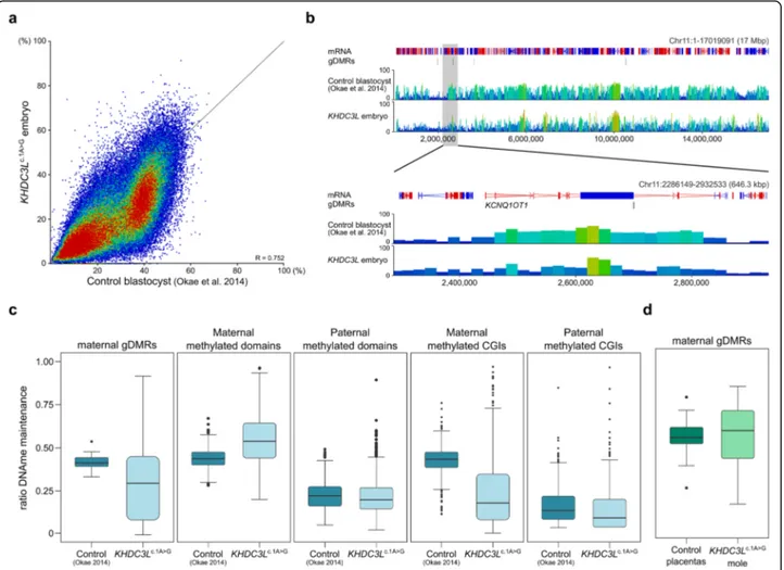
The remaining 2 MII oocytes were subject to intra-cytoplasmic sperm injection from which 1 embryo devel-oped in vitro until the late morula/early blastocyst stage at day 6 (Additional file3: Figure S8A) when it was collected for methylation analysis. Despite being developmentally less advanced than normal blastocysts, the KHDC3Lc.1A>G embryo had reduced methylation in comparison with con-trol blastocysts [30] (median methylation of 20-kb win-dows 19.4 versus 28.3%; Fig. 3a, b) and did not cluster with any embryonic stage by PCA (Additional file 3: Figure S8B), suggesting an anomalous methylation pat-tern. Maternal gDMRs showed variable LoM, as they did in oocytes. (Additional file3: Figure S8C). A residual, gene body methylation pattern inherited from oocytes is apparent in control blastocysts, and this pattern appears attenuated in the KHDC3Lc.1A>G embryo (Fig. 3b), as it does in KHDC3Lc.1A>Goocytes. We inferred the fidelity of methylation maintenance at those gDMRs that had≥ 20% residual methylation in KHDC3Lc.1A>G oocytes by calcu-lating the embryo to oocyte methylation ratio, assuming that perfect maintenance would be reflected as a ratio of 0.5. For maternal gDMRs, the actual figure was 0.44 in control blastocysts, but only 0.32 in the KHDC3Lc.1A>G embryo (median of 38 gDMRs; Fig. 3c, Additional file4: Table S3). CGIs exclusively methylated in oocytes (mater-nal methylated CGIs) showed a similar deficit, whereas other sequence features methylated in oocytes (maternal methylated domains) or methylated in sperm (paternal methylated domains and CGIs) were less affected (Fig.3c). The further reduction in gDMR and maternally methylated CGI methylation in the KHDC3Lc.1A>Gembryo could indi-cate a role for the SCMC also in methylation maintenance mechanisms. However, when we evaluated methylation at
gDMRs in the KHDC3Lc.1A>G mole, there tended to be a maintenance of the residual levels of methylation from the oocyte (Fig.3d).
Discussion
Here, for the first time, we show that an SCMC defect causing BiCHM results in a genome-wide methylation deficit in oocytes. This demonstrates an important appli-cation of single-cell bisulphite sequencing in obtaining high-resolution DNA methylation data from very rare material. With the genome-wide coverage obtained, we were able to answer key questions relating to the origins of methylation defects in BiCHM that could not previ-ously be addressed. Importantly, we identified that ma-ternal gDMRs were affected to a similar magnitude as other sequence features that become methylated de novo in oocytes, demonstrating that there is no specificity of the primary defect towards imprinted loci. A primary ef-fect in female gametes is consistent with earlier reports that methylation at the H19 paternal gDMR is un-affected in BiCHM [12]. That BiCHM tissue in compari-son has a generally preserved methylation status, apart from imprinted gDMRs, indicates that these conceptuses are competent for de novo methylation of the genome after implantation, but that defective methylation at gDMRs arising in oocytes cannot be rescued. We also observed a further reduction in gDMR methylation in the single KHDC3Lc.1A>G preimplantation embryo we were able to assess, which could indicate a role for the integrity of the SCMC also in methylation maintenance mechanisms. Alternatively, the incomplete methylation of CGIs and gDMRs in oocytes, particularly at ZFP57 sites assumed to be critical for methylation maintenance, might allow further erosion of methylation in the preim-plantation embryo during the phase of genome-wide methylation reprogramming. We should note, however, that we were only able to assess a single KHDC3Lc.1A>G preimplantation embryo, which might not be competent for implantation and further development, and may
(See figure on previous page.)
Fig. 2 Genome-wide deficit in DNA methylation inKHDC3Lc.1A>Goocytes. a Global CpG methylation as determined by scBS-seq inKHDC3Lc.1A>GMII oocytes (n = 5) compared with PBAT from a bulk population of control GV/MI oocytes (denoted by Okae et al. [30]) and scBS-seq of MII oocytes (n = 32, denoted by Zhu et al. [31]). Indicated are mean methylation levels for each single oocyte (dots) and the median of each group. b Scatterplot of methylation values of 20-kb windows in groupedKHDC3Lc.1A>Goocytes (n = 5) compared with grouped control oocytes (Zhu et al. [31];n = 32). c Seqmonk browser screenshot of genomic methylation in groupedKHDC3Lc.1A>Goocytes (n = 5) compared with bulk (Okae et al. [30]) and grouped (Zhu et al. [31];n = 32) control oocytes. The zoomed-in view shows methylation over the KCNQ1OT1 imprinted domain. Each vertical bar in both zoomed-out and zoomed-in views is a 20-kb window, height and colour-coded for % methylation. d–f Box and whisker plots showing methylation in grouped control (Zhu et al. [31];n = 32) and KHDC3Lc.1A>Goocytes (n = 5) of oocyte methylated and unmethylated domains (d), methylated and unmethylated CpG islands (CGIs; > 70 and < 20% in control oocytes, respectively) (e) and maternal gDMRs (f). Boxes represent the interquartile range; lines, the medians; whiskers, the 1.5× the interquartile range; dots beyond the whiskers, outliers. The numbers of features per category are given in Additional file4: Table S3. g Heatmap showing methylation of maternal gDMRs in groupedKHDC3Lc.1A>Goocytes (n = 5) and bulk GV/MI (Okae et al. [30]) or grouped MII (Zhu et al. [31];n = 32) control oocytes. h Stripcharts reporting percentage of methylated CpG sites (115) in ZFP57 binding motifs overlapping 32 maternal gDMRs in each single oocyte of control (Zhu et al. [31];n = 32) and KHDC3Lc.1A>Goocytes (n = 5). Dots represent the mean methylation % of all the ZFP57-CpG sites measured in a single oocyte, lines the median of each group. Comparisons in a and d–h are all statistically significant (p < 0.0001). Detailed statistical measures are given in Additional file4: Table S3
therefore manifest more severe methylation defects than an embryo from which molar tissue could arise.
How could a defect in the SCMC—or in the protein components—impair DNA methylation establishment in the oocyte? The mechanisms of de novo methylation in oocytes are best understood in the mouse, benefitting from genetic manipulations [18]. De novo methylation takes place on a genome largely demethylated after spe-cification of primordial germ cells, in the latter stages of oocyte growth (secondary to antral follicle stage), and culminates in a distinctive methylation landscape with methylation preferentially over expressed gene bodies [21,30,35, 38]. Imprinted gDMR methylation is part of this generalised transcription-dependent mechanism [35,
39, 40]. Successful methylation establishment involves the interplay of several nuclear processes. In mice, the required de novo methyltransferase proteins DNMT3A and DNMT3L become abundant in oocytes concomitant with the onset of methylation [41]. Genomic recruitment of DNMT3A/DNMT3L is assumed to depend upon an appropriate chromatin state. DNA methylation coincides with domains of enrichment of histone 3 lysine 36 tri-methylation (H3K36me3) over expressed genes, depos-ited by the unique H3K36me3 methyltransferase SETD2 [42]. Conversely, the histone mark H3K4me3 conven-tionally enriched at active promoters is antagonistic to DNMT3A/3L recruitment and activity [43, 44], and re-moval of H3K4 methylation at gDMRs requires
Fig. 3 Methylation abnormalities in aKHDC3Lc.1A>Gembryo. a Scatterplot comparing methylation determined by PBAT in a single whole
KHDC3Lc.1A>Gpreimplantation embryo with PBAT of a bulk population of control blastocysts (Okae et al. [30]). Each point is a 20-kb window. b
Screenshot of methylation in theKCNQ1OT1 imprinted domain in control blastocysts (Okae et al. [30]) and theKHDC3Lc.1A>Gembryo. Each vertical
bar is a 20-kb window, height and colour-coded for % methylation. c Box and whisker plots comparing ratio of methylation maintenance in control blastocysts and theKHDC3Lc.1A>Gembryo at maternal gDMRs retaining≥ 20% DNA methylation in the oocyte (n = 38),
maternal-methylated domains (n = 285), paternal-methylated domains (n = 2471), maternal-methylated CGIs (n = 735) and paternal-methylated CGIs (n = 299). d Box and whisker plots comparing methylation maintenance ratio in control blastocysts and theKHDC3Lc.1A>Gmole at maternal and
placenta-specific gDMRs (n = 40) retaining ≥ 20% DNA methylation in KHDC3Lc.1A>Goocytes. In c and d, boxes represent the interquartile range;
lines, the medians; whiskers, the 1.5× the interquartile range; and dots beyond the whiskers, outliers. Comparisons in c and d are all statistically significant (p = 0.0125 for maternal gDMRs in c and p < 0.0001 for all others). Detailed statistical measures are given in Additional file4: Table S3
transcription-coupled nucleosome remodelling and/or activity of H3K4 demethylases such as KDM1B [35, 45,
46]. The normal methylation pattern also depends upon the exclusion of DNMT1 and its auxiliary protein UHRF1 from the nucleus, which otherwise leads to methylation of intergenic regions: this nuclear exclusion depends on the protein STELLA/PGC7 [47]. How could the global effect on methylation we observe in KHDC3Lc.1A>G oocytes be explained? Considering the major role of transcription in specifying methylation in oocytes, a global problem in transcription could lead to a generalised deficit in methylation; however, this seems unlikely, as a major effect on transcription sufficient to attenuate methylation to the magnitude observed would likely be incompatible with full development and matur-ation of the oocyte. RNA-seq analysis of KHDC3Lc.1A>G oocytes would be required to determine whether tran-scription defects could account for the variable methyla-tion loss across maternal gDMRs and other genomic features. Gross reductions in the abundance or nuclear localisation of some of the key players above, such as DNMT3A or SETD2, could also cause the effects ob-served. However, loss of SETD2 in mouse oocytes, in addition to abrogating gene body methylation, leads to substantial methylation gain in intergenic regions [42], which we do not observe in the KHDC3Lc.1A>Goocytes. The involvement of STELLA in sequestering DNMT1/ UHFR1 from the nucleus [47] demonstrates the import-ance of regulated subcellular localisation of selected epi-genetic factors in the oocyte. Conceivably, therefore, normal nuclear localisation of DNMT3A/3L—or other as-sociated factors—in growing oocytes could depend upon an intact SCMC. However, mouse oocytes genetically ab-lated for the SCMC component NLRP2 are reported to have normal nuclear staining of DNMT3A [17], but they are also unlikely to have such pervasive methylation de-fects. Moreover, neither Dnmt3a or Dnmt3L is haploin-sufficient in mouse oocytes, indicating that their nuclear availability would need to be reduced to below 50% of normal levels to substantially affect de novo methylation [48,49]. One mouse gene knockout that phenocopies the global methylation defect of KHDC3Lc.1A>Goocytes is the oocyte-specific ablation of the histone H3 chaperone HIRA [50]. HIRA is responsible for continuous replace-ment of H3/H4 during oogenesis, particularly associated with transcription. In addition to grossly diminished de novo methylation, Hira-deficient oocytes are severely compromised in ovulation, developmental competence, and chromosome compaction and segregation [50], evi-dence of the importance of chromatin regulation during oogenesis. It is also important to bear in mind the poten-tial differences between humans and mice in relation to de novo methylation mechanisms in oocytes [18]. Al-though the striking gene body methylation pattern is
conserved, a recent study found differences in the histone regulatory landscape in human compared to mouse oo-cytes [51], and DNMT3L, an essential co-factor for DNMT3A in the mouse oocyte [48, 52, 53], is not expressed in human oocytes [30, 54]. Furthermore, be-cause KHDC3L, as well as other SCMC members such as NLRP7, do not have mouse orthologues, studying the functional mechanisms by which the SCMC regulates de novo DNA methylation remains challenging.
It was notable that the maternal gDMRs in the KHDC3Lc.1A>G oocytes, preimplantation embryo and mole exhibited a range of LoM; in addition, there was evidence that individual KHDC3Lc.1A>Goocytes varied in the severity of gDMR LoM. In comparison, molar tissue from women of the first reported KHDC3L pedigree had complete LoM at the few gDMRs evaluated [12]. At present, we do not know the reason for this variation in methylation loss. Similar to our patient D, the mutation in the original pedigree was identified in the initiation codon of KHDC3L, but at the + 3 position [4, 12]. In transfections, Parry and co-workers demonstrated the use of the next available in-frame ATG codon (at pos-ition 14) for translation of an amino-terminally trun-cated protein from this mutation [4]. If some residual, truncated protein were translated in vivo, or if other SCMC proteins partially compensate for the loss of KHDC3L, this could account for the partial LoM we ob-serve. Alternatively, if KHDC3L deficiency results in a structural defect in the SCMC that impairs the normal localisation or activity of factors essential for methyla-tion, it might manifest at a time in oocyte growth when de novo methylation is already in progress, such that it attenuates rather than completely prevents methylation. Consistent with this possibility, studies in mice indicate there is asynchrony in the acquisition of methylation of gDMRs and CGIs during oocyte growth [55–57], and re-cent single-cell analysis of growing and fully-grown oo-cytes shows some heterogeneity amongst oooo-cytes in methylation of maternal gDMRs [58]. This has some similarity with the variation in gDMR methylation we observe in KHDC3Lc.1A>Goocytes, again consistent with the notion that the progression of de novo methylation during oocyte growth is impaired. Future analysis of the de novo methylation process during normal human oo-genesis, as well as knowledge of the timing of elabor-ation of the SCMC, may help resolve this issue.
Conclusions
In conclusion, we show that a mutation causing BiCHM results in a substantial and generalised reduction in methylation in oocytes, together with impaired mainten-ance of gDMR methylation during preimplantation de-velopment, and persistence of methylation defects preferentially at imprinted loci post-implantation. The
discovery of a primary oocyte defect will now focus at-tention on how the mechanism of methylation establish-ment is globally impaired by defects in the SCMC. It also has important implications for possible therapeutic interventions in patient oocytes; these would be very challenging and, if to be considered, would have to aim to restore de novo methylation during oocyte growth, perhaps by injection of KHDC3L cRNA into oocytes growing in in vitro follicle culture systems [59–61].
Supplementary information
Supplementary information accompanies this paper athttps://doi.org/10.
1186/s13073-019-0694-y.
Additional file 1 : Table S1: List of gDMR coordinates; list of ZFP57 binding sites in gDMRs; .xls, 25 KB.
Additional file 2 : Table S2: Pyrosequencing primer sequences; .xls, 12 KB. Additional file 3: Figure S1. Characterization of Patient D: A. Sequence ofKHDC3L mutation c.1A>G in patient; B. Extended pedigree. Figure S2. Characterization of KHDC3Lc.1A>Gmolar tissue: A. Microsatellite analysis; B. Sequence ofKHDC3L mutation c.1A>G in molar tissue. Figure S3. DNA methylation analysis ofKHDC3Lc.1A>Gmole: A. Principal component analysis of EPIC array data; B. Barchart of DNA methylation changes of genomic features; C. Heatmap of methylation at imprinted DMRs; D. Pyrosequencing validation of methylation changes at imprinted DMRs; E. Barchart of methylation changes at gDMRs and CpG islands. Figure S4. Characterization ofKHDC3Lc.1A>Goocytes: A. Photomicrograph of oocytes; B. Principal component analysis of low-depth scBS-seq datasets; C. Gen-ome browser screenshot of low-depth scBS-seq datasets. Figure S5. Single-cell DNA methylation analysis ofKHDC3Lc.1A>Goocytes: A. Genome browser screenshot of deeply-sequenced scBS-seq datasets; B. Principal component analysis of low-depth scBS-seq datasets; C. Stripchard of non-CpG methylation levels inKHDC3Lc.1A>Goocytes. Figure S6. Variability of DNA methylation loss inKHDC3Lc.1A>Goocytes. A. Barchart of DNA methy-lation changes at genomic features; B. Box:whisker plot of DNA methyla-tion loss at gDMRs compared with matched non-gDMR features; C. Beanplot of DNA methylation loss over gene bodies; D. Scatterplot of DNA methylation changes correlated with transcript levels; E. Stripchart of DNA methylation changes at repetitive elements. Figure S7. Single-cell variation of DNA methylation of imprinted gDMRs inKHDC3Lc.1A>G oocytes. A: Heatmap of methylation of gDMRs and B: Stripchart of methy-lation of gDMRs in individual oocytes. Figure S8. DNA methymethy-lation ana-lysis ofKHDC3Lc.1A>Gembryo. A. Photomicrograph of preimplantation embryo; B. Principal component analysis of methylation ofKHDC3Lc.1A>G embryo and published datasets; C. Heatmap of gDMR methylation in KHDC3Lc.1A>G
embryo. (.word 17.25MB.)
Additional file 4 : Table S3. Summary of statistical analysis in Figures and Supplementary Figures; .xls, 24 KB.
Additional file 5 : Table S4. Hypomethylated regions inKHDC3Lc.1A>G
mole; .xls, 56 KB
Additional file 6 : Table S5. DNA methylation sequencing summary; .xls, 10 KB.
Abbreviations
AnCHM:Androgenetic complete hydatidiform mole; BiCHM: Biparental complete hydatidiform mole; CGIs: CpG islands; gDMRs: Germline differentially methylated regions; GV: Germinal vesicle; LoM: Loss of methylation; MI: Metaphase I; MII: Metaphase II; MLID: Multi-locus imprinting disorder; PBAT: Post-bisulphite adaptor tagging; PCA: Principal component analysis; scBS-seq: Single-cell bisulphite sequencing; SCMC: Subcortical maternal complex; WGBS: Whole-genome bisulphite sequencing
Acknowledgements
We would like to thank M. Fardaei and J. Fallahi of the Genetics Department of Shiraz University of Medical Sciences for contributing to genotyping the
patient, S. Clark for the initial assistance with the scBS-seq experimental de-sign and K. Tabbada and C. Munare of the Babraham Next Generation Se-quencing Facility for their contribution to data generation. We thank the Babraham Bioinformatics Unit for their support with data analysis, especially F. Krueger for the contribution to sequencing QC, mapping and data submis-sion; S. Wingett for the ZFP57 binding site motive search; and A. Segonds-Pichon for the input into the statistical approach. Our special thanks go to the patient and her husband for enabling this study with their oocyte donation.
Data citation
Bernstein BE, Meissner A. BI Human Reference Epigenome Mapping Project (GEO accession GSE17312). Bisulfite-seq analysis of WGBS_Lib 68 derived from human placenta cells, WGBS_Lib 68. (GSM1186665).
Demond H, Anvar Z, Jahromi BN, Davari M, Andrews S, Kelsey G. Impaired DNA methylation in oocytes with a mutation in KHDC3L causing a recurrent hydatidiform mole (GEO accession GSE122872).
Hernandez JR, Monk D. Placenta-specific DMRs maintain methylation across gestation (GEO accession GSE121056).
Huang K, Zhigang X, Fan G. Genetic Programs in Human and Mouse Early Embryos revealed by single-cell RNA sequencing (GEO accession GSE44183). Okae H, Takahiro A. DNA methylation dynamics during early human development (DDBJ accession DRP002710).
Zhu P, Guo H, Ren Y, Hou Y, Yan L, Qiao J, Tang F. Single-cell DNA methy-lome sequencing of human preimplantation embryos (GEO accession GSE81233).
Demond H, Anvar Z, Jahromi BN, Sparago A, Verma A, Davari M, Calzari L, Russo S, Jahromi MA, Monk D, Andrews S, Riccio A, Kelsey G. A KHDC3L mutation resulting in recurrent hydatidiform mole causes genome-wide DNA methylation loss in oocytes and persistent imprinting defects post-fertilisation (GEO accession GSE138864).
Authors’ contributions
ZA conceived the study in discussion with GK. BNJ, MD and MAJ provided the patient materials. HD, ZA, AS, LC and SR performed the molecular analyses. HD, AV and SA performed the bioinformatic analysis. AR advised the genetic and array analysis. DM provided the essential materials. HD, ZA, AR and GK wrote the manuscript with input from all other authors. All authors read and approved the final manuscript.
Funding
Work in GK’s lab is supported by grants from the UK Medical Research Council (MRC; MR/K011332/1, MR/S000437/1) and Biotechnology and Biological Sciences Research Council (BBSRC; BBS/E/B/000C0423). Work in AR’s and SR’s labs is supported by grants from Italian MIUR-PRIN 2015 JHLY35, Telethon-Italia GGP15131, Associazione Italiana Ricerca sul Cancro IG 2016 N.18671,‘Progetti per la ricerca oncologica della Regione Campania’ Grant: I-Cure and‘Progetti competitivi intra-Ateneo’ Programma VALERE (VAnviteLli pEr la RicErca) 2019 Grant: MIRIAM, Università degli studi della Campania‘Luigi Vanvitelli’. AS was supported by Programma VALERE (VAnvi-teLli pEr la RicErca) from Università degli studi della Campania‘Luigi Vanvi-telli’. ZA and BNJ were supported by grant 95-01-50-12690 from the Shiraz University of Medical Sciences.
Availability of data and materials
Sequence and microarray data that support the findings of this study have been deposited under accession codes GSE121056, GSE138864 and GSE122872 in the Gene Expression Omnibus database available athttps://www.ncbi.nlm. nih.gov/geo/. Publically available datasets were sourced from DDBJ (https://
www.ddbj.nig.ac.jp/index-e.html) and GEO databases: accession DRP002710
[30], GSE81233 [31], GSE17312 (GSM1186665) [27] and GSE44183 [32].
Ethics approval and consent to participate
Oocytes were obtained from Patient D and sperm from the
husband voluntarily at the IVF centre of Ghadir Mother and Child Hospital affiliated to Shiraz University of Medical Sciences with signed informed consent of the patient and her husband and the approval of the Ethics Committee of Shiraz University of Medical Sciences (ethics codes: IR.sums.rec.1395.S718 for oocyte retrieval and IR.sums.rec.1396.S779 for embryo production). All procedures performed in this study were in accordance with the Helsinki Declaration.
Consent for publication
The patient and husband consented to publication of information reported in this study.
Competing interests
The authors declare that they have no competing interests.
Author details
1Epigenetics Programme, Babraham Institute, Cambridge, UK.2Infertility
Research Center, Shiraz University of Medical Sciences, Shiraz, Iran.
3Department of Obstetrics and Gynecology, School of Medicine, Shiraz
University of Medical Sciences, Shiraz, Iran.4Institute of Genetics and Biophysics‘Adriano Buzzati-Traverso’, Consiglio Nazionale delle Ricerche (CNR), Naples, Italy.5Dipartimento di Scienze e Tecnologie Ambientali Biologiche e Farmaceutiche, Università degli Studi della Campania‘Luigi Vanvitelli’, Caserta, Italy.6IVF Section, Ghadir-Mother and Child Hospital of Shiraz, Shiraz, Iran.7Medical Cytogenetics and Molecular Genetics Laboratory,
Centro di Ricerche e Tecnologie Biomediche IRCCS, Istituto Auxologico Italiano, Milan, Italy.8Department of Pathology, Shiraz University of Medical
Sciences, Shiraz, Iran.9Imprinting and Cancer Group, Bellvitge Biomedical Research Institute (IDIBELL), L’Hospitalet de Llobregat, Barcelona, Spain.
10
Bioinformatics Group, Babraham Institute, Cambridge, UK.11Centre for Trophoblast Research, University of Cambridge, Cambridge, UK.
Received: 27 June 2019 Accepted: 15 November 2019
References
1. Li L, Baibakov B, Dean J. A subcortical maternal complex essential for preimplantation mouse embryogenesis. Dev Cell. 2008;15(3):416–25. 2. Lu X, Gao Z, Qin D, Li L. A maternal functional module in the mammalian
oocyte-to-embryo transition. Trends Mol Med. 2017;23(11):1014–23. 3. Murdoch S, Djuric U, Mazhar B, Seoud M, Khan R, Kuick R, et al. Mutations in
NALP7 cause recurrent hydatidiform moles and reproductive wastage in humans. Nat Genet. 2006;38(3):300–2.
4. Parry DA, Logan CV, Hayward BE, Shires M, Landolsi H, Diggle C, et al. Mutations causing familial biparental hydatidiform mole implicate c6orf221 as a possible regulator of genomic imprinting in the human oocyte. Am J Hum Genet. 2011;89(3):451–8.
5. Docherty LE, Rezwan FI, Poole RL, Turner CL, Kivuva E, Maher ER, et al. Mutations in NLRP5 are associated with reproductive wastage and multilocus imprinting disorders in humans. Nat Commun. 2015;6:8086. 6. Alazami AM, Awad SM, Coskun S, Al-Hassan S, Hijazi H, Abdulwahab FM,
et al. TLE6 mutation causes the earliest known human embryonic lethality. Genome Biol. 2015;16:240.
7. Xu Y, Shi Y, Fu J, Yu M, Feng R, Sang Q, et al. Mutations in PADI6 cause female infertility characterized by early embryonic arrest. Am J Hum Genet. 2016;99(3):744–52.
8. Begemann M, Rezwan FI, Beygo J, Docherty LE, Kolarova J, Schroeder C, et al. Maternal variants in NLRP and other maternal effect proteins are associated with multilocus imprinting disturbance in offspring. J Med Genet. 2018;55(7):497–504.
9. Mu J, Wang W, Chen B, Wu L, Li B, Mao X, et al. Mutations in NLRP2 and NLRP5 cause female infertility characterised by early embryonic arrest. J Med Genet. 2019;56(7):471-80.
10. Reddy R, Akoury E, Phuong Nguyen NM, Abdul-Rahman OA, Dery C, Gupta N, et al. Report of four new patients with protein-truncating mutations in C6orf221/KHDC3L and colocalization with NLRP7. Eur J Hum Genet. 2013; 21(9):957–64.
11. Zhu K, Yan L, Zhang X, Lu X, Wang T, Yan J, et al. Identification of a human subcortical maternal complex. Mol Hum Reprod. 2015;21(4):320–9. 12. Judson H, Hayward BE, Sheridan E, Bonthron DT. A global disorder of
imprinting in the human female germ line. Nature. 2002;416(6880):539–42. 13. Sanchez-Delgado M, Martin-Trujillo A, Tayama C, Vidal E, Esteller M,
Iglesias-Platas I, et al. Absence of maternal methylation in Biparental Hydatidiform moles from women with NLRP7 maternal-effect mutations reveals widespread placenta-specific imprinting. PLoS Genet. 2015;11(11):e1005644. 14. Qian J, Nguyen NMP, Rezaei M, Huang B, Tao Y, Zhang X, et al. Biallelic
PADI6 variants linking infertility, miscarriages, and hydatidiform moles. Eur J Hum Genet. 2018;26(7):1007–13.
15. Tong ZB, Gold L, Pfeifer KE, Dorward H, Lee E, Bondy CA, et al. Mater, a maternal effect gene required for early embryonic development in mice. Nat Genet. 2000;26(3):267–8.
16. Yu XJ, Yi Z, Gao Z, Qin D, Zhai Y, Chen X, et al. The subcortical maternal complex controls symmetric division of mouse zygotes by regulating F-actin dynamics. Nat Comm. 2014;5:4887.
17. Mahadevan S, Sathappan V, Utama B, Lorenzo I, Kaskar K, Van den Veyver IB. Maternally expressed NLRP2 links the subcortical maternal complex (SCMC) to fertility, embryogenesis and epigenetic reprogramming. Sci Rep. 2017;7: 44667.
18. Hanna CW, Demond H, Kelsey G. Epigenetic regulation in development: is the mouse a good model for the human? Hum Reprod Update. 2018;24(5):556–76. 19. Kelsey G, Feil R. New insights into establishment and maintenance of DNA
methylation imprints in mammals. Philos Trans R Soc London B Biol Sci. 2013;368(1609):20110336.
20. Monk D, Mackay DJG, Eggermann T, Maher ER, Riccio A. Genomic imprinting disorders: lessons on how genome, epigenome and environment interact. Nat Rev Genet. 2019;20(4):235–48.
21. Smallwood SA, Lee HJ, Angermueller C, Krueger F, Saadeh H, Peat J, et al. Single-cell genome-wide bisulfite sequencing for assessing epigenetic heterogeneity. Nat Methods. 2014;11(8):817–20.
22. Morris TJ, Butcher LM, Feber A, Teschendorff AE, Chakravarthy AR, Wojdacz TK, et al. ChAMP: 450k Chip analysis methylation pipeline. Bioinformatics. 2014;30(3):428–30.
23. Maksimovic J, Gordon L, Oshlack A. SWAN: subset-quantile within array normalization for illumina infinium HumanMethylation450 BeadChips. Genome Biol. 2012;13(6):R44.
24. Zhao H, Sun Z, Wang J, Huang H, Kocher JP, Wang L. CrossMap: a versatile tool for coordinate conversion between genome assemblies. Bioinformatics. 2014;30(7):1006–7.
25. Quinlan AR, Hall IM. BEDTools: a flexible suite of utilities for comparing genomic features. Bioinformatics. 2010;26(6):841–2.
26. Hanna CW, Penaherrera MS, Saadeh H, Andrews S, McFadden DE, Kelsey G, et al. Pervasive polymorphic imprinted methylation in the human placenta. Genome Res. 2016;26(6):756–67.
27. Bernstein BE, Stamatoyannopoulos JA, Costello JF, Ren B, Milosavljevic A, Meissner A, et al. The NIH roadmap Epigenomics mapping consortium. Nat Biotechnol. 2010;28(10):1045–8.
28. Angermueller C, Clark SJ, Lee HJ, Macaulay IC, Teng MJ, Hu TX, et al. Parallel single-cell sequencing links transcriptional and epigenetic heterogeneity. Nat Methods. 2016;13(3):229–32.
29. Hanna CW, Taudt A, Huang J, Gahurova L, Kranz A, Andrews S, et al. MLL2 conveys transcription-independent H3K4 trimethylation in oocytes. Nat Struct Mol Biol. 2018;25(1):73–82.
30. Okae H, Chiba H, Hiura H, Hamada H, Sato A, Utsunomiya T, et al. Genome-wide analysis of DNA methylation dynamics during early human development. PLoS Genet. 2014;10(12):e1004868.
31. Zhu P, Guo H, Ren Y, Hou Y, Dong J, Li R, et al. Single-cell DNA methylome sequencing of human preimplantation embryos. Nat Genet. 2018;50(1):12–9. 32. Xue Z, Huang K, Cai C, Cai L, Jiang CY, Feng Y, et al. Genetic programs in
human and mouse early embryos revealed by single-cell RNA sequencing. Nature. 2013;500(7464):593–7.
33. Quenneville S, Verde G, Corsinotti A, Kapopoulou A, Jakobsson J, Offner S, et al. In embryonic stem cells, ZFP57/KAP1 recognize a methylated hexanucleotide to affect chromatin and DNA methylation of imprinting control regions. Mol Cell. 2011;44(3):361–72.
34. Anvar Z, Cammisa M, Riso V, Baglivo I, Kukreja H, Sparago A, et al. ZFP57 recognizes multiple and closely spaced sequence motif variants to maintain repressive epigenetic marks in mouse embryonic stem cells. Nucleic Acids Res. 2016;44(3):1118–32.
35. Veselovska L, Smallwood SA, Saadeh H, Stewart KR, Krueger F, Maupetit-Mehouas S, et al. Deep sequencing and de novo assembly of the mouse oocyte transcriptome define the contribution of transcription to the DNA methylation landscape. Genome Biol. 2015;16:209.
36. Li X, Ito M, Zhou F, Youngson N, Zuo X, Leder P, et al. A maternal-zygotic effect gene, Zfp57, maintains both maternal and paternal imprints. Dev Cell. 2008;15(4):547–57.
37. Mackay DJ, Callaway JL, Marks SM, White HE, Acerini CL, Boonen SE, et al. Hypomethylation of multiple imprinted loci in individuals with transient neonatal diabetes is associated with mutations in ZFP57. Nat Genet. 2008; 40(8):949–51.
38. Kobayashi H, Sakurai T, Imai M, Takahashi N, Fukuda A, Yayoi O, et al. Contribution of intragenic DNA methylation in mouse gametic DNA methylomes to establish oocyte-specific heritable marks. PLoS Genet. 2012; 8(1):e1002440.
39. Chotalia M, Smallwood SA, Ruf N, Dawson C, Lucifero D, Frontera M, et al. Transcription is required for establishment of germline methylation marks at imprinted genes. Genes Dev. 2009;23(1):105–17.
40. Smith EY, Futtner CR, Chamberlain SJ, Johnstone KA, Resnick JL.
Transcription is required to establish maternal imprinting at the Prader-Willi syndrome and Angelman syndrome locus. PLoS Genet. 2011;7(12):e1002422. 41. Hara S, Takano T, Fujikawa T, Yamada M, Wakai T, Kono T, et al. Forced
expression of DNA methyltransferases during oocyte growth accelerates the establishment of methylation imprints but not functional genomic imprinting. Hum Mol Genet. 2014;23(14):3853–64.
42. Xu Q, Xiang Y, Wang Q, Wang L, Brind’Amour J, Bogutz AB, et al. SETD2 regulates the maternal epigenome, genomic imprinting and embryonic development. Nat Genet. 2019;51(5):844–56.
43. Ooi SK, Qiu C, Bernstein E, Li K, Jia D, Yang Z, et al. DNMT3L connects unmethylated lysine 4 of histone H3 to de novo methylation of DNA. Nature. 2007;448(7154):714–7.
44. Guo X, Wang L, Li J, Ding Z, Xiao J, Yin X, et al. Structural insight into autoinhibition and histone H3-induced activation of DNMT3A. Nature. 2015; 517(7536):640–4.
45. Ciccone DN, Su H, Hevi S, Gay F, Lei H, Bajko J, et al. KDM1B is a histone H3K4 demethylase required to establish maternal genomic imprints. Nature. 2009;461(7262):415–8.
46. Stewart KR, Veselovska L, Kim J, Huang J, Saadeh H, Tomizawa S, et al. Dynamic changes in histone modifications precede de novo DNA methylation in oocytes. Genes Dev. 2015;29(23):2449–62.
47. Li Y, Zhang Z, Chen J, Liu W, Lai W, Liu B, et al. Stella safeguards the oocyte methylome by preventing de novo methylation mediated by DNMT1. Nature. 2018;564(7734):136–40.
48. Bourc’his D, Xu GL, Lin CS, Bollman B, Bestor TH. Dnmt3L and the establishment of maternal genomic imprints. Science. 2001;294(5551): 2536–9.
49. Kaneda M, Hirasawa R, Chiba H, Okano M, Li E, Sasaki H. Genetic evidence for Dnmt3a-dependent imprinting during oocyte growth obtained by conditional knockout with Zp3-Cre and complete exclusion of Dnmt3b by chimera formation. Genes Cells. 2010;15(3):169–79.
50. Nashun B, Hill PW, Smallwood SA, Dharmalingam G, Amouroux R, Clark SJ, et al. Continuous histone replacement by Hira is essential for Normal transcriptional regulation and De novo DNA methylation during mouse oogenesis. Mol Cell. 2015;60(4):611–25.
51. Xia W, Xu J, Yu G, Yao G, Xu K, Ma X, et al. Resetting histone modifications during human parental-to-zygotic transition. Science. 2019;365(6451):353–60. 52. Smallwood SA, Tomizawa S, Krueger F, Ruf N, Carli N, Segonds-Pichon A,
et al. Dynamic CpG island methylation landscape in oocytes and preimplantation embryos. Nat Genet. 2011;43(8):811–4.
53. Shirane K, Toh H, Kobayashi H, Miura F, Chiba H, Ito T, et al. Mouse oocyte methylomes at base resolution reveal genome-wide accumulation of non-CpG methylation and role of DNA methyltransferases. PLoS Genet. 2013;9(4): e1003439.
54. Guo H, Zhu P, Yan L, Li R, Hu B, Lian Y, et al. The DNA methylation landscape of human early embryos. Nature. 2014;511(7511):606–10. 55. Hiura H, Obata Y, Komiyama J, Shirai M, Kono T. Oocyte growth-dependent
progression of maternal imprinting in mice. Genes Cells. 2006;11(4):353–61. 56. Lucifero D, Mann MR, Bartolomei MS, Trasler JM. Gene-specific timing
and epigenetic memory in oocyte imprinting. Hum Mol Genet. 2004; 13(8):839–49.
57. Gahurova L, Tomizawa SI, Smallwood SA, Stewart-Morgan KR, Saadeh H, Kim J, et al. Transcription and chromatin determinants of de novo DNA methylation timing in oocytes. Epigenetics Chromatin. 2017;10:25. 58. Gu C, Liu S, Wu Q, Zhang L, Guo F. Integrative single-cell analysis of
transcriptome, DNA methylome and chromatin accessibility in mouse oocytes. Cell Res. 2019;29(2):110–23.
59. Telfer EE, McLaughlin M, Ding C, Thong KJ. A two-step serum-free culture system supports development of human oocytes from primordial follicles in the presence of activin. Hum Reprod. 2008;23(5):1151–8.
60. McLaughlin M, Albertini DF, Wallace WHB, Anderson RA, Telfer EE. Metaphase II oocytes from human unilaminar follicles grown in a multi-step culture system. Mol Hum Reprod. 2018;24(3):135–42.
61. Sang Q, Li B, Kuang Y, Wang X, Zhang Z, Chen B, et al. Homozygous mutations in WEE2 cause fertilization failure and female infertility. Am J Hum Genet. 2018;102(4):649–57.
Publisher’s Note
Springer Nature remains neutral with regard to jurisdictional claims in published maps and institutional affiliations.