1
UNIVERSITÀ POLITECNICA DELLE MARCHE
SCUOLA DI DOTTORATO DI RICERCA DELLA FACOLTÀ DI MEDICINA E CHIRURGIA
CORSO DI DOTTORATO IN SALUTE DELL’UOMO XV CICLO
THE ROLE OF OBSTRUCTIVE SLEEP APNEA
SYNDROME IN THE PATHOGENESIS AND EVOLUTION
OF DEMENTIA
D
OCENTET
UTOR:
D
OTTORANDOChiar.mo Prof. Mauro Silvestrini
Dott. ssa Laura Buratti
C
OORDINATORE:
Chiar.mo Prof. Armando Gabrielli
2
SUMMARY
1. INTRODUCTION p. 3
1.1 SLEEP AND COGNITIVE DECLINE: A STRONG BIDIRECTIONAL RELATIONSHIP p. 3 1.2 OBSTRUCTIVE SLEEP APNEA AND COGNITIVE IMPAIRMENT p. 41.2.1 ANATOMO-PATHOLOGICAL ASPECTS AND POSSIBLE MECHANISMS INVOLVED IN OBSTRUCTIVE SLEEP APNEA SYNDROME-RELATED CEREBRAL DAMAGE p. 5
1.2.2 ISSUES UNDERLYING THE ASSOCIATION BETWEEN OBSTRUCTIVE SLEEP APNEA SYNDROME AND ALZHEIMER’S DISEASE AND THE POSSIBLE ROLE OF VASCULAR FACTORS p. 5
2. SUBJECTS AND METHODS p. 6
3. RESULTS p. 10
4. DISCUSSION p. 12
5. REFERENCES p.16
TABLE 1 p.23
TABLE 2 p.24
TABLE 3 p.25
TABLE 4 p.26
FIGURE 1 p.27
3
1. INTRODUCTION
1.1 SLEEP AND COGNITIVE DECLINE: A STRONG BIDIRECTIONAL RELATIONSHIP
Sleep exerts a significant role in the regulation of cerebral activity and in the preservation of brain anatomic integrity through different and complex mechanisms. An involvement in synaptic plasticity has been demonstrated: during the night, a remodeling of synapses occurs and, in particular, only necessary synapses are preserved.1 Moreover, a scavenger role has been suggested by recent experimental studies indicating that cortical interstitial spaces increase by more than 60% during sleep, resulting in an improved efficiency in amyloid beta (Aβ) and other compounds clearance.2 The dramatic increase in the incidence of
Alzheimer’s disease (AD) and other types of dementia and the lack of effective therapies have stimulated the search for strategies to prevent or delay cognitive decline onset and/or progression.3 Several conditions have been considered in an attempt to improve the pathophysiologic knowledge and the consequent therapeutic possibilities in demented patients. Sleep problems could contribute to the pathology of AD and to the progression of cognitive impairment.4 In mouse models of AD, chronic sleep deprivation increases the deposition of Aβ plaques, while improving sleep quality with an orexin receptor antagonist reduces Aβ plaques concentration.5
Preservation of a restorative sleep allows the memory consolidation, in particular a transition from short-term memory to the long-short-term memory, through the transfer of information from hippocampus to the anterior brain regions.6
Sleep disturbances and disruption of the neural regulation of the sleep-wake rhythm appear to be involved in the cellular and molecular mechanisms of cognitive decline. In this respect, several studies have suggested that sleep disorders are associated with an increased risk of incident dementia and cognitive decline.7-11
Sleep is a dynamic process involving a network that may be altered by degenerative processes. Cholinergic connections of the brain stem also seem to be involved in the sleep-wake rhythm dysregulation that is early detected in clinical and preclinical AD patients.12,13
4 Sleep disturbances are highly prevalent in community-dwelling patients with AD and they are also very common in fronto-temporal, vascular, Lewy body (LB) and Parkinson Disease (PD) dementia.14-17
AD patients exhibit many sleep disturbances since the early stages of the disease. In particular, the results of recent epidemiological investigations underlined a very high prevalence of sleep disordered breathing (SDB).16 In preclinical AD, Aβ deposition, as assessed by cerebrospinal fluid (CSF) Aβ 42 levels, seems to be associated with low sleep quality.18 In LB and PD dementia, sleep disorders, including excessive daytime sleepiness (EDS), nocturnal parasomnias, and rapid eye movement (REM) behavior disorder have been recognized as prodromal features that may contribute to the typical fluctuations of the diseases. In this respect treatment can be expected to improve patients’ quality of life.19,20
Despite the high prevalence, the probable pathogenic role and the clinical and social implications, sleep disturbances have not been systematically and carefully investigated in clinical settings and thus they are probably underestimated and do not receive sufficient attention in the global management of patients with dementia.4
1.2 OBSTRUCTIVE SLEEP APNEA AND COGNITIVE IMPAIRMENT
Among SDB, obstructive sleep apnea syndrome (OSAS) is a highly prevalent condition in the general population. The incidence of OSAS in the community rises steadily with age, from less than 3% in children to 4-9% in middle-aged subjects, but up to 21-44% in the elderly.21 OSAS is characterized by instability of the upper airway during sleep, which results in markedly reduced (hypopnea) or absent (apnea) airflow at the nose/mouth level and diurnal dysfunctions such as sleepiness and cognitive difficulties. Apnea/hypopnea episodes are typically accompanied by oxyhemoglobin desaturation and terminated by brief microarousals that result in sleep fragmentation and diminished amounts of slow wave and REM sleep.4 Sleep apnea is present in 33-53 % of patients with probable AD.22 In recent years, the demonstration of an association between apolipoprotein E Ɛ4 allele, the best established genetic risk factor for sporadic AD, and SDB, has raised strong interest for the possible association between OSAS and cognitive decline.23,24
5
1.2.1 ANATOMO-PATHOLOGICAL ASPECTS AND POSSIBLE MECHANISMS INVOLVED IN OBSTRUCTIVE SLEEP APNEA SYNDROME-RELATED CEREBRAL DAMAGE
OSAS-damaged brain regions are located in areas supplied by the anterior and posterior cerebral arteries. Vascular-metabolic balance in these regions is more fragile and susceptible to reach the threshold for ischemic damage during the unfavorable conditions triggered by the hypoxic periods resulting from OSAS.25,26 Further, the cholinergic innervation of the anterior and posterior cerebral arteries is preferentially affected in OSAS patients which explains an impaired reactive dilation of these arteries during apneic episodes.27,28
The different mechanisms involved in the pathogenesis of brain damage in OSAS, including oxidative stress, inflammation, increased blood viscosity due to a pro-inflammatory cytokine effect on coagulation factors and adhesion molecules and blood-brain barrier dysfunction can, at the same time, be stimulated by a critical flow condition and amplify its effects.29,30
1.2.2 ISSUES UNDERLYING THE ASSOCIATION BETWEEN OBSTRUCTIVE SLEEP APNEA SYNDROME AND ALZHEIMER’S DISEASE AND THE POSSIBLE ROLE OF VASCULAR FACTORS
OSAS is widely diffused among elderly people.31 According with the roles of sleep in maintaining cerebral anatomic integrity and functions (synaptic plasticity, memory consolidation and scavenger), the main features of OSAS—sleep fragmentation and hypoxia— may have negative effects on cognitive activity.32,33 The deconstruction of sleep architecture secondary to OSAS alters the consolidation of memories.34 Poor sleep quality in elderly is associated with increased brain levels of Aβ, measured by PET scanning.35,36 Based on the results of recent clinical investigations showing the role of vascular factors in influencing cognitive deterioration in AD patients,37,38 it has been suggested that the link between OSAS and AD may be due to the induction of circulatory unfavorable changes. In OSAS patients, different anatomic and functional unfavorable changes involving respiratory, biochemical and cerebral hemodynamic mechanisms occur during sleep. The results of experimental investigations have shown that degenerative arterial wall changes are directly related to oxygen desaturation.39 Further, pathological vascular changes may be also related to
6 the instability of cardiovascular activity as well as to changes in blood viscosity that has been described in OSAS.40,41
In summary, the interaction between sleep alterations and neurodegeneration is complex and bidirectional. OSAS patients exhibit vascular alterations and brain damages topography similar to AD patients. Based on these considerations, it can be hypothesized that an early treatment of OSAS should, slow or, at least, partially reverse the pathogenic mechanisms involved in the development of cognitive and vascular damage.
The main purposes of this study were to assess the neuropsychological patterns of patients with OSAS, and to obtain information about the impact of OSAS on the risk of developing cognitive impairment. The possibility to delay or slow the progression of cognitive impairment through an early diagnosis and treatment was evaluated. In the attempt to expand knowledge about the complex interactions among degenerative, sleep profile and vascular alterations in influencing the cognitive status in OSAS patients, a careful evaluation of cerebral hemodynamics and polysomnographic data was performed.
2. SUBJECTS AND METHODS
Patients were selected from consecutive subjects referred to our Sleep outpatient service by general practitioners from 2014 to 2015 for a suspected OSAS. The following exclusion criteria were considered:
native language different from Italian;
diagnosis of dementia;
years of education < three years;
family history of cognitive impairment;
exposure or history of substance abuse in the last 12 months;
ongoing therapy with drugs that may impair cognitive functions;
other neurological diseases;
significant neck vessels stenosis;
7
mental retardation or psychiatric disorders that might interfere on neuropsychological performances;
cancer and/or autoimmune diseases;
diseases that can lead to cognitive impairment (such as thyroid disorders, syphilis, deficiency diseases, severe liver and kidney diseases, HIV infection);
any other sleep disorders in addition to OSAS.
A structural interview was performed for each subject in order to deliver accurate information about the possible presence of nocturnal symptoms like nicturia, choking and gastroesophageal reflux and diurnal symptoms like dry mouth, morning headache, memory impairment, personality and mood changes, and EDS. In this phase, EDS and sleep quality during the last month were also investigated by means of the Epworth Sleepiness Scale and Pittsburgh Sleep quality Index respectively.42,43
Patients’ partners were also involved especially in the case of unawareness of snoring and breath interruption episodes. All patients underwent a careful physical examination and information collection to rule out other diseases potentially predisposing to OSAS. The following data were also collected: body mass index (BMI), neck circumference, Mallampati score, heart rate and oxyhemoglobin saturation (SpO2) and presence of craniofacial dysmorphism.
The Berlin questionnaire44 was administered to all patients in order to quantify the risk of OSAS: low and high risk. In high risk patients, a polysomnography (PSG) was performed. Nocturnal PSG recordings were performed using EBNeuro instrument (BE Micro–Holter EEG) The following parameters were recorded: electroencephalogram (EEG) with surface electrodes positioned in F3, F4, C3, C4, O1, O2, electrooculogram (EOG) with 2 electrodes placed 1 cm at the side and 1 cm above or below the external canthus of each eye, mastoid reference (A1 or A2), electromyogram (EMG) with 2 surface electrodes placed in submental region, in correspondence of the mylohyoid muscle. Together with PSG recordings, other parameters were recorded: airflow (using nasal pressure recording), snoring sound (by means of a vibration sensor), electrocardiogram , oxygen saturation (SpO2) of hemoglobin and heart rate obtained from pulse oximetry, thoracic and abdominal movements recorded by using inductive plethysmography, body position (by
8 means of a specific sensor), and thoracic and abdominal respiratory efforts. Sleep events were scored manually according to the American Academy of Sleep Medicine criteria.45
Evaluation of intracranial circle was performed by means of transcranial Doppler (Multidop×DWL; Elektronische Systeme, GmbH, Germany) according to validated criteria.46 Cerebrovascular reactivity to hypercapnia was measured with the Breath-Holding Index (BHI).47 This index is obtained by dividing the percentage increase in mean flow velocity (MFV), occurring during breath-holding, by the length of time (seconds) the subjects hold their breath after a normal inspiration. BHI=([breath-holding MFV - basal MFV/basal MFV]×100/seconds of breath-holding). Two transducers placed on the temporal bone window with a stable angle of insonation secured by a head frame were used to obtain a bilateral continuous measurement of flow velocity of middle cerebral arteries. Subjects were requested to hold their breath for a period of 30 seconds. Breath-holding length and efficiency was monitored by a capnometer. For each patient, three recordings were obtained, and the BHI considered was the mean of all values obtained. For the analysis, left, right and mean (right + left) values were considered.
Regarding the cognitive aspects, all study patients underwent a global cognitive status screening assessment (MMSE)48 followed by a single cognitive domain neuropsychological evaluation. In particular, all subjects were tested with specific and standardized neuropsychological tasks exploring cognitive aspects that have been frequently described as involved in OSAS patients:49
- logical-deductive abilities: Progressive Raven Matrices;50 - selective and sustained attention: Stroop Colour Word Test;51
- verbal and spatial working memory: Digit Span and Corsi Cubes respectively;52 - verbal fluency: Category Fluency Test52 and Letter Fluency Test;53
- constructive praxis: Rey Figure B copy;52,54
- spatial memory: Rey Figure B immediate (ST) and delayed (LT) recall;52,54
- verbal memory: Rey Auditory Verbal Learning Test (Rey AVLT short- term/long- term).53
As controls, a group of age- and sex-matched subjects without OSAS, with a similar vascular profile was included and submitted to the same protocol of cognitive and vascular assessment.
9 After inclusion, in each patient and control, pharmacological treatment of vascular risk factors was planned according to international guidelines.55 For patients affected by OSAS, the most appropriate treatment, including, when necessary, the use of continuous positive airway pressure (CPAP) was recommended. Six months (T1) after inclusion (T0) patients were submitted to the following evaluations:
nocturnal polysomnigraphy to assess the evolution of the sleep parameters (the exam was performed after a 7-day interruption of CPAP);
neuropsychological assessment to evaluate the onset of cognitive impairment or a possible modification of the neuropsychological profile;
ultrasonographic evaluation of intracranial vessels to assess the intracranial hemodynamics.
Clinical conditions, adherence to prescribed medical therapy and, when used, to CPAP treatment were assessed.
All participants gave their informed written consent according to the Declaration of Helsinki. Statistical Analysis
Sex, OSAS severity and OSAS improvement were synthesized as binary variables. We used the OSAS improvement at 6 months as the main grouping variable, and we treated this variable as dichotomous. Subjects who had a polysomnographic improvement after treatment were labeled as group 1, patients who remained stable were labelled as group 2.
Age, left BHI at baseline and at 6 months, right BHI at baseline and at 6 months, mean BHI at baseline and at 6 months, were collected as continuous variables. Polysomnographic parameters: apnea– hypopnea index (AHI), oxygen desaturation index (ODI), % time spent with SaO2 < 90%, % average desaturation, % sleep efficiency, total sleep time (min), % N1 sleep of total sleep time, % N2 sleep of total sleep time, % N3 sleep of total sleep time, % REM sleep of total sleep time, and sleep onset (min) were recorded as continuous variables. Neuropsychologic variables were recorded as continuous variables both at baseline and at 6 months.
Continuous variables were compared with t-test for repeated measures, dichotomous variables were cross-tabulated and compared with chi-squared test. A GLM/multivariate model for repeated
10 measures, considering group 1 and group 2, was set up to analyse the relationship between OSAS improvement after therapy and mean of BHI at baseline and at the follow-up evaluation, controlling for age and sex as covariates. The same model was then adopted to evaluate how improvement of OSAS and BHI could impact on neuropsychological outcomes, selecting only the tests that differed significantly at t-test between baseline and follow-up evaluation.
3. RESULTS
Forty-one OSAS patients and 41 control subjects were included. Mean age was 65.05±8.99 and 66.08±7.88 years respectively. Education and prevalence of hypertension, diabetes, smoking and dyslipidemia was similar in the two groups. All included subjects were right-handed. Thirty-four patients and controls (82.9% of the sample) were males. Patients with an AHI between 5 and 14 were considered to have mild OSAS (n. 6), patients with an AHI between 15 and 30 were considered to have moderate OSAS (n. 25), and patients with an AHI greater than 30 had severe OSAS (n. 10).56 Due to the presence of more than one vascular risk factors in each patients, CPAP treatment was prescribed to all patients, including those with mild OSAS.
Regarding the cognitive profile, t-test showed some significant differences between patients and controls. In particular, the cognitive performances were lower in patients with respect to controls in the following tasks: Stroop Test T1 and T2 (the time required for completion of the first and second part of the test) and E1 and E2 (the number of mistakes made in the first and second part of the test) (p=0.001), Rey AVLT short-term/long-term (p=0.0001 and 0.001, respectively) and semantic and phonetic fluency test (p=0.001). Considering cerebrovascular reactivity, we observed a statistically significant difference between patients and controls in mean BHI (p<0.05). These findings are described in details in table 1.
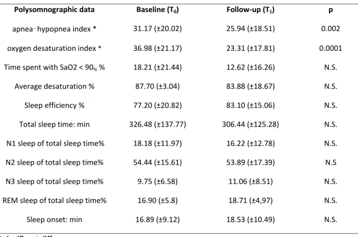
Considering polysomnographic data, a significant difference between T0 and T1 in AHI (T0:
31.17±20.02; T1:25.94±18.51;p=0.002) and ODI (T0: 36.98±21.17; T1: 23.31 ±17.81; p=0.0001) was found,
11 Regarding cerebrovascular reactivity in patients, we observed a statistically significant difference between T0 and T1 in left BHI (T0: 0.746±0.33; T1: 0.898±0.33; p=0.015) and mean BHI (T0: 0.753±0.30; T1:
0.880±0.80; p=0.0001); right BHI did not result significantly different (T0: 0.794±0.36; T1: 0.879±0.37;
p=0.155).
Then, we classified OSAS patients in two groups, group 1 (n=21): subjects who had an improvement of OSAS severity after treatment and group 2 (n=20): subjects whose OSAS severity remained stable. Classification of OSAS as improved or stable was based on the AHI values at T1 that allowed to verify
whether each single patient could be switch to a lower OSAS severity category. The two groups did not differ regarding baseline OSAS severity, body mass index, age, sex distribution and prevalence of hypertension, diabetes, smoking and dyslipidemia. In group 1 patients, we observed a significant difference between the two evaluations in left (T0: 0.660±0.36; T1: 0.917±0.43; p=0.001) and mean BHI (T0:
0.676±0.32; T1: 0.888±0.32; p=0.001). We did not find any difference of cerebrovascular reactivity in group
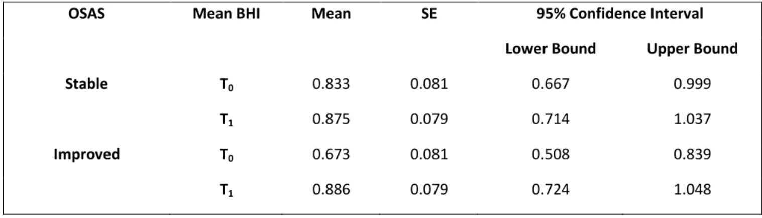
2 patients. Analysing the cognitive profile, we found a significant difference in short-term (p=0.02) and long-term Rey AVLT (p=0.001) in subjects of group 1. Among patients of group 2, we did not find any statistically significant difference between the two evaluations for neuropsychological variables (table 3). GLM/Multivariate model for repeated measures to evaluate changes in cerebrovascular reactivity and neuropsychological variables was applied to assess the differences between subjects of group 1 and group 2. In this model, mean BHI values between the two time points resulted significantly improved for subjects in group 1 (T0: 0.833; 95%CI:0.667-0.999; T1: 0.875; 95%CI:0.714-1.037; p=0.009) while they remained
stable in group 2 patients (T0: 0.833; 95%CI:0.667-0.999; T1: 0.875; 95%CI:0.714-1.037; p=0.009), as shown
in Table 4. We also applied the same multivariate model to left and right BHI, underlining that right BHI was not statistically different in the two time points in both groups, while left BHI was significantly higher at T1
12
4. DISCUSSION
The results of this study show the presence of a significant association between OSAS and reduced efficiency in some cognitive tasks in patients without a history of dementia. The link between reduced cognitive performances and alteration in cerebral hemodynamics suggests a possible pathogenic role of unfavorable circulatory changes in sustaining the cerebral dysfunction in OSAS. This conclusion is based on the simultaneous presence of a significant difference in some neuropsychological performances and extent of cerebrovascular reactivity alteration between patients and controls. Episodes of apnea-hypopnea are typically accompanied by brief microarousals that result in sleep fragmentation and diminished amounts of slow wave and REM sleep.32 Consequently, an improvement of the nocturnal respiratory status that occurs in subjects with a reduction of OSAS severity, is expected to be coupled to a favorable change in sleep architecture. In the group of our patients who had an improvement of OSAS severity, we did not observe significant changes in sleep parameters but only a slight tendency to improve. This unexpected result may have various explanations including the possibility that the different duration of the disease may have influenced the potential for reversibility of the unfavorable changes in sleep architecture despite an improvement in respiratory parameters that allowed the classification of OSAS patients in a lower class of severity. In this respect, we were unable to precisely define when each included patients had begun to suffer from OSAS. Moreover, recently, the presence of different OSAS phenotypes has been described to explain the variability in the response to treatments. Further, in our study, we only analyzed sleep macrostructure. It is possible that the study of sleep microstructure would have been able to provide greater potential in monitoring the effects of CPAP treatment.57
Alzheimer’s disease, that is the most common form of dementia, affects millions of people and its prevalence is continuously and dramatically increasing.58 The lack of effective therapies and the demonstration that the clinical manifestations of AD appear many years after the gradual occurrence of pathological changes in the brain, has underlined the necessity to shift the attention rather than on the development of therapies for cognitive impairment, on prevention measures.59 In this respect, the first step for a correct clinical approach, would be to identify the modifiable risk factors that increase susceptibility
13 for the development of cognitive impairment. Different investigations have underlined a significant correlation between OSAS and cognitive alterations.4,48 Accordingly, the possibility to consider OSAS as a predisposing factor for dementia has been suggested. The increasing amount of evidence regarding the possible involvement of vascular factors in promoting cognitive deterioration has suggested that brain hypoperfusion play a pivotal role in sustaining the pathogenic link between OSAS and cognitive deterioration.37,38 In this respect, the fragmentation of sleep and hypoxia occurring in OSAS patients, may induce an altered deposition of Aβ and a cholinergic dysfunction that are well-known conditions able to produce microvascular dysfunction that, in turn, is a risk factor for the promotion of degenerative changes.36,60 This particular aspect seems to configure a vicious circle in which neurodegenerative, vascular and respiratory pathologic changes feed each other so contributing to the development of dementia (Figure 1).
In the present study, we focused our attention on subjects without clinically relevant cognitive dysfunctions in order to verify whether the presence of OSAS was associated with reduced performances in tasks exploring the main cognitive domains. The hypothesis was that an eventual subclinical deficiency could be considered as an early stage of dementia, yet potentially reversible by an appropriate treatment. Our data, besides confirming the presence in OSAS patients of a performance reduction in tasks exploring sustained attention, verbal memory and semantic and phonetic fluency, suggest that an effective treatment may improve mental abilities, in particular as regards the memory functions. It is interesting to underline that an increased score in verbal memory tasks, observed in the group of patients who had a reduction of severity in the sleep respiratory parameters after a 6-month period of treatment, was associated with a recovery in vasomotor reactivity. The correspondence between mental performances and the hemodynamic status in cerebral areas involved in the specifically investigated cognitive activity, has been previously investigated and demonstrated.61 The possibility that the simultaneous improvement in cognitive and hemodynamic aspects has a specific pathophysiologic link, is suggested by the fact that the improvement in the scores of Rey AVLT long-term, that explore left mesial temporal lobe functions, was associated to an increase in the ipsilateral BHI. In our study population, about half of the patients did not
14 show an improvement in their OSAS severity. There are different possible explanations for this. A relevant cause might be a reduced compliance to CPAP treatment. Further, some patients did not perform the necessary instrumental controls to assess the adequacy of the CPAP treatment, as prescribed. Finally, in some cases, the compliance with additional indicated measures, such as weight loss, abstinence from consumption of toxic substances such as alcohol or tobacco, respect of sleep hygiene, correct body position in bed and use of oral appliances might have been insufficient. Further, in the present study, we did not investigate the possible presence of other subtle sleep disorders including periodic limb movements (PLM) that can be present regardless of patient’s awareness and therefore are not normally detected on history collection but require an instrumental evaluation.62 This latter is not usually performed in subjects without a positive history for restless leg syndrome which is frequently associated to PLM.63
The use of CPAP can reduce or delete respiratory unfavorable events, leading to a recovery from hypoxia and sleep fragmentation with a consequent improvement of nocturnal circulatory conditions thus explaining not only the better sleep and life quality but also the reduction in vascular morbidity and mortality. Regarding this latter issue, there is evidence that CPAP treatment is effective in improving cerebral hemodynamics.27,28 Although it has been demonstrated that CPAP treatment can increased gray matter volume in the hippocampal and frontal regions, involved in memory processes, data about the efficacy in improving cognitive activities are controversial.4,64,65 The timeliness and precocity of OSAS diagnosis and treatment is probably the main reason for the contradictory data about this issue. Accordingly, in our study, the reduction of OSAS severity was not associated to an improvement of all the cognitive performances. It is possible that in some patients, anatomic and functional alterations were already stabilized and not subject to change at the start of treatment. The same problem may also justify the finding of a partial recovery of cerebrovascular reactivity observed in patients with improvement of OSAS severity. Further, there are evidences that when successful treatment starts, recovery of both sleep parameters and cognitive functions may not be immediate and some unfavorable changes may be permanent.57 According with our results, sleep and vascular disorders should then be carefully and early investigated in subjects at risk for cognitive deterioration using an in-depth history, physical examination,
15 questionnaires and instrumental supports in specific situations. Further, a full consideration and evaluation of the possible presence of sleep disorders require a comprehensive strategy aimed at reducing the burden of cognitive deterioration. In particular, detecting and treating OSAS before it becomes severe enough to cause irreversible effects should be considered. This could be of keen interest for the clinical implications related to increase prevention strategies for reducing the risk of dementia especially due to lack of any effective medical treatment available for patients after disease occurrence. The significant link among respiratory, vascular and cognitive alterations deserves consideration for a comprehensive therapeutic strategy aimed at exploring and correcting the complex changes occurring in OSAS patients. Future research is necessary to further expand the pathophysiologic knowledge and the possible therapeutic implications in patients suffering from OSAS. Further, due to the complexity and multiplicity of the physiologic effects of sleep and of the importance, in terms of health preservation, of sleep quality, efforts should be done in order to investigate the possible negative impact, and consequently the potential benefit of correction, of the different known sleep disorders including those in which the insidiousness of the clinical picture often makes an early and accurate diagnosis very challenging.
16
5. REFERENCES
1. Tononi G, Cirelli C. Sleep and the price of plasticity: from synaptic and cellular homeostasis to memory consolidation and integration Neuron 2014;81:12-34.
2. Guarnieri B, Sorbi S. Sleep and Cognitive Decline: A Strong Bidirectional Relationship. It Is Time for Specific Recommendations on Routine Assessment and the Management of Sleep Disorders in Patients with Mild Cognitive Impairment and Dementia. Eur Neurol 2015;74:43-8.
3. de la Torre JC. Three postulates to help identify the cause of Alzheimer’s disease. J Alzheimers Dis 2011;24:657-68.
4. Buratti L, Luzzi S, Petrelli C, Baldinelli S, Viticchi G, Provinciali L, Altamura C, Vernieri F, Silvestrini M. Obstructive Sleep Apnea Syndrome: An Emerging Risk Factor for Dementia. CNS Neurol Disord Drug Targets 2016;15:678-82.
5. Kang JE, Lim MM, Bateman RJ, Lee JJ, Smyth LP, Cirrito JR, Fujiki N, Nishino S, Holtzman DM. Amyloid-beta dynamics are regulated by orexin and the sleep-wake cycle. Science 2009;13;326:1005-7.
6. Casey SJ, Solomons LC, Steier J, Kabra N, Burnside A, Pengo MF, Moxham J, Goldstein LH, Kopelman MD. Slow wave and REM sleep deprivation effects on explicit and implicit memory during sleep. Neuropsychology 2016;30:931-45.
7. Tranah GJ, Blackwell T, Stone KL, Ancoli- Israel S, Paudel ML, Ensrud KE, Cauley JA, Redline S, Hillier TA, Cummings SR, Yaffe K; SOF Research Group: Circadian activity rhythms and risk of incident dementia and mild cognitive impairment in older women. Ann Neurol 2011;70: 722–32.
8. Chen PL, Lee WJ, Sun WZ, Oyang YJ, Fuh JL: Risk of dementia in patients with insomnia and long-term use of hypnotics: a populationbased retrospective cohort study. PLoS One 2012;7:e49113.
9. Virta JJ, Heikkilä K, Perola M, Koskenvuo M, Räihä I, Rinne JO, Kaprio J. Midlife sleep characteristics associated with late life cognitive function. Sleep 2013; 36: 1533–1541.
17 10. Lim AS, Kowgier M, Yu L, Buchman AS, Bennett DA. Sleep fragmentation and the risk of incident Alzheimer’s disease and cognitive decline in older persons. Sleep 2013;36:1027-32.
11. Jaussent I, Bouyer J, Ancelin ML, Berr C, Foubert-Samier A, Ritchie K, Ohayon MM, Besset A, Dauvilliers Y. Excessive sleepiness is predictive of cognitive decline in the elderly. Sleep 2012;35:1201-7.
12. Wisor JP, Edgar DM, Yesavage J, Ryan HS, McCormick CM, Lapustea N, Murphy GM Jr. Sleep and circadian abnormalities in a transgenic mouse model of Alzheimer’s disease: a role for cholinergic transmission. Neuroscience 2005;131:375-85.
13. Cermakian N, Lamont EW, Boudreau P, Boivin DB. Circadian clock gene expression in brain regions of Alzheimer 's disease patients and control subjects. J Biol Rhythms 2011;26:160-70.
14. Gagnon JF, Vendette M, Postuma RB, Desjardins C, Massicotte-Marquez J, Panisset M, Montplaisir J. Mild cognitive impairment in rapid eye movement sleep behavior disorder and Parkinson’s disease. Ann Neurol 2009;66:39–47.
15. Ancoli-Israel S, Vitiello MV. Sleep in dementia. Am J Geriatr Psychiatry 2006;14:91-4.
16. Guarnieri B, Adorni F, Musicco M, Appollonio I, Bonanni E, Caffarra P, Caltagirone C, Cerroni G, Concari L, Cosentino FI, Ferrara S, Fermi S, Ferri R, Gelosa G, Lombardi G, Mazzei D, Mearelli S, Morrone E, Murri L, Nobili FM, Passero S, Perri R, Rocchi R, Sucapane P, Tognoni G, Zabberoni S, Sorbi S. Prevalence of sleep disturbances in mild cognitive impairment and dementing disorders: a multicenter Italian clinical cross-sectional study on 431 patients. Dement Geriatr Cogn Disord 2012;33:50-8.
17. Anderson KN, Hatfield C, Kipps C, Hastings M, Hodges JR. Disrupted sleep and circadian patterns in frontotemporal dementia. Eur J Neurol 2009;16:317–323.
18. Ju YE, McLeland JS, Toedebusch CD, Xiong C, Fagan AM, Duntley SP, Morris JC, Holtzman DM. Sleep quality and preclinical Alzheimer disease. JAMA Neurol 2013;70:587-93.
18 19. McKeith IG. Consensus guidelines for the clinical and pathologic diagnosis of dementia with Lewy bodies (DLB): report of the consortium on DLB international workshop. J Alzheimers Dis 2006;9 (3 Suppl): 417-23.
20. Postuma RB, Bertrand JA, Montplaisir J, Desjardins C, Vendette M, Rios Romenets S, Panisset M, Gagnon JF. Rapid eye movement sleep behavior disorder and risk of dementia in Parkinson’s disease: a prospective study. Mov Disord 2012;27:720-6.
21. Young T, Shahar E, Nieto FJ, Redline S, Newman AB, Gottlieb DJ, Walsleben JA, Finn L, Enright P, Samet JM; Sleep Heart Health Study Research Group. Predictors of sleep-disordered breathing in community dwelling adults: the Sleep Heart Health Study. Arch Int Med 2002;162: 893-900.
22. Bombois S, Derambure P, Pasquier F, Monaca C. Sleep disorders in aging and dementia. J Nutr Health Aging 2010;14:212-7.
23. Bliwise DL. Sleep apnea, APOE4 and Alzheimer's disease 20 years and counting? J Psychosom Res 2002;53:539-46.
24. Svatikova A, Wolk R, Shamsuzzaman AS, Kara T, Olson EJ, Somers VK. Serum amyloid a in obstructive sleep apnea. Circulation 2003;108:1451-4.
25. Balfors EM, Franklin KA. Impairment of cerebral perfusion during obstructive sleep apneas. Am J Respir Crit Care Med 1994;150:1587-91.
26. Tatu L, Moulin T, Bogousslavsky J, Duvernoy H. Arterial territories of the human brain: cerebral hemispheres. Neurology 1998;50:1699-708.
27. Placidi F, Diomedi M, Cupini LM, Bernardi G, Silvestrini M. Impairment of daytime cerebrovascular reactivity in patients with obstructive sleep apnoea syndrome. J Sleep Res 1998;7:288-92.
28. Diomedi M, Placidi F, Cupini LM, Bernardi G, Silvestrini M. Cerebral hemodynamic changes in sleep apnea syndrome and effect of continuous positive airway pressure treatment. Neurology 1998;51:1051-6.
19 29. Calvin AD, Albuquerque FN, Lopez-Jimenez F, Somers VK. Obstructive sleep apnea, inflammation, and the metabolic syndrome. Metab Syndr Relat Disord 2009;7:271-8.
30. Wang Y, Zhang SX, Gozal D. Reactive oxygen species and the brain in sleep apnea. Respir Physiol Neurobiol 2010;174:307-16.
31. Young T, Palta M, Dempsey J, Skatrud J, Weber S, Badr S. The occurrence of sleep-disordered breathing among middle-aged adults. New Engl J Med 1993;328: 1230-5.
32. Weiss MD, Tamisier R, Boucher J, et al. A pilot study of sleep, cognition, and respiration under 4 weeks of intermittent nocturnal hypoxia in adult humans. Sleep Med 2009;10:739-45.
33. Lim AS, Kowgier M, Yu L, Buchman AS, Bennett DA. Sleep Fragmentation and the Risk of Incident Alzheimer's Disease and Cognitive Decline in Older Persons. Sleep 2013;36:1027-32.
34. Born J, Wilhelm I. System consolidation of memory during sleep. Psychol Res 2012;76:192-203.
35. Spira AP, Gamaldo AA, An Y, Simonsick EM, Bilgel M, Zhou Y, Wong DF, Ferrucci L, Resnick SM. Self-reported sleep and beta amyloid deposition in community-dwelling older adults. JAMA Neurol 2013;70:1537-43.
36. Li L, Zhang X,Yang D, Luo G, Chen S, Le W. Hypoxia increases A beta generation by altering beta- and gamma- cleavage of APP. Neurobiol Aging 2009;30:1091-8.
37. de la Torre JC. Cardiovascular risk factors promote brain hypoperfusion leading to cognitive decline and dementia. Cardiovasc Psychiatry Neurol 2012;2012:367516.
38. Silvestrini M, Viticchi G, Altamura C, Luzzi S, Balucani C, Vernieri F. Cerebrovascular assessment for the risk prediction of Alzheimer’s disease. J Alzheimers Dis 2012;32:689-98.
39. Gainer JL. Hypoxia and atherosclerosis: Re-evaluation of an old hypothesis. Atherosclerosis 1987;68: 263-6.
20 40. Hla KM, Young TB, Bidwell T, Palta M, Skatrud JB, Dempsey J. Sleep apnoea and hypertension: A population-based study. Ann Intern Med 1994;120:382-8.
41. Tsivgoulis G, Alexandrov AV. Cerebral autoregulation impairment during wakefulness in obstructive sleep apnea syndrome is a potential mechanism increasing stroke risk. Eur J Neurol 2009;16:283-4.
42. Johns MW. A new method for measuring daytime sleepiness: the Epworth sleepiness scale. Sleep 1991; 14:540-5.
43. Knutson KL, Rathouz PJ, Yan LL, Liu K, Lauderdale DS. Stability of the Pittsburgh sleep quality index and the Epworth sleepiness questionnaires over 1 year in early middle-aged adults: the CARDIA study. Sleep 2006;29:1503–6.
44. Netzer NC, Stoohs RA, Netzer CM, Clark K, Strohl KP. Using the Berlin questionnaire to identify patients at risk for the sleep apnea syndrome. Ann Intern Med 1999;131:485-91.
45. Berry RB, Gamaldo CE, Harding SM, Lloyd RM, Marcus CL, Vaughn BV; for the American Academy of Sleep Medicine. The AASM Manual for the Scoring of Sleep and Associated Events: Rules, Terminology and Technical Specifications, Version 2.0.3. Darien, IL: American Academy of Sleep Medicine;2014.
46. Alexandrov AV, Sloan MA, Tegeler CH, Newell DN, Lumsden A, Garami Z, Levy CR, Wong LK, Douville C, Kaps M, Tsivgoulis G; American Society of Neuroimaging Practice Guidelines Committee 2012. Practice standards for transcranial Doppler (TCD) ultrasound. Part II. Clinical indications and expected outcomes. J Neuroimaging 2012;22:215-24.
47. Silvestrini M, Vernieri F, Pasqualetti P, Matteis M, Passarelli F, Troisi E, Caltagirone C. Impaired cerebral vasoreactivity and risk of stroke in patients with asymptomatic carotid artery stenosis. JAMA 2000;283: 2122-7.
48. Folstein MF, Folstein SE, McHugh PR. “Mini Mental State”: A practical method for grading the cognitive state of patients for clinician. J Psychiat Res 1975;12:189-98.
21 49. Buratti L, Balestrini S, Altamura C, Viticchi G, Falsetti L, Luzzi S, Provinciali L, Vernieri F, Silvestrini M. Markers for the Risk of Progression from Mild Cognitive Impairment to Alzheimer's Disease. J Alzheimers Dis 2015;45:883-90.
50. Raven JC. Advanced Progressive Matrices: Sets I and II: Plan and use of the scale with a report of the experimental work carried out by GA Foulds and A R Forbes. London: H. K. Lewis. 1965.
51. Valgimigli S, Padovani R, Budriesi C, Leone ME, Lugli D, Nichelli P. Test di Stroop: dati normativi italiani di una versione cartacea per l’uso clinico. Giornale Italiano di Psicologia 2010;37:985-93.
52. Spinnler H, Tognoni G. Standardizzazione e taratura italiana di test neuropsicologici. It J Neurol Sci 1987; Suppl 8.
53. Caltagirone C, Gainotti G, Masullo C, Miceli G. Validity of some neuropsyhological tests in the assessment of mental deterioration. Acta Psych Scand 1979;60:50-6.
54. Luzzi S, Pesallaccia M, Fabi K, Muti M, Viticchi G, Provinciali L, Piccirilli M. Non-verbal memory measured by Rey Osterrieth Complex Figure B: normative data. Neurol Sci 2011;32:1081-9.
55. Goldstein LB, Bushnell CD, Adams RJ, et al. American Heart Association Stroke Council; Council on Cardiovascular Nursing; Council on Epidemiology and Prevention; Council for High Blood Pressure Research; Council on Peripheral Vascular Disease, and Interdisciplinary Council on Quality of Care and Outcomes Research. Guidelines for the primary prevention of stroke: A guideline for healthcare professionals from the American Heart Association/American Stroke Association. Stroke 2011; 42:517-84.
56. American Academy of Sleep Medicine Task Force. Sleep related breathing disorders in adults: Recommendations for syndrome definition and measurement techniques in clinical research. The Report of an American Academy of Sleep Medicine Task Force. Sleep 1999;22:667-89.
57. Parrino L, Thomas RJ, Smerieri A, Spaggiari MC, Del Felice A, Terzano MG. Reorganization of sleep patterns in severe OSAS under prolonged CPAP treatment. Clin Neurophysiol 2005;116:2228-39.
22 58. Wimo A, Jönsson L, Bond J, Prince M, Winblad B; Alzheimer Disease International. The worldwide economic impact of dementia. Alzheimers Dement 2013;9:1-11.e3.
59. Kuehn BM. The Brain Fights Back: New Approaches to Mitigating Cognitive Decline. JAMA 2015;314:2492-4.
60. Iadecola C, Hachinski V, Rosenberg GA. Vascular cognitive impairment: introduction. Stroke 2010;41(10 Suppl):S127-8.
61. Silvestrini M, Paolino I, Vernieri F, Pedone C, Baruffaldi R, Gobbi B, Cagnetti C, Provinciali L, Bartolini M. Cerebral hemodynamics and cognitive performance in patients with asymptomatic carotid stenosis. Neurology 2009;72:1062-8.
62. Walters AS, Rye DB. Evidence continues to mount on the relationship of restless legs syndrome/periodic limb movements in sleep to hypertension, cardiovascular disease, and stroke. Sleep 2010;33:287.
63. Wijemanne S, Jankovic J. Restless legs syndrome: clinical presentation diagnosis and treatment. Sleep Med 2015;16:678-90.
64. Ayalon L, Ancoli-Israel S, Stepnowsky C, et al. Adherence to continuous positive airway pressure treatment in patients with Alzheimer's disease and obstructive sleep apnea. Am J Geriatr Psychiatry 2006;14:176-80.
65. Chong MS, Ayalon L, Marler M, et al. Continuous positive airway pressure reduces subjective daytime sleepiness in patients with mild to moderate Alzheimer's disease with sleep disordered breathing. J Am Geriatr Soc 2006;54:777-81.
Table 1. Results of the cognitive evaluation and cerebrovascular reactivity test (BHI) in patients at
23 Neuropsychological
evaluation
Patients (T0) Controls P
Mini Mental State Examination 28.58 (±1.86) 28.63 (±1.07) N.S.
Progressive Raven Matrices 28.26 (±6.01) 29.84 (±3.97) N.S. Stroop Test T1 * 36.53 (±17.46) 30.28 (±8.45) 0.001 Stroop Test T2 * 79.56 (±43.87) 66.43 (±16.36) 0.001 Stroop Test E1 * 2.35 (±1.2) 0.05 (±0.25) 0.001 Stroop Test E2 * 2.41 (±3.85) 1.95 (±2.07) 0.001 Digit Span 4.94 (±1.39) 5.20 (±0.87) N.S Corsi Cubes 5.05 (±1.13) 4.86 (±0.78) N.S.
Rey Figure short-term 20.38 (±9.07) 23.98 (±3.34) N.S. Rey Figure long-term 19.85 (±9.19) 22.90 (±3.80) N.S. Rey AVLT short-term * 25.12 (±13.75) 40.45 (±7.98) 0.0001
Rey AVLT long-term * 5.49 (±7.68) 8.02(±2.33) 0.001 Verbal Fluency * 36.61 (±10.97) 44.37(±8.13) 0.001 Phonetic Fluency * 23.29 (±10.01) 34.87(±9.87) 0.001 BHI mean value * 0.753 (±0.30) 1.160 (±0.24) <0.05
24
Table 2. Polysomnographic data at baseline (T0) and at the 6-month follow-up evaluation (T1). Values are
mean ±SD. Values are mean ±SD.
Polysomnographic data Baseline (T0) Follow-up (T1) p
apnea–hypopnea index * 31.17 (±20.02) 25.94 (±18.51) 0.002
oxygen desaturation index * 36.98 (±21.17) 23.31 (±17.81) 0.0001 Time spent with SaO2 < 90% % 18.21 (±21.44) 12.62 (±16.26) N.S.
Average desaturation % 87.70 (±3.04) 83.88 (±18.67) N.S.
Sleep efficiency % 77.20 (±20.82) 83.10 (±15.06) N.S.
Total sleep time: min 326.48 (±137.77) 306.44 (±125.28) N.S. N1 sleep of total sleep time% 18.18 (±11.97) 16.22 (±12.78) N.S. N2 sleep of total sleep time% 54.44 (±15.61) 53.89 (±17.39) N.S N3 sleep of total sleep time% 9.75 (±6.58) 11.06 (±8.51) N.S. REM sleep of total sleep time% 16.90 (±5.8) 18.71 (±4,97) N.S.
Sleep onset: min 16.89 (±9.12) 18.53 (±10.49) N.S.
25
Table 3. Hemodynamic and cognitive results obtained at entry (T0) and at the 6-month follow-up
evaluation (T1) in patients with stable or improved OSAS severity. Values are mean ±SD.
Variabile Improved (n=18) Stable (n=16)
T0 T1 p T0 T1 p Right BHI 0.769 (±0.39) 0.917 (±0.27) 0.12 0.817 (±0.352) 0.843 (±0.453) 0.74 Left BHI 0.669 (±0.36) 0.917 (±0.43) 0.001 0.818 (±0.288) 0.880 (±0.213) 0.50 Mean BHI 0.676 (±0.32) 0.888 (±0.32) 0.001 0.830 (±0.271) 0.872 (±0.254) 0.31 BMI 30.4 (±5.10) 29.4 (±4.93) 0.16 29.51 (±3.97) 30.13 (±4.49) 0.20 MMSE 28.7 (±1.48) 29.0 (±1.72) 0.22 28.43 (±2.25) 28.63 (±2.18) 0.61
Progressive Raven Matrices 27.50 (±7.33) 29.7 (±5.29) 0.23 29.13 (±4.13) 26.75 (±6.40) 0.09
Stroop Test T1 37.39 (±15.7) 34.0 (±11.61) 0.22 35.56 (±19.7) 31.69 (±7.23) 0.42 Stroop Test T2 80.94 (±50.8) 79.6 (±55.95) 0.72 78.00 (±36.2) 72.2 (±30.5) 0.22 Stroop Test E1 4.28 (±17.6) 11.0 (±46.42) 0.33 0.19 (±0.544) 0.06 (±0.250) 0.16 Stroop Test E2 1.83 (±3.68) 2.28 (±5.57) 0.69 3.06 (±4.06) 7.44 (±13.6) 0.14 Digit Span 4.83 (±1.69) 4.89 (±0.90) 0.89 5.06 (±0.998) 5.13 (±0.806) 0.72 Corsi cubes 5.15 (±1.29) 5.10 (±1.01) 0.77 4.94 (±0.929) 4.88 (±1.408) 0.87 Rey Figure short-term 22.1 (±8.13) 23.1 (±8.05) 0.18 18.44 (±9.93) 21.13 (±10.3) 0.20 Rey Figure long-term 21.4 (±8.50) 21.8 (±9.24) 0.74 18.09 (±9.89) 19.88 (±10.4) 0.33 Rey Words short-term 23.9 (±13.8) 34.1 (±13.9) 0.02 26.23 (±14.2) 23.46 (±11.6) 0.20 Rey Words long-term 5.19 (±10.6) 8.44 (±9.77) 0.001 3.92 (±2.98) 2.23 (±2.52) 0.19 Verbal Fluency 34.9 (±13.9) 38.3 (±12.90) 0.27 38.44 (±6.22) 37.94 (±8.58) 0.70 Phonetic Fluency 24.2 (±11.3) 28.0 (±10.09) 0.13 22.31 (±8.63) 25.94 (11.41) 0.14
26
Table 4. Relationship between OSAS severity changes and mean BHI.
OSAS Mean BHI Mean SE 95% Confidence Interval
Lower Bound Upper Bound
Stable T0 0.833 0.081 0.667 0.999
T1 0.875 0.079 0.714 1.037
Improved T0 0.673 0.081 0.508 0.839
27 Figure 1. The vicious circle among degenerative, vascular and respiratory changes leading to dementia.