Università Politecnica delle Marche
Research Doctorate in Life and Environmental Sciences
Curriculum of Marine Biology and Ecology
Cycle XVI
Application of nanotechnologies in
aquaculture
Supervisor:
Ph.D. candidate:
Prof. Ike Olivotto
Giulia Chemello
Co-Supervisor:
Supervisor
Prof. Ike Olivotto
Professor of Aquaculture, Reproduction and Development of ornamental and commercial species
Department of Life and Environmental Sciences Università Politecnica delle Marche (Ancona, Italy)
Co-supervisor
Prof. Giuseppe Radaelli
Professor of Anatomy of Domestic Animals
Department of Comparative Biomedicine and Food Science Università degli Studi di Padova (Legnaro, Italy)
All’amore e al sostegno di chi in questo percorso c’è stato, c’era già e ci sarà.
Contents
Abstract
IIntroduction
1Model organisms
21Nanomaterials
27References
29Chapter 1
Oxytetracycline delivery in adult female zebrafish by iron oxide nanoparticles 1.1 Introduction 451.2 Materials and Methods 47
1.3 Results 53 1.4 Discussion 55 1.5 Conclusions 59 References 60 Tables 65 Figures 66
Chapter 2
Protein hitch-hiking by iron oxide nanoparticles as an emerging concept of organotropic drug targeting: mechanism and in vivo study 2.1 Introduction 752.2 Materials and Methods 77
2.3 Results and Discussion 82
2.4 Conclusions 86
References 87
Tables 90
Chapter 3
Safety assessment of antibiotic administration by magnetic nanoparticles in ex vivo zebrafish and gilthead sea bream liver and intestine cultures
3.1 Introduction 97
3.2 Materials and Methods 100
3.3 Results 106 3.4 Discussion 109 References 114 Tables 120 Figures 121
Chapter 4
A novel photocatalytic purification system for fish culture 4.1 Introduction 1314.2 Materials and Methods 134
4.3 Results 140 4.4 Discussion 142 References 149 Tables 154 Figures 155
Chapter 5
Larvicidal activity of a new photosensitizer complex tested on Aedes aegypti larvae 5.1 Introduction 1635.2 Materials and Methods 166
5.3 Results 168 5.4 Discussion 169 5.5 Conclusions 172 References 173 Figures 177
Conclusions
181
I Abstract
Nanotechnology represents the ability to control and manipulate the matter at the atomic and molecular level and therefore hold a great potential to create new materials with enhanced properties.
Nanoparticles (NPs) play an important role in nanotechnology advances, unique NPs characteristics have accelerated the growth in the production of nanoscale materials and the rapid increase of their application in many areas. The major advantages of NPs are represented by their small size and high surface/volume ratio, which make them the key promoters of several industries and research sectors growth. Aquaculture represents the fastest growing food-producing sector in the world and significantly contributes to the world’s supply of fish for human consumption. In order to guarantee a sustainable growth that meets the global needs, aquaculture activity has to overcome some disadvantageous aspects deriving from its own practices, such as the high amount of organic compounds in untreated wastewater, the large use of antibiotics and the proliferation of disease vectors. Nanotechnology application could offer different solutions to solve such issues and ensure the sustainable development of aquaculture activity.
Thanks to a multidisciplinary approach that includes molecular, chemical and microscopy analysis, this study was able to test the innovative and safe application of two different types of nanotechnology on different aquaculture aspects.
This project addresses five topics that are described in different chapters as follows:
1. In chapter one, I focused on a new methodology for oxytetracycline (OTC) administration through the use of iron oxide nanoparticles called SAMNs (made of maghemite γ-Fe2O3) in zebrafish (Danio
II
Adult female zebrafish exposure to SAMNs allowed to test their efficacy as OTC delivery vector. The accumulation of OTC into zebrafish organs and the interaction with the biological environment were evaluated excluding possible toxic effects at a morphological and molecular level. This new OTC administration method seems much more efficient than the traditional way of exposure and has the potentiality to reduce antibiotic utilization and possible environmental impacts.
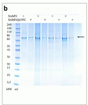
2. The second chapter described the process known as “protein hitch-hiking” as the mechanism of OTC transfer and accumulation observed in the first chapter. After bare SAMNs and SAMNs@OTC had been incubated in sea bream (Sparus aurata) blood serum, successive analysis determined the selective binding of SAMNs and SAMNs@OTC with the apolipoprotein A1, a known carrier of lipids and low-polarity molecules to fish ovary. Within the organism, the selective binding of apolipoprotein A1, which can be considered as an endogenous targeting signal, could lead to the organotropic and delivery of OTC in the fish ovary.
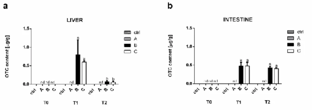
3. In the third chapter, the same oxytetracycline (OTC) delivery method previously studied in in vivo experiments reported in Chapter 1was tested using fish organ cultures from zebrafish and gilthead sea bream. This paper demonstrated that the complex SAMNs@OTC herein used may represent a valid and safe way to deliver OTC in organ cultures. However, the discrepancy observed between these results and those obtained from in vivo exposure highlighted different important factors which reveal that in vivo studies replacement with ex vivo models are not always able to represent the typical interaction complexity of a biological system. The main differences between in vivo and ex vivo
III
exposure are represented by the simplicity of the organ culture system used in this study compared to the biological complexity of the animal in toto as well as the two different experiment exposure times (hours vs days) and the direct contact of the NPs suspended in the culture medium with the organ cultures
4. The fourth chapter compared, for the first time, the effects on fish culture of a classical mechanical, biological, UV purification system to a TiO2-PCD one, with particular emphasis on water chemistry and
physiological responses in adult zebrafish. The photocatalytic system showed excellent efficiency in removing nitrogen compounds from water. Physiological analysis including histological and molecular assays demonstrated that no significant biological alterations/effects were detectable in cultured fish.
5. At last, the fifth chapter focused on assessing the efficacy of an innovative complex made of magnetic nanoparticle and a photosensitizer molecule (under evaluation for patentability) in relation to mosquitoes’ proliferation control. Aedes aegypti larvae were exposed to the complex in water solution and the larvicidal activity of the nanomaterial and photosensitizer molecule alone were compared to the new complex. In particular, tissues accumulation of the complex was evaluated by optic and transmission electron microscopy (TEM).
1
Introduction
Nanotechnology
Nanoparticles (NPs) are conventionally defined as particles with at least one or more dimensions between 1 and 100 nm that show properties we cannot attribute to bulk samples of the same material (Auffan et al., 2009). Nanotechnology investigates the integration of these nanoscale particles into larger material components and systems, keeping the control and construction of new and improved materials at the nanoscale. The peculiar properties of NPs have accelerated the growth rate growth in nanomaterials (NMs) production and the increase of their application in many areas, capturing the attention of researchers, governments, and industries worldwide.
Natural NPs have always existed on our planet, but only with the development of tools (such as scanning tunneling microscope (STM) and Atomic Force Microscope (AFM)), able to accurately detect the microscopic matter, scientists were able to study and manipulate NMs.
The term nanotechnology indeed was first introduced by Professor Norio Taniguchi from the University of Sciences in Tokyo, in 1974, in his thesis entitled “On the basic concept of Nano – Technology”, where he precisely described the formation of materials with dimensions of a nanometer (Demetzos, 2016).
Before Taniguchi’s definition, the starting point of the new field we now regard to as nanotechnology was introduced by the famous physicist Richard P. Feynman during a talk at the Annual Meeting of the American Physical Society at the Californian Institute of Technology in 1959. In his speech entitled “There’s plenty of room at the bottom” Feynman lectured about the problem of manipulating and controlling things on a small scale (Winkelmann & Editors, 2016) which is indeed the basic concept of nanotechnology studies.
2
Some years after Feynman’s speech, the National Science Foundation (NSF) established in 1991 its first program dedicated to NPs and from 1997-1998 funded a cross-disciplinary program entitled ‘‘Partnerships in Nanotechnology’’ (Roco, 2011).
Governments and companies set up funds for research and development to support the rapid worldwide expansion of nanotechnology market which now includes more than 1.000 commercially available products containing nanomaterials or made using nanotechnology (Klaine et al., 2012).
Such a rapid advancement of nanotechnology has laid the groundwork for innovation in different fields, including health, agriculture, food, transport, energy, electronics and communications, resulting in a significant increase of novel nanomaterials development (Roco, 2011).
Nanoparticles characteristic and synthesis
NPs can be spherical, tubular or irregularly shaped, and can exist in fused, aggregated or agglomerated forms (Grillo, Rosa and Fraceto, 2015). They can be categorized into carbon-based materials such as fullerenes and carbon nanotubes and inorganic nanoparticles including the ones based on metal oxides (zinc oxide, iron oxide, titanium dioxide and cerium oxide), metals (gold, silver and iron) and quantum dots (cadmium sulfide and cadmium selenide) (Ju-Nam and Lead, 2008).
Currently, NPs are commonly classified in two main categories based on their origins: non-engineered and engineered NPs. The formers are found in the environment and derive from natural events such as terrestrial dust storms, erosion, volcanic eruptions and forest fires. We can also consider as non-engineered NPs those accidentally produced and introduced into the environment by human activities such as combustion products or generated by combustion engines, power plants and other thermos-degradation systems
3
(Cupaioli et al., 2014). On the other hand, the development of nanotechnology is responsible for human exposure to a new NPs category: the engineered NPs. Engineered NPs are intentionally produced by humans using different materials, such as metals (including Ag, Zn, Au, Ni, Fe and Cu), metal oxides (TiO2, Fe3O4, SiO2, CeO2 and Al2O3), non-metals (silica and quantum dots),
carbon (nanotubes and fullerene), polymers (alginate, chitosan, hydroxymethylcellulose, polyhydroxyalkanoates and poly-e-caprolactone) and lipids (soybean lecithin and stearic acid) (Grillo, Rosa and Fraceto, 2015). The synthesis of engineered NPs produces homogenous materials with controlled characteristics, which exhibit intermediate features between atoms’ and corresponding bulk materials’. Small size and high surface/volume ratio strongly determine NPs chemical-physical properties, reactivity, and interactions with the environment.
As an example, the melting behavior is one of the chemical-physical parameters that differentiates nanoscale from the corresponding bulk material. Small particles have lower melting points than bulk material due to an increase of surface and internal ratio atoms as the size of particles decreases. Moreover, NPs shape strongly influences their interactions with the environment. For instance, the charge distribution on the surface of spherical inorganic NPs is quite different from that of rod or urchin shaped particles (Hutter et al., 2010). The chemical-physical characteristics of NPs also determine their biodistribution, biological effects and, consequently, their toxicity. In the case of engineered NPs for drug delivery, the chemical-physical characteristics influence drug loading, drug release, NPs stability and cellular uptake. NPs size, shape and composition strongly influence their interaction with biological systems. The high surface/volume ratio and the chemistry of NPs surfaces promote their aggregation and interaction with biomolecules such as proteins and DNA (Ludwig K. Limbach et al., 2005). Due to their size, NPs
4
should be easily uptaken by living organisms; smallest NPs penetrate the cellular barrier (such as Blood-Brain Barrier) more rapidly than larger ones (Chen et al., 2010).
Together with size, the shape also interferes with NPs biodistribution and tissue-specific accumulation. Recent evidence show that sub-micrometric discoidal particles accumulate in lungs, liver, heart, and spleen more than spherical and cylindrical particles (Decuzzi et al., 2010).
In addition, the hydrophobicity of NPs influences their capacity to bind proteins. It has been hypothesized that interaction of NPs with proteins might alter protein conformation leading to exposure of new epitopes and/or abnormal functions (Mu et al., 2009).
To obtain particles with specific characteristics at the nanoscale size, different production procedures have been discovered and developed. NPs production methods can be classified into top-down and bottom-up categories. Top-down approaches involve the size-reduction of large particles to the nanometer range. This can be achieved by milling or high-pressure homogenization (Chan and Kwok, 2011). The two major types of high-pressure homogenization are microfluidization and piston-gap homogenization. Microfluidization is essentially air-jet milling, in which the particles are fragmented by collision in a high-pressure air jet. On the other hand, piston-gap homogenization involves forcing a liquid suspension at high pressure through a narrow channel or gap inside a pipe (Chan and Kwok, 2011). A major drawback of the top-down approach is the imperfection obtained at the surface structure, which in metallic NPs, for example, can significantly impact NPs physical properties and surface chemistry (Thakkar, Mhatre and Parikh, 2010).
Conversely, bottom-up methods generate nanoparticles by building them from drug molecules in solution. This can be achieved by controlled precipitation
5
(or crystallization) and evaporation. These processes can occur in the bulk solution or in droplets, depending on the technique.(Chan and Kwok, 2011).
NPs in biomedicine
Nanotechnology application in the biomedical field shows interesting advancements in several specific areas such as drug targeting, biodiagnostics, bioimaging, and genetic manipulation (Chatterjee et al., 2014). Novel directions include, for example, theranostics (nanosystems capable of diagnosis, drug delivery and monitoring therapeutic responses all in a single tool) and plasmonic photothermal therapy (PPT) (Giner-Casares et al., 2016). To date, NPs biomedical applications include drug and gene delivery, labeling, pathogen or protein detection, probing of DNA structure, tissue engineering, hyperthermia treatments and magnetic resonance imaging (MRI) contrast agents (Hoet et al., 2004).
The major advantages of nanoparticles over larger sized particles are represented by their small size and high surface/volume ratio, which make them the key promoters of the development in biomedical research. A wide variety of organic and inorganic NPs is currently used for biological applications. Semiconductor quantum dots are commercially available and offer a viable alternative to fluorescently labeled particles, whereas iron oxide nanoparticles, due to their favorable features, are the only type of magnetic NPs approved for clinical use by the Food and Drug Administration (Wilczewska et al., 2012). They are currently used in magnetic resonance imaging (MRI) as contrast enhancers in place of conventional gadolinium-based contrast agents (Chatterjee et al., 2014).
A more novel group in terms of their use in biomedicine are plasmonic nanoparticles, which offer many advantages in biomedical research due to their unique characteristics that allow the display of localized surface plasmon
6
resonance (LSPR) bands in the UV-visible-near IR (infrared) spectral range (Giner-Casares et al., 2016). Whereas, carbon NPs, magnetic NPs and those based on solid lipid are considered as promising carriers for drug delivery. In particular, metal-based NPs hold the potential to carry large drug doses as well as increase drugs circulatory half-life (Ahmad et al., 2010).
Among all the techniques improved by NPs, it is surprising how nanotechnology revolutionized drug delivery procedures. The development of NPs suspensions containing medicines has made it possible to increase the therapeutic index of many components (improvement of the activity and reduction of toxicity) by selectively directing them towards the target tissues or cells (Couvreur, 2013).
The high surface area allows NPs modification to improve drugs pharmacokinetic properties (increase vascular circulation lifetime, bioavailability improvement). Thanks to the increased vascular circulation lifetime also the efficacy of the drug showed a significant increase; the enhancement of drug bioavailability means that a significantly lesser dosage than bulk drugs could effectively exert the effects.
The possibility to modify NPs surface not only helps in targeted drug delivery but can also solve the secondary purpose of monitoring drug release.
Different approaches include solid core drug delivery systems (SCDDS), which consist in coating a solid NP with a fatty acid shell to contain the drug of interest. This methodology works at relatively low temperature and pressure, making it especially useful in the case of heat-sensitive or labile drugs (Swain et al., 2014). Porous NPs can also be used as a drug delivery matrix. For example, mesoporous silica particles can be applied to control the release of different substances (Strømme et al., 2009).
7
Finally, current research has focused on taking advantages of NPs electronic, magnetic and optical properties to improve the efficiency of signal detection, transmission, and amplification.
One of the most difficult obstacle to deal with NPs biomedical application is the toxicity expressed by some type of NPs which also present, at the same time, suitable properties to their application in several biomedical techniques. Because of their easy penetration across biomembranes and interference with basal metabolic reactions, they could damage structures within the cell. Apart from their toxicity, NPs also tend to accumulate in the body, therefore, their accumulation could have serious consequences, if not in the near future most likely after a chronic exposure.
To overcome these problems, instead of simple NPs, core/shell structured NPs are often applied thanks to their additional advantages. Core-shell NPs possess a core made of a material coated with another material on top of it. In biomedical applications, these NPs show peculiar properties such as minor cytotoxicity, higher dispersion, higher biocompatibility, better conjugation with other bioactive molecules and increased thermal and chemical stability. Shell layers act as a nontoxic coating as well as to improve the core material property (Couvreur, 2013).
These NPs open the way to the synthesis of novel drug carriers with enhanced properties such as increased residence time, increased bioavailability, and reduction of dosing quantity and frequency of administration along with a higher specificity.
8
Nanotoxicology
Nanotechnology contribution and benefits in many research and industrial fields are widely demonstrated, however it is recognized that its application may raise new challenges in safety and ethical domains. As described in the previous paragraph, a broad range of engineered NPs has been designed to be introduced into the body for diagnostic and therapeutic purposes (Thanh and Green, 2010; Reddy et al., 2012; Nalwa, 2014).
To find application in biomedicine, as well as in many other sectors, NPs should satisfy specific criteria, the most important of which is the lack of toxicity. In in vivo application, besides the absence of toxic effects, NPs must avoid non-specific interactions with plasma proteins and either avoid or allow uptake by the reticuloendothelial system (RES) depending on the application and to efficiently reach their target. NPs also have to maintain colloidal stability under physiological conditions including a wide range of pH (Thanh and Green, 2010).
Progress in the development of new types of engineered NPs often results in the creation of new materials with unexpected physical and chemical characteristics and consequently in unique and often unknown biological effects.
While the toxicity of many bulk materials is well understood, the concentration or size at which NPs begin to exhibit new toxic effects are still not clear (Sharifi et al., 2012). There is a considerable gap between the available data on NMs production and their toxicity evaluation. Engineered NPs that accidentally reach the environment represent a further problem, because biological systems did not evolve, and are therefore unprepared to deal, with their presence (Moore, 2006).
Therefore, to take advantage of NPs properties, deeper studies have been performed over the past two decades to determine whether the potential
9
benefits of nanotechnology could be exploited without any adverse impact on human health as well as on the environment.
NPs cellular uptake
To date, most of NPs toxicity and ecotoxicity studies have been conducted without determining the uptake mechanisms (Zhao and Wang, 2012). A proper knowledge of the experimental conditions that influence the transport and uptake across cell membranes would improve our understanding of their toxicity as well as their properties and applications.
NPs can enter the organism through six principal routes: intravenous, transdermal, subcutaneous, inhalation, intraperitoneal and oral (Ryman-Rasmussen, Riviere and Monteiro-Riviere, 2007). NPs accidental uptake by inhalation or ingestion is likely to be the principal way in terrestrial organisms, whereas in aquatic animals there could be other routes of entry such as direct passage across gills and other external surface epithelia such as olfactory organs or body wall (Moore, 2006).
NPs bioaccumulation in aquatic organisms is already demonstrated and several data are available from different studies which analyzed NPs absorption in different aquatic species. The results revealed that the accumulation can occur in various organs (liver, intestine, gills, brain, and spleen) depending on NPs type and exposure routes (Ramsden et al., 2009; Shaw, Al-Bairuty and Handy, 2012; Hao et al., 2013; Piccinetti et al., 2014; Shang, Nienhaus and Nienhaus, 2014). Knowledge of NPs bioaccumulation is essential to detect and quantify the organisms’ uptake capacity as well as their distribution within tissues, cell and subcellular compartments (Gomes et al., 2012).
When NPs enter the organism, absorption occurs through the interaction among biological components such as proteins and cells, they can distribute
10
into various organs where they remain with unchanged, modified or metabolized structure for an uncertain amount of time before passing to other organs or be excreted (Sharifi et al., 2012).
Cell membranes tend to be impermeable to many large particles, usually, only particles in the range of 10 nm to 30 nm in size can cross the membrane through diffusion (Lead and Smith, 2009), the cell membrane acts as a barrier separating the external environment from the inside of the cell. One mechanism to overcome this barrier, which is already exploited with success by viruses is endocytosis (Smith and Helenius, 2004). Endocytosis and diffusion have been proposed as mechanisms for the uptake into cells of NPs with similar size as viruses (Kettler et al., 2014).
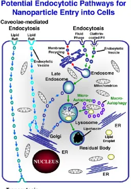
Endocytic mechanisms are normally involved in key physiological functions such as intracellular digestion and cellular immunity; endocytic pathways can either lead to the endosomal and lysosomal compartments (conventional endocytosis) or else via cell-surface lipid raft-associated domains (known as caveolae) which avoids the degradative fate of material entering the endosomal/lysosomal system (Fig.1) (Pelkmans and Helenius, 2002; Na et al., 2003; Panyam and Labhasetwar, 2003; Panyam et al., 2003). This second pathway, in medical nanotechnology, is the main route through which many NPs are designed to enter target cells (Na et al., 2003; Panyam and Labhasetwar, 2003; Panyam et al., 2003).
11
Figure 1. Possible endocytosis pathways exploited by manufactured NPs to enter the
cell. Endocytosis via clathrin-coated pits (receptor-mediated) or uncoated pits (fluid phase) transfers materials to the lysosomal degradative compartment, while caveolar endocytosis can result in translocation to the endoplasmic reticulum (ER), Golgi or through the cell by transcytosis (Moore, 2006).
NPs internalization in cells is mainly influenced by NPs different physical and chemical properties along with the conditions of the exposure medium of the cells. Recent studies have focused on in vivo biodistribution of engineered nanostructures in relation to their physical parameters such as size, shape, surface charge, surface functional groups, and hydrophilicity.
NPs size, for example, may affect their uptake efficiency, kinetics, internalization mechanism and subcellular distribution. A size-dependent uptake in different cell lines has been observed for gold, mesoporous silica, polystyrene and iron oxide NPs, with the maximum cellular uptake at NPs
12
core size in the range of 30-50 nm, which suggests that there is an optimal size for active uptake (Shang, Nienhaus and Nienhaus, 2014).
The effects of nanoparticle shape on its internalization were also examined and it was found that spherical particles of similar size were taken up 500% more than rod-shaped particles; this can be explained by a greater membrane wrapping time required for the elongated particles (Verma and Stellacci, 2010). Finally, it was observed that NPs surface charge determines aggregation in the blood or interaction with oppositely- and like-charged cell membrane surfaces. Many studies demonstrated that cationic and neutral NPs show the highest transport efficiency compared to negatively charged NPs due to the charge attraction between the positive NPs and negative cell membrane surface, thus increasing the rate and extent of absorption (Murugan et al., 2015).
NPs toxicity effects
The majority of toxicity tests suggested that generation of ROS (reactive oxygen species) and consequent oxidative stress are the most frequent effects caused by NPs exposure, which consequently mediate a series of pathological events such as inflammation, genetic damage, inhibition of cell division and cell death (Manke, Wang and Rojanasakul, 2013).
ROS are physiologically produced in trace amounts in response to various stimuli, free radicals occur as essential byproducts of mitochondrial respiration (Manke, Wang and Rojanasakul, 2013). Inflammatory phagocytes such as neutrophils and macrophages induce oxidative reactions as a defense mechanism against environmental pollutants, tumor cells and microbes (Manke, Wang and Rojanasakul, 2013).
Conversely, high ROS levels are normally indicative of oxidative stress and can damage cells by lipid peroxidation, proteins alteration, DNA damages or
13
interfering with signaling functions and modulating gene transcription (Schieber and Chandel, 2014).
The mechanism for ROS generation is different for each NP type and to date, the exact cellular mechanism is not completely understood and must be elucidated. One mechanism of NP-induced oxidative stress occurs during the dissolution of iron-based NPs, which catalyzes ROS generation and formation of OOH• and OH• radicals from H2O2 via the Fenton reaction (Sharifi et al.,
2012). Furthermore, some inert NPs do not give rise to spontaneous ROS production, yet are capable of inducing ROS production under biological conditions, based on the ability of the NPs to target mitochondria (Tian Xia et al., 2006).
Many evidence demonstrate the ability of different types of NPs to induce oxidative stress and, consequently, negative effects. For instance, CNT-induced oxidative stress activates cell signaling pathways resulting in increased expression of pro-inflammatory and fibrotic cytokines (Li et al., 2010). Some NPs have been shown to activate inflammatory cells such as macrophages and neutrophils resulting in an increased production of ROS (Kennedy et al., 2009). Other NPs such as titanium dioxide, zinc oxide, cerium oxide, and silver NPs have been shown to deposit on the cell surface or inside the subcellular organelles and induce oxidative stress signaling cascades that eventually result in oxidative stress to the cell (Manke, Wang and Rojanasakul, 2013). Oxidative stress associated with TiO2 NPs, for example,
results in early inflammatory responses, such as an increase in polymorph nuclear cells, impaired macrophage phagocytosis, and/or fibro proliferative changes in rodents (Bermudez et al., 2004). TiO2 NPs also can cause
proinflammatory effects in human endothelial cells (Sharifi et al., 2012). Carbon NPs have been shown to induce oxidative stress in fish brain cells and pulmonary inflammation in rats (Warheit et al., 2003; Oberdörster, 2004).
14 In vivo and in vitro nanotoxicity
Both in vivo and in vitro tests have been developed to evaluate the toxicity of several NPs, focusing on gaining a comprehensive understanding of the relationship between NPs properties and their potential toxicity or application in different fields.
Although any experimental analysis can be performed with cells obtained from either in vivo or in vitro experimentation, it is necessary to highlight the constant debate of the advantages and disadvantages of both methods. In vitro methods produce specific and quantitative measurements of toxicity and are extremely valid for initially evaluating the expected biocompatibility of new NPs. Different techniques can be used to assess NMs toxicity, including in vitro assays for cell viability/proliferation, mechanistic assays (ROS generation, apoptosis, necrosis, DNA damages), microscopic evaluation of intracellular localization (SEM-EDS, TEM, AFM, fluorescence spectroscopy, MRI, VEDIC microscopy), gene expression analysis, high-throughput systems, in vitro hemolysis and genotoxicity (Arora, Rajwade and Paknikar, 2012).
In vitro methods are ideal for nanotoxicology research because they can produce reproducible results rapidly and inexpensively without the use of animals. Some examples of these techniques include the lactate dehydrogenase (LDH) assay performed to test cell membrane integrity, the MTT assay of mitochondrial function and immunochemistry markers for apoptosis and necrosis (Sharifi et al., 2012). These methods, however, show some disadvantages: they provide little information on the mechanism or cause of cellular toxicity and death. Some colorimetric assays such as Live/Dead, Trypan Blue, and Neutral Red just discriminate live from dead cells but provide little information regarding the mechanisms of cell death (Sharifi et al., 2012). Moreover, recent studies suggest that classical cell-based
15
assays designed for testing chemicals may not provide reliable data since nanomaterials can interfere with assay components and readout systems (Kroll et al., 2012). The accuracy and precision of colorimetric assays for NPs in vitro toxicity, for example, are affected by the interaction between NPs and dyes. It has been demonstrated that carbon nanotubes (CNTs) adsorb MTT formazan and other dyes such as Neutral Red and Alamar Blue producing conflicting results, while the high adsorption capacity of carbon nanomaterials generates false-positive data resulting in an overestimation of their toxicity (Casey et al., 2007; Kroll et al., 2012).
On the other hand, in vivo models allow the study of aspects that cannot be otherwise observed through in vitro systems. Factors such as route of administration, biodistribution, biodegradability, long-term disposition, induction of developmental defects and activation of the immune system, are all major issues in determining in vivo nanotoxicity and cannot be properly studied by an in vitro approach (i.e. primary cell cultures, tissues and organ cultures) (Rizzo et al., 2013).
Although in vivo tests can provide information on the carcinogenicity, pulmonary, dermal and gastrointestinal toxicity related to the initial deposition of nanomaterials by considering various exposure routes, they are time-consuming, more expensive and raise ethical issues. Moreover, cultured human and animal cells can be better controlled and therefore provide more reproducible data than in vivo systems, although requiring a high standardization to maximize data reproducibility (Clift, Gehr and Rothen-Rutishauser, 2011).
At present, it is evident that one of the major gaps in the knowledge of nanotoxicity studies is the lack of correlation between in vivo and in vitro tests, which leads to discrepancy in experimental conditions.
16
Therefore, a proper development of in vitro assays as predictive tools to evaluate NPs toxicity is an important goal during early product development. If correctly validated, these initial screening tests would result useful since they are simpler, faster and less expensive than their in vivo counterparts are. Moreover, successful development of in vitro toxicity systems should reduce animal use and animal stress used in hazard screening.
Aquaculture
While the rearing of aquatic animals and plants may seem a relatively new activity, aquaculture actually represents an ancient practice that is nearly 4000-year old. Pond culture of Tilapia nilotica is engraved in an Egyptian tomb dating before 2000 B.C. and the husbandry of carp may have been the source of fresh fish for the emperor in China around 1000 B.C (Iwama, 2009). This misconception may be due to the fact that aquaculture production, as well as its utilization in human consumption, has dramatically increased in the last 3 decades. Global aquaculture production has more than doubled since 2000, increasing from 41.7 million tons to reach a new estimate of over 90.4 million tons in 2012, with production growing at an annual average rate of 6.7% since then (FAO, 2017). Such a growth is the results of the production areas expansion, increase of knowledge in husbandry systems and advancement in production technologies, but most importantly entails an increased exploitation of natural resources and hence raises concerns on environmental distress (Samuel-Fitwi et al., 2012).
Numerous are the benefits deriving from aquaculture activity:
• Production of high-quality food, increase of household food supply and improvement of nutrition.
• Strengthened marginal economies by the increase in employment and reduction of food prices.
17
• Improvement of water resources and nutrient management at household or community levels.
• Preservation of aquatic biodiversity through re-stocking and recovering of protected species.
• If done sustainably, management and preservation of wild stock stressed by commercial and sports fisheries.
• Stimulation of research and technology development. • Increased education and environmental awareness.
With a global population of nearly seven billion people, the demand for aquatic food is destined to increase and a further expansion and intensification of aquaculture production are required. To guarantee a sustainable growth that meets the global needs, aquaculture activity should overcome some disadvantageous aspects deriving from its own practice which may cause negative effects on the production itself and may have negative environmental impacts.
One of the major negative aspects that characterize intensive aquaculture practices is the large amount of uneaten feed and fish feces present in untreated wastewater (Brune et al. 2003; Piedrahita 2003; Gutierrez-Wing and Malone 2006). These waters are particularly rich in organic compounds such as nitrogen and organic nitrogen is potentially toxic for aquatic organisms especially in its anionized form NH3 (Hu et al., 2017). To date, an efficient
maintenance of the water quality in fish tanks is accomplished through “open-systems”, which consist of aquaculture facilities characterized by a constant water flow taken from a natural source (basin, lake, river, sea, and lagoon) and restituted to the main water body.
In this way, large amounts of water are used daily with substantial costs and the actual risk of organic nutrient-rich waters (nitrogen compounds,
18
phosphorus, dissolved organic carbon and organic matter) affecting the aquatic environment (Turcios and Papenbrock, 2014).
It is not rare that high amounts of organic compounds released from aquaculture intensive systems lead to eutrophication phenomena contributing to an increase in primary productivity and often generating undesirable changes in the structure and function of aquatic ecosystems (Marinho-Soriano et al., 2011). Some of the consequences deriving from the eutrophication (caused by fish farms input of organic matter), of aquatic environment, are the decrease of benthic diversity (Wu, 1995; Karakassis et al., 2000) and the alteration of sediment’s characteristics (Wu et al., 1994; Cancemi, De Falco and Pergent, 2003). Cancemi and collaborators (2003) observed the influence of a fish farm in a littoral bay of Corsica on the surrounding ecosystem, in particular, they observed the effects on the Posidonia oceanica seagrass meadows. The input of organic matter deriving from the cages caused an alteration of the bottom sediment, a high epiphyte biomass (due to the nutrient enrichment) and a plant/epiphyte competition that lead to leaf fragility and a decrease in light availability.
Also salmon farming was demonstrated to cause marked changes in species number and diversity, faunal abundance, and distribution of benthic fauna (Brown, Gowen and McLusky, 1987; Findlay, Watling and Mayer, 1995). Disease emergence is an important constraint to the growth of aquaculture, poor water quality, and high stocking density often promote outbreaks of pathogens and their potential spread in the bordering areas with serious impacts on wild populations (Thompson et al., 2002). For example, crayfish plague, introduced from the USA, has eradicated native crayfish from a large area of Europe (Alderman, 1996). Movement of stocks for aquaculture purposes have been demonstrated to increase the risk of spreading pathogens (Naylor et al., 2000). In Europe, for example, serious epidemics of
19
furunculosis and Gyrodactylus salaris, in stocks of Atlantic salmon, have been linked to movements of fish for aquaculture and re-stocking (McVicar, 1997). It has already been demonstrated that Norwegian cod and salmon farms are the principal cause of spreading pathogens from farmed fish to wild populations (Johansen et al., 2011).
Once again, many epidemics have been reported in some of the most important salmon farming regions of the world, including Chile, where infectious salmon anemia virus (ISAV) caused the most important disease and economic crisis in the history of the country’s salmon industry (2007-2009) (Mardones et al., 2014). There are evidence that anthropogenic activities, such as movement of live or harvested fish or their byproduct, may have played a more important role than the environmental or passive transmission in this outbreak (Mardones et al., 2014).
Many of these problems have been faced using large amounts of antibiotics. Their wide and frequent use has resulted in antibiotic resistance in many fish pathogens (Defoirdt, Sorgeloos and Bossier, 2011), and their massive release into the environment through faeces and uneaten antibiotic-enriched feed, causing an increased environmental antibiotic resistance (Rhodes et al., 2000; Miranda and Zemelman, 2002; Petersen et al., 2002).
Finally, another hazard, often associated to aquaculture practices, of public health interest, is represented by the proliferation of mosquitoes and others disease vectors, that may harbor their eggs and larvae on fish tanks (Erondu and Anyanwu, 2011). Mosquitoes larvae indeed feed on suspended particles or carcasses of their own or related species while floating at water surface (Merritt, Dadd and Walker, 1992).Several mosquito species are known to be vectors of many arboviruses which are important causes of human disease epidemics. Malaria, dengue, Japanese encephalitis, yellow fever, Rift Valley fever, Venezuelan equine encephalitis, and Ross River to name just a few of
20
these diseases (Penilla et al., 1998; Gubler, 2002). The geographic distribution of some mosquito vectors and some viruses have expanded globally, accompanied by more frequent and larger epidemics, e.g., dengue fever, for this reasons the controls of mosquitoes spread is of crucial importance (Gubler, 2002).
Nanotechnology could offer different solutions to overcome the problems that affect the aquaculture industry, e.g. water treatment, control of aquatic diseases, fishpond sterilization and nano-feed for fish. Some of these issues have already been investigated in the last years. For example, in aquaculture systems, fish feeding is commonly supplied as pellet food, and NPs are used to enclose or coat (nano-encapsulation technology) nutrients that would normally degrade, such as fatty acids, or have limited assimilation efficiency across the fish gut because of their poor solubility. To date, the antimicrobial properties of NPs such as titanium and silver NPs are exploited to reduce the build-up of bacteria in aquaculture systems (Sondi and Salopek-Sondi, 2004; Martínez-Castañón et al., 2008; Martinez-Gutierrez et al., 2010).
However, some others issue still need to be addressed or better investigated, and nanotechnology can offer an alternative method to guarantee water quality in aquaculture systems and can be used as drug delivery matrix that guarantees a more efficient antibiotic and vaccine delivering system as already demonstrated in human biomedicine.
Therefore, the present Ph.D. thesis examines for the first time, the possible application of different nanomaterials to antibiotic delivery, water treatment and control of mosquitoes’ proliferation in aquaculture systems.
21
Model organisms
The present Ph.D. project was conducted using different aquatic organisms (at different development stages) as experimental models, for the purpose of evaluating the effect of new nanomaterials in organisms showing different levels of complexity. Escherichia coli Class: Gammaproteobacteria Order: Enterobacterales Family: Enterobacteriaceae Genus: Escherichia
Escherichia coli is one of the most versatile gram-negative bacterial species. It alternates between its primary and secondary habitats respectively the gut of vertebrates (where it lives as a commensal), and water and sediment (which are reached after excretion from the host). It may also behave as an intra- and extra-intestinal pathogen in humans and many other animal species (Clermont et al., 2011). Such a variability is due to the high genome plasticity deriving from gene loss and gain and horizontal transfer (Touchon et al., 2009). E. coli has a core genome of less than 2000 genes, but the total number of 10000 allows a large number of different gene combinations and, therefore, the diverse phenotypes observed (Rasko et al., 2008; Touchon et al., 2009). Based on genomic information, the species has been divided into six different phylogenetic groups, denoted as A, B1, B2, C, D, and E (Touchon et al., 2009). These subgroups include saprophytic (A) and pathogenic (in particular B2, D) types and are often considered to be the result of long evolution process (van Elsas et al., 2011).
22
Pathogenic strains responsible for intestinal infections include enteropathogenic E. coli (EPEC), enterohemorrhagic E. coli (EHEC), enterotoxigenic E. coli (ETEC), enteroaggregative E. coli (EAEC), enteroinvasive E. coli, diffusely adherent E. coli, necrotoxic E. coli, and cell-detaching E. coli (Hamelin et al., 2007). Although pathogenic E. coli have been more commonly recognized as intestinal pathogens, extraintestinal infections are also a major source of morbidity and mortality (Russo, 2003). E. coli is commonly known as an important component of the gastrointestinal microflora; however, its presence as a key component of the microbial communities is crucial in the aquatic environment, especially in the sediment banks, soil and beach sands (Pachepsky and Shelton, 2011). Thanks to this and to the amount of information about its genetics, E. coli is used as a model organism in different studies to detect and test bacterial antibiotic-resistance in the aquatic environment (Costanzo, Murby and Bates, 2005; Sjölund et al., 2008; Kümmerer, 2009; Gullberg et al., 2011). Determining the presence of antibiotic-resistant bacteria in an environmental reservoir is vital, as the environmental source is not only a way of dissemination of antibiotic-resistant microorganisms among human and animals but also the route through which resistance genes are introduced into natural bacterial ecosystems (Amaya et al., 2012).
23 Aedes aegypti Class: Insecta Order: Diptera Family: Culicidae Genus: Aedes
Aedes aegypti is a tropical mosquito originating from Africa that probably spread because of international trade by sea routes through the fifteenth to the seventeenth centuries (Jansen and Beebe, 2010). Unlike many other mosquito species, A. aegypti is a day-biting mosquito and often feeds on multiple hosts during a single gonotrophic cycle. Females preferentially lay eggs in manmade or artificial containers including water tanks, flower vases, pot plant bases, discarded tires, buckets or other containers typically found in close proximity of or in houses (Christophers, 1960).
After the hatching, larvae feed on microorganisms and particulate organic matter. When larvae have acquired enough energy and size, metamorphosis to pupae occurs. Pupae do not feed but just undergo morphological changes to the adult flying mosquito. The entire life cycle lasts 8-10 days at room temperature, depending on the level of feeding.
Aedes aegypti is one of the species responsible for the transmission of yellow fever, dengue fever and dengue hemorrhagic fever (Cheng, 2003). It was also identified as one of Zika virus vectors (urban transmission) together with other species of Aedes genus (Ioos et al., 2014).
There are several aspects that make the controlling of mosquito populations a difficult task: for instance, eggs can endure desiccation for up to one year; there is no effective technique to control the eggs in water containers (Russell, Kay and Shipton, 2001); because of their high domestication, adults can rest indoor in inaccessible places, making air larvicidal useless (Ciccia, Coussio
24
and Mongelli, 2000). Finally, the prolonged application of common larvicidal substances and insect growth regulators such as organophosphates has fostered several environmental and health concerns, including widespread larval resistance, disruption of natural biological control systems, outbreaks of other insect species and undesirable effects on non-target organisms (Sen-Sung Cheng et al., 2004).
Danio rerio Class: Actinopterygii Order: Cypriniformes Family: Cyprinidae Subfamily: Danioninae Genus: Danio
Zebrafish (Danio rerio) is a small tropical freshwater cyprinoid fish hailing from India. Over the years, it has gained an increasing importance as vertebrate model in the fields of developmental biology, genetics, functional genomics, aquatic toxicology, neuroscience and many other areas of biomedical research (Arunachalam et al., 2013).
The main advantages of using zebrafish as an experimental model over other organisms derive from its high fecundity, short generation time, rapid development (hatching occurs after 2-3 days post fertilization), external fertilization, translucent embryos (suitable for observation of internal organs with conventional microscopy) and an extensive amount of information made available by genome and transcriptome sequencing (Teraoka, Dong and Hiraga, 2003). In addition, tissue types (kidney, spleen, liver, blood brain barrier) and immunogenic and physiological responses to common
xeno-25
substances such as oxidative stress and to foreign bodies are comparable to those of higher vertebrates (Rubinstein, 2003).
The transparent chorion enables an easy observation of development and a detailed knowledge of its life cycle, both for embryonic stages (Kimmel et al., 1995) and postembryonic development (Parichy et al., 2009). This has facilitated the identification of genes with important roles in development by mutagenesis screens.
Starting from the 1950s, zebrafish has been used for toxicity tests of synthetic chemicals and natural products (Battle and Hisaoka, 1952), metals (e.g., zinc, selenium, mercury, and copper) (Niimi and LaHam, 1975; Dave and Xiu, 1991; Senger et al., 2006; Johnson, Carew and Sloman, 2007) and organic solvents (e.g., phenol, aniline, and cyclohexane) (Vittozzi and De Angelis, 1991). Thanks to the inexpensive costs associated to zebrafish maintenance and handling, successive studies on different areas of toxicology were performed to understand the adverse effects of chemicals and to predict results in humans. These studies demonstrated that zebrafish is suitable for chemical screening and displays good dose responsiveness to toxicity (Zhang et al., 2003).
In recent years, along with nanotechnology development, the necessity of quick, cheap and easy protocols to conservatively assess the toxicity of nanomaterials, especially those used in biomedicine, has increasingly occurred. Zebrafish, as model organism, allows the performance of nanotoxicity tests for a wide range of nanomaterials including metal nanoparticles, carbon-based nanomaterials, and polymers, many of which are already being incorporated into nanopharmaceuticals (Fako and Furgeson, 2009).
26 Sparus aurata Class: Actinopterygii Order: Perciformes Family: Sparidae Genus: Sparus
Gilthead sea bream (Sparus aurata) is a marine teleost occupying the Mediterranean Sea and the Atlantic coasts of Europe. It is a euryhaline species moving, in early spring, towards protected coastal waters (lagoon, pond, and basins) in search for abundant food and milder temperatures while in late autumn returns to the open sea for feed purposes and because of its sensitivity to low temperatures. It is mainly carnivorous (shellfish, including mussels and oysters), occasionally herbivorous.
Gilthead sea bream is a protandrous hermaphrodite, being a functional male in the first two years and changing sex at approximately 30 cm in length. During the male phase, bisexual gonads are functionally testicles, with asynchronous spermatogenesis and no functional ovarian areas (Kadmon, Yaron and Gordin, 1985). Ovarian development is also asynchronous, and females are batch spawners that can lay 20.000-80.000 eggs per day for a period of up to 3 months. In the Mediterranean, they reproduce between October and December. The eggs are spherical and pelagic, with a diameter slightly lower than 1 mm and a single large oil droplet. The planktonic larval stage lasts about 50 days at 17-18° C.
Gilthead sea bream is economically a very important species for the Greek, and the Mediterranean in general, aquaculture (Grigorakis et al., 2002). The husbandry of this species has grown over the past two decades (FAO, 2010) and more than 200000 tons per year is now produced in the Mediterranean Sea (Arechavala-Lopez et al., 2013). Improvements in larval production
27
techniques, formulation of specialized feeds, use of cages as the main rearing technique and the substantial financing from the European Union allowed a rapid increase of sea bream production (Rodgers and Furones, 1998). Unfortunately, high-density production techniques inevitably have led to issues related to disease spreading problems in the entire Mediterranean marine aquaculture industry. Many species of marine parasites proliferate as commensal species and cause severe problems when poor on-site conditions, bad husbandry or adverse environmental factors occur (Rodgers and Furones, 1998). In particular, the most critical period in sea bream production is the larval stage, when bacterial infections commonly cause mass mortalities during the rearing (Carnevali et al., 2004). Good management techniques are therefore necessary to afford the high production rates and different studies testing innovative techniques to improve culture quality have been already performed (Hulata, 2001; Carnevali et al., 2004; Avella et al., 2010).
Nanomaterials
Surface-active magnetic nanoparticles (SAMNs)
SAMNs are magnetic nanoparticles constituted of maghemite (γ-Fe2O3) with
a mean diameter of 10 ± 2 nm (Sinigaglia et al., 2012). They were provided by the University of Padua, Italy, where a novel synthesis method that is entirely carried out in water without the need of any organic solvent or surfactant was developed (Magro et al., 2010). This new method offers several advantages: simplicity, low costs, high yield scale and, above all, it is an organic solvent free process and thus ecologically green. The final synthesis product after drying and curing at 400°C for 2 hours is a red-brown powder (Magro et al., 2010). Bare SAMNs are dispersible in water, and their suspensions are stable for several months. Because of their unique physical and chemical properties, these NPs present a high average magnetic moment
28
and their surface shows peculiar binding properties which make them suitable to be reversibly derivatized with selected organic molecules (Magro et al., 2012).
As a result, these NPs could be applied to biology and medicine, drug delivery systems, medical imaging, and protein purification (Venerando et al., 2013). SAMNs have been successfully bound to different molecules of biotechnological interest such as biotin and avidin (Magro et al., 2012), bovine serum amine oxidase (Sinigaglia et al., 2012), curcumin (Magro et al., 2014) and rhodamine (Venerando et al., 2013), showing the ability of forming a stable complex with all the target molecules. Furthermore, SAMNs cytotoxicity has been already studied by testing their effect on HeLa cells culture and demonstrating that they can be safely used at concentrations up to 100 μg/mL (Venerando et al., 2013).
In this Ph.D. project, SAMNs were bound to different molecules forming new original complexes (see Chapter 1, 2 and 3).
Titanium dioxide NPs (TiO2)
This project included also the study of a new purification system based on TiO2 photocatalytic degradation (PCD). The filtration system consisted of an
external mechanical purification system solely (EHEIM© experience 250; flow rate: 700 L/h) and a 36 W UVC sterilizer (Panaque©, Italy) equipped with 4 steel plates around the UV lamp (PCD filtration system).
The preparation method of titania paste is described in chapter 4. Final titania films obtained were deposited on stainless steel plates successively calcined in oven. Four plates were then inserted around a 36 W UVC (254 nm) lamp (Panaque©, Italy) at a 0.5 cm distance from the quartz bulb.
29
References
Ahmad, M. Z. et al. (2010) ‘Metallic nanoparticles: technology overview & drug delivery applications in oncology’, Expert Opinion on Drug Delivery. Taylor & Francis, 7(8), pp. 927–942. doi: 10.1517/17425247.2010.498473.
Alderman, D. J. (1996) ‘Geographical spread of bacterial and fungal diseases of crustaceans.’, Revue scientifique et technique (International Office of Epizootics), 15(2), pp. 603–32.
Amaya, E. et al. (2012) ‘Antibiotic resistance patterns of Escherichia coli isolates from different aquatic environmental sources in Leon, Nicaragua’, Clinical Microbiology and Infection. Blackwell Publishing Ltd, 18(9), pp. E347–E354. doi: 10.1111/j.1469-0691.2012.03930.x. Arechavala-Lopez, P. et al. (2013) ‘Differentiating the wild or farmed origin
of Mediterranean fish: a review of tools for sea bream and sea bass’, Reviews in Aquaculture, 5(3), pp. 137–157. doi: 10.1111/raq.12006. Arora, S., Rajwade, J. M. and Paknikar, K. M. (2012) ‘Nanotoxicology and in
vitro studies: The need of the hour’, Toxicology and Applied Pharmacology, 258(2), pp. 151–165. doi: 10.1016/j.taap.2011.11.010. Arunachalam, M. et al. (2013) ‘Natural History of Zebrafish ( Danio rerio )
in India’, Zebrafish. Mary Ann Liebert, Inc. 140 Huguenot Street, 3rd Floor New Rochelle, NY 10801 USA , 10(1), pp. 1–14. doi: 10.1089/zeb.2012.0803.
Auffan, M. et al. (2009) ‘Towards a definition of inorganic nanoparticles from an environmental, health and safety perspective’, Nature Nanotechnology. Nature Publishing Group, 4(10), pp. 634–641. doi: 10.1038/nnano.2009.242.
Avella, M. A. et al. (2010) ‘Application of multi-species of Bacillus in sea bream larviculture’, Aquaculture, 305(1–4), pp. 12–19. doi: 10.1016/j.aquaculture.2010.03.029.
Battle, H. I. and Hisaoka, K. K. (1952) ‘Effects of Ethyl Carbamate (Urethan) on the Early Development of the Teleost Brachydanio rerio’, Cancer Research, 12(5).
Bermudez, E. et al. (2004) ‘Pulmonary Responses of Mice, Rats, and Hamsters to Subchronic Inhalation of Ultrafine Titanium Dioxide Particles’, Toxicological Sciences. Oxford University Press, 77(2), pp. 347–357. doi: 10.1093/toxsci/kfh019.
30
farming on the benthos of a Scottish sea loch’, Journal of Experimental Marine Biology and Ecology, 109(1), pp. 39–51. doi: 10.1016/0022-0981(87)90184-5.
Brune, D. E. et al. (no date) ‘Intensification of pond aquaculture and high rate photosynthetic systems’. doi: 10.1016/S0144-8609(03)00025-6.
Cancemi, G., De Falco, G. and Pergent, G. (2003) ‘Effects of organic matter input from a fish farming facility on a Posidonia oceanica meadow’, Estuarine, Coastal and Shelf Science, 56(5–6), pp. 961–968. doi: 10.1016/S0272-7714(02)00295-0.
Carnevali, O. et al. (2004) ‘Administration of probiotic strain to improve sea bream wellness during development’, Aquaculture International, 12, pp. 377–386.
Casey, A. et al. (2007) ‘Spectroscopic analysis confirms the interactions between single walled carbon nanotubes and various dyes commonly used to assess cytotoxicity’, Carbon, 45(7), pp. 1425–1432. doi: 10.1016/j.carbon.2007.03.033.
Chan, H.-K. and Kwok, P. C. L. (2011) ‘Production methods for nanodrug particles using the bottom-up approach’, Advanced Drug Delivery Reviews, 63(6), pp. 406–416. doi: 10.1016/j.addr.2011.03.011.
Chatterjee, K. et al. (2014) ‘Core/shell nanoparticles in biomedical applications’, Advances in Colloid and Interface Science, 209, pp. 8–39. doi: 10.1016/j.cis.2013.12.008.
Chen, Y.-S. et al. (2010) ‘Size-dependent impairment of cognition in mice caused by the injection of gold nanoparticles’, Nanotechnology. IOP Publishing, 21(48), p. 485102. doi: 10.1088/0957-4484/21/48/485102. Cheng, S. (2003) ‘Bioactivity of selected plant essential oils against the yellow
fever mosquito Aedes aegypti larvae’, Bioresource Technology, 89(1), pp. 99–102. doi: 10.1016/S0960-8524(03)00008-7.
Christophers, S. (1960) ‘Aëdes aegypti (L.) the Yellow Fever Mosquito: its Life History, Bionomics and Structure.’, Aëdes aegypti (L.) the Yellow Fever Mosquito: its Life History, Bionomics and Structure. London : The Syndics of the Cambridge University Press, Bentley House, 200, Euston Road, N.W.I.
Ciccia, G., Coussio, J. and Mongelli, E. (2000) ‘Insecticidal activity against Aedes aegypti larvae of some medicinal South American plants’, Journal of Ethnopharmacology, 72(1–2), pp. 185–189. doi: 10.1016/S0378-8741(00)00241-5.
31
Clermont, O. et al. (2011) ‘Animal and human pathogenic Escherichia coli strains share common genetic backgrounds’, Infection, Genetics and Evolution, 11(3), pp. 654–662. doi: 10.1016/j.meegid.2011.02.005. Clift, M. J. D., Gehr, P. and Rothen-Rutishauser, B. (2011) ‘Nanotoxicology:
a perspective and discussion of whether or not in vitro testing is a valid alternative’, Archives of Toxicology. Springer-Verlag, 85(7), pp. 723– 731. doi: 10.1007/s00204-010-0560-6.
Costanzo, S. D., Murby, J. and Bates, J. (2005) ‘Ecosystem response to antibiotics entering the aquatic environment’, Marine Pollution Bulletin, 51(1–4), pp. 218–223. doi: 10.1016/j.marpolbul.2004.10.038.
Couvreur, P. (2013) ‘Nanoparticles in drug delivery: Past, present and future’, Advanced Drug Delivery Reviews, pp. 21–23. doi: 10.1016/j.addr.2012.04.010.
Cupaioli, F. A. et al. (2014) ‘Engineered nanoparticles. How brain friendly is this new guest?’, Progress in Neurobiology, 119, pp. 20–38. doi: 10.1016/j.pneurobio.2014.05.002.
Dave, G. and Xiu, R. (1991) ‘Toxicity of mercury, copper, nickel, lead, and cobalt to embryos and larvae of zebrafish,Brachydanio rerio’, Archives of Environmental Contamination and Toxicology. Springer-Verlag, 21(1), pp. 126–134. doi: 10.1007/BF01055567.
Decuzzi, P. et al. (2010) ‘Size and shape effects in the biodistribution of intravascularly injected particles’, Journal of Controlled Release, 141(3), pp. 320–327. doi: 10.1016/j.jconrel.2009.10.014.
Defoirdt, T., Sorgeloos, P. and Bossier, P. (2011) ‘Alternatives to antibiotics for the control of bacterial disease in aquaculture’, Current Opinion in Microbiology, pp. 251–258. doi: 10.1016/j.mib.2011.03.004.
Demetzos, C. (no date) Pharmaceutical nanotechnology : fundamentals and practical applications.
Erondu, E. and Anyanwu, P. (2011) ‘Potential hazards and risks associated with the aquaculture industry’, African Journal of Food, Agriculture, Nutrition and Development, 4(13). doi: 10.4314/ajfand.v4i13.71775. Fako, V. E. and Furgeson, D. Y. (2009) ‘Zebrafish as a correlative and
predictive model for assessing biomaterial nanotoxicity’, Advanced Drug Delivery Reviews, 61(6), pp. 478–486. doi: 10.1016/j.addr.2009.03.008.
FAO (2010) The state of world fisheries and aquaculture 2010. Food and Agriculture Organization of the United Nations.
32
FAO (2017) http://www.fao.org/fishery/statistics/software/fishstatj/en. Findlay, R. H., Watling, L. and Mayer, L. M. (1995) ‘Environmental impact
of salmon net-pen culture on marine benthic communities in Maine: A case study’, Estuaries. Springer-Verlag, 18(1), p. 145. doi: 10.2307/1352289.
Giner-Casares, J. J. et al. (2016) ‘Inorganic nanoparticles for biomedicine: where materials scientists meet medical research’, Materials Today, 19(1), pp. 19–28. doi: 10.1016/j.mattod.2015.07.004.
Gomes, T. et al. (2012) ‘Accumulation and toxicity of copper oxide nanoparticles in the digestive gland of Mytilus galloprovincialis’, Aquatic Toxicology, 118–119, pp. 72–79. doi: 10.1016/j.aquatox.2012.03.017.
Grigorakis, K. et al. (2002) ‘Comparison of wild and cultured gilthead sea bream (Sparus aurata); composition, appearance and seasonal variations’, International Journal of Food Science and Technology. Blackwell Science Ltd, 37(5), pp. 477–484. doi: 10.1046/j.1365-2621.2002.00604.x.
Grillo, R., Rosa, A. H. and Fraceto, L. F. (2015) ‘Engineered nanoparticles and organic matter: A review of the state-of-the-art’, Chemosphere, 119, pp. 608–619. doi: 10.1016/j.chemosphere.2014.07.049.
Gubler, D. J. (2002) ‘The Global Emergence/Resurgence of Arboviral Diseases As Public Health Problems’, Archives of Medical Research, 33, pp. 330–342.
Gullberg, E. et al. (2011) ‘Selection of Resistant Bacteria at Very Low Antibiotic Concentrations’, PLoS Pathogens. Edited by M. Lipsitch. Public Library of Science, 7(7), p. e1002158. doi: 10.1371/journal.ppat.1002158.
Gutierrez-Wing, M. T. and Malone, R. F. (no date) ‘Biological filters in aquaculture: Trends and research directions for freshwater and marine applications’. doi: 10.1016/j.aquaeng.2005.08.003.
Hamelin, K. et al. (2007) ‘Occurrence of Virulence and Antimicrobial Resistance Genes in Escherichia coli Isolates from Different Aquatic Ecosystems within the St. Clair River and Detroit River Areas’, Applied and Environmental Microbiology, 73(2), pp. 477–484. doi: 10.1128/AEM.01445-06.
Hao, L. et al. (2013) ‘Bioaccumulation and sub-acute toxicity of zinc oxide nanoparticles in juvenile carp (Cyprinus carpio): A comparative study