0
CHAPTER 1 ... 1
1.1 INTRODUCTION ... 1
1.1.1 Aim of the work ... 2
1.2
MTDNA VARIABILITY AND AGING ... 4
1.2.1 Mitochondrial theory of aging ... 4
1.2.2 Mitochondrial structure and function ... 6
1.2.3 The influence of mitochondrial structure and function on aging and longevity ... 10
1.2.4 Mitochondrial genome and aging ... 13
1.2.4.1 Genome structure ... 13
1.2.4.2 Mitochondrial genetics ... 15
1.2.4.3 mtDNA variability, aging and age-related diseases ... 19
1.2.4.4 Mitochondria-nucleus cross talk and aging ... 22
1.3 VARIABILITY OF NUCLEAR DNA AND AGING ... 26
1.3.1 INS/IGF1 pathways ... 28
1.3.2 Caloric restriction, and longevity ... 30
1.3.3 Cellular and systemic response to damage ... 33
1.3.4 Lipoprotein genes and longevity... 35
1.3.5 Cell cycle regulation and longevity ... 37
1.3.6 Telomere length maintenance ... 38
CHAPTER 2 ... 62
2.1 The analysis of mitochondrial DNA variability in a large sample of ultranonagenarians
across Europe allows a reappraisal of the mtDNA correlation with longevity: a GEHA
Project Study... 62
CHAPTER 3 ... 129
3.1 The interaction between Mitochondrial DNA and APOE variability in a large sample of
ultranonagenarians across Europe: a GEHA Project Study... 129
CHAPTER 4 ... 145
1
CHAPTER 1
1.1 INTRODUCTION
In the last two decades there has been an increasing interest in human aging and its genetic basis. In
the first instance, this might be explained by the increased number of elderly subjects in the
developed countries, due to the ameliorated life condition (Kannisto, 1994). But to live longer
implies higher healthcare costs necessary to treat the aging-related diseases that at present amount
to approximately 20% of total healthcare costs. The mitigation of these costs, that may increase in
the next years, seems necessary and the only way to realize this intent is to guarantee a healthy
aging.
Aging is a complex trait characterized by the interaction between environmental, genetic and
epigenetic factors (Capri M et al, 2006). Certainly, over the last centuries environmental conditions
have had an important role in the radical change in life expectancy; on the other hand, genetic
factors have also a large influence on the rate and the quality of aging (Perls et al, 2000, 2002;
Herndon et al, 2002; Kirkwood and Finch, 2002; Kirkwood, 2005; Lescai et al, 2009a, b). In
particular twin studies reported that the percentage of the variation in human life span which can be
attributed to genetic differences among individuals ranges from 22% to 33% (McGue et al, 1993;
Ljungquist et al, 1998; Bishop and Guarente, 2007;).
Longevity, defined as survival beyond the specie-specific average age of death (De Benedicis and
Franceschi, 2006), is a phenotypic expression of successful aging. Although longevity is not
sufficient for describing a successful aging process because, for example, it provides little
information about functioning and quality of life, it is the most considered and the most simple
phenotype to study. In this frame, association studies on long-lived subjects or centenarian, together
2
lifespan, and highlight genotypes that can predispose individuals to an increased or decreased risk
of reaching extreme old age in a healthy status.
In the last years, the technological progress occurring have made possible the identification of
genetic determinants of aging and longevity. Twin studies, large scale linkage studies on long-lived
families, case-control association studies on candidate gene, longitudinal studies and studies in
model organisms agree in detection of a common 'core' of genes belonging to different metabolic
pathways and defence systems and shared by all the species studied (Kenyon, 2005; Christensen et
al, 2006; Chung et al, 2010). All these studies indicate that aging is the result of the decline, that
cause a less efficient conservation, mobilization and use of nutrients, and a worse ability to respond
to external and internal stress.
1.1.1 Aim of the work
The intent of the present thesis is to highlight some genetic variants that influence this phenotype.
In particular, in my PhD researches I have studied the variability of both mitochondrial and nuclear
DNA in relation to aging and longevity. In the following chapters I’m describing the state of art of
the well known interaction between DNA variability (mitochondrial and nuclear) and aging and
longevity. Then, I report the results of the studies conducted during my PhD appointment, which are
described in three manuscripts. The first and the second manuscripts have as their topic
mitochondrial DNA variability. In particular, in the first manuscript we propose an analysis of the
whole sequences of mtDNA to evaluate if mtDNA point mutations or group of mutations occurring
in different haplogroups are involved in longevity; it is titled “The analysis of mitochondrial DNA
variability in a large sample of ultranonagenarians across Europe allows a reappraisal of the
mtDNA correlation with longevity: a GEHA Project Study” and has been submitted to the
“Hamerican Journal of Human Genetics. The second manuscript is titled “
The interaction between
3
Mitochondrial DNA and APOE variability in a large sample of ultranonagenarians across Europe:
a GEHA Project Study” and it is submitted to “Age”. In this work we analyze the interaction
between the mtDNA variability and variability of nuclear genes such as mitochondrial haplogroups
and APOE gene variability. The third manuscript titled “Two-stage case-control association study
of candidate genes and human longevity”, which is submitted to ”Age” is focused on the nuclear
DNA variability. Exactly, in this work we have conducted a multilocus analysis on hundreds of
Single Nucleotide Polymorphisms (SNPs) located in candidate genes to individuate variables
4
1.2
MTDNA VARIABILITY AND AGING
1.2.1 Mitochondrial theory of aging
Over the years more than 300 aging theories have been postulated, but an universally accepted
definition of the process of aging doesn’t exist (Vina et al, 2007). A common opinion is to consider
aging as an inevitable result of accumulation in an organism of occasional injuries. Among the
several theories proposed, the oldest and most accredited is the Harman “Free radical theory”
(Harman D, 1956). According to this theory aging is promoted by ROS (Reactive Oxygen Species)
that, over the time, lead to cellular damage, including injure to DNA, proteins, and cellular
membranes. Although several enzymes (NADPH oxidase family, enzymes of the lipid metabolism
in peroxisomes, and several other cytosolic enzymes including cyclooxygenase, ecc) contribute to
the ROS production, the 90% of ROS present in the organisms is due to the activity of the
mitochondrial respiratory chain (Balaban et al, 2005). In this frame, in 1972 Harman (Harman,
1972) proposed a new version of the “Free radical theory” where mitochondria assume a central
role in the aging process. Afterwards, in 1980 Miquel and co-workers proposed the "mitochondrial
theory of aging" based on the exclusive role of mitochondria in the aging process (Miquel et al,
1980).
However, mitochondria are not only the principal producers of ROS, but they are also the
first targets. It is supposed that the mitochondrial accumulation of damage, and in particular the
mtDNA impairment, is one of the most important factors in determining age-related cellular decline
5
Figura 1. Mitochondrial role in the energetic life and death of a cellThe diagram represents the loss of cells in a tissue over the life. The minimum number of cells for the tissue to function normally is indicated by the dashed line (modified from Wallace, 2005).
During the last decades, a number of experimental evidences supported the hypothesis of oxidative
damage accumulation as a cause of aging. It was found that levels of oxidated lipids, proteins and
DNA increased with age (Van Remmen and Richardson, 2001). This is in line with several other
literature data indicating that mitochondria of aged animals produce more ROS than mitochondria
of younger one (Sohal et al, 1990), and that the introduction in young rat cells of mitochondria
isolated from fibroblasts of aged rats cause premature aging (Corbisier and Remacle, 1990).
Another evidence derives from an experimental work in which a reduction in mitochondrial ROS
production was observed in animals with a controlled dietetic condition called caloric restriction,
characterized by a reduced food intake that cause an increase in lifespan (this will be better
explained in the next sections) (Sohal et al, 1994).
However, apart from the central role in ROS production, mitochondria carry out many crucial
activities for cell survival and, as a consequence, for aging and longevity (Passarino et al, 2010).
Thus, the next session will describe the structural and functional mitochondrial characteristics, with
6
1.2.2
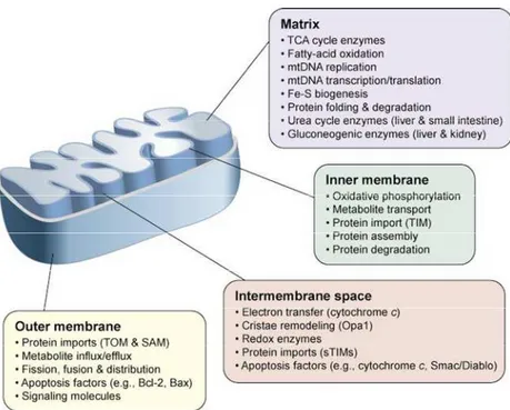
Mitochondrial structure and function
Structure
Structurally, the mitochondrion is a membrane-enclosed organelle ranging from 1–10 micrometers
in size (Henze and Martin, 2003). As shown in Fig.2, mitochondria contain both an inner and an
outer membrane, leading to the formation of two aqueous compartments, the matrix and the
inter-membrane space, where several specialized functions take place.
Each compartment, having a
specific function, cooperate to carry out all mitochondrial functions.
Figura 2. Mitochondrial structure and functions.
The compartment-specific processes and proteins are described in the colored boxes. Abbreviations: Bax, Bcl-2-associated X protein; Bcl-2, B-cell lymphoma protein 2; Opa1, Optic atrophy 1; SAM, Sorting and Assembly Machinery; sTIMs, small TIM proteins; TIM, Translocase of the mitochondrial Inner Membrane; TOM, Translocase of the mitochondrial Outer Membrane (modified from Ryan and Hoogenraad , 2007).
7
The outer membrane allows exchange of metabolites between the cytoplasm and intermembrane
space through passive exchange and protein imports (TOM-Transporter Outer Membrane and
SAM-Sorting and Assembly Machinery). Furthermore it takesa a central role in apoptosis and in
signal transduction (Ryan and Hoogenraad, 2007).
The intermembrane space contains some proteins, principally implicated in electron transfer and
apoptosis. Its most prominent member is cytochrome c, which in normal cells is involved both in
respiration and in apoptotic induction. In addition, other potential pro-apoptotic factors are present
as well as a variety of small proteins that contain cofactors or are disulfide bound (Koehler et al,
2006; Webb et al, 2006).
The inner membrane, delimiting the mitochondrial matrix, is protein rich and it has specific
transporters inside, demonstrating a highly controlled permeability. Moreover, proteic complex are
located across the inner membrane where ETC (Electron Transport Chain) take place (Arco et al,
2005).
The mitochondrial matrix contains various enzymes involved in metabolic processes: oxidation of
fatty acids and tricarboxylic acid cycle, Fe-S biogenesis, and heme synthesis. The matrix also
harbors a number of copies of mtDNA and the protein machinery involved in its maintenance and
replication as well as components involved in transcription/translation.
Mitochondria are not static organelles: they come under structural modification and turn-over.
These complex events, known as mitochondrial dynamics, allow the recruitment of mitochondria to
cellular sites where they are needed. To maintain a healthy population of mitochondria, there is in
mammalian cells a continuous turn-over: the damaged organelles are eliminated and new
mitochondria are generated through processes called mitophagy and biogenesis. The regulation of
these complex mechanisms require more than 1000 genes (located in mitochondrial and nuclear
(Lopez-8
Lluch et al, 2008). The dynamicity of mitochondria emerge also because the number and the size of
these organelles vary in the different tissues: some cells have only a single mitochondrion, whereas
others can contain several thousand mitochondria; this is principally due to cellular metabolic
demands and health status. Moreover, mitochondria are continuously in movement throughout the
cells and undergo fusion and fission modifying the inner membrane surface to readily satisfy energy
requirements (Ono et al, 2001; Bereiter-Hahn and Voth, 1994). Finally, it has been clearly
documented that in mitochondria ultrastructural change occur in response to environmental
stimulation (Bertoni-Freddari et al, 1993, 2007).
Functions
Mitochondria play an important role in cell metabolism: they are the organelles where Oxidative
Phosphorylation (OXPHOS) takes place, and occupies a key position in apoptosis regulation, heat
production, calcium homeostasis, and cellular differentiation. However the two functions
principally involved in aging and longevity are the oxidative phosphorilation and the apoptosis.
The main mitochondrial function is the production of energy through Oxidative Phosphorilation
(OXPHOS). This metabolic pathway, that through the oxidation of nutrients produces energy stored
as adenosine triphosphate (ATP) molecules, counts 80 proteins that form five complexes: NADH
dehydrogenase (complex I), succinate dehydrogenase (complex II), ubiquinone cytochrome c
oxidoreductase (complex III), cytochrome oxidase (complex IV), and, finally, F1Fo-ATP synthase
(complex V).
The OXPHOS consists of two steps: 1) a series of redox reactions along four complexes (I–IV) that
through the transfer of H
+ions (protons) across the electron transfer chain (ETC) creates an
electrochemical gradient (∆Ψ) and 2) the ATP formation by the F1Fo-ATP synthase (complex V),
9
In detail, electrons are donated to complex I from NADH+ or to complex II via succinate, and
passed to coenzymeQ (CoQ) to give ubisemiquinone (CoQH) and then ubiquinol (CoQH2).
Electrons are then donated to complex III, which transfers the electrons to cytochrome c, and,
eventually, to complex IV. Finally, they are moved to 1/2 O2 to give H2O. The energy released
along the pathway is used to pump protons across the mitochondrial inner membrane, creating the
transmembrane electrochemical gradient. Complex V uses the energy stored in the ∆Ψ to condense
ADP and Pi to make ATP. ATP is then carried to cytosol and exchanged with ADP.
Another important mitochondrial function is the regulation of apoptosis. This is a fundamental
cellular mechanism in order to remodel organisms during development or get rid of damaged or
unnecessary cells. The apoptotic cell death can be triggered by external signals detected by cell
surface membrane proteins, or alternatively by mitochondrial signals. The initiation of the
mitochondrial path for apoptosis is mainly regulated by the opening of a non-specific channel that is
located on the inner membrane of mitochondria: the mitochondrial permeability transition pore
(mtPTP) (Green et al, 1998; Chipuk et al, 2006). The opening of this pore leads to a dramatic
collapse of the transmembrane ∆Ψ. This event causes the swelling of the inner membrane, and then
the disruption of the outer membrane, with a consequent release of the pro-apoptotic molecules
enclosed in the inner membrane space, such as cytochrome c, apoptosis inducing factor (AIF),
Smac/Diablo, and several pro-caspases (Hengartner, 2000; Caroppi et al, 2009). In the cytosol,
cytochrome c, together with others factors (Apaf-1 and ATP) activates the caspases 9 and 3 that
disrupt the cytosol. In succession, AIF moves into the nucleus and disrupts the DNA.
The opening of mtPTP can be stimulated by: a) the mitochondrial uptake of excessive Ca2+, b) an
increase of the oxidative stress, c) a decrease of the mitochondrial membrane potential, d) a
decrease of the intra-mitochondrial ATP concentration (Wallace DC, 2005). It is intuitively evident
10
1.2.3 The influence of mitochondrial structure and function on aging and longevity
The importance of mitochondrial functions are underlined by the dynamicity of these organelles
that change in size and shape to satisfy cellular needs in the different physiological states. It is
obvious that a so crucial mechanism of regulation for cell activities have also an essential role in
aging and longevity. One example of this is given by the work of Kissova and colleagues (Kissova
et al, 2004), that suggest an efficient mitophagy is essential to attain longevity. In fact, in yeast the
deletion of the mitochondrial protein Uth1p was found to lead to a selective defect in mitophagy
and to a shortened lifespan in yeast during nutrient deprivation. Moreover, in recent years, studies
show that mitochondria turn over may be considerably influenced by the physiological status of
tissues and organs. For example physical activity promote mitochondrial biogenesis; on the
contrary, physiological aging is characterized by a reduction of mitochondrial biogenesis
(Holloszy, 2004), and degradation (Terman et al, 2007), causing a reduction of mitochondrial
turnover that, as consequence, lead to the accumulation of oxidized components lipids, proteins and
DNA. Also the modification of ultrastructural features is related to aging. In fact, it was observed
that in old rats less mitochondria are present than in young rats, but to compensate for the numeric
loss of organelles, there is an increase in mitochondrial size (which is presumed to increase the
potential area for respiration). Comparing the data from young and old rats, the outcome of these
balanced changes is a constancy in the overall volume fraction of cytoplasm (volume density)
occupied by mitochondria (Bertoni-Freddari et al. 1993, 2007).
Furthermore, several studies have shown how the state of the mitochondrial network morphology
influences a variety of other cellular functions: mitochondrial proliferation (Möpert et al, 2009),
propagation of signals such as those of energy deficiency (Chan 2006), apoptotic events (Suen et
al, 2008), and cell differentiation (Park et al, 2001). All these cellular functions are involved in both
11
pathological conditions such as neurodegeneration (Chen and Chan 2009), type 2 diabetes (Molina
et al, 2009), as well as normal aging (Lòpez-Lluch et al, 2008).
As previously mentioned, mitochondria play important activities essential for cell survival. So, also
the regulation of mitochondrial functions results particularly correlated to the phenotypes under
discussion.
In the energy production process, if the principal result of OXPHOS is the production of ATP
molecules, mitochondrial respiration generates ROS as by-product. In physiological conditions a
small percentage of electrons may prematurely reduce oxygen, forming toxic product such as
superoxide, and peroxyl radical. If these molecules are present in a small percentage, they are
neutralized by the scavenger molecules present in cells, but if they are present in a higher
percentage they can cause oxidative stress. Because the diffusion capability of most ROS is limited
by their lipid solubility the main harmful effects are exerted on mitochondrial molecules,
contributing to the decline in mitochondrial function and, as a consequence, to aging process.
ROS may damage lipids, proteins and nucleic acids.
ROS may act on molecules directly by peroxidation or indirectly through the production of highly
reactive aldehydes. In the direct action, ROS acts “stealing” electrons: this causes the production of
radical molecules. In turn, these molecules, that are not very stable, react with other molecules
causing a "chain reaction mechanism". In the indirect mechanism, of action the production of highly
reactive aldehydes play a central role. One example of reactive aldehydes is the
4-hydroxy-2,3-transnonenale (4HNE) aldehyde, one of the main products of lipid peroxidation. It causes a variety
of harmful effects on the molecules with which it comes into contact.
The action of ROS on phospholipids of mitochondrial inner and outer membranes cause,
principally, the decreases of their fluidity. Since the function of these membrane plays a critical role
12
nucleotide translocase (ANT) (Paradies et al, 1998), it is clear that membrane’s oxidation may have
very harmful effect on mitochondrial functions.
The effect of ROS on proteins lead to structural and functional changes (abnormal aggregation,
degradation, loss of function, etc.). Damage to mitochondrial proteins, especially those present in
the inner mitochondrial membrane, are the direct result of oxidative stress or the consequence of
lipid peroxidation. Several enzymes of the electron transport chain, such as ATPase and ANT, are
particularly susceptible to oxidative stress inactivation, and the principal consequence observed is a
decrease in OXPHOS efficiency (Le Bras et al, 2005).
Because of the proximity with ROS production, and the lack of protective structure, such as histone
proteins, the mtDNA is the primary target of ROS. The oxidative damage to mtDNA may be
detected through the presence of some indicators; an example is the presence of
8-hydroxy-2-deoxyguanosine which is the most abundant among the products of nucleotides oxidation (Chomyn
and Attardi, 2003). The accumulation of mtDNA damage may be dangerous for the cells, modifying
the efficiency of mitochondrial function directly controlled by this genome, especially OXPHOS.
The importance of apoptosis in aging is documented by several studies that show as the reduction in
number of myocytes (Kajstura et al, 1996), skeletal muscle (Dirks and Leeuwenburgh, 2002) and
T-cells (Phelouzat et al, 1997) is the result of apoptotic cell death (Higami and Shimokawa, 2000). It
is clear that the modulation of apoptosis results to be a decisive process in determining a balance
between healthy versus senescent cells and tissues. In fact, a low level of apoptosis is probably
deleterious for tissues and organs, because it leads to the accumulation of damages, but it might be
useful for attaining longevity, even though in this cause an accumulation of damaged cells and
consequent frailty is expected in oldest-olds (Rose et al, 2002). In line with this assumption is the
13
when compared with similar cells from young individuals (Salvioli et al, 2001). Moreover, the
apoptotic process is doubly correlated with aging, because it is influenced also by ROS production
(Kroemer and Reed, 2000): ROS damaging mitochondrial structure increases mitochondrial Ca2+
and consequently activates apoptosis (Lemasters et al, 2009).
1.2.4
Mitochondrial genome and aging
1.2.4.1 Genome structure
During the last decades, along with the importance of mitochondrial function, also the relevance of
the mitochondrial genome is progressively emerging. Mitochondria are the only cellular organelles,
in addition to nucleus and chloroplasts of plant cells, to possess their own genome. It is located in
the matrix and might be present in one or more copies per mitochondrion.
Mitochondrial DNA is a circular double-strand genome 16,569 bp long (Anderson et al, 1981),
composed of two strands, a heavy strand (H), rich in G and a lightweight strand (L) rich in C. It has
a compact structure, where introns are not present, the intergenic sequences are few in number and
short, and overlapping genes are present. It has been tightly conserved for more than half a billion
years, coding in every multicellular animal so far investigated for 37 genes. 2 genes code for
ribosomal RNA (rRNA), 22 for transport RNA (tRNA), and the remaining 13 genes code for all
structural subunits of oxidative phosphorylation enzyme complexes. They include 7 out 46 of the
complex I polypeptides (NADH dehydrogenase), ND1-6 and NDL 4, cytochrome b, which is one of
the 11 proteins of complex III, 3 (COI-III) out 13 proteins of complex IV (cytochrome oxidase) and
14
Figure 3. Structure and expression of the human mitochondrial DNA.The panel A represent the mitochondrial genome showing13 protein coding genes as well as 2 rRNA- and 22 tRNA-coding genes. Genes coding for subunits of different complex are shown by different colors. The origins of replication for the H- and L- (OH and OL) strands are also shown. It also shows the binding sites for the mitochondrial RNA polymerase, the mitochondrial transcription factor TFAM, the RNA processing enzyme RNAse MRP and
the transcription termination factor mTERF. In panel B the structure of the regulatory D-loop region is shown two hypervariable regions (HSV1 and HSV2) commonly used for evolutionary studies. Modified by Diaz and Moraesa, 2008.
The "displacement-loop” (D-loop), also defined as the mitochondrial regulatory region, is a
non-coding sequence which regulates mtDNA’s replication and transcription. It contains the promoters
of the H and L strands (PH and PL), four binding sites for mitochondrial transcription factor A
(mtTFA), three blocks of conserved sequences (CSB I, II, III), the origin of H strand replication
(OH) and the termination associated sequences (TAS).
The mtDNA transcription and replication take place inside the organelle (Kasamatsu et al, 1973;
Montoya et al, 1983; Clayton, 1987) although many proteins involved in these processes are
encoded from nuclear DNA (nDNA). Among others the subunits of mitochondrial DNA
15
factor (mtTFA), mitochondrial ribosomal proteins, elongation factors and metabolic enzymes are
coded by nDNA.
1.2.4.2
Mitochondrial genetics
Mitochondrial DNA shows a series of peculiar characteristics that differ from nuclear genome.
One of these characteristics is the relatively high mutation-fixation rate with respect to nuclear
genome (Brown et al, 1979; Torroni et al, 2006; Wallace, 2007). Among the reasons that explain
the presence of these peculiar feature are included: mtDNA limited repair ability, loss of histones
and physical association with the mitochondrial inner membrane where damaging reactive oxygen
species are generated (Pinz and Bogenhagen, 1998).
A second characteristic of mtDNA is its mode of inheritance. It is transmitted only maternally
through the oocyte cytoplasm (asexual reproduction). In fact, the few mitochondria from the sperm
cell that could enter the oocyte during fertilization are actively eliminated by an
ubiquitin-dependent mechanism (Sutovsky et al, 2000). Through this way of transmission mtDNA escapes
recombination and it is transmitted unaltered from mothers to children.
Finally, the cells are polyploid with respect to mtDNA: most mammalian cells contain hundreds of
mitochondria and, in turn, each mitochondrion contains several (2–10) copies of mtDNA (Wiesner
et al, 1992). If all mtDNA copies are identical, we have a condition known as homoplasmy, but if
mtDNA copies are not identical, we have a condition known as heteroplasmy. At cell division
mitochondria and their genomes are randomly distributed to daughter cells and hence, starting from
a given heteroplasmic situation, different levels of heteroplasmy up to homoplasmy can arise in
16
The uniparental mode of inheritance and the elevated mutation rate, have led to the presence in
human populations of mtDNA lineages evolved independently from each other by sequential
accumulation of mutations. Consequently, mutations which occurred ten thousands of years ago are
nowadays present in high frequency, and are population- and continent-specific. This individual
haplotypes may be grouped through phylogenetic analysis in groups, called haplogroups, sharing a
specific set of mutation (Torroni and Wallace, 1994; Wallace, 1994; Passarino et al, 1998; Richards
et al, 2000, 1996; Torroni et al, 2006, 1996; Kivisild et al, 2006; Underhill and Kivisild 2007).
The classification of mtDNA haplogroups was based on information obtained from RFLP analysis
of the coding region and from the nucleotide sequence of the control region (Torroni et al, 1996).
Today, thanks to the deep knowledge of mtDNA sequence, it is ever more frequent the
identification of haplogroups through the analysis of complete sequence or of the d-loop sequence.
The first mtDNA haplogroups, discovered in Native Americans, were named A, B, C, and D
(Torroni et al, 1993). Subsequently, detected haplogroups were designated using other letters of the
alphabet, and subcluster with a running number (Ballinger et al, 1992; Torroni et al, 1996). By now,
all letters of the alphabet, except O (although once proposed), have been used. The main identified
haplogroups are divided between the three major ethnic groups: Africans (L
1, L
2, L
3), Asians (C, D,
G, E, A, B, F,) and Europeans (H, U, T, I, J, K, V, W, X) (Torroni et al. 1994, 1996) (Fig 4), that
17
Figure 4 Mitochondrial haplogroups distribution around the world.In this image are indicated the principal mitochondrial haplogroups localization. The arrows indicate the direction of homo sapiens migrations. Modified from Bryan Syke's book: The Seven Daughters of Eve.
For example, the nine Europe haplogroups cover more than 95% of mtDNA of all subjects (Wallace
2007; Torroni et al. 2006, 1996). The philogenetics of haplogroups is complex and multiple
subclade exist. For example Haplogroup U comprises phylogenetically different subhaplogroups
such as U1, U2, U3, U4, and U5, the oldest subclade, U6, U7, U8, U9, and K (Achilli et al, 2005).
Also the prevalent European haplogroup H, that cover 30%-50% of the population, comprises
numerous sub-haplogroups (H1- H21) that have very different spatial frequency patterns in
European regions (Achilli et al, 2004). Among the sub-haplogroups, H1 and H2 have been
identified in a sample of Finns (Finnilä et al, 2001). Based on mtDNA complete sequences, two
further sub-haplogroups were described by Herrnstadt and coworkers (Herrnstadt et al, 2002): H3,
the next most common sub-haplogroup after H1, and the rare H4. Moreover, Quintàns and
colleagues (Quintàns et al, 2004) further identified H5, H6, and H7. Additional sub-haplogroups
18
Figura 5 It encompasses 62 entire myna sequences, all mutations relative to the root of R, and 15 sub-haplogrouos (H1-15) identifiedby Achilli et al, 2004.
Studies about mtDNA variability in the past decades were considered useful principally for the
reconstruction of human population history. That is possible because each lineage shares the most
ancient mutations, and distinguish itself from others for the presence of more recent ones, probably
risen during the last glaciations, when the groups of humans were isolated from each other. The
analysis of the phylogenetic relationships among haplogroups allowed some interesting inferences
about the origin of human beings: Homo sapiens appeared in Africa about 120.000-150.000 years
ago, and only 55.000-75.000 years ago he migrated first in middle east, and then in the other
country regions (Figure 4) (Cavalli Sforza et al, 1994; Torroni et al, 1996; Quintana-Murci et al.
1999; Macaulay et al 1999; Richards et al, 2000; Underhill et al, 2001; Kivisild et al. 2006; Olivieri
et al. 2006).
In the last two decades the interest of medical scientists on mtDNA is greatly increased (for a
review see Wallace and Fan 2009; Dimauro 2011) as result of the discovery of various
mitochondrial mutations leading to degenerative disorders that mainly affect the nervous and
19
other mitochondrial diseases have been identified, such as mitochondrial encephalomyopathy, lactic
acidosis (a condition however that can also be due to pharmacological treatment of infectious
diseases), myoclonic epilepsy, raggedred fibers disease (MERRF), stroke-like symptoms (MELAS),
Leber’s hereditary optic neuropathy (LHON) (Vilkki et al, 1989; Lestienne and Bataillé 1994;
Chinnery et al, 2000; for a review see Wallace 2005; Dimauro 2010).
Successively, studies have shown that also human haplogroups are qualitatively different from each
other, due to their defining mutations. For instance, two European haplogroups, H and T which
displayed a significant difference in the activity of complexes I and IV of OXPHOS (Ruiz Pesini et
al, 2000), leading to a worse motility of sperms for haplogroup T carriers. From this evidence of
different haplogroup energetic efficiency, recently confirmed by in vitro studies (Gómez-Durán et,
2010), has grown the belief that haplogroups can play an important role in predisposing to
disorders. Some examples of mtDNA haplogroups associated with particular diseases are: Wolfram
syndrome, also known as DIDMOAD syndrome (diabetes insipidus, diabetes mellitus, optic
atrophy and deafness), as well as to LHON (Hofmann et al. 1997; Torroni et al. 1997; Barrett et al.
2000; Hudson et al. 2007).
Apart from the correlation with human disease, the polymorphic variations of mtDNA are involved
in determining the inter-individual susceptibility to a number of complex traits, either pathological
(such as ophtalmolog- ical disorders, cardiovascular diseases, cancer, dementias) or physiological
such as aging (Wallace 2001; Rose et al, 2002; Santoro et al, 2006; Zeviani and Carelli 2007).
1.2.4.3 mtDNA variability, aging and age-related diseases
With aging mitochondria progressively lose their functionality. In fact, in aged tissues, a decrease in
20
accumulation of mtDNA large deletions and point mutations (Cortopassi et al, 1992, Michikawa et
al, 1999). These damages mainly originate from the impairment of respiratory function causing a
reduction in ATP production and an increase in ROS production. Today it is well established that
the enhanced production of ROS, accompanied by a decreased activity of free radical-scavenging
enzymes, is the principal cause of the age-associated decline (Di Mauro et al, 2002). As previously
mentioned, ROS attack organelle constituents by oxidizing them, and mtDNA is one of the
principal targets. ROS have a mutagenic effect on DNA and the accumulation of mutations lead to
the production of less efficient OXPHOS subunits (Drouet et al, 1999; Fannin et al, 1999; Terzioglu
and Larsson 2007), exacerbating the production of ROS that, in turn, aggravates the decay of the
organelle. This “vicious cycle” is at the basis of the aging process. Many studies on animal models
have demonstrated how ROS are responsible for mitochondrial decay of aged tissues: in vitro
studies have shown that an increased ROS production, induced pharmacologically, amplify mtDNA
impairment in fibroblasts (Esposito et al, 1999). Moreover, it is also observed that the
administration of antioxidant compounds (acetyl cysteine, GSH, vitamin C) is able to attenuate the
age-related mtDNA damage (Melov et al, 2000; Figueiredo et al, 2008, 2009). Other studies have
shown as the accumulation of mtDNA somatic mutations results to be correlated with aging
phenotypes: high levels of both point mutations and large-scale deletions of mtDNA induce in
animal models many features of premature aging. This is the case of mice expressing a
proofreading-deficient version of the catalytic subunit of mtDNA polymerase (PolgA), and of the
so-called ‘‘mutator mice’’ (Trifunovic et al, 2004; Kujoth et al, 2005; Edgar et al, 2009). Some
studies have also highlighted the correlation between the presence of mtDNA mutations and an
increase in ROS production: for example mice with an increased lifespan show a decreased damage
to mtDNA and increased mitochondrial resistance to ROS damage (Schriner et al, 2005). But this
connection is still debated because in other studies high mutation rate in mtDNA was not associated
21
Human studies have been focused on mutations that define mitochondrial haplogroups, mutations
well characterized and, for this reason, more simple to study.
In one of the first association studies, the Asian haplogroup D, which is characterized by mutations
present in protein subunits belonging to OXPHOS complex I, is overrepresented in Japanese
centenarians (Tanaka et al, 1998, 2000). In line with this finding, it was observed that in northern
Italians haplogroup J, is by far more frequent among centenarians than among younger controls (De
Benedictis et al, 1999), suggesting a haplogroup-specific effect on rate and quality of aging. Further
support for this results has been confirmed by additional studies in northern Irish (Ross et al, 2001),
and in Finns (Niemi et al, 2003),but not in southern Italians (Dato et al, 2004). Previously, a study
conducted by Torroni e colleagues (Torroni et al, 1997), revealed that the mutations causing LHON
were much more likely to cause the disease if they occurred on molecules belonging to haplogroup
J, presumptively because of the genetic background of the J haplogroup. In fact, it is characterized
by missense mutations falling in ND1 and ND5 subunits of the OXPHOS complex I (4216C,
13708A), by the 5633T-7476T-15812A haplotype that has been related to a predisposition to the
Alzheimer’s disease, and by the 3010A mutation falling in a very conserved region of the rRNA
gene 16S, and previously associated to many complex diseases (Rose et al, 2002).
From this data it seems that the same group of mutations can induce longevity or diseases. It seems
that Haplogroup J, because of the presence of missense mutations in complex I genes, have a low
efficiency of OXPHOS, putting the cell in a vulnerable situation. From a hand a low production of
ATP, may be detrimental for the cell, especially in presence of a certain environmental or genetic
condition, like the presence of a further mutation. It is the example of LHON mutations occurring in
molecules belonging to haplogroup J. On the other hand a low OXPHOS efficiency, often
associated with a higher production of ROS, may be beneficial for the cell if associated with other
environmental or genetic condition. For example, when the increased ROS production induce a
22
1999), the cell may benefit from a lower presence of ROS, that may result in a more healthy aging
(Rose et al, 2001).
The primary role occupied by mitochondria in energy production and calorie uptake is also at the
basis of several age-related diseases, in which was observed mt DNA impairment (mtDNA somatic
alterations and rearrangements) (Wallace 2001, 2005). But also in these cases the low efficiency of
OXPHOS and the consequent production of ROS may be modulated by other environmental
conditions like physical activity. For example, the availability of calories and the absence of
muscular exercise, largely diffused in human western societies, provoke a reduction of ATP
production, and an increase of heat and ROS production (Wallace 2005; Hepple 2009). The
accumulation of the problems associated with this ‘‘well being’’ combination, together with
mtDNA mutations, either inherited or acquired with aging, has important effects on the onset of
many age-related diseases as Type II Diabetes (Wilson et al, 2004), cardiac and coronary diseases
(Das et al, 1989; Corral-Debrinski et al, 1992), neurodegenerative diseases (Alzheimer Disease,
Parkinson Disease, etc.) (Zeviani and Carelli 2007), and cancer (Wallace 2005).
All these intricate events shed light on the complexity of the aging process, in which the effect of
mtDNA inter-individual variability on mitochondrial-related phenomena may be attained either
directly, or by interaction with the nuclear genome, and it may be modulated by environmental
circumstances (Rose et al, 2002).
1.2.4.4
Mitochondria-nucleus cross talk and aging
mtDNA inherited variability doesn’t operate on human phenotypes independently of nuclear
variability. One of the first evidence is the observation that when OXPHOS is temporarily reduced
23
enzymes occurs in order to get rid of ROS (Esposito et al, 1999). Today it is well known that
mtDNA works in strict connection with the nuclear genome, and all the processes involving the
mitochondrion, are regulated both by proteins coded in nuclear and mitochondrial genomes. Many
studies, dealing with the so called nucleus–mitochondria cross talk, show that a correct
communication between mtDNA and nDNA is an essential process in cell biology (Garesse et al,
2001).
One method to better discriminate how the mitochondrial and cellular functionality are due to
mitochondrial or nuclear variation is the cybrid technology, engineered cells where the
mitochondria are completely deplete of their DNA and new mitochondria are introduced into the
cells by using platelets obtained from blood donors (King and Attardi, 1989). The presence of cells
with the same nuclear genome and different mtDNA, is a good chance to understand how the
polymorphic variation of mtDNA affects mitochondria and cells health.
In the last years the idea that the combined effect of nuclear and mitochondrial DNA are often
stronger than mtDNA main effects became stronger, leading the scientist to talk of nuclear–
mitochondrial epistatic effects (Tranah, 2011). Some evidences exist about the importance of
nuclear-mitochondrial cross talk on complex traits like aging and age-related diseases in animal
models (Rand et al, 2006) like in humans (Rose et al, 2002), and their epistastic effect may explain
why some mtDNA mutations have very different phenotypic effects in different individuals.
One of first studies about the interaction between the two genomes, was conducted by De
Benedictis and collegues (2000) about a possible interaction between the mtDNA inherited variants
and a polymorphic site of tyrosine hydroxylase (THO) gene, implicated in stress-response. The
polymorphism under study is a microsatellite, that has a role in the regulation of transcription the
THO, as demonstrated by in vitro studies (Meloni et al, 1998). This study indicated that U
haplogroup was over-represented in centenarians carrying the THO genotype unfavorable to
24
An in vitro experiment that confirm the importance of the nuclear mitochondrial cross talk in
stress-response was carried out by Bellizzi and collegues (2006) on expression levels of cytokines and
cytokine receptors in cybrids cells. In fact the transcription patterns of some are specifically
modulated by the variability of the mtDNA under stress conditions (interleukin-6) and also at basal
conditions (interleukin-1 β and tumor necrosis factor receptor 2).
Another support to the influence of nucleus–mitochondria cross talk on aging and age-related
diseases are given by the results obtained by studying a large group of patients affected by sporadic
Alzheimer’s disease. Carrieri and colleagues (Carrieri et al, 2001), in a case-control study, have
demonstrated, in the Italian population, some mtDNA haplogroups, K and U, seem to neutralize the
deleterious effect of the ε4 allele, a variant of APOE, a nuclear stress-responder gene.
Unfortunately, other studies conducted in different populations (Tuscany: Mancuso et al, 2007;
Eastern European population: Maruszak et al, 2009), haven’t confirmed the idea that the
mitochondrial mtDNA variability and the APOE gene alleles interact in the modulation of some
complex traits such as the Alzheimer Disease (AD), suggesting that this interaction may be
population specific.
Other evidences are given by studies on different complex phenotype like: maternally inherited
deafness, LHON. For the development of the maternally inherited deafness associated with the
A1555G mutation in the mitochondrial 12S ribosomal RNA (rRNA) gene additional environmental
or genetic changes are required, as identified in aminoglycosides or nuclear modifier genes
(Bykhovskaya et al., 2000; 2004a; 2004b; Bindu et al, 2008). The LHON is caused by missense
mutations in mtDNA, but a recent study has shown that also a nuclear LHON susceptibility locus
on chromosome Xq25–27.2 exist (Shankar et al, 2008).
Lastly, another case of interaction between nuclear and mitochondrial genomes is shown by somatic
mutations, non-randomly distributed along the mitochondrial genome and with different rate of
25
mitochondrial genome (Gadaleta et al, 1999; Wang et al, 2001; Tanaka et al, 2000), but are also
influenced by nuclear genome (Rose et al, 2010, 2007; Attardi, 2002).
In conclusion, it is possible to contend that a complex interplay among mtDNA inherited variation,
nDNA inherited variation, and stochastic accumulation of DNA damages probably affects rate and
26
1.3 VARIABILITY OF NUCLEAR DNA AND AGING
Aging is driven by diverse molecular pathways and biochemical events; in fact, studies have shown
that during the aging process the intensity of the main cell signaling pathways change dramatically,
especially in presence of age-related diseases (Carlson and al, 2008). In this context, it is clear that
aging is not controlled by few individual genes, but rather by many genes belonging to key
signaling pathways, that many studies have demonstrated to be well-conserved in different species
from yeast to humans (Kim, 2007).
Thus, the study of the major cell signaling pathways and their specific mechanisms of transduction
have occupied a central role for the detection of genetic factors involved in aging and longevity. In
humans, studies aimed to genetic dissection of complex traits (and of longevity in particular) are
very difficult to conduct. Linkage analysis is the traditional means of genetic mapping in humans.
But in the case of longevity studies it is often difficult to use because of the scarce availability of
multi-generational DNA from long-lived individuals. The best model for the study of human
longevity are centenarians, who have avoided or survived the most important pathologies that affect
old people, causing morbidity and mortality. However, despite the increasing number of old people,
centenarians are still few (Franceschi and Bonafè, 2003). The most commonly used studies for the
individuation of genetic factor of longevity are linkage analysis, case controls studies and
longitudinal studies (Cristensen et al, 2006; Wheeler and Kim, 2011).
The candidate-gene association studies have the advantage to detect also the variants with small
effects by comparing the genotypes of centenarians at specific loci with those of younger cohorts.
Some limits of this kind of studies are that a biological knowledge of the phenomenon under study
is required, that it may suffer for population stratification, and that it is often difficult to define an
27
In longitudinal studies a cohort of individuals is followed over time, avoiding in this way the
problems about the selection of controls. However, although this methodological approach provides
a powerful opportunity to study the determinants of survival in advanced age, for longevity studies
exist some logistic difficulties, first of all the necessity to recruit thousands of people to conduct a
study on 200 centenarians.
All these studies have given more consistent results, but the only two genes associated with human
longevity that have been replicated in multiple populations are FOXO3A and APOE (Corder et al,
1993; Kervinen et al, 1994; Schachter et al, 1994; Willcox et al, 2008; Anselmi et al, 2009;
Flachsbart et al, 2009; Li et al, 2009; Pawlikowska et al, 2009), suggesting a population specific
effect.
More useful results have given the studies on animal models, where it is possible the identification
of molecular mechanisms that regulate a healthy lifespan through in vivo experiment. In fact, the
possibility to induce mutation in various genes belonging to integrated molecular pathways has
given the chance to characterize genes that dramatically increase or reduce life span. In particular,
mutations in genes affecting endocrine signaling, stress response, metabolism, or telomeres, have
been reported to increase lifespans of several model organisms (Kenyon, 2005; Fontana et al, 2010).
According to the data obtained from humans and animal models, several biological genes and
related pathways have been identified as being involved in affecting lifespan, although the
underlying mechanisms involved in the aging process are not completely understood. The intent of
the following paragraph is to summarize the main pathways and biological mechanism associated
28
1.3.1
INS/IGF1 pathways
Experimental evidence is accumulating that aging is hormonally regulated by the evolutionarily
conserved insulin/IGF-1 signalling (IIS) pathway (Kenyon, 2005; Bartke, 2008).
This IIS pathways is the first discovered to affect aging and longevity and it is also the most
prominent and thus far best studied (Kenyon, 2005; Cohen and Dillin, 2008). It involves a cascade
of phosphorylation events that include phosphatidylinositol 3-kinase (PI3K)/AKT/pyruvate
dehydrogenase kinase (PDK) which regulates the nuclear translocation and activity of FOXO (a
forkhead transcription factor) protein (Fig. 6) (Christensen et al, 2006).
Figura 6. The Insulin/IGF1 pathway involves a cascade of phosphorylation events that ultimately regulate the translocation and
activity of FOXO proteins, leading to a change in lifespan; the activation of this pathway may be induced by exogenous stimulation (e.g., diet) or growth hormone, that induce the secretion of insulin into the plasma, and the production of IGF-1. Modified from
29
In particular mutations that cause a decrease in IIS downstream cascade activity were found to
extend lifespan (Cohen and Dillin, 2008). Other transcription factors that are inhibitors of IIS
signaling, able to extend life-span are HSF-1 (the heat-shock transcription factor) and SKN-1 (a
Nrf-like xenobiotic- response factor) (Lin et al, 1997; Brunet et al, 2004; Tullet et al, 2008).
Mutations in IIS components affect lifespan in all model organisms studied (Kuningas et al, 2008).
In C. elegans, one of the first genes identified was Daf-2 which encodes for the insulin receptor-like
gene involved in insulin signaling; mutations in this gene cause a significant increase in lifespan
(Kimura et al, 1997). Following this finding, also mutations in AGE-1 gene, homologous to the
mammalian phosphatidylinositol-3-OH kinase catalytic subunits, which are located downstream of
the IR and IGF-1R, cause an increase in lifespan (Morris et al, 1996). Moreover, expression studies
have shown how the inhibition of IIS cause changes in gene expression of several transcription
factors such as DAF-16 (a FOXO transcription factor), the heat-shock transcription factor HSF-1,
and SKN-1 (a Nrf-like xenobiotic-response factor) (Tullet et al, 2008) that, as previously
mentioned, controls IIS pathways.
For D. melanogaster the increase in lifespan was observed in presence of mutations in insulin-like
receptor (InR) or in its substrate (chico), and in flies with ablated insulin-producing cells
(Giannakou & Partridge, 2007). It has been also shown that the inhibition of insulin/IGF-1
signalling or the increase of FOXO (the orthologue of DAF-16 in Drosophila) activity have as a
consequence a lifespan increase.
Mammals are more complex: they present separate receptors for insulin (IR) and IGF-1 (IGF-1R)
(Navarro et al., 1999); however, all the experimental evidence to date collected in mouse models
shows that reduced IIS can extend lifespan also in mammals. In the mouse model, the complete
disruption of the IR gene causes many pathological phenotype (insulin resistance, diabetes, etc.),
but doesn’t influence lifespan extension (Okamoto and Accili, 2003). Also tissue-specific IR
knockout mouse models doesn’t show beneficial effect for lifespan except that for fat-specific IR
30
The IGF-1 branch acts through the growth-hormone-releasing hormone, growth hormone (GH) and
IGF-1; mice mutated for the IGF-1 receptor suggest a direct role for reduced IGF-1 signalling in
mammalian longevity: in fact, Igf1r +/– females exhibit a long-lived phenotype (Holzenberger et al,
2003). Moreover, a deficiency in GH and the disruption of GH receptor, causes reduced fertility but
extends lifespan (Brown-Borg et al, 1996; Bartke et al, 2001). In mice, another gene able to
modulate aging and life span is the Klotho gene, which encodes a hormone known to inhibit IIS; in
fact, mice deficient for this hormone show an acceleration of aging and age-ralated diseases
(Kuro-o et al, 1997), (Kuro-on the c(Kuro-ontrary (Kuro-over-expressi(Kuro-on (Kuro-of Kl(Kuro-oth(Kuro-o results in IIS inhibiti(Kuro-on and increased
lifespan (Kurosu et al, 2005).
In humans, the data collected by case/control association studies, reveal a modulation of human
lifespan, but not in a magnitude that would come close to what is seen by analogous defects in some
of the model organisms.
The influence of IIS on longevity is highlighted by the evidence that longlived subjects, such as
centenarians, have decreased plasma IGF-1 levels and preserved insulin action (Paolisso et al,
1997). This result was confirmed in a number of genetic association studies; for example
polymorphisms in the IGF-1R locus, able to lower plasma IGF-1 levels, are significantly more
represented among Italian centenarians (Bonafe et al, 2003). Moreover, in Dutch population a
polymorphism in the GH1 gene is associated with longevity, and a combined effect of variation at
the GH1, IGF-1 and IRS1 loci is also suggested as associated with reduced IIS signalling on human
longevity (van Heemst et al, 2005). Genetic association studies have revealed other second
messengers of IIS associated with human longevity: INSR, AKT, FOXO1A and FOXO3A (Chung
et al, 2010).
1.3.2
Caloric restriction, and longevity
Caloric restriction (CR) is usually defined as a moderate (normally 20–40%) reduction in caloric
31
evidence of the influence of this dietary regime on longevity was observed in rats (McCay et al,
1989). Subsequently similar evidences that CR increases maximum lifespan up to 50% has been
reported for yeast, rotifers, spiders, worms, flies, fish, mice and rats (Koubova and Guarente, 2003).
In humans, very preliminary evidence based on surrogate measures show how CR exerts similar
adaptive responses as in laboratory animals, reducing the risk of developing age-associated
pathological complications (Holloszy and Fontana, 2007). Then is no doubt that CR modulates
longevity and that, from an evolutionary point of view, it represents an adaptation to food scarcity
exempt from doubt (Harrison et al, 1989; Holliday, 1989); but the mechanisms that underliethis
phenomenon is not completely understood. The class of proteins primarly correlated to CR are
sirtuins.
The Sirtuins represent an evolutionarily conserved family of silent information regulator 2 (Sir2)
gene, and are NAD+-dependent protein deacetylases, that if overexpressed extend lifespan in yeast,
worms and flies (Kenyon, 2005); moreover genetic studies have suggested that at least one member
of the SIRT family is involved in human lifespan regulation (Rose et al, 2003; Bellizzi et al, 2005).
These proteins could contribute to longevity influencing the activity of various transcription factors
and co-regulators (Bordone and Guarente, 2005). For this reason Sirtuins were heralded as the
“master controllers of a regulatory system for aging” because not only do they modify hormonal
networks (e.g. IIS pathways), inflammation (e.g. inhibition of NFKB) and other genes associated
with longevity (e.g. p53, FOXO), but they may also provide a link between diet, longevity and
epigenetic regulation (Sinclair and Guarente, 2006; Martin et al, 2007).
Many experimental evidence on animal models report the implication of this class of proteins in
CR.
In yeast Sir2 gene seems to be essential to mediate lifespan extension in CR: in fact, where the gene
coding for Sir2 was deleted the reduction in glucose was unable to increase lifespan (Lin et al,
2000). Also in D. melanogaster CR efficiently extends lifespan and increases Sir2 mRNA (Clancy
32
lifespan under a restricted feeding regime (Wood et al, 2004). In mammals, there are seven Sir2
homologues (SIRT1-7), of which SIRT1 is the most closely related to Sir2 (Frye, 2000). SIRT1,
that has been associated with glucose and fat metabolism, stress resistance and cell survival (Haigis
& Guarente, 2006), is related to CR. In fact SIRT1 protein levels are increased in response to CR in
many key metabolic tissues (Chen et al, 2008), and mice lacking SIRT1 are metabolically
inefficient and, importantly, the longevity response to CR is blunted (Boily et al, 2008).
Although many studies demonstrated that Sir2/SIRT1 is necessary for CR, not all the studies point
in that direction. The scenery seems more complex: some experiments suggest that the beneficial
effect of CR might be a consequence of the balance of more signaling networks rather than being
defined by single elements. For example, CR might not only be sensed by SIRT1 as a change in the
NAD+/NADH ratio but also by AMPK as a change in the AMP/ATP ratio. In turn, AMPK can
regulate mitochondrial respiration, which can positively regulate SIRT1. Both AMPK and SIRT1
can impact the activity of FOXO transcription factors, which also have been extensively linked to
the regulation of metabolism and longevity. Additionally, CR promotes the downregulation of
insulin-derived signals, a candidate longevity pathway, which also interacts with FOXO
33
Figure 7 Integrative view of mammalian signaling pathways involved in regulating the effects of caloric restriction (CR). CR, caloricrestriction; SIRT1, silent information regulator T1; AMPK, AMP-activated protein kinase; FOXO, forkhead box O1. Modified by Cantò and Auwerx, 2009.