©2016 JGC All rights reserved; www.jgc301.com
Research Article
Open Access
Decreased expression of Klotho in cardiac atria biopsy samples from patients
at higher risk of atherosclerotic cardiovascular disease
Giovanni Corsetti
1,*, Evasio Pasini
2,*, Tiziano M Scarabelli
3,4, Claudia Romano
1, Pratik R Agrawal
3,
Carol Chen-Scarabelli
5, Richard Knight
4, Louis Saravolatz
4, Jagat Narula
3, Mario Ferrari-Vivaldi
6,
Vincenzo Flati
7, Deodato Assanelli
8, Francesco S Dioguardi
91Department of Clinical & Experimental Sciences, Division of Human Anatomy & Physiopathology, University of Brescia, Brescia, Italy 2“S.Maugeri Foundation”, IRCCS, Cardiology Division, Medical Centre of Lumezzane, Brescia, Italy
3Zena and Michael A, Wiener Cardiovascular Institute, Mount Sinai Medical Center, New York, USA
4Center for Heart and Vessel Preclinical Studies, St. John Hospital & Medical Center, Wayne State University School of Medicine, Detroit, USA 5VA Ann Arbor Health Care System, University of Michigan, Ann Arbor, USA
6Department of Cardiovascular Surgery, “San Rocco” Hospital, Ome, Brescia, Italy
7Department of Biotechnological and Applied Clinical Sciences, University of L’Aquila, L’Aquila, Italy
8Division of Sport-Internal Medicine, Department of Clinical & Experimental Sciences, University of Brescia, Brescia, Italy 9Department of Clinical Sciences and Community Health, University of Milan, Milan, Italy
Abstract
Background Klotho proteins (α- and β) are membrane-based circulating proteins that regulate cell metabolism, as well as the lifespan modulating activity of Fibroblast Growth Factors (FGFs). Recent data has shown that higher plasma circulating Klotho levels reduce cardio-vascular risk, suggesting Klotho has a protective role in cardiocardio-vascular diseases. However, although so far it has been identified in various organs, it is unknown whether cardiomyocytes express Klotho and FGFs, and whether high cardiovascular risk could affect cardiac expres-sion of Klotho, FGFs and other molecules. Methods We selected 20 patients with an estimated 10-year high atherosclerotic cardiovascular disease and 10 age-matched control subjects with an estimated 10-year low risk undergone cardiac surgery for reasons other than coronary artery by-pass. In myocardial biopsies, we evaluated by immuno-histochemistry whether Klotho and FGFs were expressed in cardiomyo-cytes, and whether higher cardiovascular risk influenced the expression of other molecules involved in endoplasmic reticulum stress, oxida-tive stress, inflammation and fibrosis. Results Only cardiomyocytes of patients with a higher cardiovascular risk showed lower expression of Klotho, but higher expressions of FGFs. Furthermore, higher cardiovascular risk was associated with increased expression of oxidative and endoplasmic reticular stress, inflammation and fibrosis. Conclusions This study showed for the first time that Klotho proteins are pressed in human cardiomyocytes and that cardiac expression of Klotho is down-regulated in higher cardiovascular risk patients, while ex-pression of stress-related molecules were significantly increased.
J Geriatr Cardiol 2016; 13: 701711. doi:10.11909/j.issn.1671-5411.2016.08.009
Keywords: Atherosclerotic disease; Cardiomyocytes; Cardiovascular risk; Human heart; Klotho
1 Introduction
Several conditions such as diabetes, dyslipidemia, hy-pertension and obesity are well-established risk factors for
*The first two authors contributed equally to the study.
Correspondence to: Giovanni Corsetti, MSc, Division of Human Anatomy and Physiopathology, Department of Clinical & Experimental Sciences, University of Brescia, Viale Europa 11, 25124 Brescia, Italy.
E-mail: [email protected]
Telephone: +39-30-3717484 Fax: +39-30-3717486 Received: March 22, 2016 Revised: June 19, 2016
Accepted: August 2, 2016 Published online: August 28, 2016
cardiovascular damage and coronary artery disease. However, the susceptibility, severity and progression of coronary disease are not fully explicable by these established risk factors and additional biological systems could potentially be involved in the genesis and progression of vascular atherosclerotic min-eralization. Among these, the role of Klotho, fibroblast growth factors (FGFs) and related stress proteins has aroused considerable interest as mediators of coronary athe-rosclerosis and cardiomyocytes metabolic impairment.[1]
Klotho is a single-pass trans-membrane protein with a long extracellular domain and a short cytoplasmic tail. Two distinct forms have been currently identified: (1) α-Klotho
(also referred to simply as Klotho), found in many organs including kidney, parathyroid gland, brain, adipose tissue and small intestine, and (2) β-Klotho, a protein that shares 41% amino acid identity with α-Klotho, and is expressed in adipose tissue, pancreas and liver. The extra-cellular domain of both Klotho may be cleaved and released into the blood, cerebrospinal fluid and urine as a soluble form.[2] It has been
suggested that Klotho proteins (trans-membrane and circu-lating) influence the endocrine activity of FGFs, regulate mineral transport and cell metabolism preventing organ aging an death.[3]
Several experimental and clinical studies show that Klotho and FGFs are involved in the genesis and progression of various cardiovascular diseases,[3–5] preventing oxidative
stress and so apoptosis and cell death.[6–9] However, it has
not been clear whether Klotho is constitutionally expressed in human cardiomyocytes until now.
Considering the beneficial physiological functions of Klotho on cell metabolism and survival, we hypothesized that these molecules could be constitutionally expressed by human cardiomyocytes. We used immuno-histochemistry techniques, which penalizes protein quantification but lo-cates these molecules more precisely in the different cell types of the myocardial tissue. We investigated whether α- and β-Klotho, FGFs (FGF21 and FGF23) were present in human cardiomyocytes and whether patients with higher cardiovascular risk (HCVR), estimated according to the 2013 ACC/AHA guidelines on the Assessment of Cardio-vascular Risk, had altered Klotho, FGFs and any related stress molecules expression.
Recent experimental data has shown that Klotho attenu-ates endoplasmic reticulum (ER) stress and stress-induced apoptosis.[10–12] Indeed, many cardiovascular diseases affect
the ER ability to synthesize, fold and sort proteins, increas-ing the expression of molecular chaperones that promote proper folding and cellular recovery.[13–16] We therefore
evaluated the presence of chaperone GRP78, a marker of ER stress, in cardiomyocytes. The increase expression of GRP78 is induced by enhanced oxidative stress. Given that oxygen free radicals are scavenged by superoxide dismu-tase-1 (SOD1) that act as a molecular switch to activate the ER stress response and play an important role in cellular homeostasis,[17] we also examined SOD1 modulation.
Since oxidative stress is related to the inflammatory re-sponse, we also examined the presence of inducible-NOS (iNOS), endothelial-NOS (eNOS) and nuclear factor-kappa-B (NF-kB) proteins, which are molecules involved in the in-tracellular oxidative and inflammatory processes in cardio-vascular diseases.[18–20] Interestingly, these molecules are
regulated by Klotho.[4,21–23]
Finally, it has been shown that Klotho inhibits trans-forming growth factor-β1 (TGF-β1) signaling, and sup-pressed experimentally induced fibrosis.[24,25] On the basis
of these findings, we measured the degree of myocardial fibrosis by Sirius Red histochemistry and anti-TGF-β1 staining.
2 Methods
2.1 Patient population
This research was carried out in accordance with the Code of Ethics of the World Medical Association (Declara-tion of Helsinki). The study was approved by the institu-tional ethics committees of the local health authority (ASL, Brescia, Italy, No.: 5/2008). Informed consent was obtained from all individual participants included in the study.
Ten year atherosclerotic cardiovascular disease (ASCVD) risk was estimated using the Risk Estimator application available online. This Risk Estimator is intended as a com-panion tool to the 2013 ACC/AHA Guideline on the As-sessment of Cardiovascular Risk and the 2013 ACC/AHA Guideline on the Treatment of Blood Atherosclerotic Car-diovascular Risk in Adults. It is able to estimate 10-years cardiovascular disease. The information required to estimate ASCVD risk includes: age (from 40 to 79 years), sex, race, total cholesterol, high-density lipoprotein cholesterol (HDL), systolic blood pressure, blood pressure lowering medication use, diabetes status and smoking status.[26,27]
Patients who have a high 10-year ASCVD of 7.5 risk, means that 7.5% of them would be expected to have artery diseases within the next 10 years. On the contrary, 10-year ASCVD risk estimated < 5 refers to low risk patients who have a probability to developed cardiovascular diseases in the next 10 years less than 5%.
Thirty strictly selected adult patients undergoing cardiac surgery were recruited between 2012 and 2015. They had undergone cardiac surgery for reasons other than coronary artery by-pass, such as aortic aneurism or atrial myxoma. Patients (n = 20) with an estimated high 10-year ASCVD risk were included in the HCVR group and age-matched patients (n = 10) with an estimated low 10-year ASCVD risk were included in Control group. All patients in both groups were statin-naïve and had normal coronary arteries (by left heart catheterization). Patient exclusion criteria in-cluded: reduced ejection fraction, enlargement or hypertro-phy of cardiac chambers, previous myocardial infarct, ar-rhythmia, concomitant liver, inflammatory, autoimmune, endocrine, pulmonary or kidney diseases, myopathy and/or basal creatine phosphate kinase increase and chronic hy-percholesterolemia that required statin administration. These
Table 1. Patient demographics and clinical data.
CTRL, n = 10 HCVR, n = 20
Age, yrs 55.3 ± 2.2 56.0 ± 1.5
BMI, kg/m2 25.2 ± 1.5 30.3 ± 1.7*
Systolic blood pressure, mmHg 124.9 ± 3.3 120.0 ± 2.5 Diastolic blood pressure, mmHg 82.3 ± 2.4 83.5 ± 3.2 Total cholesterol, mg/dL 200.3 ± 2.6 210.5 ± 1.5 HDL, mg/dL 73.2 ± 4.0 44.9 ± 3.2 C-reactive protein, mg/dL < 2 < 2 Glucose, mg/dL 93.7 ± 5.3 90.2 ± 6.7 Creatinine, mg/dL 1.1 ± 0.3 1.0 ± 0.2 AST, U/L 19.2 ± 8.1 22.4 ± 5.3 ALT, U/L 16.5 ± 6.2 22.1 ± 7.4 Gamma-GT, U/L 32.3 ± 5.1 29.6 ± 10.3 Alkaline phosphatase, mU/mL 112 ± 34 89 ± 56 Total bilirubin, mg/dL 0.73 ± 0.1 0.69 ± 0.2
Albumin, g/L 3.9 ± 0.2 3.8 ± 0.1
Treatment for hypertension
Ca2+ channel blockers 0 12
β-blockers 0 10
ACEI 0 12
Cigarette smokers 0 0
Alcohol drinking 0 0
Values are mean ± SD. *P < 0.001. ACEI: angiotensin-converting enzyme inhibitors; ALT: alanine aminotransferase; AST: aspartate aminotransferase; BMI: body mass index; HDL: high-density lipoprotein.
very strict inclusion criteria were applied as we wanted to avoid any confounding patient-related factors. The demo-graphic and clinical patients data are shown in Table 1.
2.2 Surgical procedure
The anesthesia and cardioplegia we used are previously described.[28] The operations were all performed by the same
surgeon.
Immediately after the start of cardio-pulmonary bypass before cardioplegia, a biopsy sample was obtained from the right atrium as previously described.[28] There were no
clin-ical complications related to the sampling procedure. The samples were fixed in 4% formaldehyde and processed for paraffin embedding.
2.3 Immuno-histochemistry
Paraffin sections were incubated overnight with poly-clonal primary anti-Klotho (sc-22220), anti-β-Klotho (sc- 74343), anti-iNOS (sc-651), anti-eNOS (sc-654), anti- GRP78 (sc-1050), anti-TGF-b1 (sc-146), anti-SOD1 (FL-154) from Santa Cruz Biotechnology Inc; anti-FGF21 (ab171941) from Abcam; anti-FGF23 (bs-5768R) from BiossInc, diluted 1: 100 with PBS; and monoclonal anti-NF-kB (NB110- 57266) from Novus Biologicals, diluted 1: 250. The
sec-tions were visualized with a rabbit ABC-peroxidase staining system kit (Santa Cruz Biotechnology Inc.). In order to ex-clude incorrect interpretation of immuno-staining due to endogenous biotin, we also carried out experiments using the peroxidase-anti-peroxidase detection system. We obtained similar results with both methods. The immuno-histochemis-try control was performed by omitting the primary antibody.
The staining intensity in both histochemical and im-muno-histochemistry slides was evaluated using an optical microscope equipped with an image analysis program (Im-ageProPlus 4.5.1). The integrated optical density was calcu-lated for arbitrary areas, by measuring 10 fields for each sample. On sections stained with anti-NF-kB, we also counted the percentage of immuno-stained nuclei.
Collagen density and fibrosis were evaluated using a Sirius red staining method,[29] using a light microscope
un-der polarized light. The various thicknesses of collagen fi-bres showed a different gradient of colours. While the bire-fringent colour was more a measure of collagen fibre size than of collagen type, usually the thicker and denser type I collagen fibres ranged from orange to red, whereas the thinner type III collagen fibres appeared from yellow to green.[30]
Different researchers, blinded to the samples, independ-ently analyzed all slides.
2.4 Statistical analysis
The statistical analysis was performed by one-way ANOVA followed by the Student-Newman-Keuls test or by a Student t-test, and by multivariate regression analysis. A value of P < 0.05 was considered statistically significant.
3 Results
3.1 Patient demographic and clinical data
Table 1 shows the demographic and clinical data of pa-tients studied. Control and HCVR papa-tients were the same age and had the same chemical clinical variables. HCVR had normal values of blood pressure but all of them were in therapy with anti-hypertensive drugs.
3.2 Higher ASCVD risk influenced the expression of Klotho proteins and FGFs
In the control patients, we observed a very intense cyto-plasmic immunoreactivity of Klotho and β-Klotho proteins uniformly distributed in all cardiomyocytes (Figure1A and 1D, respectively). In HCVR patients, the Klotho immuno- staining intensity was lower. However, some clusters of cells show intense staining (Figure 1B). The β-Klotho im-muno-staining of cells from HCVR patients was uniformly diffuse inside the cytoplasm of all cardiomyocytes, but was
much lower than that seen in control patients (Figure 1E). The optical density value for Klotho and β-Klotho staining was resumed in Figure 1C and 1F, respectively.
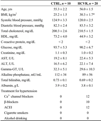
Since Klotho is the co-receptor for FGF23 and β-Klotho
is the co-receptor for FGF21, we also tested the expression of these proteins in cardiomyocytes. In the cardiomyocytes from control patients, the FGF23 immuno-staining was seen to be faint although uniformly diffuse (Figure 2A). Cardio-
Figure 1. Klotho (A-C) and β-Klotho (D-F) immuno-staining. (A): Control cardiomyocytes show intense cytoplasm staining; (B): car-diomyocytes from HCVR patients show reduced staining; however, some clusters of cells show intense staining (arrow); (C): relative values of optical density according to groups; (D): control cardiomyocytes show intense cytoplasmic staining; (E): the staining is reduced in all cardiomyocytes from HCVR patients; and (F): relative values of optical density according to groups. Original magnification 100 ×; scale bar = 20 μm; *P < 0.05; **P < 0.01. HCVR: higher cardiovascular risk; IOD: integrated optical density.
Figure 2. FGF23 (A-C) and FGF21 (D-F) immuno-staining. (A): All control cardiomyocytes show moderate staining; (B): FGF23 staining increases intensely in cardiomyocytes from HCVR patients; (C): relative values of optical density according to groups; (D): the staining in cardiomyocytes from control patients is faint; (E): the staining increased in cells from HCVR patients; and (F): relative values of optical density according to groups. Original magnification 100 ×; scale bar = 20 μm; **P < 0.01. HCVR: higher cardiovascular risk; IOD: integrated optical density.
myocytes from HCVR patients had higher FGF23 staining (Figure 2B). The mean optical density for FGF23 staining was resumed in Figure 2C. FGF21 immuno-staining from control patients showed diffuse but poor staining, which was not uniformly distributed inside the cytoplasm (Figure 2D). In contrast, cardiomyocytes from HCVR patients had more intense FGF21 staining, which was uniformly distrib-uted inside the cytoplasm (Figure 2E). The optical density for FGF21 staining was resumed in Figure 2F.
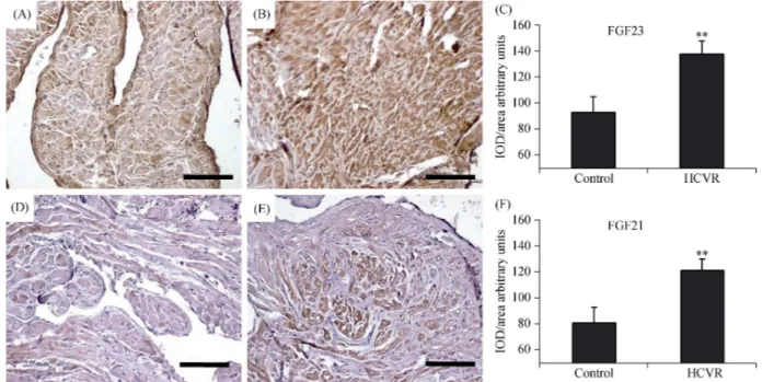
3.3 Higher ASCVD risk increases ER-stress (GRP78)
Since depletion of Klotho can promote cell stress, in or-der to evaluate ER stress and chronic oxidative alterations in cardiomyocytes from control and HCVR patients, we also detected GRP78 and SOD1. In cardiomyocytes from control patients, GRP78 staining intensity was weak to moderate and concentrated in cytoplasmic areas, sometimes near the nucleus (Figure 3A). In HCVR patients, GRP78 expression was higher and mostly concentrated in cytoplasmic areas (Figure 3B). Some cardiomyocytes were clustered and in-tensely stained in certain myocardial areas (Figure 3B, insert). The density for GRP78 staining was resumed in Figure 3C.
SOD1 staining intensity was weakly to moderately in all cells from Control patients, and localized only in certain
cytoplasmic areas (Figure 3D). In contrast, cardiomyocytes from HCVR patients showed intense immuno-staining, which was uniformly distributed throughout the cytoplasm (Figure 3E). The optical density for SOD1 staining was resumed in Figure 3F.
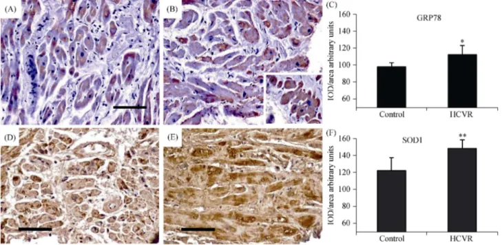
3.4 Higher ASCVD risk increases NF-kB immuno- reactivity
In order to evaluate the inflammatory alterations in car-diomyocytes from patients, we also detected NF-kB, which is a redox regulated transcription factor involved in inflam-mation, immune function, cellular growth and apoptosis. NF-kB was faintly expressed inside the cytoplasm of Con-trol, and stained nuclei were observed only occasionally (Figure 4A). However, cardiomyocytes from HCVR pa-tients had NF-kB immuno-staining which was intense and uniformly distributed not only inside the cytoplasm, but also strongly expressed in a large proportion of nuclei (Figure 4B). Large cardiomyocytes frequently containing debris and had large nuclei strongly immuno-stained (Figure 4B insert) that sometimes showed “bite-like” signs at pole extremities. The quantitative data of stained nuclei is shown in Figure 4C. The optical density for NF-kB staining is resumed in Figure 4D.
Figure 3. GRP78 (A-C) and SOD1 (D-F) immuno-staining. (A): In cardiomyocytes from Control patients, the staining is weak to mod-erate and is concentrated in the cytoplasm, sometimes near the nucleus; (B): in cells from HCVR patients, GRP78 staining is more intense. Some cells are clustered and much intensely stained (insert); (C): relative values of optical density according to groups; (D): the staining is faint to moderate in all cells from Control patients; more intense staining is concentrated in some areas of the cytoplasm; (E): all cardiomyo-cytes from HCVR patients show intense staining uniformly distributed within the cytoplasm; (F): relative values of optical density according to groups. Original magnification 100 ×; scale bar = 20 μm; *P < 0.05; **P < 0.01. GRP78: glucose-regulated protein 78; HCVR: higher cardiovascular risk; IOD: integrated optical density; SOD1: superoxide dismutase-1.
Figure 4. NF-kB immuno-staining. (A): Cardiomyocytes from control patients showed a faint signal in the cytoplasm, while the nuclei are only stained occasionally (insert). (B): cardiomyocytes from HCVR patients have intense and uniformly distributed NF-kB staining also in many nuclei (arrows). Large cardiomyocytes with very pale cytoplasm containing a lot of cellular debris have large strongly stained nuclei (insert). (C): percentage of positively NF-kB stained nuclei according to groups. (D): relative values of optical density according to groups. Original magnification 200 ×; scale bar = 10 μm; **P < 0.01. HCVR: higher cardiovascular risk; IOD: integrated optical density; NF-KB: nuclear factor kappa B.
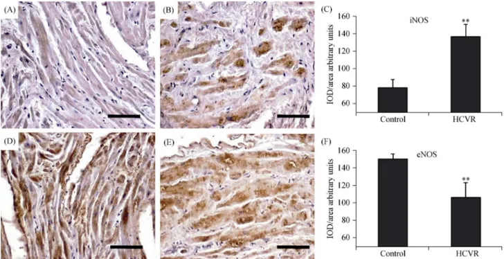
3.5 Higher ASCVD risk increases iNOS and decreases eNOS immuno-reactivity
The increased presence of ER-stress and oxidative stress markers, as well as inflammatory transcription factors in cardiomyocytes from HCVR patients, suggests a higher production of pro-inflammatory iNOS with reduced expres-sion of eNOS. iNOS had very weak staining inside the cy-toplasm of the control (Figure 5A), whereas essentially, all HCVR cardiomyocytes showed a moderate to intensely stained cytoplasm (Figure 5B). The mean optical density for iNOS staining was resumed in Figure 5C. The eNOS stain-ing was intense in the cytoplasm of cells from control tients (Figure 5D), but lower in the cells from HCVR pa-tients (Figure 5E). The optical density for eNOS staining was resumed in Figure 5F.
3.6 Higher ASCVD risk increases fibrosis and TGF-β1 immuno-reactivity
The degree of fibrosis was very poor in control patients, whereas it was more intense in HCVR cardiomyocytes, with a prevalence of thick orange-red fibres instead of thin green fibres (Figure 6A and 6B respectively). The density for Sir-ius-red staining in patient groups was resumed in Figure 6C. In line with histochemical data, the density of TGF-β1
staining, a fundamental protein for collagen synthesis, was more intense and uniformly distributed in the cytoplasm of cells from HCVR patients (Figure 6E), than in cells from control patients (Figure 6D). The optical density for TGF-β1 staining was resumed in Figure 6F.
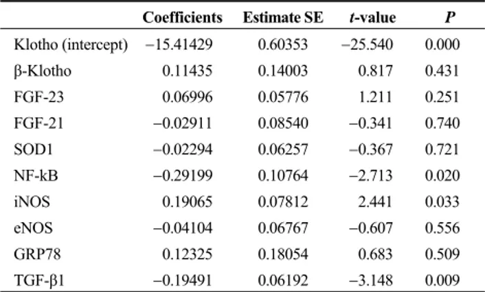
Multivariate regression analysis between klotho and the mean variation of other proteins is summarized in Table 2. The data shows that the variation of klotho was significantly related to the variation of NF-kB, iNOS, and TGF-β1.
4 Discussion
This is the first study to show that: (1) Klotho and FGF-21/FGF-23 proteins are expressed in human cardio-myocytes and (2) cardiocardio-myocytes from patients with a higher cardiovascular risk disease had reduced expression of Klotho but increased expression of FGFs and expression of related stress molecules involved in cardiomyocyte damage. Until now, Klotho had been shown to be expressed solely at the sinoatrial node in mouse hearts, where it is essential for the sinoatrial pacemaker function under stress condi-tions.[31] Here, we have shown expression of Klotho inside
human cardiomyocytes. However, it should be noted that we examined human tissue samples of cardiac atria and hence we cannot translate our results to the whole heart, or
Figure 5. iNOS (A-C) and eNOS (D-F) immuno-staining. (A): The staining is very weak in the cytoplasm of cells from Control patients; (B): cardiomyocytes from HCVR patients show intensely stained cytoplasm; (C): relative values of optical density according to groups; (D): the distribution of staining is uniform and intense in the cytoplasm of cells from Control patients; (E): cells from HCVR patients are faintly to moderately stained; and (F): relative values of optical density according to groups. Original magnification 200 ×; scale bar = 10 μm; **P < 0.01. eNOS: endothelial nitric oxide synthase; HCVR: higher cardiovascular risk; iNOS: inducible nitric oxide synthase; IOD: integrated optical density.
Figure 6. Fibrosis (A-C) and TGF-β1 (D-F) immuno-staining. (A): Control patients samples did not show any abnormal collagen fibres deposition. (B): HCVR patients had diffuse fibrosis. Type-I collagen fibres are shown as orange to red, whereas the thinner type-III collagen fibres appear yellow to green. (C): Relative values of optical density according to groups. (D): Cell staining from Control patients is faint. (E): Immuno-staining from HCVR patient cells is more intense. (F): Relative values of optical density according to groups. Original magnifica-tion 200 ×; scale bar = 10 μm; **P < 0.01. HCVR: higher cardiovascular risk; IOD: integrated optical density; TGF-β1: transforming growth factor-β1.
Table 2. Multivariate regression analysis between klotho and mean variation of other proteins.
Coefficients Estimate SE t-value P Klotho (intercept) 15.41429 0.60353 25.540 0.000 β-Klotho 0.11435 0.14003 0.817 0.431 FGF-23 0.06996 0.05776 1.211 0.251 FGF-21 0.02911 0.08540 0.341 0.740 SOD1 0.02294 0.06257 0.367 0.721 NF-kB 0.29199 0.10764 2.713 0.020 iNOS 0.19065 0.07812 2.441 0.033 eNOS 0.04104 0.06767 0.607 0.556 GRP78 0.12325 0.18054 0.683 0.509 TGF-β1 0.19491 0.06192 3.148 0.009
eNOS: endothelial nitric oxide synthase; GRP78: glucose-regulated protein 78; FGF: fibroblast growth factors; HCVR: higher cardio-vascular risk; iNOS: inducible nitric oxide synthase; NF-KB: nu-clear factor kappa B; SOD1: superoxide dismutase-1; TGF-β1: transforming growth factor-β1.
to a peculiar role in the human sinoatrial node. Nonetheless, unlike the mouse study, the abundant cytoplasmic expres-sion of Klotho proteins in human cardiomyocytes of low cardiovascular risk atria would strongly suggest the pivotal role played by such cardiomyocyte Klotho proteins in whole heart physiology. To the best of our knowledge, this is the first study that identifies Klotho proteins diffusely and in-tensely expressed inside the cytoplasm of human cardio-myocytes. The heart could therefore be regarded not only as a target of soluble Klotho produced by other organs, but also an organ that produces its own Klotho proteins. In par-ticular, we have shown that Klotho protein expression is blunted in patients with HCVR. Our data agrees with pre-vious studies showing a correlation between soluble Klotho concentration and myocardial diseases.[3,7,32,33]
Recent studies show the association between circulating FGF23 levels and pathologic cardiovascular conditions, including left ventricular hypertrophy and vascular endothe-lial dysfunction.[34–37]
We did not measure serum concentrations of FGF23, but we did demonstrate that cardiomyocytes themselves may produce detectable amounts of FGF23 protein and that this was higher in HCVR patients. FGF23 is capable of binding FGF receptors, although with low affinity, also in the ab-sence of membrane Klotho. It is therefore plausible that activation of FGF receptors also occurs in presence of high FGF23 levels in organs where Klotho is absent.[38] We
therefore believe that cardiac pathophysiology is influenced not only by the circulating FGF23, but also by FGF23 pro-duced by cardiomyocytes themselves and might play an important role in metabolic and cardiac functions, perhaps
through an Klotho-independent way and/or with another unknown co-factor.
Klotho also works as a receptor for FGF21. FGF21, ini-tially identified as a liver-derived endocrine factor and sub-sequently found in adipose tissue and muscle,[38] stimulates
glucose uptake in adipocytes (independently from insulin), and has subsequently been related to extension of animal life-span.[39,40] Experimental studies have shown that in
re-sponse to ischemia-reperfusion injury to the myocardium, FGF21 is up-regulated and released from hepatic cells and adipocytes into the circulation. It then interacts with FGFR1 on cardiomyocytes with β-Klotho, inducing phosphorylation of FGFR1. In turn, this reduces apoptosis, cell death and myocardial infarction, improving myocardial function.[39,41]
Serum FGF21 has been found to be higher in patients with chronic metabolic and heart diseases. In addition, FGF21 levels correlate positively with triglyceride, fasting blood glucose, apolipoprotein-B100 and insulin resistance, but negatively with HDL and apolipoprotein-A1.These findings indicate a positive correlation between FGF21 and adverse lipid profile in chronic heart disease patients and a possible compensatory response or resistance to FGF21.[42] Here, we
show that FGF21 is over-expressed by cardiomyocytes. This would however suggest a possible adaptation or com-pensatory elevation of FGF21 in order to overcome cardio-vascular metabolic disorders.[43]
The intracellular distribution of Klotho mostly overlaps with that of the ER and Golgi apparatus,[44] whose
expres-sion has been linked to the ability of cells to withstand stress conditions. Indeed, Klotho decreases the GRP78 expression and related apoptosis in mice.[10] Our data agree with these
observations. Indeed, cardiomyocytes from HCVR patients had reduced Klotho expression and higher increases of GRP78 expression. GRP78 recognizes misfolded proteins inside endoplasmic reticulum and dissociates its binding from its molecular partners. This in turn triggers the un-folded protein response by regulating gene responses aimed at restoring endoplasmic reticulum proteome stability.[13]
Failure to promote cell survival by decreasing misfolded proteins and oxidative stresses may promote apoptotic cell death.[45]
GRP78 is induced by increased oxidative stress related to the inflammatory response, and inflammatory cytokines are known to down-regulate Klotho through NF-kB.[21,22] We
therefore examined the presence and modulation of SOD1, iNOS and NF-kB proteins. Our results demonstrated an increase of SOD1, iNOS and NF-kB inside the cardiomyo-cytes from HCVR patients, suggesting a chronic pro-oxida-tive, pro-inflammatory and pro-apoptotic status. This is in line with previous research and shows that there is a
correla-tion between obesity, diabetes, decrease in Klotho and in-crease in NF-kB and oxidative stress. In addition, we ob-served reduced immuno-staining of eNOS in HCVR car-diomyocytes. Notably, cytoplasmic eNOS improves ener-getic cellular metabolisms and stimulates mitochondrial biogenesis.[46] So, there also exists the possibility of a
dis-ruption of energetic metabolism and altered mitochondrial biogenesis in HCVR patients cardiomyocytes.
Not surprisingly, we detected fibrosis and higher TGF-β1 expression in cardiomyocytes of HCVR patients. We be-lieve that the reduced Klotho proteins expression found in cardiomyocytes from HCVR patients, could be related to the modulation of TGF-β1 synthesis and subsequent fibrosis. Indeed, Klotho is able to interfere with TGF-β1 signaling.[24]
Furthermore, enhanced synthesis of Klotho improves the arrangement of cardiomyocytes and myocardial fibres in animal models.[10] Although these studies show an
associa-tion between both Klotho deficiency and metabolic and cardiac diseases, the cause-effect relationship remains to be clarified.
4.1 Clinical implications
It is accepted that diabetes, obesity and hypertension are risk factors for both coronary calcification and cardiomyo-cyte pathology. However, the susceptibility, severity and progression of cardiovascular diseases are not completely explained by these risk factors. Therefore, identification of additional molecules involved in the pathophysiology and the evolution of cardiovascular diseases is fundamental to achieve important objectives such as the development of specific new therapies, identification of biomarkers for grading cardiovascular risk and monitoring the effects of therapy including physical rehabilitation.
4.2 Limitations
Certain limitations of this study need to be recognized. Firstly, we collected atrial biopsies but atrial tissues were considered myocardium and have the same metabolism and cellular signaling as the cardiomyocytes. In addition, sam-pling aimed to provide an appropriate amount of tissue without compromising the left ventricular function, and consequently the patient’s survival.
Secondly, only a limited number of patients were en-rolled in this study. The patients were however very highly selected to avoid any confounding co-morbidity and, in ad-dition, the sample was adequate for us to identify the major relationship between Klotho and related protein presence and cardiovascular risk.
Thirdly, we only used immuno-histochemistry to support
our results. This was dictated by the rules in force in our operating theatre, which excluded the possibility of using liquid nitrogen and then immediately freeze the biopsies for molecular biology evaluations. However, although the im-muno-histochemistry does not quantify molecules as accu-rately as molecular biology, we believe that this technique has certain advantages. Indeed, the standard fixation main-tains the architecture of the tissue, allowing adequate mor-phological assessments. In addition, immuno-histochemistry allows us to localize the analyzed molecules both between different cells types and inside the cells. In contrast, mo-lecular biology requires immediate freezing and homogeni-zation of the sample. As a consequence, it does not take into account the specificities of the location of immuno-staining, and so the morphology of the tissue.
4.3 Conclusions
In conclusion, we have reported for the first time the presence of Klotho proteins in human hearts in vivo, and that HCVR cardiomyocytes have deranged expressions of Klotho, FGF23, FGF21 and related stress molecules in-volved in cellular injury. We believe that modulation of these molecules could play a pivotal role in heart patho-physiology. We therefore suggest that preventing the Klotho decrease or promoting its increase, and/or FGFs modulation inside cardiomyocytes, could prevent and/or reduce cardio-vascular diseases in high risk patients.
Acknowledgments
We would like to thank Prof. Robert Coates (Centro Lingusitico, Bocconi University, Milan, Italy) for his lin-guistic revision and Prof. Stefano Calza (Department of Molecular and Translational Medicine, University of Bre-scia, Italy) for his statistical advice. The authors declare that they have no conflict of interests. This study was funded by grants from the University of Brescia (to Corsetti G).
References
1 EvrardS, DelanayeP, KamelS, et al. Vascular calcification: from pathophysiology to biomarkers. Clin Chim Acta 2015; 438: 401–414.
2 Kuro-o M. Klotho. Pflugers Arch 2010; 459: 333–343. 3 Navarro-Gonzalez JF, Donate-Correa J, Muros de Fuentes M,
et al. Reduced Klotho is associated with the presence and
se-verity of coronary artery disease. Heart 2014; 100: 34–40. 4 Ikushima M, Rakugi H, Ishikawa K, et al. Anti-apoptotic and
anti-senescence effects of Klotho on vascular endothelial cells.
Biochem Biophys Res Commun 2006; 339: 827–832.
against a direct role of Klotho in insulin resistance. Pflugers
Arch 2010; 459: 465–473.
6 Yamamoto M, Clark JD, Pastor JV, et al. Regulation of oxi-dative stress by the anti-aging hormone klotho. J Biol Chem 2005; 280: 38029–38034.
7 Semba RD, Cappola AR, Sun K, et al. Plasma Klotho and cardiovascular disease in adults. J Am Geriatr Soc 2011; 59: 1596–1601.
8 Majumdar V, Christopher R. Association of exonic variants of Klotho with metabolic syndrome in Asian Indians. Clin Chim
Acta 2011; 412: 1116–1121.
9 Moe SM. Klotho: a master regulator of cardiovascular disease?
Circulation 2012; 125: 2181–2183.
10 Song S, Gao P, Xiao H, et al. Klotho suppresses cardiomyo-cyte apoptosis in mice with stress-induced cardiac injury via downregulation of endoplasmic reticulum stress. PLoS One 2013; 8: e82968.
11 Szabadkai G, Rizzuto R. Participation of endoplasmic reticu-lum and mitochondrial calcium handling in apoptosis: more than just neighborhood? FEBS Lett 2004; 567: 111–115. 12 Xu C, Bailly-Maitre B, Reed JC. Endoplasmic reticulum
stress: cell life and death decisions. J Clin Invest 2005; 115: 2656–2664.
13 Kaufman RJ. Orchestrating the unfolded protein response in health and disease. J Clin Invest 2002; 110: 1389–1398. 14 Okada K, Minamino T, Tsukamoto Y, et al. Prolonged
endo-plasmic reticulum stress in hypertrophic and failing heart after aortic constriction: possible contribution of endoplasmic re-ticulum stress to cardiac myocyte apoptosis. Circulation 2004; 110: 705–712.
15 Martindale JJ, Fernandez R, Thuerauf D, et al. Endoplasmic reticulum stress gene induction and protection from ischemia/ reperfusion injury in the hearts of transgenic mice with a tamo-xifen-regulated form of ATF6. Circ Res 2006; 98: 1186–1193. 16 Hamada H, Suzuki M, Yuasa S, et al. Dilated cardiomyopathy caused by aberrant endoplasmic reticulum quality control in mutant KDEL receptor transgenic mice. Mol Cell Biol 2004; 24: 8007–8017.
17 Homma K, Fujisawa T, Tsuburaya N, et al. SOD1 as a mo-lecular switch for initiating the homeostatic ER stress re-sponse under zinc deficiency. Mol Cell 2013; 52: 75–86. 18 Roberts CK, Barnard RJ, Sindhu RK, et al. A high-fat,
re-fined-carbohydrate diet induces endothelial dysfunction and oxidant/antioxidant imbalance and depresses NOS protein ex-pression. J Appl Physiol 2005; 98: 203–210.
19 Li R, Wang WQ, Zhang H, et al. Adiponectin improves endo-thelial function in hyperlipidemic rats by reducing oxidative/ nitrative stress and differential regulation of eNOS/iNOS activity.
Am J Physiol Endocrinol Metab 2007; 293: E1703–E1708.
20 Toda N, Tanabe S, Nakanishi S. Nitric oxide-mediated coro-nary flow regulation in patients with corocoro-nary artery disease: recent advances. Int J Angiol 2011; 20: 121–134.
21 Moreno JA, Izquierdo MC, Sanchez-Nino MD, et al. The inflammatory cytokines TWEAK and TNFalpha reduce renal
klotho expression through NFkappaB. J Am Soc Nephrol 2011; 22: 1315–1325.
22 Thurston RD, Larmonier CB, Majewski PM, et al. Tumor necrosis factor and interferon-gamma down-regulate Klotho in mice with colitis. Gastroenterology 2010; 138: 1384–1394. 23 Teocchi MA, Ferreira AE, da Luz de Oliveira EP, et al. Hip-pocampal gene expression dysregulation of Klotho, nuclear factor kappa B and tumor necrosis factor in temporal lobe ep-ilepsy patients. J Neuroinflammation 2013; 10: 53.
24 Doi S, Zou Y, Togao O, et al. Klotho inhibits transforming growth factor-beta1 (TGF-beta1) signaling and suppresses re-nal fibrosis and cancer metastasis in mice. J Biol Chem 2011; 286: 8655–8665.
25 Kararigas G, Dworatzek E, Petrov G, et al. Sex-dependent regulation of fibrosis and inflammation in human left ven-tricular remodelling under pressure overload. Eur J Heart Fail 2014; 16: 1160–1167.
26 Andrus B, Lacaille D. 2013 ACC/AHA guideline on the as-sessment of cardiovascular risk. J Am Coll Cardiol 2014; 63: 2886.
27 Stone NJ, Robinson JG, Lichtenstein AH, et al. 2013 ACC/AHA guideline on the treatment of blood cholesterol to reduce atherosclerotic cardiovascular risk in adults: a report of the American College of Cardiology/American Heart Asso-ciation Task Force on Practice Guidelines. J Am Coll Cardiol 2014; 63: 2889–2934.
28 Lalu MM, Pasini E, Schulze CJ, et al. Ischaemia-reperfusion injury activates matrix metalloproteinases in the human heart.
Eur Heart J 2005; 26: 27–35.
29 Dayan D, Hiss Y, Hirshberg A, et al. Are the polarization colors of picrosirius red-stained collagen determined only by the diameter of the fibers? Histochemistry 1989; 93: 27–29. 30 Koren R, Yaniv E, Kristt D, et al. Capsular collagen staining
of follicular thyroid neoplasms by picrosirius red: role in dif-ferential diagnosis. Acta Histochem 2001; 103: 151–157. 31 Takeshita K, Fujimori T, Kurotaki Y, et al. Sinoatrial node
dysfunction and early unexpected death of mice with a defect of klotho gene expression. Circulation 2004; 109: 1776–1782. 32 Ichikawa S, Imel EA, Kreiter ML, et al. A homozygous
mis-sense mutation in human KLOTHO causes severe tumoral calcinosis. J Clin Invest 2007; 117: 2684–2691.
33 Angelin B, Larsson TE, Rudling M. Circulating fibroblast growth factors as metabolic regulatorsa critical appraisal.
Cell Metab 2012; 16: 693–705.
34 Gutierrez OM, Januzzi JL, Isakova T, et al. Fibroblast growth factor 23 and left ventricular hypertrophy in chronic kidney disease. Circulation 2009; 119: 2545–2552.
35 Mirza MA, Larsson A, Lind L, et al. Circulating fibroblast growth factor-23 is associated with vascular dysfunction in the community. Atherosclerosis 2009; 205: 385–390.
36 Shibata K, Fujita S, Morita H, et al. Association between circulating fibroblast growth factor 23, alpha-Klotho, and the left ventricular ejection fraction and left ventricular mass in cardiology inpatients. PLoS One 2013; 8: e73184.
37 Faul C, Amaral AP, Oskouei B, et al. FGF23 induces left ventricular hypertrophy. J Clin Invest 2011; 121: 4393–4408. 38 Martin A, David V, Quarles LD. Regulation and function of
the FGF23/Klotho Endocrine pathways. Physiol Rev 2012; 92: 131–155.
39 Kharitonenkov A, Shiyanova TL, Koester A, et al. FGF-21 as a novel metabolic regulator. J Clin Invest 2005; 115: 1627–1635.
40 Kenyon C. Could a hormone point the way to life extension?
eLife 2012; 1: e00286.
41 Liu SQ, Roberts D, Kharitonenkov A, et al. Endocrine protec-tion of ischemic myocardium by FGF21 from the liver and adipose tissue. Sci Rep 2013; 3: 2767.
42 Lin Z, Wu Z, Yin X, et al. Serum levels of FGF-21 are
in-creased in coronary heart disease patients and are independ-ently associated with adverse lipid profile. PLoS One 2010; 5: e15534.
43 Unger RH. Klotho-induced insulin resistance: a blessing in disguise? NatMed 2006; 12: 56–57.
44 Li SA, Watanabe M, Yamada H, et al. Immunohistochemical localization of Klotho protein in brain, kidney, and reproduc-tive organs of mice. Cell Struct Funct 2004; 29: 91–99. 45 Niforou K, Cheimonidou C, Trougakos IP. Molecular
chap-erones and proteostasis regulation during redox imbalance.
Redox Biol 2014; 2: 323–332.
46 Nisoli E, Clementi E, Paolucci C, et al. Mitochondrial bio-genesis in mammals: the role of endogenous nitric oxide.