Arch Orthop Trauma Surg ( 1987) 106:209-219 Archives of Orthopaedicand Traumatic Surgery © Springer-Verlag 1987
The Foreign Body Reaction in Total Hip Arthroplasties
A Correlated Light-Microscopy, SEM, and TEM Study
U E Pazzaglial, C Dell'Orbo 2, and M J Wilkinson 3
1 Institute of Orthopaedics of the University of Pavia, Via Taramelli, -27100 Pavia, Italy 2 Institute of Anatomy of the University of Pavia, Via Taramelli, I-27100 Pavia, Italy 3
Department of Biomedical Engineering, Brockley Hill, Stanmore, Middlesex HA 7 4 LP, United Kingdom
Summary An in vivo histological and ultrastructural study of the cellular reaction to particulate material currently used in orthopaedic surgery produced evi-dence that, on a strictly cellular level, the main dam-age is done by the smallest particles produced by hip prostheses, i e metal particles, irrespective of differ-ences in their chemical composition Particle size and release rate are the critical factors, although other mechanisms of cellular damage may be active once granulation tissue is formed.
Candidate biomaterials are evaluated by a number of methods to assess their mechanical and biological suitability for implantation With regard to biological suitability, the factors of size, shape and temporal ef-fects are of particular importance.
The relevance of the form and size of foreign ma-terials has been observed in tumor induction studies l 12,13 l These factors are also of significance in the foreign body reaction to wear and corrosion debris released from hip prostheses l 1,15 l.
The time parameter is also relevant: If particles are presented to cells at a rapid rate they will be en-veloped by a capsule of connective tissue l 4 l If par-ticles are released slowly into tissue they will be phagocytosed with a significantly greater degree of associated cellular reaction The latter condition is typical of the mode of release of wear and corrosion debris in vivo.
Retrieved implants and associated tissue are an important source of material for the study of the bio-logical reaction to particulates l 3, 7, 14, 15 l The histology of the foreign body reaction in hip
arthro-Offprint requests to: U E Pazzaglia
plasties is well documented l 5, 8, 15 l Along with routine light microscopy, a limited amount of ultra-structural work has been performed in this area l 17 l. Up to now, no attempt has been made to correlate size, shape and composition of particles to cell re-sponse at both the histological and the ultrastructural level.
In this study we have employed several methods of light microscopy and electron microscopy in an at-tempt to achieve this.
Materials and Methods
The material for this work was obtained from 12 cemented hip prostheses removed following aseptic loosening Case reports are not included because the study concerns exclusively the cell response to foreign materials Femoral components included stainless-steel and cast Co-Cr alloy prostheses of different de-signs The cups were all of high-density polyethylene Radio-opaque cement had been used in all cases The main data on these prostheses are summarized in Table 1.
Tissue specimens were obtained from the capsule sur-rounding the head of the prosthesis and from the bone-cement interfaces of the stem and the cup For light microscopy, speci-mens of tissue were fixed in 10 % formaldehyde buffered at p H 7.4 before embedding in paraffin Sections 5-7 tlm thick were stained with haematoxylin-eosin Other specimens were fresh frozen to -700C and sections were then prepared with a cryo-tome These sections were stained for lipids and acid phos-phatase l 2 l Scanning electron microscopy and X-ray micro-analysis was performed on routine histology sections; the cover-slip was removed with xylene and the sections were coated with a thin layer of carbon or gold For transmission electron microscopy specimens were fixed in 4 % buffered glutaral-dehyde and post-fixed in 1 % osmium tetroxide solution They were dehydrated in alcohol and embedded in Epon 812 resin. Some of the thin sections prepared by ultramicrotomy were stained with uranyl acetate and lead citrate before examination in a Philips 300 TEM.
U E Pazzaglia et al : Foreign Body Reaction in Total Hip Arthroplasty
Table 1 Data on implants which supplied tissue specimens for the study
Patient Prosthesis Alloy Removed after Stem surface signsa
CGB Charnley Stainless steel 5 years 1 month Corrosion
NE Charnley Stainless steel 2 years 1 month Breakage, corrosion and wear
EG Miiller Co-Cr 9 years 6 months Wear
CW M Uller Co-Cr 2 years Wear
MP Charnley Stainless steel 5 years 2 months Corrosion
BR GR (Rizzoli) Co-Cr 11 years 3 months Wear
RA Charnley Stainless steel 6 years 8 months Corrosion
PM Charnley Stainless steel 11 years 5 months Breakage, corrosion and wear
AP Muller Co-Cr 6 years 3 months Breakage, wear
SF Charnley Stainless steel 6 years 10 months Breakage, corrosion and wear FP Charnley Stainless steel 7 years 5 months Breakage, corrosion and wear PF Charnley Stainless steel 11 years 1 month Breakage, corrosion and wear
a No sign of abnormally high wear was observed on the sockets
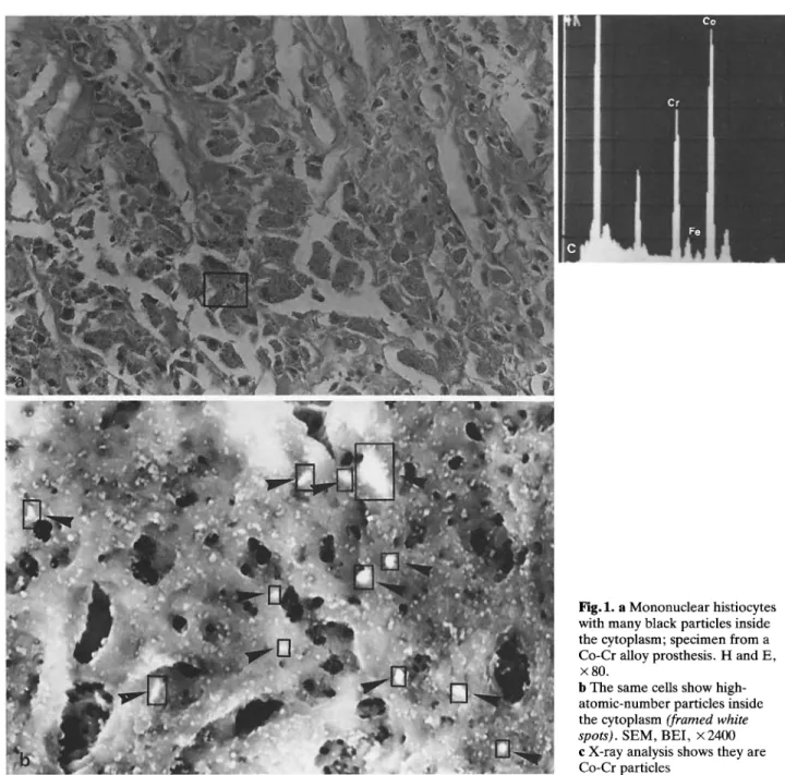
Fig 1 a Mononuclear histiocytes
with many black particles inside the cytoplasm; specimen from a Co-Cr alloy prosthesis H and E, x 80.
b The same cells show high-atomic-number particles inside the cytoplasm (framed white
spots) SEM, BEI, x 2400 c X-ray analysis shows they are
Co-Cr particles 210
U E Pazzaglia et al : Foreign Body Reaction in Total Hip Arthroplasty
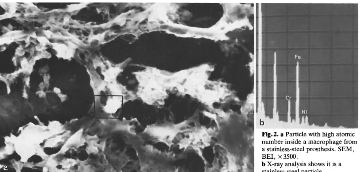
Fig 2 a Particle with high atomic
number inside a macrophage from a stainless-steel prosthesis SEM, BEI, x 3500.
b X-ray analysis shows it is a stainless steel particle
Results
Three classes of material in particulate form pro-duced in vivo by hip prostheses were observed in this study: metal particles, polyethylene fibres and methyl-methacrylate globules Each class is well character-ized, not only by the chemical composition but also by the shape and size range In these respects all cases were similar.
Metal particles were always observed inside the cytoplasm of cells; only where an acellular and not yet organized biological material (fibrin-like mate-rial) was present were the particles cell free.
The metal particles were phagocytosed by mono-nuclear histiocytes, which exhibited a foamy cyto-plasm The particles appeared black when viewed by transmitted light microscopy and had an irregular shape They were not larger than 1 im (Fig 1) Metal particles are not birefringent in polarized light; in-deed, they are opaque to the light, but the smallest refract the light on their edges, so they appear weakly luminous The same refraction is evident when they are observed in a dark field.
Acid phosphatase and lipid staining showed a very positive reaction in the cells which phagocytosed metal particles.
A scanning electron microscope with a backscat-tered electron detector revealed the particles as bright spots, and simultaneous X-ray analysis con-firmed their elemental composition: Ni, Cr, Mn and Fe were detected in particles from stainless-steel prostheses, Co, Cr and Mo from Co-Cr alloy prosth-eses (Fig 2) In ultrathin sections, both stained and
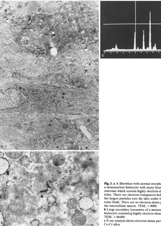
unstained, electron-opaque particles were contained in numerous secondary lysosomes, which dominated the cytoplasm X-ray analysis by TEM was used to confirm the metallic nature of this opaque material (Fig 3) While some cells had uninterrupted cyto-plasmic and lysosomal membranes, other cells ex-hibited disruption of these structures, having suffered various degrees of cellular damage, and in some cases appeared frankly necrotic (Fig 4) Such macrophages were observed both in soft tissue specimens and in bone specimens, where they had penetrated the haversian canals of diaphyseal cortical bone and the medullary spaces of cancellous bone.
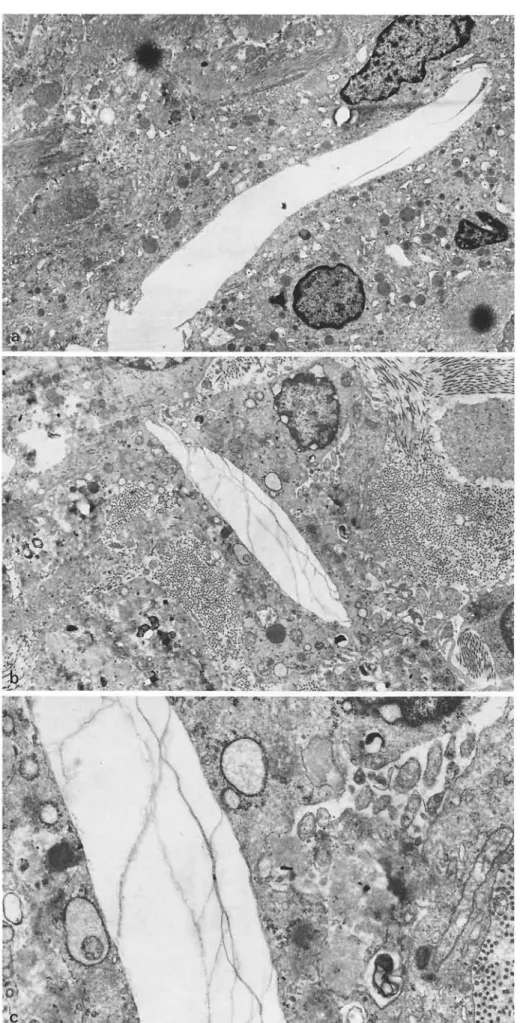
Polyethylene particles are transparent and there-fore cannot be directly observed by bright-field light microscopy In polarized light they are birefringent; their shape is that of elongated, irregular fibres or flattened chips; the size range is wide, but they are never less than 3 m The smallest particles are phagocytosed by mononuclear cells, but as the size increases they are engulfed by multinucleated giant cells or surrounded by a fibrous tissue capsule.
Polyethylene fibers inside the cells cannot be de-tected by scanning electron microscopy because their low atomic number gives rise to a poor backscattered electron signal Larger particles, which lie outside the cells, may be observed directly by the SEM operated in the secondary electron mode They are elongated like stripes or look like flattened chips; their surface is rough and frayed (Fig 5) In TEM polyethylene fibres inside the cells look like electron-transparent areas, rounded or elongated in shape according to the plane of the section, the smallest reaching 211
U E Pazzaglia et al : Foreign Body Reaction in Total Hip Arthroplasty
Fig 3 a A fibroblast with normal morphology and
a mononuclear histiocyte with many lysosomal cisternae which contain highly electron-dense par-ticles There are electron-transparent holes where the largest particles tore the slice under the micro-tome blade There are no electron-dense particles in the intercellular matrix TEM, x 9000.
b Large secondary lysosomes of a mononuclear
histiocyte containing highly electron-dense particles. TEM, x 46000.
c X-ray analysis shows electron-dense particles to be
Co-Cr alloy 212
U E Pazzaglia et al : Foreign Body Reaction in Total Hip Arthroplasty
Fig 4 a A mononuclear histiocyte
in very poor condition with many large lysosomes, widespread myelinic figures and discontinuous plasma membrane TEM, x 4000. b Mononuclear histiocytes show a strongly positive reaction for acid phosphatase x 175
30000
A
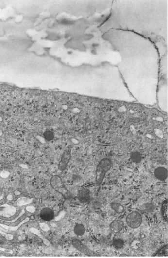
The edges may be irregular, with deep gaps where cytoplasmic substance penetrates No evidence of membrane structure is observable around the par-ticles, and they appear to be in intimate contact with the cytoplasm The cells have well-developed cyto-plasmic organelles and appear healthy.Often, it is possible to observe cells which have phagocytosed both small polyethylene particles and metal particles In this case, metal particles are al-ways observed inside secondary lysosomes, while polyethylene particles do not exhibit any definite re-lation with the cytoplasmic membranes (Fig 6).
Methylmethacrylate particles are solubilized by solvents used in tissue processing Their presence is implied by rounded, empty holes in the section. These are surrounded by large multinucleated giant cells or by a fibrous connective capsule The size range is wide, the smallest being about 50-100 pm in diameter and engulfed by a single giant cell, while the
largest may be 3-5 mm in diameter surrounded by a fibrous capsule Both giant cells and fibroblasts of the capsule lack acid-phosphatase staining activity.
Some of the globules may contain a fine granular material which refracts the light in dark field and in polarized light The corresponding SEM picture is of empty cavities inside one giant cell or between two or more giant cells Bright material looks like granular particles, O 1 tm in size, and with a high atomic number if observed in BEI and with the characteristic spectrum of barium in X-ray analysis (Fig 7).
The same granular material may be observed in the general vicinity of the acrylic globule and is pre-sent only on the surface of the adjoining cells, never inside the cells This is due to the fact that Ba par-ticles become dislodged during processing of the tis-sue.
Transmission electron microscopy of thin sections showed the cells surrounding the acrylic globule to 213
U E Pazzaglia et al : Foreign Body Reaction in Total Hip Arthroplasty
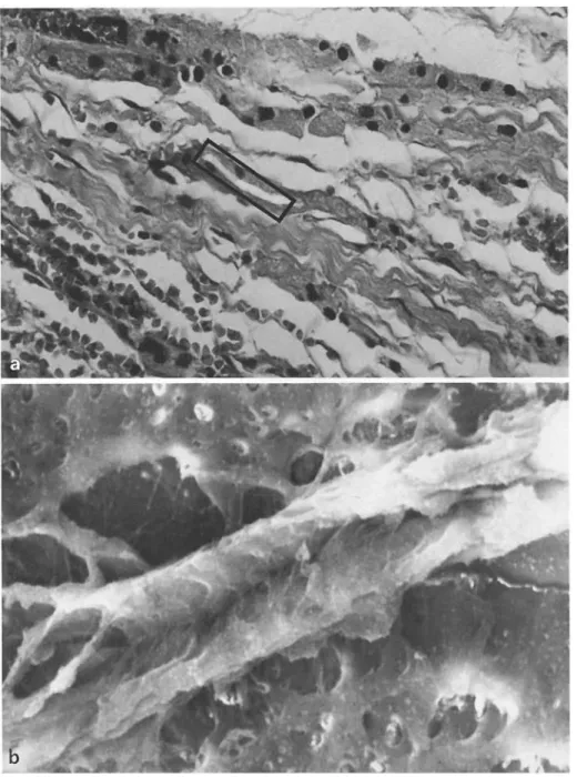
Fig 5 a A large polyethylene
fibre surrounded by histiocytes; double exposition in polarized light and bright field H and E, x 80.
b The same fibre in scan SEM,
SEI, x 1600
have a well-developed endoplasmic reticulum, free ribosomes, mitochondria and a well-developed Golgi apparatus The plasma membrane adjacent to the methylmethacrylate was complete, with a thin layer of filamentous material (fibrin-like) interposed be-tween the cell surface and the acrylic cement Thin sheets of the same material appeared inside what could have been gaps in the mass of the acrylic globule The cells had a healthy appearance (Fig 8). Discussion
Three types of foreign bodies are considered, which are produced by hip prostheses comprising a metal
stem with a high-density polyethylene cup and fixed by acrylic bone cement Identification of particles by routine light-microscope histology is easily achieved l 1,8,15 l, but little is known about the cellular re-sponse to each type of foreign material as it can be visualized by electron microscopy and histochemical methods.
Several factors influence the cellular reaction to foreign material:
The Chemical Composition of the Material
Toxic materials may produce intracellular damage and necrosis Toxicity of metals used in orthopaedic 214
U E Pazzaglia et al : Foreign Body Reaction in Total Hip Arthroplasty
Fig 6 a A large polyethylene
fibre surrounded by several mono-nuclear histiocytes or giant cells. No lysosomes are observed inside the cells TEM, x 9000.
b A small polyethylene fibre
inside the cytoplasm of a mono-nuclear histiocyte TEM, x 10200.
c Detail of b: The same cell has
phagocytosed metal particles and they are observed inside lyso-somes TEM, x 26100
U E Pazzaglia et al : Foreign Body Reaction in Total Hip Arthroplasty
Fig 7 a A methylmethacrylate globule
with opaque particles on the borders and inside H and E, x 300.
b The same methylmethacrylate globule;
particles have high-atomic-number ele-ments SEM, BEI, x 220.
c Higher magnification of particles. SEM, BEI, x 2100.
d X-ray analysis of particles shows they
are barium crystals 216
U E Pazzaglia et al : Foreign Body Reaction in Total Hip Arthroplasty
Fig 8 Ultrastructural detail of a cell surrounding a methyl-methacrylate globule The cell shows a well-developed granu-lar endoplasmic reticulum, ribosomes and mitochondria The cell membrane is uninterrupted and a thin fibrin-like material is interposed between the cell membrane and the acrylic Fila-ments of this material spread inside thin cracks in the acrylic. TEM, x 27000
implants has been observed for tissue culture cells (Co-Cr alloy being more toxic than stainless steel), but the metals seem to be well tolerated by human tissues l 9, 16 l.
The results of this study agree with the latter ob-servation: cells which have phagocytosed or are in contact with particles of the studied materials do not show ultrastructural lesions of cytoplasmic structures. Necrosis was observed only in cells which had phago-cytosed large amounts of metal particles either stain-less steel or Co-Cr alloy), while small quantities of the same materials left the cells healthy.
Two explanations are possible: (a) It is necessary to accumulate large quantities of particles to release soluble ions sufficient to reach the threshold of toxic-ity, but in this case the behaviour of cells which phago-cytosed stainless steel or Co-Cr alloy particles should
have been different (b) There is a mechanism of cel-lular damage other than direct poisoning by metal ions; this will be discussed later.
Also, barium particles were identified in contact with cells surrounding acrylic globules, but these were more likely an artefact related to processing of
the specimens.
The Form, the Size and the Location of Particles
Methylmethacrylate particles are typically globular; polyethylene particles form fibres or chips; metal particles are very irregular in shape The form of the particles is related to the mechanism of production. Methylmethacrylate particles are derived from free acrylic globules or from pieces of cement abraded by bone Polyethylene particles are produced by proces-ses of wear of the socket Metal particles result from both wear and corrosion of the stem.
For each type, size shows a wide range of varia-tion Nevertheless, it is possible to characterize ap-proximately the limits for each type, based on the structural properties of the original material and the mechanism of particle production.
Metal particles are the smallest, varying from 50-100 A to a maximum of 1 xm The size of polyethyl-ene particles is difficult to evaluate in histological sec-tions because of their fibre shape; they are randomly orientated in the tissue, and it is impossible to know if the section is cut perpendicular to the main axis of the fibre or parallel to it Although with this limit the smallest polyethylene fibres observed in this work were about 3-5 Lm long, while the longest reached 3-5 mm The size of methylmethacrylate particles is easily evaluated on account of their globular shape with seriate sections: they vary from 50-100 tm to
several millimeters.
The size of the particles is an important factor de-termining the behaviour of the cells; small particles (i.e all the metal particles and the smallest polyethyl-ene particles) are phagocytosed by mononuclear his-tiocytes, while larger particles are engulfed by multi-nucleated giant cells When particles are very large they are surrounded by a fibrous capsule or by sev-eral giant cells.
This cellular behaviour was also observed in pre-vious light-microscopy studies of the tissue reaction to foreign material l 8,15 l Metal particles in the same size range but of different composition (stainless steel and Co-Cr alloy) have always been observed inside secondary lysosomes and appear to elicit the same intracellular response, i e development of the lyso-somal system l 6 l and synthesis of lysolyso-somal enzymes in an attempt to digest the foreign material Small polyethylene particles have also been observed inside 217
U E Pazzaglia et al : Foreign Body Reaction in Total Hip Arthroplasty the cell cytoplasm of mononuclear histiocytes, but
they are not surrounded by any membrane structure. This is particularly evident in macrophages which have engulfed both metal and plastic particles.
The different reaction of the cells to the two types of material may be related to the chemical composi-tion or to the different size; the smallest polyethylene particles are much larger than metal debris and well above the size of the lysosomal structures.
Neither large polyethylene fibres nor methyl-methacrylate globules stimulate lysosome develop-ment or cytoplasmic organelle alterations in the phagocytosing or adjoining giant cells; indeed, lyso-somes in multinucleated giant cells were observed to be not larger than those in mononuclear histiocytes. This leads to the hypothesis that for the material con-sidered in this study the critical factor is size.
The Time Factor
It has been observed in experimental studies that small particles of metal debris implanted in the tissue of experimental animals are engulfed en bloc by a fi-brous capsule l 4 l Large masses of small particles presented to the cells at a rapid rate elicit a tissue reaction to the whole mass Only if particles are re-leased slowly in the tissue may the reaction develop through the phagocytosis processes, and this is the case with particles released from hip prostheses in vivo.
Cellular damage was observed to occur only in those cells which phagocytosed the smallest material particles, i e metal particles, while small polyethyl-ene and acrylic particles were also phagocytosed by cells did not lead to lysosomal development The fol-lowing chain of events is suggested by the ultrastruc-tural study of the foreign body reaction in peri-prosthetic tissues: In the attempt to digest the foreign material of suitable size, development of the lyso-somal system and synthesis of lysolyso-somal enzymes takes place; a single enzyme marker (acid phospha-tase) was used here, but the result can be extrapo-lated to a wider range of lysosomal enzymes l 10 l Be-cause the material remains undigested, the cells be-come autolytic, and lytic enzymes, together with un-digested particles, are ultimately released into the surroundings More macrophages move in and so more lytic enzymes are produced, activating a self-perpetuating cycle.
The results of this study suggest that for materials currently in orthopaedic use the critical factors may be quantity, size and particle release rate; in the cases studied this critical combination was reached exclu-sively by metal particles (both stainless steel and Co-Cr alloy).
Previous papers concerning the failure of hip prostheses have emphasized the role of plastic wear particles in the foreign body reaction l 1,5,11 l This is not in contrast with our results; we also observed a cellular reaction to these particles with stimulation of a granulation tissue, but in this case the mechanism of cellular damage is different from that of metal par-ticles As described by Buchhorn and Willert l 1 l, an excessive proliferation of the granulation tissue tends to produce cellular necrosis if the blood supply can-not follow the expansion of the granulomas In this case the critical factor becomes the quantity of wear particles when they exceed the capacity of transport and storage of the drainage system.
Conclusions
This study of the foreign body reaction in hip prosthe-ses employing electron-microscopy methods has shown that metal particles are potentially more harmful at the cellular level than plastic or acrylic be-cause of the factor of size In this regard, differences between stainless steel and Co-Cr alloy particles were not observed.
Acknowledgements This work was supported by grant no.
82.02034 04 of the Italian National Research Council The au-thors acknowledge Mrs G Bodini for her technical assistance.
References
1 Buchhorn GH, Willert HG ( 1982) Effect of plastic wear particles on tissue In: Williams DF (ed) Biocompatibility of orthopedic implants, vol I CRC Press, Boca Raton, Florida
2 Burstone MS ( 1962) Enzyme histochemistry Academic Press, New York
3 Charosky CB, Bullough PG, Wilson PD ( 1973) Total hip replacement failures A histological evaluation J Bone Joint Surg lAml 55: 49-58
4 Cohen J ( 1959) Assay of foreign body reaction J Bone Joint Surg lAml 41:152-166
5 Freeman MAR, Vernon-Roberts B ( 1977) The tissue
re-sponse to total joint replacement prostheses In: Freeman MAR, Swanson SAV (eds) Scientific basis of joint replace-ment Pitman Medical Publishing, London
6 Goldner RD, Adams DO ( 1977) The structure of mono-nuclear phagocytes differentiating in vitro III The effect of particulate foreign substances Am J Pathol 89: 335-350 7 Gui L, Pizzoferrato A, Ranieri L, Gualtieri T ( 1978)
Rea-zione tissutale articolare e para-articolare negli impianti protesici G Ital Ortop Traum lSuppll 4:215-227
8 Mirra JM, Amstutz HC, Matos M, Gold R ( 1976) The pa-thology of the joint tissue and its clinical relevance in pros-thesis failure Clin Orthop 117:221-240
U E Pazzaglia et al : Foreign Body Reaction in Total Hip Arthroplasty 9 Pappas AM, Cohen J ( 1968) Toxicity of metal particles in
tissue culture J Bone Joint Surg lAml 50:535-556 10 Rae T ( 1975) A study on the effects of particulate metals of
orthopaedic interest on murine macrophages in vitro J Bone Joint Surg lBrl 57: 444-450
11 Revell PA, Weightman B, Freeman MAR, Vernon-Ro-berts B ( 1978) The production and biology of polyethylene wear debris Arch Orthop Trauma Surg 91: 167-181 12 Stinson NE ( 1963) The tissue reaction induced in rats and
guinea-pigs by polymethylmethacrylate (acrylic) and stain-less steel Br J Exp Pathol 45:21-29
13 Stinson NE ( 1964) Tissue reaction induced in guinea-pigs by particulate polymethylmethacrylate, polyethylene and nylon of the same size range Br J Exp Pathol 46:135-146
14 Willert HG ( 1973) Tissue reactions around joint implants and bone cement In: Chapchal G (ed) Arthroplasty of the hip Thieme, Stuttgart
15 Willert HG, Semlitsch M ( 1976) Tissue reactions to plastic and metallic wear products of joint endoprostheses In: Gschwend N, Debrunner HV (eds) Total hip prosthesis. Huber, Bern
16 Williams DF ( 1982) Tissue reaction to metallic corrosion products and wear particles in clinical orthopedics In: Wil-liams DF (ed) Biocompatibility of orthopedic implants, vol I CRC Press, Boca Raton, Florida
17 Winter GD ( 1974) Tissue reactions to metallic wear and corrosion products in human patients J Biomed Mater Res 5: 11-26