Abstract - Mast cell and basophils express the high
affinity receptor for IgE (FcRI) and are primary
effector cells of allergic disorders. The urokinase
(uPA)-mediated plasminogen activation system is
involved in physiological and pathological events
based on cell migration and tissue remodelling, such
as inflammation, wound healing, angiogenesis and
metastasis. uPA is a serine protease that binds uPAR,
a high affinity glycosyl-phosphatidyl-inositol
(GPI)-anchored receptor. uPAR focuses uPA activity at the
cell surface and activates intracellular signaling
through lateral interactions with integrins, receptor
tyrosine kinases and the G-protein-coupled family of
fMLF chemotaxis receptors (FPRs).
We investigated the expression of the
uPA-uPAR system and its functional interaction with
FPRs in human mast cells (MCs). Differently from
basophils, MCs produced uPA that was able to
induce their chemotaxis. Indeed, MCs also expressed
uPAR, both in the intact and in a cleaved form
(DII-DIII-uPAR) that can expose, at the N-terminus, the
SRSRY sequence, able to interact with FPRs and to
mediate cell chemotaxis. MCs also expressed
mRNAs for FPRs that were functionally active;
indeed, uPA and a soluble peptide (uPAR
84-95),
containing the SRSRY chemotactic sequence of
uPAR and able to interact with FPRs, were able to
induce MCs chemotaxis.
Thus, uPA is a potent chemoattractant for
MCs acting through the exposure of the chemotactic
epitope of uPAR, that is an endogenous ligand for
FPRs. The same mechanism could be involved in
VEGF-A secretion by human MCs, also induced by
uPA and uPAR
84-95stimulation.
Keywords: Mast Cells, FPRs, uPA/uPAR, VEGF-A I. INTRODUCTION
Mast cells (MCs) are haematopoietic cells widely distributed in vascularized tissues, at the interface with the
external environment. Unlike other immune cells, MCs normally circulate through the vascular system as immature progenitors and undergo the terminal stages of their differentiation and/or maturation locally, after migration into vascularized tissues or serosal cavities, in a process regulated by multiple local factors [1]. The main factors that influence MCs number and phenotype include c-Kit ligand stem-cell factor (SCF) [2] and the chief survival and/or developmental factors for the MCs, IL-3 and T helper type 2 (Th2)-associated cytokines such as IL-4 and IL-9, but the complete list comprises a wide panel of other growth factors, cytokines and chemokines [1].
MCs abound especially near surfaces exposed to the environment, including the gastrointestinal and airways tract and skin, where pathogens, allergens and other environmental agents are frequently encountered [3-5]. Due to their specific anatomical location, MCs have numerous functions; in particular they are responsible for the first line of defence against external pathogens and other environmental insults [6]. MCs are well known for their versatile role in allergic responses through the binding of specific antigens to the FcRI-IgE complex [1]. However, in recent years, it has been demonstrated that MCs contribute to a variety of non-allergic immunoregulatory reactions. MCs infiltrate the sites of chronic inflammation [7]; increased numbers of MCs have been found in the synovial tissues and fluids of patients with rheumatoid arthritis (RA), and at sites of cartilage erosion [8]. It has been reported that MC density is increased at the margins of various tumors in humans [9, 10], modulating many aspects of the tumor natural history [11-13] and correlating with angiogenesis through the synthesis and release of a wide spectrum of angiogenic factors, such as Vascular Endothelial Growth Factor-A (VEGF-A) and Vascular Endothelial Growth Factor-B (VEGF-B), [14,15] and tumor invasion by releasing cytokines and proteases [12].
The urokinase-type plasminogen activator receptor (uPAR, CD87) is a GPI-anchored protein that functions as the receptor for urokinase (uPA) [16]. uPAR, expressed by a wide variety of cells, including monocytes, macrophages, neutrophils and basophils [17,18], is formed by three homologous domains (DI, DII, DIII) [19]. The uPAR can be cleaved within the DI/DII linker region by several proteolytic enzymes, including uPA itself [20,21].
The Urokinase/Urokinase Receptor System in Mast Cells: Effects of its
Functional Interaction with fMLF Receptors
Francesca Wanda Rossi
1, Nella Prevete
1, Felice Rivellese
1,2, Filomena Napolitano
1,
Nunzia Montuori
1, Loredana Postiglione
1, Carmine Selleri
3, and Amato de Paulis
11Department of Translational Medical Sciences, Federico II University of Naples, Naples, Italy
2Centre for Experimental Medicine and Rheumatology, William Harvey Research Institute, Barts and The London,
School of Medicine and Dentistry, Queen Mary University of London, London, UK
3Department of Medicine and Surgery, University of Salerno, Baronissi, Italy
The cleavage causes the release of DI from the molecule. Therefore, uPAR can exist on the cell surface in either a three-domain form (DI-DII-DIII-uPAR), which is capable of binding uPA, or a two-domain form (DII-DIII-uPAR), which does not bind uPA [20].
uPAR has important roles in both physiological and pathological processes; in addition to its regulatory role in fibrinolysis and inflammation, it has been implicated in tumor invasion, metastasis, fibrosis, and in the development of protective immunity in infections. In particular, uPAR is strongly up-regulated in several cancers where represents a negative prognostic factor [22]. uPAR traditional role was considered the focusing of proteolytic uPA activity on the cell membrane, however uPAR also binds vitronectin (VN), a component abundant in tumor-associated ECM [23], and interacts with various integrins regulating their activity. In addition uPAR mediates uPA-dependent cell migration and is required for chemotaxis induced by fMet-Leu-Phe (fMLF), a potent leukocyte chemoattractant. Through a specific site corresponding to amino acids 88-92 (SRSRY), located in the region linking uPAR domain 1 (DI) to uPAR domain 2 (DII), the cell-surface uPAR functionally interacts with the N-formyl peptide receptors (FPRs) [24].
FPRs are a family of pattern recognition receptors. It is now well known that, by interacting with several structurally diverse pro- and anti-inflammatory ligands, FPRs seem to possess important regulatory effects in multiple pathological conditions, including inflammation and cancer. FPRs are expressed in abundance on cells of the host defense system; in addition, all FPRs expressed on epithelia seem to be required for wound repair and restitution of barrier integrity, by facilitating epithelial cell migration, proliferation, and neo-angiogenesis [25]. Three variants of FPRs have been identified in humans: the high and low affinity receptors, FPR1 and FPR2, and the FPR3, which does not bind fMLF [26].
Recently, new insight on the diversity of MC products, signalling mechanisms, and interactions with other cell types has led to many attractive hypotheses about the diverse potential effector and immunoregulatory roles of MCs in physiological and pathological conditions. In particular, several authors focused their attention on the role of MCs in tumor growth, starting from the observation that mast cell deficient mice show a reduced cancer infiltration [27]. Based on such evidences, in this study we investigated whether MCs, by expressing and modulating the FPRs and the uPA/uPAR system, could represent a novel target in several inflammatory and neoplastic diseases.
II. METHODOLOGY Peptides and chemicals
The following were purchased: di-isopropyl fluorophosphate (DFP; Fluka, Buchs, Switzerland); fMLF was from Calbiochem (La Jolla, CA); the peptide uPAR84–
95 was synthesized by PRIMM (Milan, Italy), the
59-(N-ethylcarboxamido) adenosine (NECA) was from Sigma-Aldrich (St. Louis, MO, USA), the phorbol myristate acetate (PMA) was obtained from LC Services (Woburn, MA), human uPA and the uPA N-terminal fragment (ATF) were from Sekisui Diagnostics (Lexington, MA, USA); PE-labeled anti-IgE Abs (Caltag Laboratories, Burlingame, CA); FITC-labeled goat anti-rabbit IgG (Abcam, Cambridge, U.K.). For chemotaxis assay 8-mm-pore polycarbonate membranes (Nucleo8-mm-pore, Pleasanton, CA), TRIzol solution was from Invitrogen FischerScientific (Illkirch,France), and DNA ladder and Moloney leukemia virus reverse transcriptase were from Promega (Madison, WI). Protein concentration was estimated with a modified Bradford assay (Bio-Rad Laboratories). ECL Plus was from GE Healthcare (Buckinghamshire, UK). The mixture of protease and phosphatase inhibitors was from Calbiochem. Rabbit polyclonal anti-uPAR and monoclonal anti-uPA antibodies were from Sekisui Diagnostics, rabbit anti-actin was from Sigma-Aldrich (St. Louis, MO). Secondary anti-mouse and anti-rabbit Abs coupled to HRP were from Bio-Rad (Munchen, Germany). RANTES was from PeproTech EC LTD (London, UK) and SCF was from recombinant human stem cell factor (SCF) from Amgen (Thousand Oaks, CA).
Cell culture
Human mast cell line HMC-1 was kindly donated by Dr. J.H. Butterfield (Mayo Clinic, Rochester, MN); cells were maintained in suspension culture at a density of 3-9 x 10⁵ cells/ml of IMDM supplemented with 10% FCS,
2 mM L-glutamine, 1.2 mM monothioglicerol.
The THP-1 monocyte-like cell line was grown in RPMI 1640 medium supplemented with 10% heat-inactivated FCS [17,18].
Isolation and purification of human lung mast cells (HLMC)
Lung tissue was obtained from patients undergoing thoracotomy and lung resection, after obtaining their informed consent according to the guidelines of the institutional review board. Macroscopically normal parenchyma was dissected free from pleura, bronchi, and blood vessels and minced into a single-cellsuspension as previously described [28]. Yields ranged between 3x10⁶ and 18x106 mast cells, and purity
was between 1 and 8%. Lung mast cells were purified by countercurrent elutriation (J2/21; Beckman) and then by discontinuous Percoll density gradient as previously described. Mast cells were further purified to near homogeneity by positive selection: incubation with anti-FcRI (IgG1) was followed by the exposure to magnetic beads coated with MACS goat anti-mouse IgG. Labeled cells wereenriched by positive selection columns (MACS system; MiltenyiBiotec). The final preparations contained >95% viable cells, as assessed by the trypan blue exclusion method, and purity was >98% mast cells.
RNA purification and analysis
Total cellular RNA was isolated by lysing cells in TRIzol solution, according to the supplier’s protocol [29]. RNA was precipitated and quantitated by spectroscopy. Five micrograms of total RNA was reversely transcribed with random hexamer primers and 200 U murine Moloney leukemia virus reverse transcriptase. One microliter of reverse-transcribed DNA was then amplified for FPR1, FPR2, FPR3, uPA, uPAR and GAPDH using specific primers. The primers for FPR1 were 5’-ATGGAGACAAATTCCTCTCTC (sense) and 3’-CACCTCTGCAGAAGGTAAAGT (antisense); for FPR2 were 5’-CTTGTGATCTGGGTGGCTGGA (sense) and 3’CATTGCCTGTAACTCAGTCTC (antisense); and for FPR3 were 5’AGTTGCTCCACAGGAATCCA (sense)
and 3’-GCCAATATTGAAGTGGAGGATCAGA
(antisense), for uPA were 5’-AAAATGCTATGTG CTGCTGACC (sense) and 3’CCCTGCCCTGAAG TCGTTAGTG (antisense) [24], for uPAR were 5’ CTGCGGTGCATGCAGTGT AAG (sense) and 3’GGTCCAGAGGAGAGTGCCTCC (antisense) [18], for VEGF-A were 5’TCTTCAAGCCATCCTGTGTG (sense) and 3’GCCTCGGCTTGTCACATC (antisense) [30].
The primers for GAPDH were 5’GCCAAAGGG TCATCA TCTC (sense) and 3’-GTAGAGGCAGGGA TGATGTTC (antisense). PCR products, together with a DNA ladder as a size standard, were separated on a 1% agarose gel, stained with ethidium bromide, and quantified with the image analysis system ChemiDoc XRSn (Bio-Rad Laboratories).
Western blot
Immunoblotting experiments were performed according to standard procedures [31]. Briefly, cells were harvested in lysis buffer (50 mM HEPES, 150 mM NaCl, 10% glycerol, 1% Triton X-100, 1 mM EGTA, 1.5 mM MgCl2, 10 mM NaF, 10 mM sodium pyrophosphate, and 1 mM Na3VO4) supplemented with a mixture of proteases and phosphatases inhibitors. The protein content was measured by a colorimetric assay. Fifty micrograms of protein was electrophoresed on a 10% SDS-PAGE under non reducing conditions and transferred onto a polyvinylidene fluoride membrane.
The membrane was blocked with 5% nonfat dry milk and probed with specific Abs: mouse anti-uPA (1g/ml), rabbit anti uPAR (1g/ml), and rabbit anti--actin (0.5 g/ml). Finally, washed filters were incubated with HRP-conjugated anti-rabbit or antimouse Abs. The immunoreactive bands were detected by a chemiluminescence kit and quantified by densitometry (ChemiDoc XRS, BioRad).
Flow cytometric analysis of surface molecules
Flow cytometric analysis of cell surface molecules was performed as previously described [18]. Briefly, after saturation of non specific binding sites with total rabbit IgG, cells were incubated for 20 min at +4°C with specific or isotype control antibodies. For indirect staining this step was followed by a second incubation for
20 min at +4°C with an appropriate anti-isotype-conjugated antibody. Finally, cells were washed and analyzed with a FACSCalibur Cytofluorometer using Cell Quest software (Becton & Dickinson, San Fernando, CA). A total of 104 events for each sample were acquired in all
cytofluorimetric analyses. Chemotaxis assay
Human lung mast cell (HLMC) chemotaxis was performed using a modified Boydenchamber technique as previously described [18]. Briefly, 25 µl of PACGM buffer or various concentrations of the chemoattractantsin the same buffer were placed in triplicate in the lower compartment of a 48-well microchemotaxis chamber (Neuroprobe, Cabin John,MD). The lower compartments were covered with polycarbonate membraneswith a two-filter sandwich constituted by 5-µm (lower) and 8-µm (upper) pore size polycarbonatemembranes (Nucleopore, Pleasanton, CA). Fifty microlitersof the cell suspensions (5x10⁴/well) resuspended in PACGMwas pipetted into the
upper compartments. The chemotactic chamberwas then incubated for 3 h at37°C in a humidified incubator with 5% CO2 (automatic CO2 incubator, model 160 IR,
ICN/Flow Laboratories). At the endof the incubation the upper polycarbonate filter wasdiscarded, while the lower nitrate cellulose filter was fixedin methanol, stained with Alcian Blue, and then mounted on a microscopeslide with Cytoseal (Stephen Scientific, Springfield, NJ).
HLMC chemotaxis was quantitated microscopically by countingthe number of cells attached to the surface of the 5-µm cellulosenitrate filter. In each experiment 10 fields/triplicate filterwere measured at x 40 magnification. The results were compared with buffer controls.
Check board analysis was performed to discriminate between chemotaxis and nondirected migration (chemokines) of HLMC. In these experiments mast cells were placed in the upper chambers, and various concentrations of stimuli or buffer were added into the upper or lower wells or both. Spontaneous migration (chemokinesis) was determined in the absence of chemoattractant or when stimuli were added toboth lower and upper chambers.
The mast cellmigratory responses to the stimuli were largelydue to chemotaxis and not to chemokinesis. Indeed, a check boardanalysis, in which chemoattractants above and below the filters were varied, resulted in significant migration only when there was a gradient of the factor below the filters (data not shown).
ELISA for VEGF-A
VEGF-A content in cell lysates and in culture supernatants of human mast cells was measured in duplicate determinations with a commercially available ELISA (R&D Systems) [32].
Inactivation of uPA
The uPA was inactivated by incubation with 10 mM DFP for 2 h at 4°C [18].
III. RESULTS
Expression of uPAR and FPRs in the human mast cell line HMC-1
In several conditions, such as in the fibroblast-to-myofibroblast transition involved in the pathogenesis of inflammatory and neoplastic disorders, the uPA/uPAR system acts through the structural and functional interaction with FPRs [24].
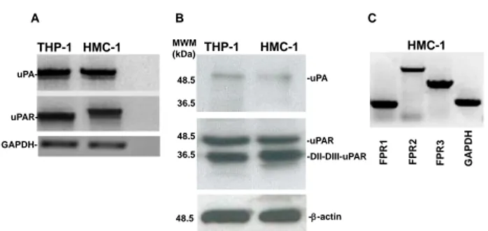
To investigate the existence of a functional interaction between the uPA-uPAR system and FPRs in MCs, we first evaluated the expression at mRNA and protein level of uPA and uPAR in HMC-1 cells. uPA and uPAR mRNAs were detected by RT-PCR analysis of RNAs from HMC-1 cells and THP-1 monocyte-like cells, used as a positive control (Fig. 1A).
Western blot analysis of HMC-1 cell lysates with a monoclonal anti-uPA antibody confirmed the production of uPA. The same analysis with a polyclonal anti-uPAR antibody demonstrated that HMC-1 cells expressed uPAR in the intact (DI-DII-DIII-uPAR) and cleaved (DII-DIII-uPAR) forms (Fig. 1B). This result was previously described in other cell types, including monocyte-like THP-1 cells [18].
DII-DIII-uPAR can interact with FPRs through its SRSRY chemotactic domain, exposed at the N-terminus [21,33]. Moreover, upon binding uPA, intact uPAR can expose the same chemotactic domain, located in the DI-DII linker region, through a conformational modification [18]. Then, we examined FPRs expression in HMC-1 cells by RT-PCR analysis. Electrophoresis in agarose gel showed the presence of mRNAs for all three receptors: FPR1, FPR2 and FPR3 (Fig. 1C).
These experiments demonstrated that MCs synthesized uPA and its specific receptor uPAR as well as FPRs, most important mediators of uPA/uPAR-induced cell responses in cancer and inflammation [16,22,24,31]. Effects of DFP-uPA and uPAR84-95 on VEGF-A expression
and production in the HMC-1 human mast cell line It has been reported that human mast cells can express and synthesize VEGF-A and VEGF-B, as well as Vascular Endothelial Growth Factor-C (VEGF-C) and Vascular Endothelial Growth Factor-D (VEGF-D) [14, 15].
Thus, we evaluated whether uPA, both directly and/or by inducing uPAR interaction with FPRs, could stimulate VEGF-A expression and production by HMC-1 cells.
To this aim, HMC-1 cells were stimulated for 2 hours with DFP inactivated-uPA (10-8 M), able to bind
uPAR but devoid of enzymatic activity, and with the
uPAR84-95 peptide (10-9 M), containing the SRSRY
chemotactic sequence of uPAR and able to bind FPRs. RT-PCR analysis of VEGF-A mRNA expression showed that HMC-1 cells synthetized mRNA for VEGF-A after 2 hours stimulation with both ligands (Fig. 2A).
-uPAR -DII-DIII-uPAR A B C THP-1 HMC-1 MWM (kDa) 36.5 48.5 36.5 48.5 -uPA F P R1 F P R3 F P R2 G A P DH HMC-1 THP-1 HMC-1 - GAPDH- uPA- uPAR---actin 48.5
Figure 1. uPA, uPAR and FPRs expression in HMC-1 cells.
A: mRNA expression of uPA and uPAR in HMC-1 cells. Total RNA of THP-1 monocyte-like cells as a positive control (lane 1)
and HMC-1 cells (lane 2) were prepared, reverse transcribed, and amplified by 40 PCR cycles in the presence of uPA and
uPAR-specific primers and GAPDH primers, as a loading control. PCR products were analyzed by electrophoresis in 1%
agarose gel containing ethidium bromide, followed by photography under UV illumination.
B: Western blot analysis of uPA and uPAR in HMC-1 cells. THP-1 monocyte-like cells as a positive control (lane 1) and HMC-1 cells (lane 2) were lysed in Triton X-100 and 50 g of total protein were analyzed by 9% SDS-PAGE and Western blot
with an anti-uPA and anti-uPAR polyclonal antibody. C: mRNA expression of FPRs in HMC-1 cells. Total RNA was prepared, reverse transcribed and amplified by 40 PCR cycles in the presence of FPR1- (lane 1), FPR2- (lane 2), and FPR3- (lane 3) specific primers and GAPDH (lane 4) primers, as loading control. PCR products were analyzed by electrophoresis in 1%
agarose gel containing ethidium bromide, followed by photography under UV illumination.
To evaluate whether DFP-uPA and uPAR84-95
also induced VEGF-A production, we investigated the kinetics of VEGF-A release from HMC-1 cells. Both DFP-uPA and uPAR84-95 induced a time-dependent release
of VEGF-A in HMC-1 cells (not shown) that was significant after 6 hours of stimulation (Fig 2B).
Effect of DFP-uPA, and uPAR84–95 on primary human
lung mast cells (HLMC) chemotaxis
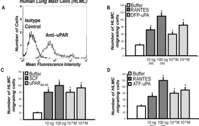
We demonstrated that uPA can induce human basophil chemotaxis through uPAR functional interaction with FPRs [18].
Thus, we the studied uPAR expression and the existence of a functional interaction with FPRs also in primary human MCs. To this aim, human lung mast cells (HLMC) were purified from normal subjects and analyzed by flow cytometry with a polyclonal anti-uPAR antibody or with purified control IgG, then stained with FITC-conjugated goat rabbit IgG and PE-FITC-conjugated anti-IgE. The vast majority (80–94%) of HLMC showed uPAR expression on the cell surface (Fig. 3A).
In order to elucidate the potential interaction of cell-surface uPAR and FPRs, we tested the effects of DFP-inactivated uPA (10-9-10-8 M) and of different
concentrations of the uPAR-derived chemotactic peptide
---0 20 40 60 80 100 120 140 160 180 0 20 40 60 80 100 120 140 160 180 V E G F -A mR NA E xpre ss ion (o ve r con tr o l) 10-10M 10-9M ATF-uPA uPAR84-95 PMA 10-5M V E G F -A p ro tein rele as e (o ve r con tr o l) * * * Buffer 2h 6h A B 10-9M 10-10M 25 ng/ml NECA * * * ATF-uPA uPAR84-95 Buffer
Figure 2. Effect of ATF-uPA and uPAR84-95 on
VEGF-A expression and release by HMC-1 cells.
A: effect of ATF-uPA, uPAR84-95 on VEGF-A mRNA expression in HMC-1 cells. HMC-1 cells were cultured with cell medium alone, ATF-uPA (10-9 M), uPAR
84-95 (10-10 M), or PMA
(25 ng/ml), as a positive control, for 2 hours at 37°C in a humidified (5% CO2) incubator. Total RNA of HMC-1 cells was
prepared, reverse transcribed and amplified by 40 PCR cycles in the presence of VEGF-A specific primers and GAPDH primers,
as a loading control. PCR products were analyzed by electrophoresis in 1% agarose gel containing ethidium bromide,
followed by photography under UV illumination. Results are expressed as percentage of VEGF-A expression increase in 10-9
M ATF-uPA-treated (grey column), 10-10 M uPAR
84-95-treated
(light grey column), or 25 ng/ml PMA-treated (black column) HMC-1 cells, relative to untreated HMC-1 cells (white column),
after normalization to GAPDH. Values are the mean ± SEM of three experiments. * p: <0.05.
B: effect of ATF-uPA and uPAR84-95 on VEGF-A synthesis by HMC-1 cells. 106 cells/samples were incubated for 6 hours
without (white column) or with ATF-uPA (10-9 M) (grey column), uPAR84-95 (10-10 M) (light grey column), or NECA (10-5
M) (black column) at 37°C in a humidified (5% CO2) incubator. Results are expressed as percentage of increase of VEGF-A release relative to untreated cells. HMC-1 supernatants were collected and VEGF-A was determined by ELISA assay. Values
are the mean ± SEM of three experiments. * p: <0.05.
Both DFP-uPA and uPAR84-95 caused HLMC migration
(Fig. 3B, C). Both the chemokine RANTES (10 and 100 ng/ml) and the major chemotactic factor SCF (10 and 100 ng/ml), used as a positive control, showed to be potent factors in HLMC migratory activity (Fig. 3B, C).
To determine whether migration of HLMC resulted from chemotaxis or chemokinesis, checkerboard analysis was performed and showed that both stimuli in a dose dependent manner induced HLMC migration when added to the lower wells of the chemotaxis chamber. An optimal concentration of the stimuli added with the cells to the upper wells or to both compartments did not induce directional HLMC migration. Thus, DFP-uPA and
uPAR84-95-induced migration of HLMC resulted from
chemotaxis, rather than from chemokinesis (data not shown).
In addition, experiments with different concentration of ATF (aa 1–143) (10-9-10-8 M), which
consists only of the uPAR-binding region of uPA and is
devoid of enzymatic activity [18], demonstrated that this peptide retained its chemotactic properties (Fig. 3D). Taken together, these results indicated that uPA induced MCs migration by binding uPAR and stimulating its interaction with FPRs, most probably through the exposure of the uPAR84-95 epitope; indeed, the enzymatic
activity of uPA was not primarily responsible for inducing HLMC chemotaxis. A B C D 0 20 40 60 80 100 120 140 Num be r of HLM C m ig rati ng c e ll s Buffer SCF uPAR84-95 * * * * 0 20 40 60 80 100 120 140 * Num be r of HLM C mi grating ce lls Buffer RANTES DFP-uPA 10 ng /ml 100 ng/ml 10 -8 M 10-9 M * * *
Human Lung Mast Cells (HLMC)
Num be r of Cel ls
Mean Fluorescence Intensity
Isotype Control Anti–uPAR 0 20 40 60 80 100 120 140 Num be r of HLM C m ig rati ng c e ll s Buffer RANTES ATF-uPA * * * * 10 ng /ml 100 ng/ml 10 -8 M 10-9 M 10 ng /ml 100 ng /ml 10-8 M 10-9 M
Figure 3. uPAR expression and function in primary HLMC cells.
A: cytofluorometric analysis of uPAR expression on HLMC cell surface. HLMC were incubated with anti-IgE PE monoclonal
antibody, anti-uPAR polyclonal antibody, isotype-matched control polyclonal antibody and FITC-conjugated goat
anti-rabbit antibody.
B: effect of DFP-uPA on HLMC cell chemotaxis. HLMC were allowed to migrate in response to cell medium alone (white column), or with the indicated concentrations of RANTES (grey
column) or DFP-uPA (light grey column) for 3 h at 37°C in a humidified (5% CO2) incubator. Values are the mean ± SEM of
three experiments. *p: <0.05.
C: effect of uPAR84-95 on HLMC cell chemotaxis. HLMC were allowed to migrate in response to cell medium alone (white column), or with the indicated concentrations of SCF (grey column) or uPAR84-95 (light grey column) for 3 h at 37°C in a humidified (5% CO2) incubator. Values are the mean ± SEM of
three experiments. * p: <0.05.
D: effect of ATF-uPA on HLMC cell chemotaxis. HLMC were allowed to migrate in response to cell medium alone (white column), or with the indicated concentrations of RANTES (grey
column) or ATF-uPA (light grey column) for 3 h at 37°C in a humidified (5% CO2) incubator. Values are the mean ± SEM of
three experiments. * p: <0.05.
IV. DISCUSSION
A straight relation between cancer and flogosis has been extensively described, starting from the consideration that inflammatory cells are localized all around tumors [9, 34-36].
The interaction between cancer cells and their microenvironment are multiple and can result in both progression and arrest of tumor growth [37]. However tumor microenvironment is constituted not only by cells of the innate and adaptive immune system but also from
stromal cells [38]. It has been described that stromal cells, from different tumors, are able to synthetize mRNA for diverse molecules, for example collagenase, matrix metalloproteases and proteases, such as plasmin, all responsible of the early stages of tumor growth through the degradation of the extracellular matrix components (ECM) that is one of the mechanisms that neoplastic cells use to invade the interstitial tissue [39].
Furthermore stromal cells, by producing chemoattractant molecules, recruit inflammatory cells into tumor sites, influencing them in a way that ultimately promotes cancer progression and prognosis [38,40,41]. In these context the detection of MCs within the tumor argues for their role in the modulation of neoplasia biology and hypothize a possible cross-talk with stromal cells localized within the tumor [27].
The three N-formyl receptors named FPR1, FPR2 and FPR3 are innate immune receptors belonging to the pattern recognition receptors and associated to G-proteins. Beside their involvement in inflammatory disorders FPRs have been described as regulating receptors involved in wound healing [42], angiogenesis [25], and myofibroblast activation occurring in the context of fibrotic disorders [24]. The involvement of FPRs in tumors has been studied by few authors and in some animal models where their role seems to differ in relation of the ligands or of the affected tissue [25]. We have recently showed that FPRs, as previously demonstrated in epithelial cells, could be involved in the pathogenesis of SSc, an autoimmune disease, through different mechanisms, including the interaction with the uPA-uPAR system [24]. In this paper we describe for the first time the expression of all the three FPRs in the human mast cell line HMC-1 (Fig. 1).
The uPA-uPAR system is an important and complex cellular recognition system that mediates different activities such as fibrinolysis, cell adhesion and migration, and tissue remodeling. In particular uPA, uPAR and the plasminogen activator inhibitor type-1 (PAI-1) are not only strongly upregulated in a wide variety of cancer types but their biological levels correlate with a poor neoplastic outcome and a more rapid tumor progression [22,43]. Nielsen and colleague described through in situ hybridization and immunohistochemical studies of invasive ductal breast cancer tissue that uPA, uPAR and PAI-1 are expressed in stromal cells immediately surrounding the invasive cancer cells [44].
We therefore studied the expression, at mRNA and protein levels, of uPA and uPAR in HMC-1 cells. HMC-1 cells, differently from basophils, synthetized uPA and expressed uPAR both in the intact and in a cleaved form (DII-DIII-uPAR), which is able to bind FPRs through the SRSRY sequence (residues 88-92) (Fig. 1). Interestingly, uPA itself can cleave uPAR in the DI-DII linker region, thus exposing the SRSRYsequence.
Moreover, by cytofluorimetric analysis using HLMC isolated from donors undergoing thoracotomy and lung resection, we confirmed that uPAR was expressed on the cell surface of the vast majority of HLMC (80-94%). We also found that enzymatically inactive uPA
(DFP-uPA) and ATF, which is devoid of enzymatic activity, were potent chemoattractants for HLMC as well as the uPAR-derived peptide uPAR84–95, containing the SRSRY
sequence (Fig. 3). Thus, uPA released by MCs can act in an autocrine fashion both by cleaving uPAR and exposing the SRSRY sequence than, similarly to what happens in basophils, by triggering uPAR conformational changes able to mediate binding to FPRs and the transmission of signals from the cell surface to the inner domains involved in MCs chemotaxis.
We have previously demonstrated that the H. pylori-derived peptide Hp(2-20) stimulated eosinophil migration through the engagement of FPR2 and FPR3, and also induced production of VEGF-A and TGF-beta, two key mediators of tissue remodeling [32]. Thus, we evaluated the possibility that FPRs stimulation through uPA and uPAR84-95 triggering was able to induce synthesis
and release of the Vascular endothelial growth factor-A (VEGF-A). VEGF-A is the most potent proangiogenic mediator known so far, involved in endothelial proliferation, migration, and survival. It also acts as a proinflammatory cytokine [45]. Both DFP-uPA and the
uPAR84-95 peptide proved to be potent stimuli for VEGF-A
synthesis and release. It has been demonstrated that, in murine models of glioma and meningioma, uPAR and cathepsin B knock-out inhibited angiogenesis by disrupting the JAK/STAT pathway-dependent VEGF expression [46,47]. Thus, uPAR could represent a useful target to inhibit VEGF-A mediated angiogenesis both in neoplastic and inflammatory diseases [48].
V. CONCLUSION
Our work suggests the possibility that MCs, through the expression of the uPA/uPAR system and its interaction with FPRs, can be responsible of chronic inflammation, tumor progression and angiogenesis. Moreover, it can be hypothesized that stromal cells, by secreting chemotactic factors, can additionally contribute to the recruitment of MCs, for example, in the tumor surroundings.
Finally, the results described in this study may have practical implications in inflammatory and neoplastic disorders where MCs infiltration play a prominent role. In fact, several author recently suggested that the crosstalk between MCs and other tumor-infiltrating cells could be a potential target for anticancer therapies [13], or it is conceivable that agents acting on uPAR-mediated chemotaxis (i.e., by blocking the chemotactic epitope) may be used to modify the MCs driven inflammatory and tumor promoting reactions.
REFERENCES
[1] Reber LL, Sibilano R, Mukai K, Galli SJ. Potential effector and immunoregulatory functions of mast cells in mucosal immunity. Mucosal Immunol 2015;8(3):444-463.
[2] Galli SJ, Zsebo KM, Geissler EN. The kit ligand, stem cell factor. Adv Immunol 1994;55:1-6.
[3] Bradding P, Arthur G. Mast cells in asthma--state of the art. Clin Exp Allergy 2016;46(2):194-263.
[4] Marone G, Triggiani M, Genovese A, De Paulis A. Role of human mast cells and basophils in bronchial asthma. Adv Immunol 2005;88:97-160.
[5] Sehra S, Serezani AP, Ocaña JA, Travers JB, Kaplan MH. Mast Cells Regulate Epidermal Barrier Function and the Development of Allergic Skin Inflammation. J Invest Dermatol 2016;136(7):1429-1437.
[6] Taylor ML, Metcalfe DD. Mast cells in allergy and host defense. Allergy Asthma Proc 2001;22(3):115-119. [7] Bischoff SC. Role of mast cells in allergic and non-allergic immune responses: comparison of human and murine data. Nat Rev Immunol 2007;7(2):93-104.
[8] Suurmond J, van der Velden D, Kuiper J, Bot I, Toes RE. Mast cells in rheumatic disease. Eur J Pharmacol 2016;778:116-124.
[9] Varricchi G, Galdiero MR, Marone G, Granata F, Borriello F, Marone G. Controversial role of mast cells in skin cancers. Exp Dermatol 2016;15:doi: 10.1111/exd.13107.
[10] Ribatti D. Mast cells as therapeutic target in cancer. Eur J Pharmacol 2016;778:152-157.
[11] Soucek L, Lawlor ER, Soto D, Shchors K, Swigart LB, Evan GI. Mast cells are required for angiogenesis and macroscopic expansion of Myc-induced pancreatic islet tumors. Nat Med 2007;13(10):1211-1218.
[12] Liu J, Zhang Y, Zhao J, Yang Z, Li D, Katirai F, Huang B. Mast cell: insight into remodeling a tumor microenvironment. Cancer Metastasis Rev 2011;30(2):177-184.
[13] Ammendola M, Sacco R, Sammarco G, Luposella M, Patruno R, Gadaleta CD, et al. Mast Cell-Targeted Strategies in Cancer Therapy. Transfus Med Hemother 2016;43(2):109-113.
[14] Marone G, Varricchi G, Loffredo S, Granata F. Mast cells and basophils in inflammatory and tumor angiogenesis and lymphangiogenesis. Eur J Pharmacol 2016;778:146-151.
[15] Detoraki A, Staiano RI, Granata F, Giannattasio G, Prevete N, de Paulis A, et al. Vascular endothelial growth factors synthesized by human lung mast cells exert angiogenic effects. J Allergy Clin Immunol 2009;123(5):1142-1149.
[16] Ragno, P. The urokinase receptor: a ligand or a receptor? Story of a sociable molecule. Cell. Mol. Life Sci 2006;63(9):1028-1037.
[17] Ragno P, Montuori N, Rossi G. Urokinase-type plasminogen-activator receptor associates to a cell surface molecule in monocytic cells. Biochem Biophys Res Commun 1996;224(1):252-257.
[18] de Paulis A, Montuori N, Prevete N, Fiorentino I, Rossi FW, Visconte V, et al. Urokinase induces basophil chemotaxis through a urokinase receptor epitope that is an endogenous ligand for formyl peptide receptorlike 1 and -like 2. J Immunol 2004;173(9):5739-5748.
[19] Danø K, Behrendt N, Høyer-Hansen G, Johnsen M, Lund LR, Ploug M, et al. Plasminogen activation and cancer. Thromb Haemost 2005;93(4):676-681.
[20] Høyer-Hansen G, Rønne E, Solberg H, Behrendt N, Ploug M, Lund LR, et al. Urokinase plasminogen activator cleaves its cell surface receptor releasing the ligand-binding domain. J Biol Chem 1992;267(25):18224-18229. [21] Montuori N, Carriero MV, Salzano S, Rossi G,. Ragno P. The cleavage of the urokinase receptor regulates its multiple functions. J Biol Chem 2002;277(49):46932-46939.
[22] Su SC, Lin CW, Yang WE, Fan WL, Yang SF. The urokinase-type plasminogen activator (uPA) system as a biomarker and therapeutic target in human malignancies. Expert Opin Ther Targets 2016;20(5):551-566.
[23] Montuori N, Ragno P. Multiple activities of a multifaceted receptor: roles of cleaved and soluble uPAR. Front. Biosci (Landmark Ed.) 2009;14:2494-2503. [24] Rossi FW, Napolitano F, Pesapane A, Mascolo M, Staibano S, Matucci-Cerinic M, et al. Upregulation of the N-formyl Peptide receptors in scleroderma fibroblasts fosters the switch to myofibroblasts. J Immunol 2015;194(11):5161-5173.
[25] Prevete N, Liotti F, Marone G, Melillo RM, de Paulis A. Formyl peptide receptors at the interface of inflammation, angiogenesis and tumor growth. Pharmacol Res 2015;102:184-191.
[26] Ye RD, Boulay F, Wang JM, Dahlgren C, Gerard C, Parmentier M, et al. International Union of Basic and Clinical Pharmacology. LXXIII. Nomenclature for the formyl peptide receptor (FPR) family. Pharmacol Rev 2009;61:119-161.
[27] Visciano C, Prevete N, Liotti F, Marone G. Tumor-Associated Mast Cells in Thyroid Cancer. Int J Endocrinol 2015;2015:705169. doi: 10.1155/2015/705169.
[28] Marone G, Genovese A, Granata F, Forte V, Detoraki A, de Paulis A, Triggiani M. Pharmacological modulation of human mast cells and basophils. Clin Exp Allergy 2002;32(12):1682-1689.
[29] Prevete N, Salzano FA, Rossi FW, Rivellese F, Dellepiane M, Guastini L, et al. Role(s) of formyl-peptide receptors expressed in nasal epithelial cells. J Biol Regul Homeost Agents 2011;25(4):553-564.
[30] de Paulis A, Prevete N, Fiorentino I, Rossi FW, Staibano S, Montuori N, et al. Expression and functions of the vascular endothelial growth factors and their receptors in human basophils. J Immunol 2006;177(10):7322-7331. [31] Postiglione L, Montuori N, Riccio A, Di Spigna G, Salzano S, Rossi G, et al. The plasminogen activator system in fibroblasts from systemic sclerosis. Int J Immunopathol Pharmacol 2010;23(3):891-900.
[32] Prevete N, Rossi FW, Rivellese F, Lamacchia D, Pelosi C, Lobasso A, et al. Helicobacter pylori HP(2-20) induces eosinophil activation and accumulation in superficial gastric mucosa and stimulates VEGF-alpha and TGF-beta release by interacting with formyl-peptide receptors. Int J Immunopathol Pharmacol 2013;26(3):647-662.
[33] Resnati M, Pallavicini I, Wang J M, Oppenheim J, Serhan CN, Romano M, et al. The fibrinolytic receptor for urokinase activates the G protein coupled chemotactic receptor FPRL1/lXA4R. Proc Natl Acad Sci USA 2002;99(3):1359.
[34] Mantovani A. Cancer: inflaming metastasis. Nature 2009;457(7225):36-37.
[35] Grivennikov SI, Greten FR, Karin M. Immunity, inflammation, and cancer. Cell 2010;140(6):883-899. [36] Pesic M, Greten FR. Inflammation and cancer: tissue regeneration gone awry. Curr Opin Cell Biol 2016;43:55-61.
[37] de Visser KE, Eichten A, Coussens LM. Paradoxical roles of the immune system during cancer development. Nat Rev Cancer 2006;6(1):24-37.
[38] Bussard KM, Mutkus L, Stumpf K, Gomez-Manzano C, Marini FC. Tumor-associated stromal cells as key contributors to the tumor microenvironment. Breast Cancer Res 2016;18(1):84. doi: 10.1186/s13058-016-0740-2.
[39] He Y, Liu XD, Chen ZY, Zhu J, Xiong Y, Li K, et al. Interaction between cancer cells and stromal fibroblasts is required for activation of the uPAR-uPA-MMP-2 cascade in pancreatic cancer metastasis. Clin Cancer Res 2007;13(11):3115-3124.
[40] Coussens LM, Werb Z. Inflammation and cancer. Nature. 2002;420(6917):860-867.
[41] Massara M, Bonavita O, Mantovani A, Locati M, Bonecchi R. Atypical chemokine receptors in cancer: friends or foes? J Leukoc Biol 2016;99(6):927-933. [42] de Paulis A, Prevete N, Rossi FW, Rivellese F, Salerno F, Delfino G, et al. Helicobacter pylori Hp(2-20) promotes migration and proliferation of gastric epithelial cells by interacting with formyl peptide receptors in vitro and accelerates gastric mucosal healing in vivo. J Immunol 2009;183(6):3761-3769.
[43] Duffy MJ, McGowan PM, Harbeck N, Thomssen C, Schmitt M. uPA and PAI-1 as biomarkers in breast cancer: validated for clinical use in level-of-evidence-1 studies. Breast Cancer Res 2014;16(4):428. doi: 10.1186/s13058-014-0428-4.
[44] Nielsen BS, Rank F, Illemann M, Lund LR, Danø K. Stromal cells associated with early invasive foci in human mammary ductal carcinoma in situ coexpress urokinase and urokinase receptor. Int J Cancer 2007;120(10):2086-2095.
[45] Ferrara N. VEGF-A: a critical regulator of blood vessel growth. Eur Cytokine Netw 2009;20(4):158-163. [46] Malla RR, Gopinath S, Gondi CS, Alapati K, Dinh DH, Gujrati M, et al. Cathepsin B and uPAR knockdown inhibits tumor-induced angiogenesis by modulating VEGF expression in glioma. Cancer Gene Ther 2011;18(6):419-434.
[47] Gupta R, Nalla AK, Gogineni VR, Chetty C, Bhoopathi P, Klopfenstein JD, et al. uPAR/cathepsin B overexpression reverse angiogenesis by rescuing FAK phosphorylation in uPAR/cathepsin B down regulated meningioma. PLoS One 2011;6(2):e17123.
[48] Rea VE, Lavecchia A, Di Giovanni C, Rossi FW, Gorrasi A, Pesapane A, et al. Discovery of new small molecules targeting the vitronectin-binding site of the urokinase receptor that block cancer cell invasion. Mol Cancer Ther 2013;12(8):1402-1416.