PROCEEDINGS OF THE 1
stINTERNATIONAL CONFERENCE ON DESIGN AND
PROCESSES FOR MEDICAL DEVICES PROMED,
PADENGHE SUL GARDA (BRESCIA), ITALY
2 – 4 MAY 2012
Editors
Elisabetta Ceretti – Università di Brescia
Antonio Fiorentino– Università di Brescia
Luca Giorleo– Università di Brescia
Claudio Giardini– Università di Bergamo
Technology and Manufacturing System Group
copyright NEOS EDIZIONI srl
Via Genova 57, Rivoli (TO) - tel 011 9576450
e-mail [email protected] - http: www.neosedizioni.it
ISBN 978 88 6608 058 9
IV
Information Technology Supporting Organ Printing
53
Rodrigo A Rezende, Vladimir Mironov, Turlif Vilbrandt, Júlia A Nogueira, Frederico D A S
Pereira, Daniel T Kemmoku, Jorge V L da Silva
Towards Large-Scale Biofabrication of Tissue Spheroids for Organ Printing
57
Vladimir Mironov, Frederico D A S Pereira, Rodrigo A Rezende, Vladimir Kasyanov, Jorge
V L da Silva
Study of Poly(3-Hydroxybutyrate) scaffolds fabrication by selected laser sintering for use in
tissue engineering
61
T. F. Pereira, M. A. C. Silva, M. F. Oliveira, I. A. Maia, J. V. L. Silva, M. F. Costa, R.M. S. M.
Thiré
Producing polylactic acid scaffolds (PLA) by bioextrusion and degradation study during the
manufacturing process
65
Fermín C, Z., Lopez da Silva, J. V.; Sena F. D., Inforçatti, P., Sabino, M.A.
An Algorithm to Obtain Support Structures for Additive Manufacturing
69
N. Volpato, A. Franzoni, L.M.T. da Silva, A.A. Silva
Rapid Tooling using 3D printing system for manufacturing of customized tracheal stent
73
Evila L. Melgoza, G. Vallicrosa, L. Sereno, A. Rosell, C. Rodríguez, A. Elias, J. Ciurana
An Investigation into the Viability of a 3D Printing Technology for Reconstructive Surgical
Applications
79
D.M. Dimitrov, J.R. Honiball, N. de Beer
Prosthesis design based on virtual prototyping techniques
83
Walter Albisetti, Giorgio Colombo, Omer De Bartolomeo, Giancarlo Facoetti, Stella Gabbiadini,
Roberto Morotti, Daniele Regazzoni, Caterina Rizzi
Bicell printing: scaffold characterization for cells growth
87
Tatiana Patrício, Marco Domingos, Jorge Coelho, Antonio Gloria, Paulo Jorge Bártolo
Processing and Biomaterials
Grinding of cobalt-chromium alloy medical implants
95
C. Tournier, C. Iabassene, A. Guiot and Y. Quinsat
Rehological model for muscle stretching characterization
99
A. Fiorentino, A. Elias, E. Ceretti
Fabrication of a Biopsy Micro-Forceps (BMF) prototype with Two Point Incremental Sheet
Forming
103
M. L. Garcia-Romeu, R. Pérez-Santiago, I. Bagudanch , L. Puigpinós
Computational fluid dynamics (CFD) in synthesis of phema hydrogels by laser infrared
107
____________________
* Corresponding author: Antonio Fiorentino
Via Branze 38, 25123 Brescia (Italy), ph: +39 0303715522, fax: +39 0303702448, [email protected]
RHEOLOGICAL MODEL FOR MUSCLE STRETCHING
CHARACTERIZATION
A. Fiorentino
1*, A. Elias
2, E. Ceretti
1 1Dept. of Mechanical and Industrial Engineering – University of Brescia
2
Mechanical Engineering Department
Tecnológico de Monterrey, Campus Monterrey
ABSTRACT: Engineering and Medicine are two research fields that rarely join their competences since they focus on
very different topics. Anyway, problems that Physicians encounter are not so different from the Engineer’s ones if they are considered under the solving methods point of view. In fact, FEM codes are widely used to simulate material deformation processes for product manufacturing or to estimate elastic stresses acting on a structure and these codes can be used to simulate the stresses acting on a prosthesis and the surrounding tissues (ie. stresses on hip prosthesis and femur). Diseases like Pelvic Organ Prolapse are frequent in the female population and the vaginal birth is thought to be the main risk factor. For this reason, the knowledge of the dynamic that takes place during the vaginal delivery is very important to understand the causes of POP and many information (such as stresses or deformations) can be obtained from FEM simulations. Unfortunately, very few is known about biological tissues mechanical properties and this makes their simulation more difficult. With the aim to identify a rheological model for pelvic muscle characterization, in this paper, tensile tests performed on vaginal muscle have been analyzed identifying a common behaviour and a constitutive law able to represent the stress-strain relation during the virgin tissue traction. In particular, this law resulted to have a very simple formulation and demonstrated a good agreement with the experimental results.
KEYWORDS: Rheological model, vaginal tissue, tensile test
1 INTRODUCTION
Diseases like Pelvic Organ Prolapse (POP, loss of vaginal support) is frequent in the female population (43%-76%) [1] and the vaginal birth is thought to be the main risk factor [2]. Moreover, other health problems, such as urinary incontinence or stress urinary incontinence, are associated to POP [3]. For these reasons, the knowledge of the dynamics that take place during the vaginal delivery is very important to understand the causes of POP. Recent studies [4-5] performed on the FEM simulation of vaginal delivery show interesting results, but a lack is still present in terms of exploitable results since no comparison with experiments is performed. In particular, FEM models require the materials constitutive law and information about biological tissues flow-stress behaviour.
Studies on cyclic stretching of biological soft tissues infact, [6-8] showed that their mechanical behaviour is non linear, strongly anisotropic and subjected to the strain induced softening effect called Mullins effect (Figure 1). The effect takes its name from the British rubber scientist Leonard Mullin who studied the phenomenon on rubbers [9,10] and it consists of an irreversible softening of the stress-strain curve that
occurs whenever the load increases beyond its prior maximum value ever reached. Moreover, this effect can be idealized as instantaneous. As results (Figure 1), during the load application to the virgin (never loaded) material, one stress-stretch curve is followed, while during load removal the material follows a different and softened path (stresses are lower than in the loading phase). When the material is reloaded, the new curve is followed backward till the original stress strain-curve is reached and so on. Models for tissues characterization have been derived from polymers rheological models, which well fit the experimental data, but they require a lot of constants to be estimated [8,11]. In particular, the softened path modelling strongly depends on the initial point, i.e. the point on the virgin material curve when the load is released. For this reason, virgin path correct modelling is fundamental for tissue characterization. In this paper, data from vaginal muscle cyclic tensile tests reported in [12] were analyzed identifying a simple model for vaginal tissue virgin path characterization. The proposed model identifies two behaviours of the tissue, for low and high deformations and, and it is able to give a good representation of experimental data using a limited number of constants.
PROCEEDINGS OF THE 1st INTERNATIONAL CONFERENCE ON DESIGN AND PROCESSES FOR MEDICAL DEVICES – Ceretti et alii. PROMED, ISBN: 978 88 6608 058 9
Figure 1 Comparison of the Mullins effect model with experimental data on skin samples from a) male and b) female mice [13] and c) experimental stress–stretch curves for vaginal tissue specimens [8].
2 DATA ANALYSIS
To assess the accuracy of the proposed constitutive model to describe the behavior of the vaginal tissue during loading and unloading cycles, uniaxial extension experimental data collected by Peña et. al. in [8-12] have been used. During the experimental test, Peña and co-workers tested tissue samples, longitudinal and transverse strips (approximately 6 mm wide and 15 mm long), that were taken from seven postmenopausal patients, with a mean age of 66.5 ± 11.7 years. All details about temperature of the samples, humidity, test conditions and so on, are clearly stated in [8].
The virgin material path showed a viscoelastic-like behavior (Figure 2) and a high scattering (Figure 3). In fact, for a fixed stretch value, the initial stress (peak) asymptotically decreases (equilibrium). In order to identify a constitutive law able to represent the vaginal muscle behaviour, data were initially observed and then interpolated using different strategies.
From preliminary observations on both equilibrium and peak data sets no significant difference can be noticed, except in Test III where a probable improper set-up (pretension) or clamping (specimen sliding) occurred (Figure 3).
- Test IV curve is very different from the other. This can be related to a proper behaviour of the vaginal tissue, to the sample preparation (dehydration) or foreign tissues (tendons, muscle, ligaments,…) could be present.
Figure 2 Initial and equilibrium normalized stress results for all the tests [12].
3 THE PROPOSED MODEL
The rheological model here described starts from a similitude between the Mullins effect and the metal strain-stress curve when subjected to load-unload cycles beyond the yield strength. In fact, loading and unloading
paths (respectively paths1 and 2 in Figure 4) are
different and when the load is reapplied, the unloading path is followed backwards (path 3). Differently from metals, in Mullins effect the strain-stress relation along the unloading path is non linear. Because of this similitude, it was chosen to derive a constitutive model for soft biological tissues from the metallic ones.
Figure 3 Stress-stretch behaviour of vaginal muscle and detail on equilibrium data[12]. 0.0 0.1 0.2 0.3 0.4 0.5 0.6 0.7 0.8 0.0 0.1 0.2 0.3 0.4 s e
Equilibrium - All Test
I II III IV V VI VII VIII DETAILS 0 0.01 0.02 0.03 0.04 0.0 0.1 0.2 0.3 0.4 s e Equilibrium - All Test (Details)
I II III IV V VI VII VIII 0.3 0.4 0.5 0.6 0.7 0.8 0.9 1.0 0 100 200 300 400 500 600 700 800 900 N o rm a liz e d s tr e s s Time II VI,III IV I,VII V Peak Equilibrium CYCLE 3` CYCLE 2 CYCLE 1 CYCLE 4 a) b) c)
100
Therefore, an initial linear behaviour followed by a non-linear one were researched. A non-linear law was used to interpolate the data for low deformations (ε εlin) and an
exponential law was used for high deformations (ε > εlin)
as reported in (1).
The idea is that muscle fibres and metal dislocations behave in a similar way during deformation. Initially (εεlin) the tissue fibres are elastically stretched without moving, then (εεlin) the fibres start moving
and hampering each other. As the deformation proceeds, the movement becomes more and more difficult, maybe due to a reduction of possible fibres movement
(saturation). Moreover, in both cases, residual
deformations are present.
lin ε n linε
ε
e
F
ε
ε
ε
E
ε
σ
(1)where: E linear coefficient
F, n exponential coefficients
εlin low deformation limit
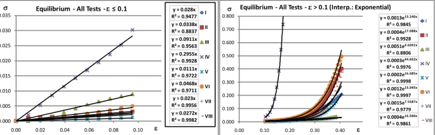
Figure 5 shows the interpolation and fitting results for equilibrium values (Figure 2). All tests show a linear (or linear-like) behaviour for low strain values. A limit for the linearity was identified equal to εlin = 0.1 for all the
tests. The exponential interpolation shows a good approximation of the results, even if the correlation values (R2) show that test I, VII and VIII have a worse interpolation. Accuracy in Test III results is poor due to the previously discussed problems (section 2).
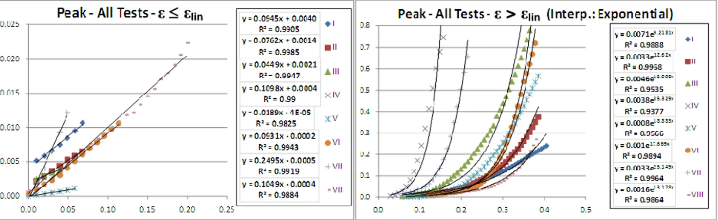
Similar considerations can be done for results (Figure 6) on peak values (Figure 2). This case differs for having
different εlin values and a worse fitting. Being their
values strongly time dependant (Figure 2), their estimation is, in fact, affected by a lower accuracy.
Figure 4 Similitude between strain hardening effect and residual strain in metals (bottom) and Mullins effect in soft biological tissues (top).
Figure 5 Stress-stretch results interpolation (equilibrium).
y = 0.0013e13.240x R² = 0.9845 y = 0.0004e17.088x R² = 0.9928 y = 0.0051e4.6091x R² = 0.8806 y = 0.0003e44.652x R² = 0.9976 y = 0.0002e16.685x R² = 0.9998 y = 0.0012e15.045x R² = 0.9997 y = 0.0015e7.5687x R² = 0.9779 y = 0.0004e16.560x R² = 0.9861 0.000 0.100 0.200 0.300 0.400 0.500 0.600 0.700 0.800 0.00 0.10 0.20 0.30 0.40 s e
Equilibrium - All Tests - e > 0.1 (Interp.: Exponential)
I II III IV V VI VII VIII y = 0.028x R² = 0.9477 y = 0.0338x R² = 0.8837 y = 0.0911x R² = 0.9563 y = 0.2955x R² = 0.9928 y = 0.0111x R² = 0.9722 y = 0.0468x R² = 0.9711 y = 0.023x R² = 0.9956 y = 0.0272x R² = 0.9982 0.000 0.005 0.010 0.015 0.020 0.025 0.030 0.035 0.00 0.02 0.04 0.06 0.08 0.10 s e
Equilibrium - All Tests -e≤ 0.1 I
II III IV V VI VII VIII ε ε s ε O A B 1 2 3 3 O’ ε0 (residual strain) Metals
(curve 1) O-A: Load on the virgin material (curve 2) A-O’: Unload
(curve 3) O-B: Load re-application
s ε VIRGIN MATERIAL O A B SOFTENED MATERIAL 1 2 3 3 Soft tissues Permanent set
101
Figure 6 Stress-stretch results interpolation (peak).
4 CONCLUSIONS
Pelvic Organ Prolapse is a disease that affects a high percentage of women and childbirth is one of the main risk cause. FE analysis allows to study stresses acting on pelvic tissues during the delivery but the reliability of the results requires the proper definition of the tissues characteristics. The Rheological models available in literature are accurate, but are based on a high number of material constants which requires a high number of tests and computational time for their estimation.
This work proposes a simpler model based on the combination of a linear and an exponential behaviors (respectively for low and high deformations) allowing to strongly reduce the number of material constants to be estimated (3 in the proposed model, more than 10 in literature).
The comparison with experimental data from tensile tests on vaginal muscle samples showed that the proposed model is able to describe the virgin material behavior with a good accuracy.
Since direct observation of the muscle fibers movement during stretching are necessary to physically explain the behavior in low and high deformation fields, a test campaign will be conducted. Moreover, other non linear functions and tissue anisotropy will be considered in order to improve and extend the validity of the actual model.
ACKNOWLEDGEMENT
The present research was possible thanks to IREBID project founded by FP7 – Marie Curie Actions – IRSES – 247476.
The Authors would like to thank prof. E. Peña for kindly providing the experimental data.
5 REFERENCES
[1] Barber M.: Symptoms and outcome measures of pelvic floor prolapse. Clin. Obstet. Gynecol, 48: 648-661, 2005.
[2] Lukacz E., Lawrence J., Contreras R., Nager C., Luber K.: Parity, mode of delivery and pelvic floor disorders. Obstet. Gynecol., 107:1253-1260, 2006.
[3] Jelovesk J., Maher C., Barber, M.: Pelvic organ prolapse. Lancet, 369:1027-1038, 2007.
[4] Parente M., Natal J.R., Mascarenhas T., Martins J.: The influence of the material properties on the biomechanical behaviour of the pelvic floor miscles diring vaginal delivery. Journal of Biomechanics 42:1301-1306, 2009.
[5] Li X., Kruger J., Nash P., Nielsen, P.: Effects of nonlinear muscle elasticity on pelvic floor mechanics during vaginal delivery. Journal of Biomechanical Engineering, 132, 2010.
[6] Peña E., Doblarè M.: An anisotropic pseudo-elastic approach for modelling Mullins effect in fibrous
biologial materials. Mechanics Research
Communications, 36:784-790, 2009.
[7] Ehret A., Itskov M.: Modeling of anisotropic softening phenomena: Application to soft biologial tissues. International Journal of Plasticity, 25:901-919, 2009.
[8] Peña E., Martins P., Mascarenhas T., Natal J.R.,
Ferreira A., Doblaré, M.: Mechanical
characterization of the softening behaviour of human vaginal tissue. Journal of the Mechanical behaviour of Biomedical Materials, 4:275-283, 2011.
[9] Mullins L.: Effect of stretching on the properties of rubber. Journal of Rubber Research, 16:275–289, 1947.
[10] Mullins L.: Softening of rubber by deformation. Rubber Chemistry and Technology, 42:339-362, 1969.
[11] Dorfmann A., Ogden R.: A pseudo-elastic model for loading, partial unloading and reloading of particle-reinforced rubber. International Journal of Solids Structures, 40: 2699–2714, 2003.
[12] Peña E., Peña J., and Doblaré M. On the Mullins effect and hysteresis of fibred biological materials:
A comparison between continuous and
discontinuous damage models. International
Journal of Solids and Structures, 46 (7-8) 1727-1735, 2010.
[13] Muñoz M., Bea J., Rodríguez J., Ochoa I., Grasa J., Pérez A., et al.: An experimental study of the mouse skin behaviour: damage and inelastic aspects. Journal of Biomechanics, 41:93–99, 2008.
![Figure 1 Comparison of the Mullins effect model with experimental data on skin samples from a) male and b) female mice [13] and c) experimental stress–stretch curves for vaginal tissue specimens [8]](https://thumb-eu.123doks.com/thumbv2/123dokorg/5512089.63858/6.892.94.798.106.292/figure-comparison-mullins-experimental-samples-experimental-vaginal-specimens.webp)