Hepatic Metastases from Gastric Carcinoma
Guido Alberto Massimo Tiberio, Nazario Portolani
and Stefano Maria Giulini
University of Brescia
Italy
1. Introduction
“Aprioristic passive attitude”: this is the mode that correctly defines the standard approach to gastric cancer patients presenting with hepatic metastases.
This behaviour is deeply motivated by the aggressive biology of the disease that so often frustrates any therapeutic approach. In fact, at diagnosis liver metastases are often multiple and associated to other extra-hepatic metastatic sites [D’Angelica et al., 2004; Dicken et al., 2005]. Furthermore, in the rare cases submitted to ablative treatments, hepatic and systemic recurrence has been experienced in the majority of cases.
However, considering survival performances extrapolated from a cohort of 1452 patients submitted to hepatic resection for noncolorectal nonendocrine liver metastases [Adam et al.,2006], it was observed that metastases from gastric adenocarcinoma performed in an intermediate way, ranking 10th in a list of 18 primaries. In fact, in selected cases an
aggressive treatment can achieve unexpected results: 5-year survival rates from 10% to 40% have consistently been reported in surgical series considering patients with liver metastases as sole metastatic site.
An aggressive attitude, however, doesn’t really penetrate into clinical practice and passivity still prevails, as depicted by an Italian survey reporting over 60% of patients not receiving specific treatments, including 30% of cases affected by 1 or 2 small metastases, and by therapeutic indication being influenced by patients’ determination [Tiberio et al., 2008]. This attitude is rather diffused if a recent review reported 436 surgically treated cases [Kerkar, 2010] and a French survey recruited 101 patients from 41 centres [Chiche, 2005], numbers to be faced, for example, to more than 14700 resections for metastases from colo-rectal cancer enrolled in LiverMetSurvey as to December 2010.
With this contribution we will attempt to promote a pragmatic approach to these patients, knowing that only a μετάνοια (change of mind) may lead to the recognition of cases that can benefit from a tailored treatment and thus to better results. This attitude seems particularly important in these days in which promising therapeutic improvements are announced by state-of-art multimodal treatments favouring local and systemic control of the disease.
2. Clinical considerations
The dimension of the phenomenon is difficult to assess. It is influenced by a number of factors such as the incidence of gastric cancer in different geographical areas, the
characteristics and quality of different health-care systems, the deployment of effective mass-screening programs capable to detect -and cure- gastric malignancies in an early phase. It seems reasonable to assume that the incidence of hepatic metastases from gastric cancer during the course of the disease figures around 10%-20% in eastern countries like Japan and South Korea, rises to 30%-40% in Western Europe and Anglo-Saxon world were an increase of advanced stages at diagnosis is recorded despite a steady and significant reduction of the incidence of gastric carcinoma in the last 20 years [Fayçal et al., 2005] and lies unmeasured -but probably over 50%- in other less monitored countries.
According to the presentation, hepatic metastases can be synchronous or metachronous. Synchronous metastases are detected during routine workup of gastric primaries or -unexpectedly- at surgical exploration, but in some cases their detection leads to diagnosis of gastric cancer. They must be differentiated from the direct infiltration of liver parenchyma originating from gastric cancer itself (T4). They can originate from unresectable gastric cancers but also from resectable primaries.
Metachronous metastases are detected in up to 25-30% of patients submitted to curative gastrectomy. Eighty percent of them appear within the first 2 postoperative years, but it must be known that a wide consensus exists in considering synchronous those lesions diagnosed during the very first postoperative period (~ 6 months).
At diagnosis metastases-related symptoms and signs are generally observed when hepatic metastases are discovered first and lead to the diagnosis of gastric cancer; in the other cases patients are asymptomatic or may display the signs and symptoms of gastric tumor.
Clinical examination searches for epigastric or hypochondriac masses and hepatomegaly; at rectal or vaginal examination signs of peritoneal carcinosis are looked-for.
When Tumor markers CA 19-9, CEA and CA 72-4 are simultaneously positive they are strongly suggestive of liver involvement [Marrelli et al., 2004].
At US and CT imaging metastases from gastric cancer display aspecific hypo-dense and hypo-vascularised patterns and can’t be distinguished from hepatic metastases originating from other gastro-enteric primaries.
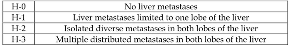
An adequate radiological report must enumerate their number, measures and location, the latter in reference to Couinaud’ segmentation. A good report allows the stadiation of hepatic disease according to the Japanese Gastric Cancer Association [10], a simple and practical classification, with direct therapeutic impact (table 1). In fact, H-3 hepatic involvement generally excludes ablative treatments, which are considered only for H-1 or H-2 cases.
H-0 No liver metastases
H-1 Liver metastases limited to one lobe of the liver H-2 Isolated diverse metastases in both lobes of the liver H-3 Multiple distributed metastases in both lobes of the liver
Table 1. Classification of hepatic metastases from gastric cancer as proposed by the Japanese Gastric Cancer Association, 1998.
3. Therapeutic approach
3.1 Systemic chemotherapy
At diagnosis liver metastases are often multiple and associated to other extra-hepatic metastatic sites such as peritoneal dissemination, extensive lymph-node and/or systemic
metastases. In these conditions nothing more than palliative or supportive treatments can be proposed, without appreciable long term results. Chemotherapy achieves median survival ranging from 7 to 15 months but long term survival remains anecdotal [Cocconi et al., 2003; Lee et al., 2007; Cao et al., 2009]. In particular, considering the few trials evaluating systemic chemotherapy in the subset of patients with liver-only metastatic involvement, 5-years survival rates do not reach 2% [Yoshida et al., 2004].
3.2 Surgical treatment
Resection of liver metastases from gastric cancer is indicated in absence of extra-hepatic disease, if a complete ablation of the metastases can be achieved while preserving postoperative liver function [Ambiru et al, 2001; Okano et al., 2002]; it must also be associated to curative gastrectomy in case of synchronous lesions. In these conditions hepatectomy is a low-risk procedure, with negligible mortality and morbidity rates.
The surgical literature shows that several clinical and pathological parameters correlate with survival; among these, staging factors of the primary tumor, metastases-related and surgery-related variables have been reported more often than others (table 2).
Author No. T N G H Ø metastasis Timing* Margin§ (months) MST survivors No. > 5 Years
Ochiai ’94 21 √ √ - - n.a. n.a. n.a. 18 2 (19 %) Miyazaki ’97 21 - - - √ n.a. - √ n.a. 2 (9.5 %) Fujii ’01 10 - - - - √ √ n.a. 16 1 (10 %) Ambiru ’01 40 - - - - - √ - 12 6 (15 %) Imamura ’01 17 - √ √ - n.a √ √ 12 0 Okano ’02 19 - - √ √ - √ n.a. 21 4 (21 %) Zacherl ’02 15 - - - √ - √ - 8.8 2 (13 %)^ Saiura ’02 10 - - - - - - n.a. 25 2 (20 %) Shirabe ’03 36 - ly - √ - - - n.a. 4 (11 %) Roh ’05 11 n.a. - - n.a. - - - 19 2 (18 %) Chiche ’05 101 - - - √ √ - √ 14,5 11 (10 %)
Sakamoto ’07 37 √ - - √ √ - - 31 2 (5,4 %) Koga ’07 42 √ - - √ - - - 34 8 (19%) Tsujimoto ’10 17 √ ly n.a. - - - n.a. 34 5 (29%)
MST: mean survival time Timing: synchronous vs.metachronous; § resection margin: + vs -; √ = prognostic factor; n.a.= not available; ly = lymphatic invasion; ^ = alive after 3 years.
Table 2. Prognostic factors and survival from series of patients submitted to surgical treatment of hepatic metastases.
However, data concerning long-term survivors demonstrate that, if we exclude bi-lobar spread of metastases (H 3), none of the reported predictive factors -alone or in combination- can deprive a patient of the possibility of long-term survival after hepatic resection, raising concern about the clinical value of prognostic factors emerging from small and super-selected populations submitted to liver resection.
3.2.1 Results from unselected populations
The correct approach to these particular patients can be extrapolated from a handful of key papers that addressed the topic analyzing unselected populations of gastric cancer patients presenting hepatic metastases as sole site of metastatic disease (table 3). From a cohort of 58 patients, the Korean group of Cheon and coll. did not extrapolate any primary-related or metastasis-related factor showing prognostic significance. On the very same line is the group of Makino and coll. from Japan, who studied 63 patients.
Ueda and colleagues, again from Japan, studied a cohort of 73 patients presenting synchronous metastases. Their data show that factors influencing survival where the extent of hepatic involvement (H1-2 vs H3) and macroscopic peritoneal dissemination (P0 vs P1) detected at surgical exploration. When focusing on the subgroup of H1-2 and P0 patients, they showed that number (1 vs >1) and size of hepatic metastases and N status of gastric cancer (N0-1 vs N2-3) influenced survival. An Italian survey performed under the auspices of the Italian Research Group on Gastric Cancer [Tiberio et al., 2008] studied an unselected cohort of 73 patients presenting metachronous metastases after curative D ≥ 2 gastrectomy. It was found that the factors T, N and G of the gastric primary, when rated T3b-T4, N+ and G3, independently display a clear negative prognostic value with cumulative effect.
These parameters may be helpful in order to appropriately select the therapeutic approach or, at least, to submit the cases to multidisciplinary evaluation, being aware that the prognosis of these patients is directly influenced by therapeutic choices. In fact, all the 4 above mentioned studies strongly highlight that the main factor influencing long-term survival (p ranging from 0.01 to 0.001) is the therapeutic approach to the liver metastases, in particular when hepatectomy is performed.
Author No Timing Prognostic factors
Cheon 2008 58 Synchronous +
metachronous R0 resection of hepatic metastases
Ueda 2009 72 Synchronous R0 Resection of hepatic metastases H; P;
Tiberio 2008 73 Metachronous T; N; G of gastric primary; Resection of hepatic metastases
Hwang 2009 73 Metachronous
Stage of gastric primary Extrahepatic metastases; H Treatment of hepatic metastases Table 3. Prognostic factors from series considering unselected populations.
In the Italian study hepatectomy was associated to a five-fold increase in survival of the less favourable patients (>1 negative prognostic factor) and achieved a 5-year survival rate of
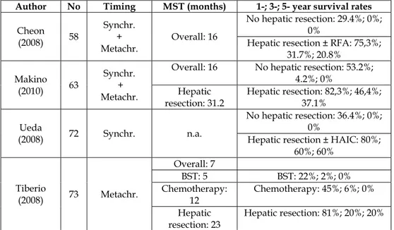
20%. Furthermore, Cheon and coll. and Ueda and coll. evidenced that the possibility to perform a radical operation (R0 vs R1) affects long-term survival and report overall 5-year survival rates of 20% and 60%, respectively. It is worth of note that in synchronous cases radical surgery was intended not only in regard to hepatic lesions, to be resected as prescribed by good surgical practice, but also in regard to gastric tumors, to be treated by standard curative D ≥ 2 gastrectomy (table 4).
Data reported in table 2 and 4 show that surgical therapy offers interesting and sound results. On a more realistic level, we must however recognize that these patients, despite all efforts, generally die of cancer progression. Hepatic recurrence is observed in about 70% of cases and in about half of them is associated to extra-hepatic relapse (table 5); if the literature and our experience do not permit an insight into the role of repeated hepatectomies in case of exclusive hepatic recurrence, this observation raises concern about the timing of treatment, in order to avoid superfluous operations. A simple time-test can achieve an acceptable selection once a potential candidate to curative surgery is encountered. It can be easily suggested in case of metachronous lesions with favourable location but it can be contraindicated in case of critically located metastases and, in general, in case of synchronous metastases, especially if associated to symptomatic or resectable gastric primaries. The French school [Adam et al., 2006] strongly suggests a multidisciplinary approach to these patients and, in particular, favours a systemic chemotherapy, to be started at diagnosis whenever possible, in order to offer its advantages to the greatest number of patients and to effectively select cases for surgery.
Author No Timing MST (months) 1-; 3-; 5- year survival rates
Cheon (2008) 58 Synchr. + Metachr. Overall: 16 No hepatic resection: 29.4%; 0%; 0%
Hepatic resection ± RFA: 75,3%; 31.7%; 20.8% Makino (2010) 63 Synchr. + Metachr.
Overall: 16 No hepatic resection: 53.2%; 4.2%; 0% Hepatic resection: 31.2 Hepatic resection: 82,3%; 46,4%; 37.1% Ueda (2008) 72 Synchr. n.a. No hepatic resection: 36.4%; 0%; 0%
Hepatic resection ± HAIC: 80%; 60%; 60% Tiberio (2008) 73 Metachr. Overall: 7 BST: 5 BST: 22%; 2%; 0% Chemotherapy: 12 Chemotherapy: 45%; 6%; 0% Hepatic
resection: 23 Hepatic resection: 81%; 20%; 20%
n.a. = not available; RFA = radio-frequency-ablation; HAIC = hepatic artery infusion chemotherapy; BST = best supportive treatment;
Author No. recurrence (%) Hepatic recurrence (%) Overall Miyazaki 1997 21 76.2 81 Fujii 2001 10 50 80 Ambiru 2001 40 72.5 77.5 Ambiru 2001 40 72.5 77.5 Okano 2002 19 63.2 73.7 Okano 2002 19 63.2 73.7 Saiura 2002 10 - 80 Shirabe 2003 36 61.1 83.3 Roh 2005 11 72.7 91 Sakamoto 2007 37 62.2 86.5 Koga 2007 42 50 66.7 Cheon 2008 22 50 63.6 Tiberio 2008 11 62.2 86.5 Makino 2010 16 25 63.6 Table 5. Recurrence after curative surgery.
3.3 Multimodal treatment
Multimodal treatments can further enhance survival rates, in particular if modern, state-of-art chemotherapy protocols are employed. For example we signal that Ueda and coll. reported in the cited work an outstanding 75% 5-year survival rate in a subgroup of 8 patients submitted to radical surgery followed by hepatic artery infusion chemotherapy.
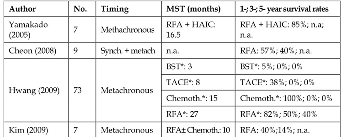
Radio-frequency ablation is another front-line, state-of-art technique to be considered in designing the treatment strategy of hepatic metastases from gastric cancer. This ablative technique is employed either as alternative or in association to hepatectomy, following the guidelines for HCC or for metastases from colo-rectal cancer. It is a minimally invasive and low-cost procedure whose interest is particularly enhanced in case of poor general conditions contraindicating surgery, which is often the case in gastric cancer patients. Its exact role is yet to be defined, as the number of reported procedures is low, follow-up short, data can’t always be effectively extrapolated from the context and reports are not unanimous as far as survival results are concerned. However, one paper by Hwang et coll., considered 72 patients with metachronous metastases submitted to different treatments but not to hepatectomy (table 5). They showed that 15 patients without extrahepatic disease treated by RFA ± chemotherapy displayed a median survival of 22 months, with 3- and 5-year survival rates of 50% and 40%, respectively, similar to those reported in the best surgical series. These data are coherent to those by Yamakado et al. which, however, suffer of short follow-up, and to those by Cheon et al.: in their experience a subgroup of 9 patients submitted to RFA compared favourably with 22 patients submitted to radical surgery, with a 4-year survival of 40% and 20%, respectively. Kim and collaborators appear less enthusiastic and report survival results superimposable to those of classic systemic chemotherapy alone. These data need further confirmation but highlight the interest of RFA in the management of these particular cases.
Author No. Timing MST (months) 1-; 3-; 5- year survival rates
Yamakado
(2005) 7 Methachronous
RFA + HAIC: 16.5
RFA + HAIC: 85%; n.a; n.a.
Cheon (2008) 9 Synch. + metach n.a. RFA: 57%; 40%; n.a.
Hwang (2009) 73 Metachronous
BST*: 3 BST*: 5%; 0%; 0% TACE*: 8 TACE*: 38%; 0%; 0% Chemoth.*: 15 Chemoth.*: 100%; 0%; 0% RFA*: 27 RFA*: 82%; 50%; 40% Kim (2009) 7 Metachronous RFA± Chemoth.: 10 RFA: 40%;14%; n.a.
n.a. = not available; HAIC = hepatic artery infusion chemotherapy; BST = best supportive treatment; TACE = trans arterial catheter embolization; * = patients without extrahepatic metastases.
Table 6. Radio-Frequency-Ablation (RFA) of hepatic metastases from gastric cancer.
3. Conclusion
Unexpected 5-year survival rates can be achieved in a subgroup of gastric cancer patients presenting hepatic metastases as sole metastatic site if an adequate treatment is selected. Simple clinical and biological characteristics of both the gastric primary and hepatic involvement display prognostic value which may be helpful in choosing therapeutic strategy, keeping in mind that the best results are achieved by aggressive treatments, which must be proposed whenever possible.
4. References
Adam R, Chiche L, Aloia T, Elias D, Salmon R, Rivoire M, Jaeck D, Saric J, Le Treut YP, Belghiti J, Mantion G and Mentha G for the Association Française de Chirurgie. Hepatic resection for noncolorectal nonendocrine liver metastases. Ann Surg 2006; 244: 524-535.
Ambiru S, Miyazaki M, Ito H, Nakagawa K, Shimizu H, Yoshidome H, Shimizu Y & Nakajima N. Benefits and limits of hepatic resection for gastric metastases. Am J Surg 2001;181:279-283.
Cao W, Yang W, Lou G, Jilang J, Geng M, Xi W, Li H, Ma T& Jin Y. Phase II trial of infusional fluorouracil, leucovorin, oxaliplatin and irinotecan (FOLFOXIRI) as first-line treatment for advanced gastric cancer. Anticancer Drugs 2009; 20: 287-293.
Cheon SH, Rha SY, Jeung H-C, Im C-K, Kim SH, Kim HR, Ahn JB, Roh JK, Noh SH & Chung HC. Survival benefit of combined curative resection of the stomach (D2 resection) and liver in gastric cancer patients with liver metastases. Ann Onc 2008; 19: 1146-1153.
Chiche L, Ducreux M, Lebreton G, Alkofer B & Rouleau V. Métastases hépatiques des cancers de l’estomac. In : Adam R, Chiche L, eds. Chirurgie des métastases
hépatiques de cancers non colorectaux non endocrine. Monographies de l’association Française de Chirurgie. Paris: Arnette; 2005: 45-59.
Cocconi G, Carlini P, Gamboni A, Gasperoni S, Rodinò C, Zironi S, Bisagni G, Porrozzi S, Cognetti F, Di Costanzo F, Canaletti R, Ruggeri EM, Camisa R & Pucci F for the Italian Oncology Group for Clinical Research. Cisplatin, epirubicin, leucovorin and 5-fluorourail (PELF) is more active than 5-fluorouracil, doxorubicin and methotrexate (FAMTX) in advanced gastric carcinoma. Ann Oncol 2003; 14: 1258-1263.
D'Angelica M, Gonen M, Brennan MF, Turnbull AD, Bains M & Karpeh MS. Patterns of initial recurrence in completely resected gastric adenocarcinoma. Ann Surg 2004; 240: 808-816.
Dicken BJ, Bigam DL, Cass C, Mackey JR, Joy AA & Hamilton SM. Gastric adenocarcinoma. Review and considerations for future directions. Ann Surg 2005; 241: 27-39.
Faiçal J, Bessaguet C, Nousbaum JB, Cauvin JM, Cholet F, Bideau K, Robaszkiewicz M & Gouerou H. Epidemiology and long term survival of gastric carcinoma in the French district of Finistère between 1984 and 1995. Gastroenterol Clin Biol 2005; 29: 23-32
Fujii K, Fujioka S, Kato K, Machiki Y, Kutsuna Y, Ishikawa A, Takamizawa J, Ko K, Yoshida K, & Nimura Y. Resection of liver metastasis from gastric adenocarcinoma. Hepatogastroenterology 2001; 48: 368-371.
Hwang S-E, Yang D-H & Kim C-Y. Prognostic factors for survival in patients with hepatic recurrence after curative resection of gastric cancer. World J Surg 2009; 33: 1468-1472.
Imamura H, Matsuyama Y, Shimada R, Kubota M, Nakayama A, Kobayashi A, Kitamura H, Ikegami T, Miyagawa SI & Kawasaki S. A study of factors influencing prognosis after resection of hepatic metastases from colorectal and gastric carcinoma. Am J Gastroenterol 2001; 96: 3178-3184.
Japanese Gastric Cancer association. Japanese classification of gastric carcinoma. 2nd English
edition. Gastric Cancer 1998; 1: 10-24.
Kerkar SP, Kemp CD & Avital I. Liver resections in metastatic gastric cancer. HPB 2010; 12: 589-596.
Kim HO, Hwang SI, Hong HP & Yoo CH: Radiofrequency ablation for metachronous hepatic metastases from gastric cancer. Surg Laparosc Endosc Percutan Tech 2009; 19 (3): 208-12
Koga R, Junji Y, Shigekazu O, Akio S, Makoto S, Yasunyuki S & Toshiharu Y. Liver resection for metastatic gastric cancer: experience with 42 patients including eight long-term survivors. JJCO 2007; 37: 836-842
Lee J, Kang WK, Kwon JM, Oh SY, Lee HR, Kim HJ Park BB, Lim HY, Han MJ, Park JO & Park YS. Phase II trial of irinotecan plus oxaliplatin and 5-fluorouracil/leucovorin in patients with untreated metastatic gastric adenocarcinoma. Ann Oncol 2007; 18: 88-92.
Makino H, Kunisaki C, Izumisawa Y, Tokuhisa M, Oshima T, Nagano Y, Fujii S, Kimura J, Takagawa R, Kosaka T, Ono HA, Akiyama H, Tanaka K & Endo I. Indication for hepatic resection in the treatment of liver metastasis from gastric cancer. Anticanc Res 2010; 30: 2367-2376.
Marrelli D, Roviello F, De Stefano A, Fotia G, Giliberto C, Garosi L & Pinto E. Risk factors for liver metastases after curative surgical procedures for gastric cancer: a prospective study of 208 patients treated with surgical resection. J Am Coll Surg 2004; 198: 51-58.
Miyazaki M, Itoh H, Nakagawa K, Ambiru S, Shimizu H, Togawa A, Shiobara M, Ohtsuka M, Sasada K, Shimizu Y, Yoshioka S, Nakajima N, Suwa T & Kimura F. Hepatic resection of liver metastases from gastric carcinoma. Am J Gastroenterol 1997; 92: 490-493.
Ochiai T, Sasako M, Mizuno S, Kinoshita T, Takayama T, Kosuge T, Yamazaki S & Maruyama K. Hepatic resection for metastatic tumours from gastric cancer: analysis of prognostic factors. Br J Surg 1994; 81: 1175-1178
Okano K, Maeba T, Ishimura K, Karasawa Y, Goda F, Wakabayashi H, Usuki H & Maeta H. Hepatic resection for metastatic tumors from gastric cancer. Ann Surg 2002; 235: 86-91.
Roh HR, Suh KS, Lee HJ, Yang HK, Choe KJ & Lee KU. Outcome of hepatic resection for metastatic gastric cancer. Am Surg 2005; 71: 95-99.
Saiura A, Umekita N, Inoue S, Maeshiro T, Miyamoto S, Matsui Y, Asakage M & Kitamura M. Clinicopathological features and outcome of hepatic resection for liver metastasis from gastric cancer. Hepatogastroenterology 2002; 49: 1062-1065.
Sakamoto Y, Sano T, Shimada K, Esaki M, Saka M, Fukagawa T, Katai H, Kosuge T & Sasako M. Favorable indications for hepatectomy in patients with liver metastasis from gastric cancer. J Surg Oncol 2007; 95: 534-539.
Shirabe K, Shimada M, Matsumata T, Higashi H, Yakeishi Y, Wakiyama S, Ikeda Y, Ezaki T, Fukuzawa S, Takenaka K, Kishikawa K, Ikeda T, Taguchi K, Maehara Y & Sugimachi K. Analysis of the prognostic factors for liver metastasis of gastric cancer after hepatic resection: a multi-institutional study of the indications for resection. Hepatogastroenterology 2003; 50: 1560-1563.
Tiberio GAM, Coniglio A, Marchet A, Marrelli D, Giacopuzzi S, Baiocchi L, Roviello F, de Manzoni G, Nitti D & Giulini SM. Metachronous hepatic metastases from gastric carcinoma: a multicentric survey. EJSO 2009; 35: 486-491.
Tsujimoto H, Ichikura T, Ono S, Sugasawa H, Hiraki S, Sakamoto N, Yaguchi Y, Hatsuse K, Yamamoto J & Hase K. Outcomes for patients following hepatic resection of metastatic tumors from gastric cancer. Hepatol Int 2010; 4: 406-413.
Ueda K, Iwahashi M, Nakamori M, Nakamura M, Naka T, Ishida K, Ojima T & Yamaue H. Analysis of the prognostic factors and evaluation of surgical treatment for synchronous liver metastases from gastric cancer. Langenbecks Arch Surg 2009; 394: 647-653.
Yamakado K, Nakatsuka A, Takaki H; Mori Y; Tonouchi H, Kusunoki M, Kida H & Takeda K: Prospective Study of Arterial Infusion Chemotherapy Followed by Radiofrequency Ablation for the Treatment of Liver Metastasis of Gastric Cancer. J Vasc Interv Radiol 2005; 16 (12): 1747-1751.
Yoshida M, Ohtsu A, Boku N, Miyata Y, Shirao K, Shimada Y, Hyodo I, Koizumi W, Kurihara M, Yoshida S & Yamamoto S. Longterm survival and prognostic factors in patients with metastatic gastric cancers treated with chemotherapy in the Japan Clinical Oncology Group (JCOG) study. JJCO 2004; 34: 654-659.
Zacherl J, Zacherl M, Scheuba C, Steininger R, Wenzl E, Muhlbacher F, Jakesz R & Längle F. Analysis of hepatic resection of metastasis originating from gastric adenocarcinoma. J Gastrointest Surg 2002; 6: 682-689.