Efficacy and Safety of Trabeculectomy
vs Nonpenetrating Surgical Procedures
A Systematic Review and Meta-analysis
Eliana Rulli, ScD; Elena Biagioli, ScD; Ivano Riva, MD; Giovanni Gambirasio, MD; Irene De Simone, ScD; Irene Floriani, PhD; Luciano Quaranta, MDIMPORTANCETo date, only a few studies have directly compared nonpenetrating surgery (NPS) and trabeculectomy (TE). Therefore, there is no strong evidence as to which surgical technique leads to the best results in terms of ocular hypotensive effect and safety. OBJECTIVETo compare the hypotensive effect and safety of NPS and TE in terms of intraocular pressure (IOP) reduction and incidence of complications.
DATA SOURCESThe MEDLINE and EMBASE databases were searched for studies potentially eligible in any language published up to March 31, 2013.
STUDY SELECTIONSystematic review and meta-analysis of comparative studies of 2 or more surgical techniques (1 of which had to be TE), including patients with open-angle glaucoma. DATA EXTRACTION AND SYNTHESISThe considered interventions were TE, deep sclerectomy (DS), viscocanalostomy, and canaloplasty.
MAIN OUTCOMES AND MEASURESThe primary outcome was the mean between-group difference in the reduction in diurnal IOP from baseline to the 6- or 12-month follow-up evaluation. We also considered the incidence of complications, expressed as relative risk. RESULTSEighteen articles, accounting for 20 comparisons, were selected for data extraction and analysis. Analysis of the 6-month follow-up data showed that the pooled estimate of the mean between-group difference was −2.15 mm Hg (95% CI, −2.85 to −1.44) in favor of TE. There was no difference between the NPS subgroups. In the subgroup antimetabolite analysis, the addition of mitomycin C to TE and DS decreased the difference in the reduction in IOP (TE and DS without mitomycin C: −2.65 mm Hg [95% CI, −3.90 to −1.39]; TE and DS with mitomycin C: −0.83 mm Hg [95% CI, −2.40 to 0.74]). In the subgroup analysis by implant addition, no significant difference induced by DS with or without drainage devices was detected (test for subgroup differences: χ2
1= 0.24; P = .62). The absolute risk of hypotony, choroidal effusion, cataract, and flat or shallow anterior chamber was higher in the TE group than in the NPS group.
CONCLUSIONS AND RELEVANCETrabeculectomy seems to be the most effective surgical procedure for reducing IOP in patients with open-angle glaucoma. However, as expected, it was associated with a higher incidence of complications when compared with NPS.
JAMA Ophthalmol. 2013;131(12):1573-1582. doi:10.1001/jamaophthalmol.2013.5059
Published online October 24, 2013.
Supplemental content at jamaophthalmology.com
Author Affiliations: Clinical Research Laboratory, IRCCS–Istituto di Ricerche Farmacologiche “Mario Negri,” Milan, Italy (Rulli, Biagioli, De Simone, Floriani); Department of Ophthalmology, University of Brescia, Brescia, Italy (Riva, Gambirasio, Quaranta).
Corresponding Author: Eliana Rulli, ScD, Clinical Research Laboratory, IRCCS–Istituto di Ricerche Farmacologiche “Mario Negri,” Via La Masa 19, Milan, Italy (eliana.rulli @marionegri.it).
T
rabeculectomy (TE) involves draining aqueous humor from the anterior chamber into the subconjunctival spaces through a sclerostomy and requires full-thickness penetration of the anterior chamber under a partial-thickness scleral flap.1It is considered to be the standardpro-cedure for lowering intraocular pressure (IOP) in patients with glaucoma,1-3but it is frequently accompanied by short- and
long-term complications such as hypotony,4,5bleb leaks,6-9
ac-celerated cataract progression,10choroidal effusion and
hemorrhaging,11and prolonged or permanent visual
impair-ment due to hypotony maculopathy.4,12These complications
are generally increased by the use of antifibrotics (also called antimetabolites) such as 5-fluorouracil or mitomycin C (MMC), but without them, the incidence of short-term failure is rela-tively high.13,14
There has recently been renewed interest in nonpenetrat-ing surgery (NPS) for glaucoma, which was developed to im-prove the safety of conventional filtering procedures. The 3 main variations of NPS are deep sclerectomy (DS),15-18
visco-canalostomy (VCO),19and canaloplasty (CP).20,21Deep
scle-rectomy is a filtering procedure for which success often re-quires bleb formation, which is infrequent in the case of VCO and CP as the reduction in IOP is mainly due to the opening of previously nonfunctional areas of the Schlemm canal. Antifi-brotics are frequently used in DS but never in VCO and CP. Moreover, it has been suggested that the use of collagen drain-age devices positioned under the scleral flap would improve aqueous humor filtration.22-24
As only a few studies have directly compared NPS and TE, there is no strong evidence as to which surgical technique leads to the best results in terms of hypotensive effects and safety. The aim of this systematic review and meta-analysis of aggre-gate data is to fill this gap.
Methods
To avoid the bias induced by post hoc decisions, the eligibil-ity criteria and methods of analysis were specified in advance and documented in a protocol described here. As this is a sys-tematic review analyzing data already published, we did not enroll patients and institutional review board approval was not required.
Eligibility Criteria
We selected experimental and observational comparative stud-ies of 2 or more surgical techniques (1 of which had to be TE). Patients
The studies involved patients with open-angle glaucoma (OAG), regardless of age, race/ethnicity, or sex. Open-angle glau-coma was defined as the following: (1) an untreated mean IOP greater than 21 mm Hg; (2) open drainage angles detected by gonioscopy; (3) typical optic disc damage with glaucomatous cupping and loss of the neuroretinal rim; and (4) visual field defects compatible with glaucomatous optic neuropathy.25
Studies including patients with exfoliative glaucoma (XFG) or pigment dispersion syndrome were considered eligible,
whereas those including patients with neovascular glau-coma, secondary glauglau-coma, or normal-tension glaucoma were excluded. Mixed study populations of patients with OAG and patients with chronic angle-closure glaucoma were also con-sidered eligible.
Interventions
The considered interventions were TE, DS, VCO, and CP, in-cluding TE and DS with intraoperative antimetabolite aug-mentation and DS with a scleral implant. Studies including combined cataract and glaucoma surgery were excluded, un-less the data relating to glaucoma surgery were described sepa-rately.
Outcome Measures
The primary outcome was the mean between-group differ-ence (MeD) in the reduction in diurnal IOP from baseline to the 6- or 12-month follow-up evaluation. Unfortunately, even if pro-gression of visual field damage is the main outcome of glau-coma treatments, there is a lack of literature on comparison of surgical techniques (TE vs NPS) and progression of visual field. Thus, it was not possible to perform such systematic review with this outcome. Six months was considered to be the minimal fol-low-up period to ensure IOP stabilization after surgery; there-fore, studies with a shorter follow-up were excluded. We also considered the incidence of complications, expressed as rela-tive risk (RR). Significant complications (including hypotony, choroidal effusion, cataract, and flat or shallow anterior cham-ber) were prespecified in the data extraction form.
Search Strategies
The MEDLINE and EMBASE databases were searched for stud-ies in any language published up to March 31, 2013 (eTable 1 in Supplement). However, non-English-language articles for which no full-text translation or evaluation was available were excluded during the screening phase. The reference lists of trial reports as well as narrative and systematic reviews were hand
Figure 1. Study Selection
210 Records identified through PubMed search
278 Records screened after duplicates removed
40 Full-text articles assessed for eligibility
18 Studies included in analysis
238 Records excluded 128 Records identified through
EMBASE search
22 Full-text articles excluded 6 Other types of surgery 3 Closed-angle or secondary
glaucoma 1 Other end point 8 Not clinical study 3 Chinese or Romanian
language 1 Short follow-up
searched to identify additional studies. Three reviewers (a bio-statistician [I.F.] and 2 ophthalmologists [L.Q. and I.R.]) inde-pendently checked the titles, abstracts, and keywords of the identified studies to ensure eligibility and then read the full articles to identify those that met the inclusion criteria; any disagreements were resolved by consensus. A κ statistic was calculated for measuring agreement between the reviewers.26
Data Extraction
The study design, patient characteristics, interventions, and outcomes were independently recorded by 2 reviewers (I.D.S. and G.G.) using a data extraction form that had been pilot tested on 4 randomly selected studies and subsequently refined. Any differences in data extraction were resolved by a third re-viewer (I.R.), who referred back to the original article.
Risk of Bias Assessment
The quality of the studies was independently evaluated by 2 reviewers (E.R. and E.B.) using the approach proposed by the Cochrane Collaboration for the experimental studies27and the
Newcastle-Ottawa Scale for the observational studies.28
Statistical Analysis
The reduction in IOP in each eligible arm of the individual stud-ies was calculated as the difference between the values at base-line and the different follow-up times, and its variance was computed as the weighted mean of their variances. The MeD was then computed as the between-treatment difference in the IOP reduction from baseline.
For studies with more than 2 eligible groups to be in-cluded in the same meta-analysis, the control group was split into 2 or more groups with smaller sample size to overcome a unit of analysis error.27
To evaluate the assumptions in the variance computa-tion, the ρ correlation coefficient for paired data (baseline and follow-up IOP) was calculated and assessed using the ap-proach suggested by the Cochrane Collaboration.27
The absolute value of the MeD indicates the size of the ef-fect difference, whereas the sign indicates the direction of this effect. A value of 0 suggests no difference in efficacy be-tween the groups. Pooled MeD estimates were calculated using the 2-step method for the random-effects model proposed by DerSimonian and Kacker.29
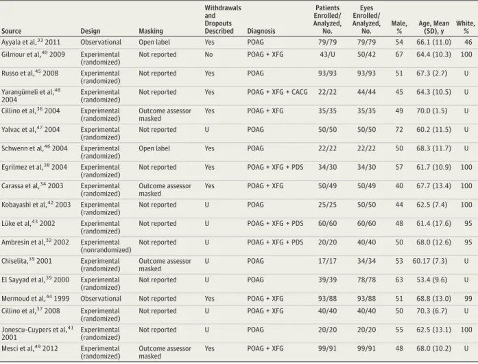
Table 1. Description of Studies and Patients
Source Design Masking
Withdrawals and Dropouts Described Diagnosis Patients Enrolled/ Analyzed, No. Eyes Enrolled/ Analyzed, No. Male, % Age, Mean (SD), y White, %
Ayyala et al,332011 Observational Open label Yes POAG 79/79 79/79 54 66.1 (11.0) 46
Gilmour et al,402009 Experimental
(randomized)
Not reported No POAG + XFG 43/U 50/42 67 64.4 (10.3) 100
Russo et al,452008 Experimental
(randomized)
Not reported Yes POAG 93/93 93/93 51 67.3 (2.7) U
Yarangümeli et al,48
2004
Experimental (randomized)
Not reported Yes POAG + XFG + CACG 22/22 44/44 45 64.3 (10.5) U
Cillino et al,362004 Experimental
(randomized)
Outcome assessor masked
Yes POAG + XFG 35/35 35/35 49 70.0 (1.5) U
Yalvac et al,472004 Experimental
(randomized)
Not reported U POAG 50/50 50/50 72 60.2 (11.5) U
Schwenn et al,46
2004 Experimental
(randomized)
Open label Yes POAG 22/22 22/22 50 68.3 (11.7) U
Egrilmez et al,382004 Experimental
(randomized)
Not reported Yes POAG + XFG + PDS 34/30 34/30 57 61.7 (10.9) 100
Carassa et al,342003 Experimental
(randomized) Outcome assessor masked Yes POAG + XFG 50/49 50/49 40 67.7 (13.4) 100 Kobayashi et al,42 2003 Experimental (randomized)
Not reported U POAG 25/25 50/50 44 62.5 (7.4) 100
Lüke et al,432002 Experimental
(randomized)
Not reported U POAG + XFG + PDS 60/60 60/60 48 61.4 (17.6) 95
Ambresin et al,322002 Experimental
(nonrandomized)
Not reported U POAG + XFG + PDS 20/20 40/40 50 68.0 (12.6) 95
Chiselita,35 2001 Experimental (randomized) Outcome assessor masked U POAG 17/17 34/34 53 60.17 (7.3) U
El Sayyad et al,392000 Experimental
(randomized)
Not reported U POAG 39/39 78/78 63 53.4 (9.6) U
Mermoud et al,441999 Observational Not reported Yes POAG + XFG 93/88 93/88 51 68.8 (13.0) 99
Cillino et al,372008 Experimental
(randomized)
Not reported U POAG + XFG 40/40 40/40 50 70.3 (6.7) U
Jonescu-Cuypers et al,41
2001
Experimental (randomized)
Not reported U POAG 20/20 20/20 55 62.5 (13.1) 100
Mesci et al,49 2012 Experimental (randomized) Outcome assessor masked Yes POAG + XFG 99/91 99/91 48 68.0 (10.2) U
Abbreviations: CACG, chronic angle-closure glaucoma; PDS, pigment dispersion syndrome; POAG, primary open-angle glaucoma; U, unknown; XFG, exfoliative glaucoma.
Safety was assessed in terms of the incidence of intraop-erative and postopintraop-erative complications in each group. The dif-ference between groups was expressed as the RR.
All results were expressed as a point estimate and its 95% confidence interval.
Statistical heterogeneity was quantified using the I2
sta-tistic, which indicates the percentage of variability due to heterogeneity rather than to chance alone: 0% indicates no heterogeneity, greater values indicate increasing heteroge-neity, and values greater than 50% imply substantial heterogeneity.30
We also used χ2tests for homogeneity. The assumption of
homogeneity was deemed not valid if P < .10.
All of the statistical analyses were performed using Re-view Manager version 5.1 software (Nordic Cochrane Centre, Cochrane Collaboration).
Efficacy Analysis
The primary analysis compared TE and NPS after 6 and 12 months of follow-up in terms of the MeD. In the case of stud-ies with no 6- or 12-month assessment, we considered the IOP recorded at the nearest subsequent evaluation. Subgroup analyses were made by type of NPS (DS, VCO, and CP), anti-metabolite augmentation during surgery (only for studies com-paring TE and DS), and the use of implants (only for studies comparing TE and DS). To verify the robustness of the results
of the primary analysis, the analyses were repeated by exclud-ing the studies not assessexclud-ing IOP at 6 or 12 months and by ex-cluding nonrandomized studies.
Safety Analysis
The safety analysis compared TE with one of the NPS proce-dures in terms of the incidence of complications. Each com-plication was analyzed separately.
Publication Bias Assessment
To exclude the presence of publication bias (ie, the bias due to the fact that studies with positive results are more likely to be published than those with negative results) and small-study effects (the tendency for treatment effect estimates to be different in small and larger studies), we visually explored any asymmetry using a funnel plot in which study size was plot-ted as a function of the measure of interest.31
Results
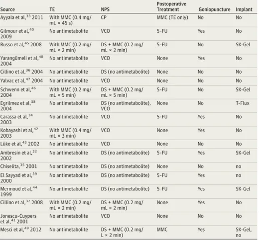
Figure 1 shows the study selection process. The electronic searches identified 278 abstracts, 238 of which did not meet the eligibility criteria; the full texts of the remaining 40 ar-ticles were examined. No additional studies were identified from the references of the selected articles, and no relevant Table 2. Surgery Characteristics
Source TE NPS
Postoperative
Treatment Goniopuncture Implant
Ayyala et al,33 2011 With MMC (0.4 mg/ mL × 45 s) CP MMC (TE only) No No Gilmour et al,40 2009
No antimetabolite VCO 5-FU Yes No
Russo et al,452008 With MMC (0.2 mg/
mL × 2 min) DS + MMC (0.2 mg/ mL × 2 min) 5-FU No SK-Gel Yarangümeli et al,48 2004
No antimetabolite VCO None Yes No
Cillino et al,362004 No antimetabolite DS (no antimetabolite) None No No
Yalvac et al,472004 No antimetabolite VCO None No No
Schwenn et al,46 2004 With MMC (0.2 mg/ mL × 5 min) DS + MMC (0.2 mg/ mL × 5 min) 5-FU No SK-Gel Egrilmez et al,38 2004
No antimetabolite DS (no antimetabolite), VCO
None No T-Flux
Carassa et al,34
2003
No antimetabolite VCO 5-FU Yes No
Kobayashi et al,42
2003
With MMC (0.4 mg/ mL × 3 min)
VCO None Yes No
Lüke et al,432002 No antimetabolite VCO None No No
Ambresin et al,32
2002
No antimetabolite DS (no antimetabolite) 5-FU Yes SK-Gel
Chiselita,352001 No antimetabolite DS (no antimetabolite) None No no
El Sayyad et al,39
2000
No antimetabolite DS (no antimetabolite) 5-FU Yes no
Mermoud et al,44
1999
No antimetabolite DS (no antimetabolite) 5-FU Yes SK-Gel
Cillino et al,372008 With MMC (0.2 mg/
mL × 2 min) DS + MMC (0.2 mg/ mL × 2 min) None Yes No Jonescu-Cuypers et al,41 2001
No antimetabolite VCO None No No
Mesci et al,49 2012 No antimetabolite DS + MMC (0.2 mg/ L × 2 min) MMC Yes SK-Gel, no Abbreviations: CP, canaloplasty; DS, deep sclerectomy; MMC, mitomycin C; NPS, nonpenetrating surgery; TE, trabeculectomy; VCO, viscocanalostomy; 5-FU, 5-fluorouracil.
unpublished studies were found. A further 22 studies were sub-sequently excluded (eTable 2 in Supplement): 6 because they investigated other types of surgery, 3 because they included patients with secondary glaucoma or closed-angle glaucoma, 1 because it had a different end point, 8 because they were not clinical studies, 1 because the follow-up was too short, and 3 because they were not written in English and no full-text trans-lation or evaluation was available. For the abstract screening, the agreement was good (κ = 0.70); regarding the full-text ar-ticle screening, we obtained full agreement between the re-viewers.
Eighteen articles were therefore selected for data extraction and analysis,32-49but as 2 of them provided 2 comparisons,38,49
the total number of comparisons was 20.
Study Characteristics
Table 1 shows the characteristics of the 18 studies. Fifteen were randomized clinical trials,34-43,45-491 was a
nonran-domized experimental study,32and 2 were observational
studies.33,44
Information about the blindness assessment was not clearly described in 12 articles.32,37-45,47,48One article40did not
describe withdrawals or dropouts, and 8 articles32,35,37,39,41-43,47
did not clearly indicate the presence of withdrawals or drop-outs. Eight studies33,35,39,41,42,45-47only included patients with
primary OAG (POAG), 6 studies34,36,37,40,44,49included
pa-tients with POAG and XFG, 3 studies32,38,43included patients
with POAG, XFG, and pigment dispersion syndrome, and 1 study48included patients with POAG, XFG, and chronic
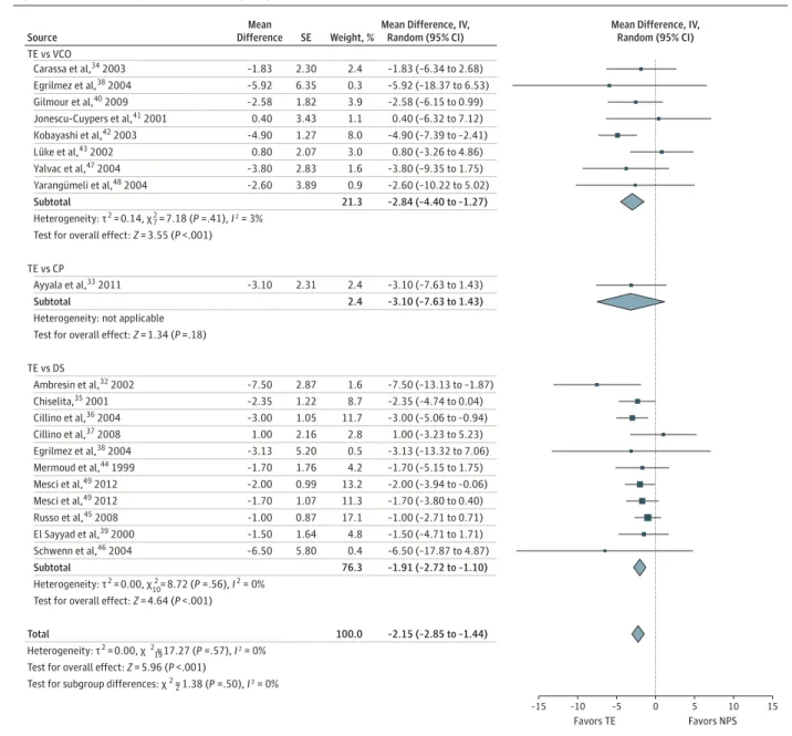
angle-Figure 2. Trabeculectomy vs Nonpenetrating Surgery at 6-Month Follow-up
–15 –5 0 5 10 15 Favors TE Favors NPS –10 Source TE vs VCO TE vs DS
Mean Difference, IV, Random (95% CI)
Mean Difference, IV, Random (95% CI) Carassa et al,34 2003 –1.83 (–6.34 to 2.68) Egrilmez et al,38 2004 –5.92 (–18.37 to 6.53) Gilmour et al,40 2009 –2.58 (–6.15 to 0.99) Jonescu-Cuypers et al,41 2001 0.40 (–6.32 to 7.12) Kobayashi et al,42 2003 –4.90 (–7.39 to –2.41) Lüke et al,43 2002 0.80 (–3.26 to 4.86) Yalvac et al,47 2004 –3.80 (–9.35 to 1.75) Yarangümeli et al,48 2004 –2.60 (–10.22 to 5.02) Subtotal –2.84 (–4.40 to –1.27) Heterogeneity: τ2 = 0.14, χ 2 = 7.18 (P =.41), I 2 = 3%
Test for overall effect: Z = 3.55 (P <.001)
Egrilmez et al,38 2004 –3.13 (–13.32 to 7.06) Mermoud et al,44 1999 –1.70 (–5.15 to 1.75) Mesci et al,49 2012 –2.00 (–3.94 to –0.06) Mesci et al,49 2012 –1.70 (–3.80 to 0.40) Cillino et al,36 2004 –3.00 (–5.06 to –0.94) Cillino et al,37 2008 1.00 (–3.23 to 5.23) Russo et al,45 2008 –1.00 (–2.71 to 0.71) El Sayyad et al,39 2000 –1.50 (–4.71 to 1.71) Schwenn et al,46 2004 –6.50 (–17.87 to 4.87) Subtotal –1.91 (–2.72 to –1.10) Heterogeneity: τ2 = 0.00, χ 2 = 8.72 (P =.56), I 2 = 0% 10
Test for overall effect: Z = 4.64 (P <.001)
TE vs CP
Ayyala et al,33 2011 –3.10 (–7.63 to 1.43)
Subtotal –3.10 (–7.63 to 1.43)
Heterogeneity: not applicable Test for overall effect: Z = 1.34 (P =.18)
Mean Difference –1.83 –5.92 –2.58 0.40 –4.90 0.80 –3.80 –2.60 –3.13 –1.70 –2.00 –1.70 –1.00 –1.50 –6.50 –3.00 1.00 –3.10 –7.50 –2.35 SE 2.30 6.35 1.82 3.43 1.27 2.07 2.83 3.89 5.20 1.76 0.99 1.07 0.87 1.64 5.80 1.05 2.16 2.31 2.87 1.22 Weight, % 2.4 0.3 3.9 1.1 8.0 3.0 1.6 0.9 21.3 0.5 4.2 13.2 11.3 17.1 4.8 0.4 76.3 Total –2.15 (–2.85 to –1.44) Heterogeneity: τ2 = 0.00, χ 2 = 17.27 (P =.57), I 2 = 0%
Test for overall effect: Z = 5.96 (P <.001)
2 19
Test for subgroup differences: χ 2 = 1.38 (P =.50), I 2 = 0%
100.0 11.7 2.8 2.4 2.4 1.6 8.7 Ambresin et al,32 2002 –7.50 (–13.13 to –1.87) Chiselita,35 2001 –2.35 (–4.74 to 0.04) 7
closure glaucoma. There were more men than women in 9 stud-ies, and the patients’ ages ranged from 53.4 to 70.3 years.
Risk of Bias
Selection bias could not be excluded in 15 of the 16 experimen-tal studies because of the absence of adequate sequence gen-eration or concealed allocation (3 studies)32,36,45or lack of
in-formation (12 studies).34,35,37-41,43,46-49Attrition bias could be
excluded in 6 experimental studies34,38,45,46,48,49that
ad-dressed the question of incomplete outcome data; the rates of follow-up and the number of withdrawals were similar be-tween the groups. Only 4 of the 16 experimental studies34-36,49
stated that the outcome assessors were unaware of the as-signed intervention.
The 2 observational studies33,44had a low risk of bias, but
the observational design limitations should be taken into con-sideration.
Effects of Interventions
The 18 studies made a total of 20 comparisons and involved 945 eyes: 7 studies34,40-43,47,48compared TE with VCO (315 eyes); 1
study33compared TE with CP (79 eyes); 8 studies32,35-37,39,44-46
compared TE with DS (430 eyes); 1 study38compared TE with
DS and VCO (30 eyes); and 1 study49compared TE with DS with
or without an implant (91 eyes). Mitomycin C was the only an-timetabolite added at the time of surgery in 7 comparisons of TE and DS (Table 2).
TE vs NPS
Analysis of the 6-month follow-up data showed that the pooled estimate of the MeD was −2.15 mm Hg (95% CI, −2.85 to −1.44; test for overall effect: Z = 5.96, P < .001); no heterogeneity was detected (I2= 0%; test for heterogeneity: χ2
19= 17.27, P = .57)
(Figure 2).
There was no difference between the surgical subgroups indicating that TE led to a greater IOP reduction.
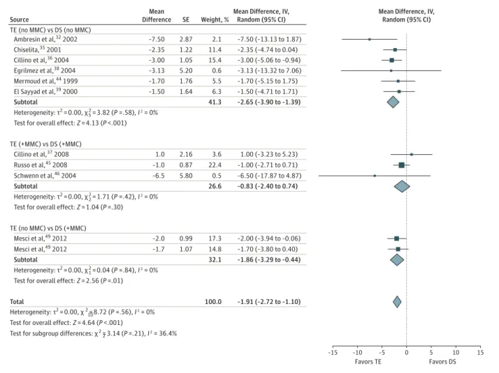
In the subgroup antimetabolite analysis (Figure 3), 6 studies32,35,36,38,39,44compared TE and DS without MMC
(MeD = −2.65 mm Hg [95% CI, −3.90 to −1.39]), 3 studies37,45,46
compared TE and DS with MMC (MeD = −0.83 mm Hg [95% CI, −2.40 to 0.74]), and 1 study49compared TE without MMC with
DS with MMC (MeD = −1.86 mm Hg [95% CI, −3.29 to −0.44]). In the subgroup analysis of the studies in which an im-plant was added to DS, there were 5 studies35-37,39,49of DS
with-Figure 3. Subgroup Analysis of Antimetabolite Addition for Trabeculectomy vs Deep Sclerectomy at 6-Month Follow-up
–15 –5 0 5 10 15
Favors TE Favors DS –10
Source
TE (no MMC) vs DS (no MMC)
Mean Difference, IV, Random (95% CI)
Mean Difference, IV, Random (95% CI) Ambresin et al,32 2002 –7.50 (–13.13 to 1.87) Chiselita,35 2001 –2.35 (–4.74 to 0.04) Cillino et al,36 2004 –3.00 (–5.06 to –0.94) Egrilmez et al,38 2004 –3.13 (–13.32 to 7.06) Mermoud et al,44 1999 –1.70 (–5.15 to 1.75) El Sayyad et al,39 2000 –1.50 (–4.71 to 1.71) Subtotal –2.65 (–3.90 to –1.39) Heterogeneity: τ2 = 0.00, χ 2 = 3.82 (P =.58), I 2 = 0%
Test for overall effect: Z = 4.13 (P <.001)
5
TE (+MMC) vs DS (+MMC)
Cillino et al,37 2008 1.00 (–3.23 to 5.23)
Subtotal
Heterogeneity: τ2 = 0.00, χ 2 = 1.71 (P =.42), I 2 = 0%
Test for overall effect: Z = 1.04 (P =.30)
Mean Difference –7.50 –2.35 –3.00 –3.13 –1.70 –1.50 1.0 SE 2.87 1.22 1.05 5.20 1.76 1.64 2.16 Weight, % 2.1 11.4 15.4 0.6 5.5 6.3 41.3 2 1 10 3.6 –1.00 (–2.71 to 0.71) –1.0 0.87 22.4 –6.50 (–17.87 to 4.87) –6.5 5.80 0.5 –0.83 (–2.40 to 0.74) 26.6 –1.91 (–2.72 to –1.10) 100.0 Russo et al,45 2008 Schwenn et al,46 2004 –1.86 (–3.29 to –0.44) 32.1 –1.70 (–3.80 to 0.40) –1.7 1.07 14.8 TE (no MMC) vs DS (+MMC) Mesci et al,49 2012 –2.00 (–3.94 to –0.06) Subtotal Heterogeneity: τ2 = 0.00, χ 2 = 0.04 (P =.84), I 2 = 0%
Test for overall effect: Z = 2.56 (P =.01)
–2.0 0.99 17.3 Mesci et al,49 2012
Total
Heterogeneity: τ2 = 0.00, χ 2 = 8.72 (P =.56), I 2 = 0%
Test for overall effect: Z = 4.64 (P <.001)
Test for subgroup differences: χ 2 = 3.14 (P =.21), I 2 = 36.4%
2
out an implant and 6 studies32,38,44-46,49of DS with an
im-plant. There was no between-group difference in the comparison of TE and DS without an implant (MeD = −2.10 mm Hg [95% CI, −3.20 to −1.00]) or in the comparison of TE and DS with an implant (MeD = −1.79 mm Hg [95% CI, −3.13 to −0.45]). The result of the test for subgroup difference was χ2
1= 0.24
(P = .62).
Two studies38,41were excluded from the analysis of the
12-month follow-up data because of an insufficient duration of fol-low-up. Overall, TE led to a greater reduction in IOP than NPS (Figure 4). There was a difference between the subgroups de-fined by type of surgery (χ2
2= 8.96; P = .01): the MeD
de-creased in favor of TE in the VCO group (from −2.84 mm Hg [95% CI, −4.40 to −1.27] at 6 months to −3.84 mm Hg [95% CI, −5.34 to −2.34] at 12 months) and the CP group (from −3.10 mm Hg [95% CI, −7.63 to 1.43] at 6 months to −4.40 mm Hg [95% CI, −8.89 to 0.09] at 12 months) but increased in favor of DS in
the DS group (from −1.91 mm Hg [95% CI, −2.72 to −1.10] at 6 months to −1.53 mm Hg [95% CI, −2.59 to −0.47] at 12 months). Sensitivity Analysis
Two studies32,45in which the first IOP evaluation was made
after 6 months were excluded from the 6-month sensitivity analysis, and 1 study45in which the first IOP evaluation was
made after 12 months was excluded from the 12-month sen-sitivity analysis. A further 3 studies32,33,44were excluded from
the analyses of only randomized clinical trials. The results of these sensitivity analyses were always consistent with those of the primary analysis.
Safety Evaluation
The results of the analysis of postoperative complications are shown in Table 3. The absolute risk of hypotony (RR = 2.3 [95% CI, 1.3-3.8]), choroidal effusion (RR = 3.9 [95% CI, 2.0-7.5]), cata-Figure 4. Trabeculectomy vs Nonpenetrating Surgery at 12-Month Follow-up
–15 –5 0 5 10 15
Favors TE Favors NPS –10
Source TE vs VCO
Mean Difference, IV, Random (95% CI)
Mean Difference, IV, Random (95% CI) Carassa et al,34 2003 –1.47 (–6.12 to 3.18) Gilmour et al,40 2009 –4.56 (–7.89 to –1.23) Kobayashi et al,42 2003 –4.30 (–6.53 to –2.07) Lüke et al,43 2002 –1.80 (–6.09 to 2.49) Yalvac et al,47 2004 –5.70 (–11.13 to –0.27) Yarangümeli et al,48 2004 –3.90 (–11.43 to 3.63) Subtotal –3.84 (–5.34 to –2.34) Heterogeneity: τ2 = 0.00, χ 2 = 2.66 (P =.75), I 2 = 0%
Test for overall effect: Z = 5.01 (P <.001)
5
TE vs CP
Ayyala et al,33 2011 –4.40 (–8.89 to 0.09)
Subtotal
Heterogeneity: not applicable Test for overall effect: Z = 1.92 (P =.05)
Mean Difference –1.47 –4.56 –4.30 –1.80 –5.70 –3.90 –4.40 SE 2.37 1.70 1.14 2.19 2.77 3.84 2.29 Weight, % 3.4 5.6 9.1 3.9 2.6 1.5 26.1 16 3.6 –4.40 (–8.89 to 0.09) 3.6 –2.22 (–3.18 to –1.27) 100.0 Subtotal Heterogeneity: τ2 = 0.99, χ 2 = 14.48 (P =.11), I 2 = 38%
Test for overall effect: Z = 2.83 (P =.005)
–1.53 (–2.59 to –0.47) 70.2 –3.16 (–5.85 to –0.47) –3.16 1.37 7.4 TE vs DS Ambresin et al,32 2002 –7.50 2.87 2.5 –7.50 (–13.13 to –1.87) Chiselita,35 2001 3.20 (–1.19 to 7.59) 3.20 2.24 3.8 Cillino et al,36 2004 –2.70 1.08 9.5 –2.70 (–4.82 to –0.58) Cillino et al,37 2008 –0.40 (–2.11 to 1.31) –0.40 0.87 11.4 Mermoud et al,44 1999 –0.60 1.78 5.3 –0.60 (–4.09 to 2.89) Mesci et al,49 2012 –1.00 (–2.71 to 0.71) –1.00 0.87 11.4 Mesci et al,49 2012 –1.50 0.81 12.0 –1.50 (–3.09 to 0.09) Russo et al,45 2008 –6.60 (–17.73 to 4.53) –6.60 5.68 0.7 El Sayyad et al,39 2000 –1.80 1.57 6.3 –1.80 (–4.88 to 1.28) Schwenn et al,46 2004 Total Heterogeneity: τ2 = 1.33, χ 2 = 26.10 (P =.05), I 2 = 39%
Test for overall effect: Z = 4.56 (P <.001)
Test for subgroup differences: χ 2 = 8.96 (P =.01), I 2 = 77.7%
2 9
ract (RR = 3.4 [95% CI, 2.1-5.5]), or a flat or shallow anterior chamber (RR = 4.3 [95% CI, 2.3-8.0]) was higher in the TE group than in the NPS group.
In the comparison of TE and VCO, the RRs for all of the con-sidered complications increased when MMC was added to TE (RR of hypotony increased from 2.3 to 11.0; RR of cataract in-creased from 3.6 to 5.0; and RR of a flat or shallow anterior chamber increased from 4.7 to 9.0).
In the comparison of TE and DS, the RRs of all of the con-sidered complications except cataract (RR increased from 3.0 to 3.9) decreased when MMC was added to both (RR of hy-potony decreased from 2.2 to 1.7; RR of choroidal effusion de-creased from 6.1 to 2.4; and RR of a flat or shallow anterior chamber decreased from 8.3 to 3.1).
Discussion
To our knowledge, this is the first meta-analysis that assesses efficacy and safety of TE vs all of the available NPS proce-dures. The results of this systematic review suggest that TE is more effective in reducing IOP than NPS 6 and 12 months af-ter surgery (−2.15 mm Hg [95% CI, −2.85 to −1.44] and −2.22 [95% CI, −3.18 to −1.27], respectively). The significance of such differences in IOP is likely to be clinically relevant especially in patients requiring a greater IOP reduction or those at greater risk for glaucoma progression. Among the NPS procedures, there was less difference in efficacy between DS and TE, al-though the superiority of TE was statistically significant.
These results are in line with what is generally believed about so-called canal surgery (VCO and CP), which cannot be expected to lower IOP as much as the bulk flow of the full-thickness perforation created in the eye by means of TE. Deep sclerectomy seems to be a clinically reasonable compromise in terms of reducing IOP.
With regard to the efficacy of TE and DS with or without MMC, the addition of MMC to both decreased the difference in the reduction in IOP: TE and DS without MMC, −2.65 mm Hg (95% CI, −3.90 to −1.39); TE and DS with MMC, −0.83 mm Hg
(95% CI, −2.40 to 0.74). This indicates that the use of MMC is advisable when performing DS because the nature of the pro-cedure (filtering surgery: filtration of aqueous humor into the subconjunctival spaces) means that antimetabolites can avoid conjunctival healing and optimize surgical outcomes.
The implantation of drainage devices during DS has been advocated as a means of increasing the success rate of the procedure,50,51but our meta-analysis shows no
signifi-cant difference in the reduction in IOP induced by DS with or without drainage devices (test for subgroup differences: χ2
1= 0.24; P = .62). This finding has important implications
in clinical practice because the use of implants significantly increases the cost of DS, which is otherwise the same as that of standard TE.
As expected, TE was associated with a higher incidence of short- and long-term complications. Viscocanalostomy had a better safety profile than TE, although the evidence concern-ing CP was not sufficient to draw any conclusion.
It has been claimed that the additional use of intraop-erative MMC increases surgery-related complications. Our findings show that without MMC, TE led to a higher inci-dence of complications than DS, but when both procedures were supplemented with MMC, the rate of complications (except cataract progression) increased in the DS group. One possible explanation is that DS has not been modified from the original technique (with or without the use of MMC), whereas standard TE has been substantially improved since the advent of antimetabolites. The term safe trabeculectomy reflects a development that can be considered as optimizing the technique and postoperative management (suture lysis, bleb manipulation).52-54
In conclusion, TE still offers the possibility of obtaining ex-cellent IOP control at the long-term follow-up in patients with OAG. Success may vary depending on glaucoma form, ie, it may work better in XFG, whereas NPS procedures are not a viable option in chronic angle-closure glaucoma. Moreover, NPS pro-cedures are more difficult to perform, require a long learning curve even for an experienced glaucoma surgeon, and are more costly. Despite a higher incidence of postoperative complica-Table 3. Complications
Subgroup
Hypotony Choroidal Effusion Cataract
Flat or Shallow Anterior Chamber Studies, No. (Eyes, No.) RR (95% CI) Studies, No. (Eyes, No.) RR (95% CI) Studies, No. (Eyes, No.) RR (95% CI) Studies, No. (Eyes, No.) RR (95% CI) TE vs VCO 6 (303) 2.6 (1.2-5.6) 3 (159) 6.0 (1.1-33.8) 4 (204) 3.8 (1.5-9.5) 3 (154) 5.5 (1.2-25.1) TE (no MMC) vs VCO 5 (253) 2.3 (1.1-5.0) 3 (159) 6.0 (1.1-33.8) 3 (154) 3.6 (1.4-9.7) 2 (104) 4.7 (0.7-32.6) TE (+MMC) vs VCO 1 (50) 11.0 (0.6-188.9) NA NA 1 (50) 5.0 (0.3-99.2) 1 (50) 9.0 (0.5-158.9) TE vs DS 7 (399) 2.1 (0.9-4.6) 7 (409) 3.8 (1.6-9.0) 6 (424) 3.3 (1.8-5.8) 9 (521) 4.1 (2.1-8.0) TE (no MMC) vs DS (no MMC) 3 (153) 2.2 (0.5-8.8) 3 (163) 6.1 (1.9-19.9) 4 (240) 3.0 (1.3-6.6) 5 (275) 8.3 (2.6-26.7) TE (no MMC) vs DS (+MMC) 1 (91) 3.1 (1.6-6.3) 1 (91) 15.6 (0.9-281.3) 1 (91) 3.5 (1.3-9.4) 1 (91) 2.8 (1.0-7.9) TE (+MMC) vs DS (+MMC) 3 (155) 1.7 (0.4-6.3) 3 (155) 2.4 (0.6-9.8) 1 (93) 3.9 (0.9-17.0) 3 (155) 3.1 (0.6-16.7) TE vs CP NA NA 1 (79) 12.3 (0.7-205.9) NA NA NA NA TE vs NPS 13 (702) 2.3 (1.3-3.8) 11 (647) 3.9 (2.0-7.5) 10 (628) 3.4 (2.1-5.5) 12 (675) 4.3 (2.3-8.0)
Abbreviations: CP, canaloplasty; DS, deep sclerectomy; MMC, mitomycin C; NA, not applicable; NPS, nonpenetrating surgery; RR, relative risk; TE, trabeculectomy; VCO, viscocanalostomy.
tions when compared with NPS, the advent of the safe tra-beculectomy technique offers the possibility of tailoring the IOP postoperatively with minimal postoperative
complica-tions. Therefore, further studies are needed to assess the safety profile of the current TE procedure compared with NPS techniques.
ARTICLE INFORMATION
Submitted for Publication: February 21, 2013; final revision received April 22, 2013; accepted April 23, 2013.
Published Online: October 24, 2013. doi:10.1001/jamaophthalmol.2013.5059. Author Contributions: Dr Rulli had full access to all of the data in the study and takes responsibility for the integrity of the data and the accuracy of the data analysis. Drs Rulli and Biagioli contributed equally to this work.
Study concept and design: Rulli, Floriani, Quaranta. Acquisition of data: Biagioli, Riva, Gambirasio,
De Simone.
Analysis and interpretation of data: Rulli, Biagioli,
Riva, Floriani, Quaranta.
Drafting of the manuscript: Rulli, Biagioli, Riva,
De Simone, Quaranta.
Critical revision of the manuscript for important intellectual content: Rulli, Biagioli, Riva, Gambirasio,
Floriani, Quaranta.
Statistical analysis: Rulli, Biagioli.
Administrative, technical, or material support: Riva. Study supervision: De Simone, Floriani, Quaranta.
Conflict of Interest Disclosures: None reported.
REFERENCES
1. Razeghinejad MR, Fudemberg SJ, Spaeth GL. The changing conceptual basis of trabeculectomy: a review of past and current surgical techniques. Surv
Ophthalmol. 2012;57(1):1-25.
2. Netland PA; Ophthalmic Technology Assessment Committee Glaucoma Panel, American Academy of Ophthalmology. Nonpenetrating glaucoma surgery.
Ophthalmology. 2001;108(2):416-421.
3. Cairns JE. Trabeculectomy: preliminary report of a new method. Am J Ophthalmol.
1968;66(4):673-679.
4. Azuara-Blanco A, Katz LJ. Dysfunctional filtering blebs. Surv Ophthalmol. 1998;43(2):93-126. 5. Burney EN, Quigley HA, Robin AL. Hypotony and choroidal detachment as late complications of trabeculectomy. Am J Ophthalmol. 1987;103(5):685-688.
6. Fluorouracil Filtering Surgery Study Group. Fluorouracil Filtering Surgery Study one-year follow-up. Am J Ophthalmol. 1989;108(6):625-635. 7. Belyea DA, Dan JA, Stamper RL, Lieberman MF, Spencer WH. Late onset of sequential multifocal bleb leaks after glaucoma filtration surgery with 5-fluorouracil and mitomycin C. Am J Ophthalmol. 1997;124(1):40-45.
8. Singh J, O’Brien C, Chawla HB. Success rate and complications of intraoperative 0.2 mg/mL mitomycin C in trabeculectomy surgery. Eye (Lond). 1995;9(pt 4):460-466.
9. Greenfield DS, Liebmann JM, Jee J, Ritch R. Late-onset bleb leaks after glaucoma filtering surgery. Arch Ophthalmol. 1998;116(4):443-447. 10. Feiner L, Piltz-Seymour JR; Collaborative Initial Glaucoma Treatment Study. Collaborative Initial Glaucoma Treatment Study: a summary of results to date. Curr Opin Ophthalmol. 2003;14(2):106-111.
11. Edmunds B, Thompson JR, Salmon JF, Wormald RP. The National Survey of Trabeculectomy, III: early and late complications. Eye (Lond).
2002;16(3):297-303.
12. Rahman A, Mendonca M, Simmons RB, Simmons RJ. Hypotony after glaucoma filtration surgery. Int Ophthalmol Clin. 2000;40(1):127-136. 13. Wormald R, Wilkins MR, Bunce C.
Post-operative 5-fluorouracil for glaucoma surgery.
Cochrane Database Syst Rev. 2001;(3):CD001132.
14. Wilkins M, Indar A, Wormald R. Intra-operative mitomycin C for glaucoma surgery. Cochrane
Database Syst Rev. 2005;(4):CD002897.
15. Smith R. A new technique for opening the canal of Schlemm: preliminary report. Br J Ophthalmol. 1960;44:370-373.
16. Krasnov MM. Externalization of Schlemm’s canal (sinusotomy) in glaucoma. Br J Ophthalmol. 1968;52(2):157-161.
17. Bylsma S. Nonpenetrating deep sclerectomy: collagen implant and viscocanalostomy procedures.
Int Ophthalmol Clin. 1999;39(3):103-119.
18. Fedorov SN, Ioffe DI, Ronkina TI. Glaucoma surgery: deep sclerectomy [in Russian]. Vestn
Oftalmol. 1982;(4):6-10.
19. Stegmann R, Pienaar A, Miller D.
Viscocanalostomy for open-angle glaucoma in black African patients. J Cataract Refract Surg. 1999;25(3):316-322.
20. Lewis RA, von Wolff K, Tetz M, et al. Canaloplasty: circumferential viscodilation and tensioning of Schlemm’s canal using a flexible microcatheter for the treatment of open-angle glaucoma in adults: interim clinical study analysis.
J Cataract Refract Surg. 2007;33(7):1217-1226.
21. Grieshaber MC, Pienaar A, Olivier J, Stegmann R. Canaloplasty for primary open-angle glaucoma: long-term outcome. Br J Ophthalmol.
2010;94(11):1478-1482.
22. Ateş H, Uretmen O, Andaç K, Azarsiz SS. Deep sclerectomy with a nonabsorbable implant (T-Flux): preliminary results. Can J Ophthalmol.
2003;38(6):482-488.
23. Dahan E, Ravinet E, Ben-Simon GJ, Mermoud A. Comparison of the efficacy and longevity of nonpenetrating glaucoma surgery with and without a new, nonabsorbable hydrophilic implant.
Ophthalmic Surg Lasers Imaging. 2003;34(6):
457-463.
24. Ravinet E, Bovey E, Mermoud A. T-Flux implant vs Healon GV in deep sclerectomy. J Glaucoma. 2004;13(1):46-50.
25. European Glaucoma Society. Terminology and
Guidelines for Glaucoma. 3rd ed. Genoa, Italy:
European Glaucoma Society; 2008. 26. Cohen J. A coefficient of agreement for nominal scales. Educ Psychol Meas. 1960;20(1):37-46. doi:10.1177 /001316446002000104.
27. Higgins JPT, Green S, eds. Cochrane Handbook
for Systematic Reviews of Interventions. Chichester,
England: John Wiley & Sons; 2010.
28. Wells GA, Shea B, O'Connell D, et al. The Newcastle-Ottawa Scale (NOS) for assessing the quality of nonrandomised studies in meta-analyses. http://www.ohri.ca/programs/clinical_epidemiology /oxford.asp. Accessed October 19, 2012. 29. DerSimonian R, Kacker R. Random-effects model for meta-analysis of clinical trials: an update.
Contemp Clin Trials. 2007;28(2):105-114.
30. Higgins JP, Thompson SG, Deeks JJ, Altman DG. Measuring inconsistency in meta-analyses.
BMJ. 2003;327(7414):557-560.
31. Sterne JA, Egger M. Funnel plots for detecting bias in meta-analysis: guidelines on choice of axis.
J Clin Epidemiol. 2001;54(10):1046-1055.
32. Ambresin A, Shaarawy T, Mermoud A. Deep sclerectomy with collagen implant in one eye compared with trabeculectomy in the other eye of the same patient. J Glaucoma. 2002;11(3):214-220. 33. Ayyala RS, Chaudhry AL, Okogbaa CB, Zurakowski D. Comparison of surgical outcomes between canaloplasty and trabeculectomy at 12 months’ follow-up. Ophthalmology.
2011;118(12):2427-2433.
34. Carassa RG, Bettin P, Fiori M, Brancato R. Viscocanalostomy vs trabeculectomy in white adults affected by open-angle glaucoma: a 2-year randomized, controlled trial. Ophthalmology. 2003;110(5):882-887.
35. Chiselita D. Non-penetrating deep sclerectomy vs trabeculectomy in primary open-angle glaucoma surgery. Eye (Lond). 2001;15(pt 2):197-201. 36. Cillino S, Di Pace F, Casuccio A, et al. Deep sclerectomy vs punch trabeculectomy with or without phacoemulsification: a randomized clinical trial. J Glaucoma. 2004;13(6):500-506. 37. Cillino S, Di Pace F, Casuccio A, Cillino G, Lodato G. Deep sclerectomy vs trabeculectomy with low-dosage mitomycin C: four-year follow-up.
Ophthalmologica. 2008;222(2):81-87.
38. Egrilmez S, Ates H, Nalcaci S, Andac K, Yagci A. Surgically induced corneal refractive change following glaucoma surgery: nonpenetrating trabecular surgeries vs trabeculectomy. J Cataract
Refract Surg. 2004;30(6):1232-1239.
39. El Sayyad F, Helal M, El-Kholify H, Khalil M, El-Maghraby A. Nonpenetrating deep sclerectomy vs trabeculectomy in bilateral primary open-angle glaucoma. Ophthalmology. 2000;107(9):1671-1674. 40. Gilmour DF, Manners TD, Devonport H, Varga Z, Solebo AL, Miles J. Viscocanalostomy vs trabeculectomy for primary open angle glaucoma: 4-year prospective randomized clinical trial. Eye
(Lond). 2009;23(9):1802-1807.
41. Jonescu-Cuypers C, Jacobi P, Konen W, Krieglstein G. Primary viscocanalostomy vs trabeculectomy in white patients with open-angle glaucoma: a randomized clinical trial.
Ophthalmology. 2001;108(2):254-258.
42. Kobayashi H, Kobayashi K, Okinami S. A comparison of the intraocular pressure-lowering effect and safety of viscocanalostomy and trabeculectomy with mitomycin C in bilateral
open-angle glaucoma. Graefes Arch Clin Exp
Ophthalmol. 2003;241(5):359-366.
43. Lüke C, Dietlein TS, Jacobi PC, Konen W, Krieglstein GK. A prospective randomized trial of viscocanalostomy vs trabeculectomy in open-angle glaucoma: a 1-year follow-up study. J Glaucoma. 2002;11(4):294-299.
44. Mermoud A, Schnyder CC, Sickenberg M, Chiou AGY, Hédiguer SEA, Faggioni R. Comparison of deep sclerectomy with collagen implant and trabeculectomy in open-angle glaucoma. J Cataract
Refract Surg. 1999;25(3):323-331.
45. Russo V, Scott IU, Stella A, et al.
Nonpenetrating deep sclerectomy with reticulated hyaluronic acid implant vs punch trabeculectomy: a prospective clinical trial. Eur J Ophthalmol. 2008;18(5):751-757.
46. Schwenn O, Springer C, Troost A, Yun SH, Pfeiffer N. Deep sclerectomy using a hyaluronate implant vs trabeculectomy: a comparison of two
glaucoma operations using mitomycin C [in German]. Ophthalmologe. 2004;101(7):696-704. 47. Yalvac IS, Sahin M, Eksioglu U, Midillioglu IK, Aslan BS, Duman S. Primary viscocanalostomy vs trabeculectomy for primary open-angle glaucoma: three-year prospective randomized clinical trial.
J Cataract Refract Surg. 2004;30(10):2050-2057.
48. Yarangümeli A, Güreser S, Köz OG, Elhan AH, Kural G. Viscocanalostomy vs trabeculectomy in patients with bilateral high-tension glaucoma. Int
Ophthalmol. 2004;25(4):207-213.
49. Mesci C, Erbil HH, Karakurt Y, Akçakaya AA. Deep sclerectomy augmented with combination of absorbable biosynthetic sodium hyaluronate scleral implant and mitomycin C or with mitomycin C vs trabeculectomy: long-term results. Clin Experiment
Ophthalmol. 2012;40(4):e197-e207.
50. Sanchez E, Schnyder CC, Sickenberg M, Chiou AG, Hédiguer SE, Mermoud A. Deep sclerectomy:
results with and without collagen implant. Int
Ophthalmol. 1996-1997;20(1-3):157-162.
51. Shaarawy T, Nguyen C, Schnyder C, Mermoud A. Comparative study between deep sclerectomy with and without collagen implant: long term follow up. Br J Ophthalmol. 2004;88(1):95-98. 52. Stalmans I, Gillis A, Lafaut AS, Zeyen T. Safe trabeculectomy technique: long term outcome. Br J
Ophthalmol. 2006;90(1):44-47.
53. Wells AP, Bunce C, Khaw PT. Flap and suture manipulation after trabeculectomy with adjustable sutures: titration of flow and intraocular pressure in guarded filtration surgery. J Glaucoma.
2004;13(5):400-406.
54. Papadopoulos M, Khaw PT. Improving glaucoma filtering surgery. Eye (Lond). 2001;15 (pt 2):131-132.
OPHTHALMIC IMAGES
Entopic Image of Dislocated Intraocular Lens
Jennifer Tordilla-Wadia, MD; Sejal Shah, MD; Curtis E. Margo, MD, MPHA B
C
A 67-year-old man, with history of right cataract extraction surgery 10 years earlier then retinal detachment with therapeutic vitrectomy 1 year later, experienced severe blurred vision in the eye for 1 week. Visual blur was interrupted with the transient image of a “gasket,” which he could draw better than explain verbally (A). Visual acuity was 20/25 with +8-diopter correction. On reclined fundus examination, a plate haptic intraocular lens (IOL) was observed floating in front of the macula (B). The dislocated IOL was removed surgically (C) and replaced with an anterior chamber IOL. The cloudy edge of the entopic image corresponded to adherent lens capsule to the IOL (C, insert; periodic acid–Schiff; bar = 20 μm).