7. Eisenhauer EA, Therasse P, Bogaerts J et al. New response evaluation criteria in solid tumours: revised RECIST guideline (version 1.1). Eur J Cancer 2009; 45: 228–247. 8. Cella D, Nichol MB, Eton D et al. Estimating clinically meaningful changes for the
Functional Assessment of Cancer Therapy-Prostate: results from a clinical trial of patients with metastatic hormone-refractory prostate cancer. Value Health 2009; 12: 124–129.
9. Yount S, Cella D, Banik D et al. Brief assessment of priority symptoms in hormone refractory prostate cancer: the FACT Advanced Prostate Symptom Index (FAPSI). Health Qual Life Outcomes 2003; 1: 69.
10. Cella D, Hahn EA, Dineen K. Meaningful change in cancer-specific quality of life scores: differences between improvement and worsening. Qual Life Res 2002; 11: 207–221.
11. Yost KJ, Eton DT. Combining distribution- and anchor-based approaches to determine minimally important differences: the FACIT experience. Eval Health Prof 2005; 28: 172–191.
12. Mallinckrodt CH, Sanger TM, Dube S et al. Assessing and interpreting treatment effects in longitudinal clinical trials with missing data. Biol Psychiatry 2003; 53: 754–760.
13. Molenberghs G, Kenward M. Missing Data in Clinical Studies. Chichester, UK: John Wiley & Sons Ltd, 2007.
14. Ratitch B, O’Kelly M, Tosiello R. Missing data in clinical trials: from clinical assumptions to statistical analysis using pattern mixture models. Pharm Stat 2013; 12: 337–347.
15. Thompson JC, Wood J, Feuer D. Prostate cancer: palliative care and pain relief. Br Med Bull 2007; 83: 341–354.
16. Tannock IF, Osoba D, Stockler MR et al. Chemotherapy with mitoxantrone plus prednisone or prednisone alone for symptomatic hormone-resistant prostate cancer: a Canadian randomized trial with palliative end points. J Clin Oncol 1996; 14: 1756–1764.
17. Kornblith AB, Herndon JE, Zuckerman E et al. The impact of docetaxel, estramustine, and low dose hydrocortisone on the quality of life of men with hormone refractory prostate cancer and their partners: a feasibility study. Ann Oncol 2001; 12: 633–641.
18. Berthold DR, Pond GR, Roessner M et al. Treatment of hormone-refractory prostate cancer with docetaxel or mitoxantrone: relationships between prostate-specific antigen, pain, and quality of life response and survival in the TAX-327 study. Clin Cancer Res 2008; 14: 2763–2767.
19. Basch E, Autio K, Ryan CJ et al. Abiraterone acetate plus prednisone versus prednisone alone in chemotherapy-naive men with metastatic castration-resistant prostate cancer: patient-reported outcome results of a randomised phase 3 trial. Lancet Oncol 2013; 14: 1193–1199.
20. Siddiqui O, Hung HM, O’Neill R. MMRM vs. LOCF: a comprehensive comparison based on simulation study and 25 NDA datasets. J Biopharm Stat 2009; 19: 227–246.
21. Siddiqui O. MMRM versus MI in dealing with missing data—a comparison based on 25 NDA data sets. J Biopharm Stat 2011; 21: 423–436.
Annals of Oncology 26: 185–192, 2015 doi:10.1093/annonc/mdu490 Published online 30 October 2014
Differences among young adults, adults and elderly
chronic myeloid leukemia patients
F. Castagnetti
1*, G. Gugliotta
1, M. Baccarani
2, M. Breccia
3, G. Specchia
4, L. Levato
5, E. Abruzzese
6,
G. Rossi
7, A. Iurlo
8, B. Martino
9, P. Pregno
10, F. Stagno
11, A. Cuneo
12, M. Bonifacio
13, M. Gobbi
14,
D. Russo
15, A. Gozzini
16, M. Tiribelli
17, A. de Vivo
1, G. Alimena
3, M. Cavo
1, G. Martinelli
1, F. Pane
18,
G. Saglio
19& G. Rosti
1on behalf of the GIMEMA CML Working Party
1
Department of Experimental, Diagnostic and Specialty Medicine, Institute of Hematology‘L. and A. Seràgnoli’, ‘S. Orsola-Malpighi’ University Hospital;2
Department of Hematology and Oncology‘L. and A. Seràgnoli’, University of Bologna, Bologna;3
Hematology Section, Department of Biotechnologies and Cellular Hematology, ‘La Sapienza’ University, Rome;4
Chair of Hematology, University of Bari, Bari;5
Hematology Unit,‘Pugliese-Ciaccio’ Hospital, Catanzaro;6
Hematology Unit,‘S. Eugenio’ Hospital, Rome;7
Hematology Unit, Azienda Ospedaliera‘Spedali Civili’, Brescia;8
Oncohematology of the Elderly Unit, Division of Oncohematology, IRCCS Ca’ Granda —Maggiore University Hospital, Milan;9
Hematology Unit, Azienda Ospedaliera‘Bianchi-Melacrino-Morelli’, Reggio Calabria;10
Hematology Unit, Azienda Ospedaliero-Universitaria Città della Salute e della Scienza, Turin;11
Hematology Section, Department of Biomedical Sciences, University of Catania, Catania;12 Chair of Hematology, Azienda Ospedaliero-Universitaria Arcispedale S. Anna, University of Ferrara, Ferrara;13
Hematology Section, Department of Medicine, University of Verona, Verona;14
Clinical Hematology Unit, IRCCS AOU San Martino-IST, Genoa;15
Blood Diseases and Stem Cell Transplantation Unit, Azienda Ospedaliera‘Spedali Civili’, University of Brescia, Brescia;16
Hematology Unit,‘Careggi’ University Hospital, Florence;17
Hematology Unit,‘S. Maria Della Misericordia’ University Hospital, Udine; 18
Hematology Section, Department of Biochemistry and Medical Biotechnologies,‘Federico II’ University, Naples;19
Department of Clinical and Biological Sciences, ‘S. Luigi Gonzaga’ University Hospital, University of Torino, Orbassano, Italy
Received 20 August 2014; revised 22 September 2014; accepted 2 October 2014
Background:The incidence of chronic myeloid leukemia (CML) increases with age, but it is unclear how the charac-teristics of the disease vary with age. In children, where CML is very rare, it presents with more aggressive features, includ-ing huge splenomegaly, higher cell count and higher blast cell percentage.
*Correspondence to: Dr Fausto Castagnetti, Institute of Hematology‘L. & A. Seràgnoli’, Department of Experimental, Diagnostic and Specialty Medicine,‘S. Orsola-Malpighi’ University Hospital, University of Bologna, Via Massarenti, 9 - 40138 Bologna, Italy. Tel: +39-349-319-9142; Fax: +39-051-636-4037; E-mail: [email protected]
© The Author 2014. Published by Oxford University Press on behalf of the European Society for Medical Oncology. All rights reserved. For permissions, please email: [email protected].
Patients and methods:To investigate if after childhood the disease maintains or loses these characteristics of aggres-siveness, we analyzed 2784 adult patients, at least 18 years old, registered by GIMEMA CML WP over a 40-year period.
Results:Young adults (YAs: 18–29 years old) significantly differed from adults (30–59 years old) and elderly patients (at least 60 years old) particularly for the frequency of splenomegaly (71%, 63% and 55%, P < 0.001), and the greater spleen size (median value: 4.5, 3.0 and 1.0 cm, P < 0.001). According to the EUTOS score, that is age-independent, high-risk patients were more frequent among YAs, than among adult and elderly patients (18%, 9% and 6%, P < 0.001). In tyrosine kinase inhibitors-treated patients, the rates of complete cytogenetic and major molecular response were lower in YAs, and the probability of transformation was higher (16%, 5% and 7%, P = 0.011).
Conclusions:The characteristics of CML or the host response to leukemia differ with age. The knowledge of these differences and of their causes may help to refine the treatment and to improve the outcome.
Clinical trial numbers:NCT00510926, NCT00514488, NCT00769327, NCT00481052.
Key words:chronic myeloid leukemia, BCR-ABL, prognosis, young adults, tyrosine kinase inhibitors
introduction
Chronic myeloid leukemia (CML) is characterized by a specific genomic abnormality, the BCR-ABL1 gene, coding for a tyro-sine kinase protein (PTK) and causing the leukemic transform-ation of hemopoietic stem cells [1]. In absence of treatment, the outcome is fatal and the course varies from few months to many years. Also with very effective treatments, as tyrosine kinase inhibitors (TKIs), about 20% of patients are still at risk of dying of leukemia [1–5]. The prognosis of CML is still based on three
scoring systems including clinical and hematologic variables, such as spleen size, platelet count, and the percentage of blast cells, eosinophils and basophils in the peripheral blood. The Sokal [6] and EURO [7] scoring systems, based on the overall survival (OS) of CML patients treated with conventional chemo-therapy and with recombinant interferon-α (rIFN-α), respect-ively, also include the age: some patients are classified in the high Sokal and EURO risk category mainly because of old age, and some patients are not classified in the high Sokal and
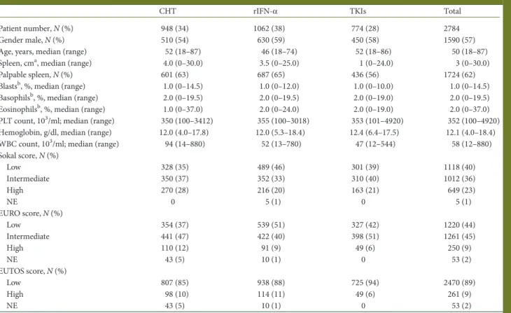
Table 1. Baseline demographic and hematologic characteristics, according to the frontline treatment
CHT rIFN-α TKIs Total
Patient number, N (%) 948 (34) 1062 (38) 774 (28) 2784
Gender male, N (%) 510 (54) 630 (59) 450 (58) 1590 (57)
Age, years, median (range) 52 (18–87) 46 (18–74) 52 (18–86) 50 (18–87)
Spleen, cma, median (range) 4.0 (0–30.0) 3.5 (0–25.0) 1 (0–24.0) 3 (0–30.0)
Palpable spleen, N (%) 601 (63) 687 (65) 436 (56) 1724 (62)
Blastsb, %, median (range) 1.0 (0–14.5) 1.0 (0–12.0) 1.0 (0–10.0) 1.0 (0–14.5) Basophilsb, %, median (range) 2.0 (0–19.5) 2.0 (0–19.5) 2.0 (0–19.0) 2.0 (0–19.5) Eosinophilsb, %, median (range) 1.0 (0–37.0) 2.0 (0–24.0) 2.0 (0–19.0) 2.0 (0–37.0) PLT count, 103/ml; median (range) 350 (100–3412) 355 (100–3018) 353 (101–4920) 352 (100–4920) Hemoglobin, g/dl, median (range) 12.0 (4.0–17.8) 12.0 (5.3–18.4) 12.4 (6.4–17.5) 12.1 (4.0–18.4) WBC count, 103/ml; median (range) 94 (14–880) 52 (13–780) 47 (12–544) 58 (12–880) Sokal score, N (%) Low 328 (35) 489 (46) 301 (39) 1118 (40) Intermediate 350 (37) 352 (33) 310 (40) 1012 (36) High 270 (28) 216 (20) 163 (21) 649 (23) NE 0 5 (1) 0 5 (1) EURO score, N (%) Low 354 (37) 539 (51) 327 (42) 1220 (44) Intermediate 441 (47) 422 (40) 398 (51) 1261 (45) High 110 (12) 91 (9) 49 (6) 250 (9) NE 43 (5) 10 (1) 0 53 (2) EUTOS score, N (%) Low 807 (85) 938 (88) 725 (94) 2470 (89) High 98 (10) 114 (11) 49 (6) 261 (9) NE 43 (5) 10 (1) 0 53 (2)
aMaximum distance below the costal margin, assessed by manual palpation. b
Percentage in peripheral blood.
CHT, chemotherapy; rIFN-α, recombinant interferon-α; TKIs, tyrosine kinase inhibitors; PLT, platelets; WBC, white blood cells; NE, not evaluable.
EURO risk category mainly because of young age. The more recent EUTOS scoring system, designed to predict the complete cytogenetic response (CCyR) probability at 18 months in TKIs-treated CML patients, did not account for age [8,9]. It is known since many years that, in children Ph+, BCR-ABL-positive, CML have more aggressive clinical and hematologic features, particularly huge splenomegaly, high white blood cell (WBC) count and higher blast cell percentage [10–13]. It is unclear if and how much these characteristics of aggressiveness are still present after childhood and, if so, how long. For this purpose, we reviewed all the GIMEMA CML databases, spanning over 40 years, to investigate if in adults the characteristics of the disease and the risk score distribution differ according to the age.
methods
The GIMEMA CML Working Party, previously Italian Cooperative Study Group on CML, promoted several multicentric prospective clinical studies since 1972 to today. A total number of 2784 adult,≥18 years old, chronic phase (CP) CML patients were enrolled: 948 (34%) treated frontline with chemotherapy between 1972 and 1988, 1062 (38%) treated frontline with rIFN-α-based regimes between 1986 and 2002, 774 (28%) treated frontline with TKIs, mainly imatinib and nilotinib, between 2003 and 2010. An allo-geneic stem-cell transplantation (alloSCT) was carried out in 83/948 patients
(9%) in the chemotherapy cohort, in 292/1062 patients (27%) in the rIFN-α cohort, and in 22/774 patients (3%) in the TKIs cohort, respectively.
The diagnosis of CML was based on the identification of the Ph chromo-some by chromochromo-some banding analysis of marrow cell metaphases, or on interphasefluorescence in situ hybridization in case of masked Ph, and after 2001 also on the demonstration of the BCR-ABL transcript by polymerase chain reaction, as recommended [5]. The European LeukemiaNet (ELN) cri-teria for distinguishing CP from accelerated and blastic phase (AP and BP) were used [4,5]. The hematologic, cytogenetic and molecular responses were defined as recently recommended by ELN [5]. Baseline data were obtained before any treatment. The spleen size was always reported as maximum dis-tance in centimeters below the costal margin, as assessed by manual palpa-tion. Risk scores were calculated according to the original reports [6–8]. All patients provided written informed consent before the enrollment. All studies were approved by the Institutional Review Board of all participating institutions and carried out in accordance with the Declaration of Helsinki, but only the most recent studies could be preregistered at NCT.
Baseline characteristics and hematological data were compared by theχ2 or the Kruskal–Wallis tests, as appropriate. The survival times were calcu-lated from the date of treatment start until death for any cause (OS) or leuke-mia-related death (leukeleuke-mia-related survival). The death was attributed to leukemia in all the patients who died after progression to AP or BP or with hematologic evidence of leukemia, who died after alloSCT, or who died due to unknown reasons. The cumulative incidence of progression was calculated from the date of start of treatment until the date of transformation to AP or BP. The patients who underwent alloSCT were not censored at transplant.
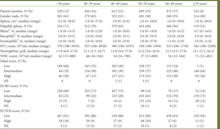
Table 2. Baseline demographic and hematologic characteristics, according to age, at diagnosis
<30 years 30–39 years 40–49 years 50–59 years 60–69 years ≥70 years Patient number, N (%) 329 (12) 444 (16) 613 (21) 693 (25) 473 (17) 232 (8)
Gender male, N (%) 201 (61) 279 (63) 335 (55) 401 (58) 260 (55) 114 (49)
Spleen, cma, median (range) 4.5 (0–28.0) 5.0 (0–27.0) 3.0 (0–25.0) 2.0 (0–28.0) 1.8 (0–30.0) 1.0 (0–20.0) Palpable spleen, N (%) 234 (71) 312 (70) 379 (62) 414 (60) 266 (56) 119 (51) Blastsb, %, median (range) 1.0 (0–14.5) 1.0 (0–12.0) 1.0 (0–10.0) 1.0 (0–14.0) 1.0 (0–14.5) 0.7 (0–14.0) Basophilsb, %, median (range) 2.0 (0–19.5) 2.0 (0–19.0) 2.0 (0–19.5) 2.0 (0–19.5) 2.0 (0–16.0) 2.0 (0–16.0) Eosinophilsb, %, median (range) 2.0 (0–16.0) 2.0 (0–10.0) 2.0 (0–23.0) 2.0 (0–37.0) 1.3 (0–12.0) 2.0 (0–11.0) PLT count, 103/ml; median (range) 370 (100–3018) 355 (100–4920) 380 (100–4553) 350 (100–3594) 333 (100–1750) 345 (100–2208) Hemoglobin, g/dl, median (range) 11.8 (6.0–17.0) 12.1 (5.3–16.7) 12.0 (4.0–17.8) 12.2 (5.6–18.4) 12.3 (4.5–17.0) 12.1 (5.3–16.2) WBC count, 103/ml; median (range) 62 (15–880) 66 (16–516) 54 (14–780) 57 (13–600) 54 (12–544) 71 (12–481) Sokal score, N (%) Low 199 (60) 243 (55) 302 (49) 258 (37) 115 (24) 1 (1) Intermediate 64 (19) 134 (30) 182 (30) 259 (37) 225 (48) 148 (64) High 66 (20) 67 (15) 127 (21) 173 (25) 133 (28) 83 (36) NE 0 0 2 (1) 3 (1) 0 0 EURO score, N (%) Low 228 (69) 325 (73) 457 (75) 99 (14) 79 (17) 32 (14) Intermediate 82 (25) 99 (22) 125 (20) 453 (65) 332 (70) 170 (73) High 15 (5) 7 (2) 14 (2) 131 (19) 54 (11) 29 (12) NE 4 (1) 13 (3) 17 (3) 10 (1) 8 (2) 1 (1) EUTOS score, N (%) Low 267 (81) 392 (88) 539 (88) 615 (89) 438 (93) 219 (94) High 58 (18) 39 (9) 57 (9) 68 (10) 27 (6) 12 (5) NE 4 (1) 13 (3) 17 (3) 10 (1) 8 (2) 1 (1)
aMaximum distance below the costal margin, assessed by manual palpation. b
Percentage in peripheral blood.
PLT, platelets; WBC, white blood cells; NE, not evaluable.
The survival probabilities and the cumulative incidences were estimated using the Kaplan–Meier method. Survival times and times to event were compared by the log-rank test.
results
baseline characteristics
The baseline demographic and hematologic characteristics are shown in Table1. The frequency of high-risk patients decreased over the years: from 28% to 21% by Sokal score; from 12% to 6% by EURO score and from 10% to 6% by EUTOS score.
The patient characteristics according to the age are shown in Table2. Only the spleen size showed an impressive variabil-ity, ranging from a median value of 4.5 cm in the youngest group (18–29 years old) to a median value of 1.0 cm in the oldest one (≥70 years old), with a progressive decrease, start-ing at the age of 40. As expected, the proportion of high Sokal and high EURO score patients increased with age, rising from 20% and 5%, respectively, in the youngest patients, to 36% and
12%, respectively, in the oldest patients. Conversely, the pro-portion of high EUTOS score patients was highest (18%) in the youngest age group (18–29 years), intermediate (9–10%) in patients 30–59 years old, and lowest (5–6%) in patients ≥60 years old. Based on the EUTOS score distribution, in all subse-quent calculations, the patients were divided into three age groups, 18–29 years old, 30–59 years old and ≥60 years old; high-risk patients, were 18%, 9% and 6%, respectively, P < 0.001. An analysis of baseline characteristics within the three age groups has been carried out (Table3 and supple-mentary Table S1, available at Annals of Oncology online). The main difference among the three age groups was the pro-portion of patients with a palpable spleen (71%, 63% and 55%, respectively, P < 0.001) and the spleen size (median, 4.5, 3.0 and 1.0 cm below costal margin respectively, P < 0.001). Moreover, the patients <30 years old were more frequently males, had a higher blast cell percentage in the peripheral blood, a lower hemoglobin concentration and a slightly higher platelet count (Table3).
Table 3. Comparison of baseline demographic and hematologic characteristics among young adults, adults and elderly (18–29 years, 30–59 years, ≥60 years)
<30 years years ≥60 years P
Patient number, N (%) 329 (12) 1750 (63) 705 (25)
-Gender male, N (%) 201 (61) 1015 (58) 374 (53) 0.024
Spleen, cma, median (range) 4.5 (0–28.0) 3.0 (0–28.0) 1.0 (0–30.0) <0.001
Palpable spleen, N (%) 234 (71) 1105 (63) 385 (55) <0.001
Blastsb, %, median (range) 1.0 (0–14.5) 1.0 (0–14.0) 1.0 (0–14.5) 0.003
Basophilsb, %, median (range) 2.0 (0–19.5) 2.0 (0–19.5) 2.0 (0–16.0) 0.485
Eosinophilsb, %, median (range) 2.0 (0–16.0) 2.0 (0–37.0) 2.0 (0–12.0) 0.410 PLT count, 103/ml, median (range) 370 (100–3018) 358 (100–4920) 336 (102–2208) 0.041 Hemoglobin, g/dl, median (range) 11.8 (6.0–17.0) 12.1 (4.0–18.4) 12.2 (4.5–17.0) 0.019
WBC count, 103/ml, median (range) 61 (15–880) 57 (13–780) 59 (12–544) 0.627
e13a2 transcript presentc, N (%)d 30 (54) 202 (44) 122 (47) 0.382
CCA Ph+, N (%)d 2 (4) 22 (5) 8 (3) 0.514 Variant translocation, N (%)d 0 27 (6) 21 (8) 0.071 Sokal score, N (%) Low 199 (60) 803 (46) 116 (16) <0.001 Intermediate 64 (19) 575 (33) 373 (53) High 66 (20) 367 (21) 216 (31) NE 0 5 (1) 0 EURO score, N (%) Low 228 (69) 881 (50) 111 (16) <0.001 Intermediate 82 (25) 677 (39) 502 (71) High 15 (5) 152 (9) 83 (12) NE 4 (1) 40 (2) 9 (1) EUTOS score, N (%) Low 267 (81) 1546 (88) 657 (93) <0.001 High 58 (18) 164 (9) 39 (6) NE 4 (1) 40 (2) 9 (1)
aMaximum distance below the costal margin, assessed by manual palpation. bPercentage in peripheral blood.
cPatients with e13a2 transcript or with both e13a2/e14a2 transcripts were included.
dAvailable data in 774 patients: age <30 years, 56 patients; age 30–59 years, 457 patients; age >60 years, 261 patients.
PLT, platelets; WBC, white blood cells; e13 a2 transcript, b2a2 BCR-ABL transcript; CCA Ph+, clonal chromosomal abnormalities in Philadelphia positive cells; NE, not evaluable.
response to therapy and outcome
The response to the therapy was fully evaluable only in the TKIs cohort. In this cohort, the cumulative incidence of CCyR by 5 years was lower in the young adult (YA) group (80%) than in the remaining two age groups (90% in both groups) (P = 0.041, YA compared with all others). Also the cumulative incidence of major molecular response (MMR) by 5 years was lower in the YA group (71%) than in the two other age groups (86% and 88%) (P = 0.004, YA compared with all others).
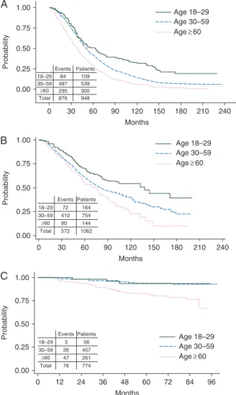
The OS according to the frontline treatment and by age group is shown in Figure1. In the chemotherapy and in the rIFN-α
cohorts, the OS was better in YA than in adult patients, and it
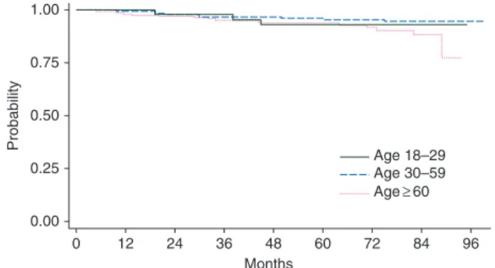
was better in adult than in elderly patients. In the TKIs cohort, the difference between YA and adults disappeared, and both groups had higher OS than the elderly group (P < 0.001). In the chemotherapy and in the rIFN-α cohorts, the leukemia-related deaths were 97% and 96%, respectively. In the TKIs cohort, all deaths in the YA group were leukemia-related, while the leuke-mia-related deaths occurring in the adult and elderly groups were 69% and 40%, respectively (Table4). The 5-year estimated probability of progression to AP and BP was 16% in the YA group, 5% in the adult group and 7% in the elderly group (P = 0.011) (Figure2). Therefore, the leukemia-related survival was much less affected by age: 93% in the YA group, 95% in the adult group and 89% in the elderly group, P = 0.079 (Figure3).
discussion
This study of 2784 Ph+ CML patients in early CP, enrolled in mul-ticentric studies over a long period of time, shows that the clinical
0 30 60 90 120 150 180 210 240 Months Probability 0.00 0.25 0.50 0.75 1.00 Age 18–29 Age 30–59 Age≥ 60 18–29 30–59 ≥60 Total 84 497 295 876 109 539 300 948 Events Patients 0 30 60 90 120 150 180 210 240 Months Probability 0.00 0.25 0.50 0.75 1.00 18–29 30–59 ≥60 Total 72 410 90 572 164 754 144 1062 Events Patients Age 18–29 Age 30–59 Age≥ 60 0 12 24 36 48 60 72 84 96 Months Probability 0.00 0.25 0.50 0.75 1.00 18–29 30–59 ≥60 Total 3 26 47 76 56 457 261 774 Events Patients Age 18–29 Age 30–59 Age≥ 60 C B A
Figure 1. Overall survival (OS) by age groups (<30, 30–50, >60 years) in the chemotherapy cohort (A), the rIFN-α cohort (B) and the TKIs cohort (C). In the chemotherapy cohort, the 15-year survival was 20% [95% confi-dence interval (CI) 12% to 28%] in young adults, 7% (95% CI 5% to 9%) in adults and 2% (95% CI 0 to 4%) in the elderly (P < 0.001). In the rIFN-α cohort, the 15-year survival was 39% in young adults (95% CI 25% to 53%), 22% in adults (95% CI 16% to 30%) and 10% (95% CI 3% to 24%) in the elderly (P < 0.001). In the TKIs cohort, the 8-year survival was 93% (95% CI 80% to 98%) in young adults, 93% (95% CI 90% to 95%) in adults, and 77% (95% CI 69% to 83%) in the elderly (P < 0.001). These survival calculations were made counting all deaths, for any reason and without any censoring.
Table 4. TKIs cohort Age group Patients,
N Progressions, N (%) Deaths, N (%) Leukemia-related Leukemia-unrelated Total <30 years 56 8 (14) 3 (5) 0 3 (5) 30–59 years 457 21 (5) 18 (4) 8 (2) 26 (6) >60 years 261 14 (5) 19a(7) 28 (11) 47 (18) Total 774 43 (6) 40 (5) 36 (5) 76 (10) Progression to AP and BP, as defined by ELN criteria (6), and death, by age group. All leukemia-related deaths occurred after transformation to AP or BP, with the exception of five elderly patients who did not die in remission, but did notfit the criteria of AP or BP.
a
Five patients died because of leukemia withoutfitting the criteria of AP and BP.
TKIs, tyrosine kinase inhibitors; AP, accelerated phase; BP, blastic phase; ELN, European LeukemiaNet.
0 12 24 36 48 60 72 84 96 Months Probability 0.00 0.25 0.50 0.75 1.00 Age 18–29 Age 30–59 Age≥ 60
Figure 2. Estimated cumulative probability of progression to AP and BP by age groups (<30, 30–50, >60 years) in the TKIs cohort. At 8 years, it was 16% (95% CI 8% to 31%) in young adults, 5% (95% CI 3% to 8%) in adults and 7% (95% CI 4% to 11%) in the elderly (P = 0.011).
presentation of CML varies consistently and unidirectionally with age, particularly because of differences in spleen enlargement, leading to a relative excess of high-risk patients among YA. It was already well known that the disease is more aggressive in children [10–13]. A recent study on 150 patients younger than 18 years old reported that the spleen was palpable in 78% of children, with a median value of 8 cm below costal margin [13]. For children, a high WBC, a high platelet count, and a high blast cell percentage at the diagnosis have been reported [10–13]. Also a multicentric study, including 120 patients 16–29 years old, reported that, in adolescents and YA, the median value of spleen below costal margin was 5versus 0 cm in the elderly group [14].
The splenomegaly is almost universally attributed to the ex-pansion of myelopoiesis in the spleen. A classic textbook says: ‘In chronic phase the spleen is enlarged due to infiltration of the red pulp cords by granulocytes in different maturation stages…’ [15]. Spleen is a hematopoietic organ in the fetus. In children, adolescents and YA, the spleen niche may offer a more favorable environment to Ph+ cells, than in older patients; alternatively, it is possible that in younger patients Ph+ stem or progenitor cells could be qualitatively different. There are few, not recent, studies reporting on differences between marrow and spleen hemato-poiesis in CML [16–18], but there are not studies investigating the molecular profile of Ph+ cells in different age groups. The spleen enlargement could be not only caused by myeloid meta-plasia, but it could also be an expression of host reaction. In a normal spleen, the lymphoid tissue account for about 65% of the whole organ [15]. In CML patients, the exact proportion of myeloid and lymphoid cells in the spleen is not known, apart from an old report regarding 12 CML patients splenectomized at the onset of the disease [16]: based on cytologic evaluation of spleen touches (smears), the median lymphocyte proportion was 40% of all nucleated cells, suggesting that spleen enlargement was due at least in part also to lymphocytes. This may represent an inflammatory reaction, reflecting an age-related difference of the immune-inflammatory response of the host.
The splenomegaly has been always recognized as a negative prognostic factor [6–8,15]. Also in our series of 2784 patients, enrolled over a long period time, a multivariate analysis confirmed that spleen size was a significant and independent predictor of survival, regardless of age (data not shown). In the pre-TKIs era, the great majority of deaths were preceded by
progression, so that they were attributed to leukemia. In the TKIs era, the deaths not related to leukemia are also uncommon in younger patients, but account for at least 50% of all deaths in the elderly. The concept of leukemia-related survival raises obvious difficulties, suspicions and resistances, because it implies a shared definition, a detailed knowledge of the course of the disease and of the death, and requires other specific bio-statistical methods of analysis [19,20]. However, in the current TKIs era, the problem can no longer be ignored. It is not easy to detect differences in response and outcome according to age, since treatment is so effective and the number of YA patients is small; 12% in our multicentric series of 2784 patients and 8%, 11%, 13% and 8%, respectively, in other reports, where adoles-cents and YA were frequently grouped in the same age class [14,
21–23]. A difference in the probability of achieving a CCyR and an MMR, according to age, was found in our series and in an in-dependent single-center study [23], but not in another multi-centric study on patients from 16 to 29 years old [14]. In all the studies, the OS and the progression-free survival were not affected by age. The more aggressive disease characteristics in YA may explain the lower response rates and the relatively higher probability early disease progression. The impact of a poorer adherence, if any, should be clarified.
In conclusion, we extended to YA the observations already made in children. The most relevant difference was in the spleen size, and a continuous decrease of spleen size by age has been observed. It is acknowledged that the age is a continuous vari-able, and that cutoffs are artificial. Moreover, selection biases could not be excluded, because patients enrolled in clinical trials may not reflect the characteristics of the whole CML population. We repeated all the calculations reported in this paper, also con-sidering different cutoffs, particularly 18–39, 40–59, 60–69 and ≥70 years old: the results did not change. In the current TKI era, multiple treatment choices are available, the differences are small, and the treatment goals may be different according to the age of CML patients [5,24]. Our data suggest that investigating the differences according to age may be important for under-standing the biology of the disease and the response of the host, in order to design age-adapted strategies. A careful monitoring of YA patients should be carried out. It is likely that in YA an extended use of second-generation TKIs may improve the outcome.
acknowledgements
The following members of the ‘GIMEMA Working Party on CML’, formerly ‘ICSG on CML’ actively participated in this study, enrolling the study patients and collecting clinical data:
F. Salvi, M. Pini (Hematology Unit, Ospedale Civile, Alessandria); P. Leoni, S. Rupoli (Hematology Unit, Ospedale Regionale di Torrette, Ancona); P. Galieni, C. Bigazzi (Hematology Unit, Presidio Ospedaliero ‘C. e G. Mazzoni’, Ascoli Piceno); N. Cantore, F. Palmieri (Hematology Unit, Ospedale Civile ‘San Giuseppe Moscati’, Avellino); F. Albano, A. Russo Rossi (Chair of Hematology, University of Bari, Bari); A. Rambaldi, T. Intermesoli (Hematology Unit, Ospedali Riuniti, Bergamo); F. Palandri, N. Testoni, S. Luatti, S. Soverini, I. Iacobucci, M. T. Bochicchio, M. Apolinari, M. Fogli,
0 12 24 36 48 60 72 84 96 Months Probability 0.00 0.25 0.50 0.75 1.00 Age 18–29 Age 30–59 Age≥ 60
Figure 3. Estimated probability of CML-related survival by age groups in the TKIs cohort. The 8-year survival was 93% (95% CI 80% to 98%) in young adults, 95% (95% CI 92% to 97%) in adults and 89% (95% CI 81% to 93%) in the elderly (P = 0.079).
I. Cervello (Institute of Hematology‘Seràgnoli’, Department of Experimental, Diagnostic and Specialty Medicine, University of Bologna, Bologna); A. Capucci (Hematology Unit, Azienda Ospedaliera ‘Spedali Civili’, Brescia); M. Malagola (Blood Diseases and Stem Cell Transplantation Unit, Azienda Ospedaliera ‘Spedali Civili’, Brescia); A. Malpignano, M. Girasoli (Hematology Unit, Ospedale ‘Perrino’, Brindisi); E. Angelucci, E. Usala (Hematology Unit, Ospedale Oncologico ‘A. Businco’, Cagliari); S. Storti (Hematology and Oncology Unit, Università Cattolica del Sacro Cuore‘S.S. Giovanni Paolo II’, Campobasso); E. De Biasi (Hematology Unit, Presidio Ospedaliero Camposampiero, Camposampiero, PD); G. Tagariello, R. Sartori (Hematology Unit, ‘San Giacomo’ Hospital, Castelfranco Veneto); F. Di Raimondo, P. Vigneri (Hematology Unit, ‘Ferrarotto’ Hospital, Catania); S. Impera (Hematology Unit, Policlinico Garibaldi-Nesima, Catania); S. Molica (Hematology Unit, ‘Pugliese’ Hospital, Catanzaro); F. Lanza, C. Viganò (Hematology and Stem Cell Transplantation Unit, Istituti Ospedalieri, Cremona); M. Grasso, D. Rapezzi (Hematology Unit, ‘Santa Croce’ Hospital, Cuneo); F. Cavazzini (Chair of Hematology, University of Ferrara, Ferrara); A. Bosi, V. Santini (Chair of Hematology, University of Firenze, Firenze); S. F. Capalbo, G. Spinosa (Hematology Unit, Ospedali Riuniti, Foggia); I. Pierri, M. Bergamaschi (Chair of Hematology, IRCCS San Martino, Genova); A. M. Carella (Hematology Unit, IRCCS San Martino, Genova); A. Bacigalupo (Hematology and Stem Cell Transplantation Unit, IRCCS San Martino, Genova); A. De Blasio, F. Ciccone (Hematology Unit, Ospedale Civile, Latina); N. Di Renzo (Hematology Unit,‘Vito Fazzi’ Hospital, Lecce); C. Musolino, S. Russo (Chair of Hematology, University of Messina, Messina); A. Cortelezzi (Oncohematology Division, IRCCS Ca’ Granda—Maggiore University Hospital, Milano); E. Morra, E. M. Pungolino (Hematology Unit, Azienda Ospedaliera ‘Niguarda-Cà Granda’, Milano); M. Luppi, R. Marasca (Chair of Hematology, University of Modena and Reggio Emilia, Modena); E. M. Pogliani, C. Gambacorti-Passerini (Chair of Hematology, ‘San Gerardo’ Hospital, Monza); L. Luciano (Department of Biochemistry and Medical Biotechnologies, ‘Federico II’ University, Napoli); F. Ferrara, M. Annunziata (Hematology and Bone Marrow Transplantation Unit, ‘Cardarelli’ Hospital, Napoli); G. Latte, D. Noli (Hematology Unit,‘San Francesco’ Hospital, Nuoro); G. Rege-Cambrin, C. Fava (Department of Clinical and Biological Sciences,‘San Luigi Gonzaga’ University Hospital, Orbassano, TO); G. Semenzato, G. Binotto (Department of Internal Medicine, University of Padova, Padova); F. Fabbiano, D. Turri (Hematology Unit,‘V. Cervello’ Hospital, Palermo); S. Siragusa, C. Caracciolo (Chair of Hematology, University of Palermo, Palermo); M. Musso, F. Porretto (Oncology and Bone Marrow Transplantation Unit, ‘La Maddalena’ Hospital, Palermo); F. Aversa, M. Crugnola (Chair of Hematology, University of Parma, Parma); M. Cazzola, E. Orlandi (Hematology Unit, ‘S. Matteo’ University Hospital, Pavia); B. Falini, F. Falzetti (Sezione di Ematologia ed Immunologia Clinica, Policlinico ‘Monteluce’, Perugia); G. Visani, A. Isidori (Hematology Unit, ‘San Salvatore’ Hospital, Pesaro); G. Fioritoni, R. Di Lorenzo (Hematology Unit, Ospedale Civile dello Spirito Santo, Pescara); D. Vallisa, E. Trabacchi (Hematology Unit,‘Guglielmo
da Saliceto’ Hospital, Piacenza); M. Petrini, S. Galimberti (Hematology Unit, Azienda Ospedaliero-Universitaria Pisana, Pisa); M. Pizzuti (Hematology Unit, ‘San Carlo’ Hospital, Potenza); A. Zaccaria, M. Salvucci (Hematology Unit, ‘Santa Maria delle Croci’ Hospital, Ravenna); F. Ronco, D. Ielo (Hematology Unit, Ospedali Riuniti, Reggio Calabria); F. Merli, P. Avanzini (Hematology Unit, Arcispedale Santa Maria Nuova, Reggio Emilia); P. Tosi, A. Merli (Hematology Unit, Ospedale Infermi Azienda Unità Sanitaria, Rimini); P. Musto (Department of Onco-Hematology, IRCCS Centro di Riferimento Oncologico della Basilicata, Rionero in Vulture, PZ); V. De Stefano, S. Sica (Istituto di Semeotica Medica, Università Cattolica, Roma); R. Latagliata (Chair of Hematology, ‘La Sapienza’ University, Roma); P. De Fabritiis, M. Trawiska (Department of Hematology, ‘Tor Vergata’ University, Roma); I. Majolino, L. Pacilli (Hematology and Stem Cell Transplantation Unit, Azienda Ospedaliera San Camillo Forlanini, Roma); B. Ronci, M. Cedrone (Hematology Unit, Ente Ospedaliero San Giovanni Addolorata, Roma); M. C. Petti, F. Pisani (Hematology Unit, Istituto Regina Elena, Roma); A. Tafuri, E. Montefusco (Hematology Unit, ‘Sant’Andrea’ Hospital, Roma); F. Iuliano (Hematology Unit, Presidio Ospedaliero ‘N. Giannettasio’, Rossano Calabro); F. Dore, S. Pardini (Institute of Hematology, University of Sassari, Sassari); M. Bocchia, M. Defina (Chair of Hematology, University of Siena, Siena); A. M. Liberati, D. Luzzi (Hematology and Oncology Unit, Azienda Ospedaliera ‘S. Maria’, Terni); M. Boccadoro, D. Ferrero (Section of Hematology, Department of Molecular Biotechnology and Health Sciences, University of Torino, Torino); U. Vitolo (Hematology Unit, Azienda Ospedaliero-Universitaria Città della Salute e della Scienza, University of Torino, Torino); F. Gherlinzoni, E. Calistri (Hematology Unit, ‘Ca’ Foncello’ Hospital, Treviso); R. Fanin (Chair of Hematology, University of Udine, Udine); G. Pizzolo, V. Meneghini (Chair of Hematology, University of Verona, Verona); F. Rodighiero, A. D’Emilio (Hematology Unit, Ospedale Civile, Vicenza).
funding
This study has been supported by: BolognAIL, GIMEMA Onlus, European LeukemiaNet (LSHC-CT-2004–503216).
disclosure
FC has acted as a consultant for Novartis, Bristol-Myers Squibb and Pfizer and received honoraria from Novartis and Bristol-Myers Squibb; GG has acted as a consultant and received honor-aria from Novartis and Bristol-Myers Squibb; MB has acted as a consultant for Bristol-Myers Squibb and Novartis; EA has acted as a consultant for Novartis and Bristol-Myers Squibb; MB received research funding from Novartis and received honoraria from ARIAD Pharmaceuticals and Pfizer DR received research funding from Celgene and Gilead, has acted as a paid expert testimony for Novartis and served on the speakers’ bureaus of Novartis; GM served on the speakers’ bureaus of Novartis, Bristol-Myers Squibb and Pfizer. GR has acted as a consultant for Novartis, Bristol-Myers Squibb and ARIAD Pharmaceuticals and served on the speakers’ bureaus of Novartis, Bristol-Myers Squibb and Roche;
FP received research support from Novartis, served as advisor for Novartis, Bristol-Myers Squibb and ARIAD Pharmaceuticals and received lecture fees from Novartis and Bristol-Myers Squibb; GS has acted as a consultant for and received honoraria from Bristol-Myers Squibb, Novartis, ARIAD Pharmaceuticals and Celgene; MB received honoraria from Novartis, Bristol-Myers Squibb, Pfizer and ARIAD Pharmaceuticals and served on the speakers’ bureaus of Novartis and Bristol-Myers Squibb; GR has acted as a consultant for Novartis, Bristol-Myers Squibb and ARIAD Pharmaceuticals and served on the speakers’ bureaus of Novartis, Bristol-Myers Squibb and Roche.
references
1. Hehlmann R, Hochhaus A, Baccarani M. Chronic myeloid leukemia. Lancet 2007; 370: 342–350.
2. Kantarjian H, O’Brien S, Jabbour E et al. Improved survival in chronic myeloid leukemia since the introduction of imatinib therapy: a single-institution historical experience. Blood 2012; 119: 1981–1987.
3. Baccarani M, Saglio G, Goldman J et al. Evolving concepts in the management of chronic myeloid leukemia: recommendations from an expert panel on behalf of the European LeukemiaNet. Blood 2006; 108: 1809–1820.
4. Baccarani M, Cortes J, Pane F et al. Chronic myeloid leukemia: an update of concepts and management recommendations of European LeukemiaNet. J Clin Oncol 2009; 27: 6041–6051.
5. Baccarani M, Deininger MW, Rosti G et al. European LeukemiaNet recommendations for the management of chronic myeloid leukemia: 2013. Blood 2013; 122: 872–884.
6. Sokal JE, Cox EB, Baccarani M et al. Prognostic discrimination in“good-risk” chronic granulocytic leukemia. Blood 1984; 63: 789–799.
7. Hasford J, Pfirmann M, Hehlmann R et al. A new prognostic score for survival of patients with chronic myeloid leukemia treated with interferon alfa. J Natl Cancer Inst 1998; 90: 850–858.
8. Hasford J, Baccarani M, Hoffmann V et al. Predicting complete cytogenetic response and subsequent progression-free survival in 2060 patients with CML on imatinib treatment: the EUTOS score. Blood 2011; 118: 686–692.
9. Gugliotta G, Castagnetti F, Palandri F et al. Frontline imatinib treatment of chronic myeloid leukemia: no impact of age on outcome, a survey by the GIMEMA CML working party. Blood 2011; 117: 5591–5599.
10. Millot F, Traore P, Guilhot J et al. Clinical and biological features at diagnosis in 40 children with chronic myeloid leukemia. Pediatrics 2005; 116: 140–143. 11. Hasan SK, Sazawal S, Kumar B et al. Childhood CML in India: b2a2 transcript is
more common than b3a2. Cancer Gen Cytogen 2006; 169: 76–77.
12. Andolina JR, Neudorf SM, Corey SJ. How I treat childhood CML. Blood 2012; 119: 1821–1830.
13. Millot F, Suttorp M, Guilhot J et al. The international registry for chronic myeloid leukemia in children and adolescents (I-CML-Ped-Study): objectives and preliminary results. Blood 2012; 120: abstract 3741.
14. Kalmanti L, Saussele S, Lauseker M et al. Younger patients with chronic myeloid leukemia do well in spite of poor prognostic indicators: results from the randomized CML study IV. Ann Hematol 2014; 93: 71–80.
15. Chapman WC, Newman M. Disorders of the spleen. In: Wintrobe’s, Clinical Hematology, 10th edition. Philadelphia, PA: Williams & Wilkins 1999; 1969–1989.
16. Baccarani M, Zaccaria A, Santucci MA et al. A simultaneous study of bone marrow, spleen and liver in chronic myeloid leukemia. Evidence for differences in cell composition and karyotypes. Ser Haemat 1975; 8: 81–112.
17. Sjogren U, Brandt L. Different composition and mitotic activity of the hemopoietic tissue in bone marrow, spleen, and liver, in chronic myeloid leukaemia. Acta Haematol 1976; 55: 73–80.
18. Muller-Bérat CN, Wantzin GL, Philip P et al. Agar culture studies of bone marrow, spleen and liver in chronic myeloid leukemia. Leukemia Res 1977; 1: 123–131.
19. Pfirrmann M, Hochhaus A, Lauseker M et al. Recommendations to meet statistical challenges arising from endpoints beyond overall survival in clinical trials of chronic myeloid leukemia. Leukemia 2011; 25: 1433–1438.
20. Guilhot J, Baccarani M, Clark RE et al. Definitions, methodological and statistical issues for phase 3 clinical trials in chronic myeloid leukemia: a proposal by the European LeukemiaNet. Blood 2012; 119: 5963–5971.
21. Medical Research Council’s Working Party for Therapeutic Trials in Leukaemia. Chronic granulocytic leukaemia: comparison of radiotherapy and busulfan therapy. Brit Med J 1968; 1: 201–207.
22. Monfardini S, Gee T, Fried J, Clarkson B. Survival in chronic myelogenous leukemia: influence of treatment and extent of disease at diagnosis. Cancer 1973; 31: 492–501.
23. Pemmaraju N, Kantarjian H, Shan J et al. Analysis of outcomes in adolescents and young adults with chronic myelogenous leukemia treated with upfront tyrosine kinase inhibitor therapy. Haematologica 2012; 97: 1029–1035.
24. O’Brien S, Radich JP, Abboud CN et al. Chronic Myelogenous Leukemia, Version 1.2014. J Natl Compr Canc Netw 2013; 11: 1327–1340.