UNIVERSITÀ
DEGLI
STUDI
DEL
MOLISE
Department of Agricultural, Environmental and Food Sciences
International PhD Course in:
A
GRICULTURE
T
ECHNOLOGY AND
B
IOTECHNOLOGY
CURRICULUM:WELFARE, BIOTECHNOLOGY AND QUALITY OF ANIMAL PRODUCTION
(C
YCLEXXXI)
Related disciplinary scientific section: AGR/19 (Zootecnia Speciale)
PhD thesis
STUDY
OF
THE
CHICKEN
PHYSIOLOGICAL
PROFILE
IN
DIFFERENT
GUT
SITES
AND
UPON
DIFFERENT
ENVIRONMENTAL
FACTORS
Coordinator of the PhD Course: Prof. Giuseppe Maiorano
Supervisor: Prof. Giuseppe Maiorano
Co-Supervisor: Prof. Paolo Trevisi
PhD student: Micol Bertocchi
155869
I want to thank my family, my friends, my supervisors and my colleagues in having attended me during this important experience
Abstract
Physiological aspects of chicken intestinal tracts are key targets of study due to their important repercussion on productive yield and animal health. Characterization of chicken gut, including spatial variations in microbial community, gut-associated immunity and relation with diseases has highlighted the importance of gut homeostasis and its sensitivity to external factors like stressors or dietary strategies. In this context, a survey on molecular mechanisms and differential gene expression along gut sites may deepen knowledge on functional aspects and provide hints for actions for maintaining gut homeostasis and testing various experimental factors.
This thesis aimed to analyze at molecular level chicken gut physiological profile and possible functional differences between tracts, to highlight potential biomarkers linked to factors influencing gut status. In this view, microarray analysis has been chosen to realize a wide exploration of chicken gut in its gene landscape.
Trial 1: in an exploratory perspective, aim of the study was to evidence the differential tissue gene expression of jejunum and of cecum of chickens at 42 days of age. Genes and enriched gene clusters for biological functions were differentially represented in the two tissues. In jejunum, expected genes and functions related to nutrient metabolism emerged, with interesting correlations to mammals and new key aspects like sulfur transport-related genes and immune pathways tuning. In cecum, gene clusters emerged, as already seen in humans, such as cell turnover related-genes, and genes linked to sulfation for gut barrier maintenance. Results indicated the different functions of the two tissues and revealed key aspects for possible new in-depth investigations in chickens.
Trial 2: to apply former trial hints to possible in-field conditions affecting poultry gut and health, the study aimed to investigate effects of a GOS prebiotic in ovo injected in counteract the detrimental effect of the heat-stress in growing chickens. Energetic metabolism-related gene sets were enriched in GOS, mainly in jejunum, and lipid metabolism-related gene sets in GOS might have contributed in gut barrier maintenance with less immune system activation, mainly in cecum. No differences in blood parameters were seen. Only few butyrate-related bacteria were increased by GOS, while heat stress confirmed its effect on microbial imbalance. Without considering thermal treatment, GOS showed to induce a long-term effect on transcriptomic profile of jejunum and cecum. Instead, GOS had no additional efficacy in counteracting heat stress compared to control group.
In conclusion, the microarray utilization was confirmed to differentially highlight functional characteristics between gut tracts in chickens, with possibility to identify useful gene biomarkers. Different functions along gut tracts were highlighted also in the second study with the in ovo injection of GOS, with positive effects on gut transcriptome. When applied to the experimental model of heat stress, chosen as stress model due to its important role in production diseases, especially in the
Mediterranean area, the intestine seems to response in a homogeneous way along the different tracts, without considering the in ovo treatment. The concomitant analysis of microbiota in cecum offered the possibility to correlate changes in microbial community with functional genes, to identify new potential biomarkers of gut homeostasis in different conditions.
Abstract
La caratterizzazione fisiologica dei diversi tratti intestinali del pollo è un importante oggetto di studio viste le sue ripercussioni sulle prestazioni produttive e sulla salute degli animali. Tale caratterizzazione, dalla variazione nella composizione microbica lungo i tratti intestinali, alla risposta immunitaria fino al coinvolgimento dell’intestino in diverse patologie, ha evidenziato l’importanza dell’omeostasi intestinale in relazione a fattori esterni, quali stress o strategie nutrizionali. In tale contesto, un’analisi sui meccanismi molecolari e di espressione genica nei diversi tratti intestinali potrebbe rivelare nuovi aspetti funzionali utili al mantenimento dell’omeostasi e per testare diversi fattori sperimentali.
Questa tesi ha analizzato a livello molecolare il profilo fisiologico intestinale nel pollo considerando le possibili differenze tra i tratti e con lo scopo di evidenziare possibili marcatori collegati a fattori che possono influenzare lo stato dell’intestino. A tal fine, l’analisi tramite microarray è stata scelta per effettuare un’approfondita esplorazione a livello genico nel pollo.
Studio 1: a scopo esplorativo, si è cercato di evidenziare possibili differenze a livello di espressione genica e relativa funzione biologica tra digiuno e cieco in polli di 42 giorni di età. Nel digiuno si sono evidenziati dei gene set arricchiti nell’espressione di geni relativi al metabolismo dei nutrienti, riscontrati anche i mammiferi, così come relativi all’assorbimento dello zolfo e all’attivazione del sistema immunitario. Nel cieco sono emersi alcuni gene set riscontrati anche nell’uomo, inclusi geni relativi al turnover cellulare e all’utilizzo dello zolfo nel mantenimento della barriera intestinale. Le differenziazioni emerse fra i due tessuti possono rappresentare punti di partenza e spunti per ulteriori studi nel pollo.
Studio 2: volendo applicare le potenzialità del primo studio, in questa prova si è analizzato l’effetto di un prebiotico (galacto-oligosaccaride, GOS), iniettato in ovo, in polli successivamente allevati in condizioni da stress da caldo, uno dei maggiori problemi nell’area mediterranea. L’iniezione in ovo ha avuto un effetto a lungo termine del GOS sul trascrittoma intestinale, evidenziando gene set arricchiti relativi al metabolismo energetico soprattutto nel digiuno, così come gene set relativi al metabolismo lipidico. Questi ultimi, insieme con gene set impoveriti relativi all’attivazione immunitaria, soprattutto nel cieco, potrebbero aver contribuito al mantenimento della barriera intestinale, anche senza evidenti differenze nei parametri ematici di infiammazione. Il prebiotico, nel cieco, sembra aver stimolato alcuni batteri coinvolti nel metabolismo dell’acido butirrico. Il prebiotico non è stato efficace nel contrastare lo stress da caldo.
In conclusione, il microarray ha evidenziato differenze funzionali tra i tratti intestinali nel pollo, sottolineandole anche nella prova di somministrazione in ovo del GOS, i cui effetti hanno influenzato positivamente lo stato intestinale in termini di trascrittoma. Lo stress da caldo in generale sembra aver
avuto un effetto costante rispetto ai parametri considerati, a prescindere dal trattamento in ovo, se si esclude il dato relativo al microbiota. La concomitante analisi di questo nel cieco ha permesso di formulare ipotesi su possibili correlazioni geniche funzionali, con la possibilità di identificare potenziali marcatori dello stato intestinale in diverse condizioni.
INDEX
Introduction
1. Poultry industry and production ... 1
1.1 Fast growing selection-and related myopathies ... 2
2. Gastro-intestinal tracts ... 5
2.1 Digestive tract characteristics ... 5
2.1.1 Post-hatching digestive tract development ... 7
2.1.2 Intestinal tract of fast-growing hybrids ... 8
2.2 Intestinal microbial community ... 10
2.2.1 Spatial bacterial composition along the gastro-intestinal tract of adult chickens ... 10
2.2.2 Factors affecting gut microbiota ... 15
2.2.3 Temporal microbial community development in growing chickens ... 16
2.2.4 Functions of gut microbial population ... 18
2.3 Gut homeostasis and determinant variables ... 19
2.3.1 Gut barrier structure ... 19
2.3.2 Relationship between host immune system and bacteria in gut health ... 23
3. Feeding strategies to improve poultry health ... 26
3.1 Feed additives to prevent the use of antibiotics ... 27
3.1.1 Exogenous enzymes ... 27 3.1.2 Organic acids ... 28 3.1.3 Phytogenics ... 29 3.1.4 Amino acids ... 30 3.1.5 Probiotics ... 31 3.1.6 Prebiotics ... 34 3.1.7 Synbiotics ... 37
3.2 Early nutritional supplementations ... 38
3.2.1 Feeding of the hens ... 38
3.2.2 Post-hatch early feeding ... 40
3.2.3 In ovo feeding ... 42
4. Animal health: stress and disease susceptibility ... 49
4.1 Cardiopulmonary disease ... 49
4.2 Stress susceptibility and enteric diseases ... 49
4.3 Impact of heat stress on poultry production ... 53
4.3.2 Heat stress on chickens health ... 55
4.4 Feeding strategies to counteract heat stress ... 58
5. Molecular markers related to gut health: perspectives and application ... 60
Objectives of the Thesis
... 62Study 1:
Exploring differential transcriptome between jejunum and cecum
tissue of broiler
... 63Aim of the study ... 64
Materials and Methods ... 65
Results ... 66
Discussion ... 68
Conclusions... 72
Tables and Figures ... 74
Study 2:
In ovo injection of a galacto-oligosaccharide prebiotic in broiler
chickens submitted to heat-stress: impact on intestinal microbiota,
transcriptomic profile and plasma immune parameters
... 82Aim of the study ... 83
Materials and Methods ... 84
Results ... 87
Discussion ... 90
Conclusions... 99
Tables and Figures ... 101
General Discussion
... 121General Conclusion
... 124Study 1 – Supplementary Tables
... 1481
Introduction
1. Poultry industry and production
In the last decades poultry industry has faced an incredible growth thanks to the industry advances in genetics, feeding and disease control, so much so that poultry products (meat and eggs) have become pivotal in human nutrition as one of the most important sources for protein of animal origin worldwide. The economic interest of poultry industry experienced several changes along about the latter sixty years. The most important advantage for poultry industries was the progress on the genetic selection that allow in producing animals that yielded products of highest value, increases the body weight (BW), feed efficiency and carcass composition. The genetic selection led to a production cost lowering and allowed to obtain chicken products, defined as some of the least expensive protein sources in the world (Hammerstedt, 1999; Petracci and Cavani, 2012). From the initial demand for whole carcasses, about forty years ago market demand started moving towards cut-up portions, leading to a bird selection more focused on the carcass part yields (breast and legs). After, in the last two decades, market demand shifted more towards processed products and breast meat with a consequent adaptation by the poultry industry, which moved to a selection of heavier animals with high breast development and weight (Petracci et al., 2015). The nutritional profile (such as the high protein and low-fat content), sensory properties and flexibility for processing, combined with competitive prices compared to other meats have constantly increased the market demand from the middle of the last century until today. Poultry companies faced this increment increasing both the number of reared animals as well as its growth rate (GR) and feed efficiency. Together with the genetic enhancement, the efficiency of the vertical integration of the poultry supply chain has contributed in the modern poultry industry development. As further consequence of the growing poultry products consumption, and also considering the concept of poultry meat as “functional food” (due to its content in conjugated linoleic acid, polyunsaturated fatty acids, vitamins and antioxidants), higher standards of quality to improve meat sensory characteristics and functional properties are required (Havenstein et al., 2003a; b; Petracci and Cavani, 2012; Petracci et al., 2013a). Currently, the genetic progress has brought to have birds weighting around 3 kg of BW that can grow and become ready for the market in about one-half of the time compared to sixty years ago and having a breast yield of 3-4% higher than ten years ago (Havenstein et al., 2003a; b; Aviagen, 2014, 2017) (Table 1). In fact, most of the broiler selection has been directed towards the muscle mass increase, especially focused on breast muscle size, mainly due to the hypertrophy in muscle fibers that has been connected to the less blood supply and possible oxygen deficiency observed in new broiler lines.
2 Insufficient oxygen supply can also cause intestinal metabolic disorders, such as malabsorption syndrome, as well as higher susceptibility to infections (Scheele, 1997). Furthermore, a concomitant decrease in heart muscle size has been also observed: this condition obviously explains the less cardiac capacity observed in modern broiler lines and has been connected to the cardiovascular problems and relative disease susceptibility (Schmidt et al., 2009; Petracci et al., 2015). These problems are highly accentuated in broilers than in laying hen because of different genetic selection that, in meat-type chicken, has been addressed toward a higher feed consumption and very rapid development of muscle. As an example, laying hen (selected mostly for egg production) stop eating when their metabolism needs are met, while broilers (selected for meat production) continue in eating until their gut is not completely full. (Buzala and Janicki, 2016).
Table 1- Differences in body weight (BW) and breast yield (% of BW) in male chicken hybrids from 1957 up to 2017
Year Hybrid Age (d) Body Weight (g) Breast Yield (% of BW)
19571,2 Athens Canadian randombred 42-43 591 11.5
20011,2 Ross 308 42-43 2903 19.5
20143 Ross 308 42 3023 22.5
20174 Ross 308 42 3103 23.1
1Havenstein et al. (2003a); 2Havenstein et al. (2003b); 3Aviagen (2014); 4Aviagen (2017)
1.1 Fast growing selection and related myopathies
Together with the strictly targeted selection for faster growing and heavier birds, it has been observed an increase in pectoralis major muscle abnormalities (i.e. deep pectoral disease, principal growth-associated myopathy), stress-induced myopathies and higher stress and disease susceptibility (Petracci and Cavani, 2012; Petracci et al., 2015). As reported by Sandercock et al., (2006), the high metabolic rate typical of these new heavier broilers hybrids may lead to metabolic disorders due to imbalances between energy and metabolites supply, resulting in a homeostatic dysregulation and cellular and tissue damage. However, it is not yet clear if only genetic selection is the main cause of these problems: in fact, as reported by Bailey et al. (2015), it seems that non genetic-environmental factors play an equally important or greater role in triggering myopathies.
In any case, it is well understanding that these problems have a high impact on product quality, economic aspect and animal health.
Compared to slower-growing birds, fast-growing hybrids show a modified skeletal muscle structure, with higher number of muscle fibers, fiber hypertrophy and abnormal fibers, which have been associated with the onset of inflammatory status and necrosis (Petracci et al., 2013b). In fact, Mazzoni
3 et al. (2015) observed pectoralis major muscle myodegeneration accompanied by necrotic fibers, fibrosis and inflammatory infiltration as indicators of chronic inflammation, along with structural and chemical composition abnormalities in heavy broilers reared in intensive systems. Indeed, the higher lipid and lower protein content as possible effect of myodegeneration can compromise meat quality (Mazzoni et al., 2015). The main growth-associated myopathy, deep pectoral disease (DPM), also named Oregon disease or green muscle disease, is a “degenerative” myopathy having as triggering factor ischemia and that has spread rapidly and exclusively in modern selected broiler strains characterized by a high breast yield. It is defined as an ischemic necrosis following a muscle suffering caused by the blood vessel occlusion due to too much pressure exerted by the muscle itself. Consecutively, necrosis is replaced by fibro-adipose tissue with negative effect on meat composition and value (Petracci and Cavani, 2012; Kuttappan et al., 2016).
The other very well-known breast abnormality associated with the intensive selection is the PSE-like meat (pale, soft and exudative, being similar in the aspect to what observed in PSE pork meat). While in pigs have been found the genetic single mutation responsible for this abnormality, in poultry both genetic selection and environmental factors have been considered as main triggering aspects, having an increased post mortem acidification as common point (Petracci and Cavani, 2012). This muscle acidification is can occur differently depending on the two possible triggering factors: genetic role (i)Genetic: the higher metabolic rate in breast of fast-growing broilers favors the achievement of extreme acid conditions, in which too much glycogen content is degraded to lactic acid, reaching a too low pH post mortem (below 6) close to the isoelectric point of myofibrillar proteins; (ii) Environment: stress conditions affect and increase the muscle acidification rate through release of calcium ions that stimulate enzyme activity, with denaturation of sarcoplasmic proteins and loss of membrane integrity. In both cases, meat results in pale color, soft consistency and poor water holding capacity (Petracci and Cavani, 2012; Petracci et al., 2015).
Recently, other two muscle myopathies have been described, even if their pathogenesis is poorly known: white striping (WS) and wooden breast (WB), sharing some main histological characteristics. WS and WB are both characterized by multifocal degeneration and atrophic fibers, loss of cross striations and vacuolar degeneration. Then, in WS there is tissue regeneration characterized by adipocyte infiltration and fibrosis, resulting in white striations parallel to the muscle fiber direction (Russo et al., 2015). Fibrosis regeneration has been also observed in WB, where accumulation of interstitial loose connective or collagen-rich connective tissue are present, with a final meat hardness typical of this muscle abnormality; in WB it has also been observed white striping presence, with a final product resulting hard and with pale areas. WB is furthermore characterized by interstitial inflammatory cell infiltration, which can be also present in WS (Sihvo et al., 2014; Soglia et al.,
4 2015). Thus, in WS and WB myopathies an important negative nutritional value change occurs, with a decrease in protein content and increase in collagen and fat contents, which negativally affect the meat quality and its economic value (Petracci et al., 2015; Russo et al., 2015).
Since the incidence of myopathies DPM, WS and WB is higher in heavy birds with high breast yield (Bailey et al., 2015), the hypothesis that genetic direction towards a faster GR may favour these alterations is understandable. However, Russo et al. (2015) reported no direct genetic effect, even if they found that BW and average daily gain (ADG) are both predisposing variables influencing WS pathology and that WS is correlated to DPM, sharing ADG as risk factor (Russo et al., 2015). Hence, it is plausible that high GR plays a role as indirect effect of the genetic drive. Both growth and stress-related myopathies may also occur as consequence of stress conditions, which affect cellular balance, stimulating protease and lipase activity (Soglia et al., 2015) and other plasmatic enzymes, such as creatine kinase (CK): as reported by Sandercock et al. (2009), release of CK in circulation indicates alteration in muscle membrane permeability (due to alteration in fiber membrane integrity by protease activity), so CK could be considered as marker for tissue damage. Furthermore, these authors also observed a strong correlation between CK plasma concentration and BW in heavy broilers and higher ion muscle concentration compared to laying hens and traditional chickens at the same age, indicating a possible onset of muscle degeneration (Sandercock et al., 2009).
5
2. Gastro-intestinal tracts 2.1 Digestive tract characteristics
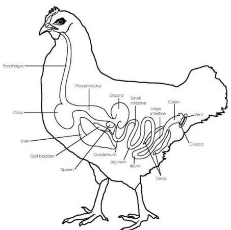
Digestive tract of poultry is generally similar to that of the other vertebrate species, except some peculiarities and the faster transit of food. From a structural point of view, bird gastro-intestinal tract (GIT) consists of beak, esophagus, crop, proventriculus (glandular stomach), gizzard (muscular stomach), small intestine (duodenum, jejunum and ileum), paired isolated ceca, colon-rectum and cloaca (Figure 1). After a first stay in crop, a feed storage organ where fermentation by lactic acid bacteria (LAB) occurs, food is then digested and mechanically grinded in proventriculus and gizzard, respectively, which act as stomachs. Gizzard is recognized as “teeth” of poultry GIT where feed breakdown occurs, and thanks to its low pH, gizzard acts also as microbial barrier (Oakley et al., 2014b; Stanley et al., 2014). Compared to mammals, bird digestive tract is shorter, consequently implying a limited retention time of digesta, reason why it needs to be extremely efficient (Klasing, 1999; Rodrigues and Choct, 2018). After swallow, food moisturization and grounding occur not in mouth, differently from mammals: after crop fermentation, feed continues towards proventriculus and gizzard, where digestion comes with gastric juices. Then, nutrients are mainly digested and absorbed in small intestine, with a short retention time in duodenum, which, anatomically, forms a loop around pancreas from which receives digestive enzymes. After that digestion goes on up to the end of jejunum, recognized as the major site of absorption of small intestine. As last small intestine segment, ileum has the role for nutrients, water and mineral absorption, even if a part of nutrient digestion can also occurs (Svihus, 2014). Like in mammals, small intestine is characterized by a single epithelial cell layer (absorptive, goblet and entero-endocrine cells) that lines villi and crypts, overlaid by a mucus layer, representing the interface between gut and microbiome and formed by mucins secreted by goblet cells. Intestinal villi, defined as protrusions of lamina propria into gut lumen to amplify the absorptive area, are the functional units of small intestine and change along the different segments, becoming shorter and smaller in ileum, where minor digestive functions are required compared to the proximal part. However, since chickens do not have much of a large intestine, ileum represents the last useful tract to adsorb nutrients. Going on with the large intestine, another main distinctive feature in birds are the two ceca, blind-ended bags located at the junction between ileum and colon, usually long and well developed and characterized by a meshwork of long interdigitating villi, of which the majority filters, as a sieve, fluids and small particles coming from ileum. Presence of this villus meshwork is an avian peculiarity (Clench, 1999). Avian ceca have key role in microbial carbohydrate degradation and fermentation, microbial vitamin and amino acid (AA) synthesis, nitrogen compounds degradation, urea recycling into AA and water absorption and balance (Clench,
6 1999; Klasing, 1999; Yamauchi, 2002; Oakley et al., 2014b; Svihus, 2014). Cecum is the most diverse gut section, characterized by the longest feed retention time compared to the upper parts and by the highest short chain fatty acid (SCFA) concentration that are absorbed by the host. In fact, cecum represents the principal site where complex nutrients such as cellulose and other non-starch polysaccharides are fermented, with high fermentation rate of facultative anaerobes and strictly anaerobic bacteria (Józefiak et al., 2011; Borda-Molina et al., 2018). Large intestine ends with a very short rectum, extending between the ileo-cecal junction and the cloaca. For histological structure, avian rectum is very similar to small intestine, but with shorter villi and richer in lymphoid follicles (Klasing, 1999).
Another avian noteworthy peculiarity in is that digesta moves in both peristaltic and anti-peristaltic ways along the GIT. Except the ordinary progression and transport of feed from duodenum to colon, thanks to intestinal caudal peristaltic contractions, in poultry also a reverse peristalsis (cranial) occurs, to improve feed retention time and digestion. Anti-peristaltic movements and reflux happen at three levels: (i) from gizzard to proventriculus due to gizzard contractions (for a greater feed exposure to proventriculus enzymes); (ii) from jejunum and duodenum back to stomachs (to enhance digestion in fasting); (iii) from cloaca to ceca as physiological continuous process (Klasing, 1999; Rodrigues and Choct, 2018).
7
2.1.1 Post-hatching digestive tract development
Already during incubation, and specifically in the late embryonic development, small intestine weight grows with a greater rate than chick body: at day 15 of incubation villi start in growing and shaping and between day 17 and 20 of incubation three different stage of villus development and maturity can be visualized, from elongated pear-shaped villi, to shorter ones and to nascent villi. Then, from this period up to the hatch, small intestine to body weight ratio increases from 1 to 3.5% and a minimal enzyme activity is developed (Uni et al., 2003). During incubation, yolk provides the embryo much of nutrition through circulation while, close to hatch and after, yolk is delivered to the GIT, contributing in small intestine development up to 48 hours post-hatching (Yegani and Korver, 2008). It is in this period that chick faces up the transition from the use of lipid-rich yolk as nutrient source to carbohydrate and protein-rich feed (Uni et al., 2003). So, the proportional growth of intestine is higher than that of BW at hatching because chicks, after a first uptake of yolk nutrients, rapidly need to develop an efficient nutrient uptake capacity supported by a proper gut maturation. During the early post-hatch period, distribution of nutrients follows a rigorous partitioning between “demand” of the tissues as largely users of energy and proteins, and “supply” of the tissues. The intestine is the primary nutrient supply organ and small intestinal epithelium determines growth potential of chicks (Hu and Guo, 2008). The sooner gut achieves its full functional capacity, sooner the chicks can exploit the diet for an efficient physiological development, including a complete achievement of immune competence , important for disease resistance (Uni et al., 1995; Lilburn and Loeffler, 2015). Intake of exogenous feed accompanies a fast development of digestive organs, with great and critical importance of timing and form of the diet and its nutrients available to chick on gut development (Yegani and Korver, 2008).Indeed, immediately after hatching, intestinal absorption rate is higher for fatty acids (mostly for unsaturated fatty acids), followed mainly by glucose, and, in few days, absorption rate for AA and carbohydrates highly increases (Cardeal et al., 2015). Early access to feed has been recognized as fundamental factor affecting intestinal function also at later stage of in life, since feeding delayed has been seen to slow down gut development (Geyra et al., 2001; Yegani and Korver, 2008). Compared to mammals, the faster gut development in chicks is reflected in the increasing number of enterocytes during first few days after hatching (Uni et al., 2003). While at hatching enterocytes are immature like in mammals post-farrowing, appearing small, round-shaped and without the typical polarized brush-border, they acquired polarity in only 24 hours and, in duodenum and jejunum, epithelial surface increases rapidly through cell hypertrophy (Geyra et al., 2001). In particular, jejunum seems to account for the greatest increase in absorption post hatching, developing a greater absorptive area with higher and denser villi already after 72 hours compared to
8 duodenum which, in turn, expands denser villi than ileum (Uni et al., 1999). In fact, differently from the preceding segments, chicken ileum enterocytes seem to be morphologically mature already at hatching, and in this segment, hypertrophy is limited and slow (Geyra et al., 2001). Next to these morphological changes, also digestive and absorptive capacity increase, with a greater functional maturation appearing first in duodenum than in the other distal segments. This maturation for digestive activity occurs during enterocyte migration from crypt to villus with increase in expression of gut nutrient transporters and pancreatic and brush-border enzymes, like disaccharidases, aminopeptidases and alkaline phosphatase, this latter recognized as enterocyte maturation marker (Uni et al., 1998). The presence of proliferative enterocytes not only at crypt level but also along the villus has been reported in all segments in chicks (while in mature bird, like in mammals, cell renewal is guaranteed only by proliferating crypt stem cells that migrate up to villus tip) (Geyra et al., 2001). After, cell proliferation undergoes a subsequent gradual decrease with age, with a slowest decline in jejunum, meaning that, here, villus mitosis is more important for growth compared to the other segments. Regarding crypts development, not-well defined crypts at hatching become distinct in 24 hours, increasing in number and size to provide enterocytes for villus growth and increase cell renewal rate (Geyra et al., 2001). Villus volume and crypt depth increase, result complete in duodenum around days 6 and 7 and after they become less considerable, while in jejunum and ileum this increase continues up to day 14 (Uni et al., 1998, 1999).
Next to the development of digestive functions and structures, contemporary development of gut-associated lymphoid tissue (GALT) happens: GALT is a set of lymphoid structures connected to intestine and representing immune system at gut level. Its development in chicks occurs during the late embryogenesis (Yegani and Korver, 2008). As further component of innate host response, gut mucus layer is formed by goblet cells that release mucins: goblet cells start their development in the late embryonic and immediate post-hatch period, firstly containing only acidic mucins and after hatch producing also neutral mucins (Smirnov et al., 2006).
2.1.2 Intestinal tract of fast-growing hybrids
Selection for GR in modern chickens has been positive correlated to feed intake and feed efficiency, and changes in production-related traits seem to have implied changes at intestinal morphological level and function (Yamauchi, 2002). Since the growing period of broiler has been drastically reduced to reach a market weight in half time compared broiler predecessors, post-hatch period has become proportionally an important phase of the total bird growing period and life. In heavy birds gut increases much more its development rate at hatching compared to non-selected birds, since selection
9 for metabolic characteristics led to a necessary early development of energy-supplying organs, first of all the digestive tract, which has adapted to the rapid GR of heavy broiler chickens in order to provide the proper nourishment (Scheele, 1997; Geyra et al., 2001; Lilburn and Loeffler, 2015). In fact, compared to non-selected light types chickens, which have a lower GR, modern fast-growing heavy broiler lines show higher crypt depth and villus surface at earlier age, meaning a more extensive absorptive surface area and higher intestinal function. Moreover, along with more matured and activated epithelial cells already at hatching and faster enterocyte migration rate, higher absorption of starch and nitrogen has been reported (Uni et al., 1995; Yamauchi, 2002). Despite broiler digestive tract must face a very high rate feed consumption, nutrient absorption and metabolism necessary for the rapid growth that characterizes these new genetic lines, small intestine length results shorter compared to the predecessor broiler strains, with higher villus height in jejunum and ileum (Lumpkins et al., 2010; Svihus, 2014). Mott and colleagues (2008) observed a decrease in expression of some nutrient transporter-related genes in small intestine of chicks selected for high BW, assuming that a higher digestive efficiency might require less nutrient absorption maximization compared to low BW chicks (Mott et al., 2008). Further, in fast-growing broilers it has been also seen that ileum has a role in starch digestion and absorption: in fact, ileum seems to be able to cope an increase need for digestive capacity through increasing its villus surface, like in case of jejunal dysfunction (Svihus, 2014). More, increase in jejunal villus height observed in fast-growing broiler upon short-term fasting has been pointed up as inverse response compared to other genetic lines (such as laying hens) where, instead, fasting usually induces decrease of jejunal villus height. This divergence may mean a greater capacity of this high selected lines in contrasting fasting period by preparing gut for a successive nutrient uptake maximization. This is also supported by the fact that in these birds, villi slough very often, indicating a high presence of proliferative cells and cell turnover in the small intestine (Thompson and Applegate, 2006). The diversified genetic selection for different lines affected not only growth performance but also digestive tract, as it is for broilers and laying hens. Compared to broilers, laying hens have a large and more muscular gizzard and longer intestine in relation to body weight, as well as longer digesta retention time in crop and gizzard and higher pancreas growth. These characteristics stand for a different digestive behavior, meaning an increasing in feed breakdown and digestibility in laying hens compared to broilers. This is already notable at hatching, where activity of disaccharidases is higher in broiler than laying hen chicks, later this enzymatic activity is completely reversed between the two lines (Buzala and Janicki, 2016).
10
2.2 Intestinal microbial community
Gastro-intestinal microbiota represents one of the denser ecosystems, showing complex and high microbial diversity (with bacteria as predominant microorganisms) and harboring from 107 to 1011 bacteria per gram of gut content in poultry, where each gut tract counts as a separate section (Stanley et al., 2012).
Methods of study of gut microbial profile have changed over time: classical microbiological culture techniques are unable in identifying most of the bacteria, since most of them are unculturable. New modern techniques culture-independent have been developed making possible to deepen the knowledge of intestinal microbial community. In the early 2000s, community-fingerprinting techniques have been introduced to provide bacterial profiles by assaying genomic DNA with PCR amplification, most of them based on 16S rRNA, such as denaturing gradient gel electrophoresis (DGGE), terminal restriction fragment length polymorphism (T-RFLP), temporal temperature gradient gel electrophoresis (TTGE) and others (Stanley et al., 2014; Shaufi et al., 2015). More recently, the advent of next generation sequencing (NGS)-based methods, from 16S rRNA amplicon sequencing to metagenomic analyses, has revolutionized the research approach in studying diversity and function of gut microbiota. 16S rRNA targeted amplicon sequencing technique allowed a more in-depth analysis to characterize the complex microbial composition through amplification and sequencing of the 16S small subunit ribosomal genes of bacteria, in particular the hypervariable regions (Borda-Molina et al., 2018). Processing of raw sequences from 16S rRNA genes allows to produce clusters of almost identical sequences, referred to operational taxonomic units (OTUs) providing taxonomic information (Choi et al., 2015). Metagenomics is the highest throughput NGS approach with high scale analysis and a large volume of sequence data that can provide more insights into gut microbial communities through targeted functional gene amplification or through directly sequencing of whole genomes with unprecedent depth and coverage. It allows to explore in depth metabolic pathways correlated to different ecological functions in the gut (Choi et al., 2015; Shaufi et al., 2015).
2.2.1 Spatial bacterial composition along the gastro-intestinal tract of adult chickens
Also due to high costs of the metagenomic approach, most of actual knowledge on bacteria diversity and functionality along chickens GIT is attributed to 16S rRNA targeted amplicon sequencing-based studies (Table 2). Microorganisms of GIT can be located in the lumen, under the mucus layer or can adhere to the mucosa, forming a cell layer. These strictly mucosa-associated bacteria play a pivotal role in host-microbiota interaction (Gabriel et al., 2006). Generally, in adult and healthy chickens,
11 except for the wide spectrum of microbial fluctuations occurring during the short and rapid growth, microbiota population has reached a steady balance in its composition, which differs between gastro-intestinal compartments according to gut section functions. The most abundant bacteria in adult chickens’ intestine are gram positive (gram +) and the five most common phyla are Firmicutes, Bacteroidetes, Proteobacteria, Actinobacteria and Cyanobacteria, with Firmicutes as the most representative phylum (Lu et al., 2003; Xiao et al., 2017). These main phyla account for more than 90% of all sequences, among which Lactobacillus, Clostridium, Ruminococcus, and Bacteroides are the most relevant genera, next to a wide proportion of bacteria belonging to unclassified species or genera (Rubio, 2018). At crop level, where starch breakdown and lactate fermentation occur, bacterial community (composed by both associated-mucosa bacteria and bacteria in digesta) is highly dominated by Lactobacillus genus, with a concentration of 109 colony forming units (CFU) /g content and including species such as L. salivarius, L. fermentum, L. reuteri and L. acidophilus (Rehman et al., 2007; Ranjitkar et al., 2016) The non-secretory and squamous crop epithelium allows lactobacilli adhesion and biofilm formation, making a hardly accessible environment for other bacteria, also due to the high lactic acid concentrations (pH ~5). From here, lactobacilli diffuse to proventriculus, a thick-walled stomach where they again dominate, and to the remainder of the gut to establish their dominance in the small intestine. In the gizzard there is an increase in bacterial diversity, always dominated by species of Lactobacillus genus, but a decrease in bacteria number and activity, due to the very low pH (~3). Actinobacteria and Proteobacteria phyla are present in minor proportion (Ranjitkar et al., 2016).
As for the crop, duodenum and jejunum are dominated by lactobacilli, even if minor species such as Enterococcus spp. can be found. Furthermore, Proteobacteria and Actinobacteria phyla are present along with Bacteroidetes, even if in less extent (Stanley et al., 2012; Awad et al., 2016; Xiao et al., 2017). Due to the high transit rate, reflux movements from jejunum, low pH and bile salt dilution, bacterial density is particularly low in duodenum, where the rapid feed passage rate allows only a small proportion of carbohydrate fermentation (Rehman et al., 2007). Due to the rapid transit of digesta and to high concentration of gastric acid, microbiota in jejunum is limited to acid-tolerant bacteria, mostly Lactobacillus species (Lan et al., 2004). In ileum, most of microbial population is again made up by Lactobacillus genus, followed by Clostridiaceae family, with Clostridium as main genus represented; then, except unclassified bacteria, a less proportion of Streptococcus spp. and Enterococcus spp. have been detected and also coliforms can be present (Lu et al., 2003; Ranjitkar et al., 2016; Xiao et al., 2017). In contrast to these studies, a recent study on microbial dynamics along chickens’ gut tracts highlighted a prevalence of Clostridiaceae and a very low percentage of Lactobacillus spp. in ileum (Shaufi et al., 2015). The other most present phyla in ileum are
12 Proteobacteria and Bacteroidetes (Shaufi et al., 2015; Xiao et al., 2017). Compared to previous tracts, Actinobacteria phylum has been found at very low level (Xiao et al., 2017). As main group dominating small intestine, the important role of Lactobacillus spp. is strictly linked to the nutrient metabolism and it has been hypothesized that lactobacilli might be also involved in nutrient absorption (Xiao et al., 2017). At small intestinal level, the absorption of bacterial fermentation products occurs, such as lactic and volatile fatty acids, and the host can use them as energy source. In addition, small intestinal bacteria population degrade and process different substances such as mucus produced by goblet cells and sloughed epithelial cells (Lu et al., 2003). Moreover, at ileal level bile salts coming from duodenum and jejunum are deconjugated by microbiota, then they are in part reabsorbed and in part delivered to ceca (Volf et al., 2017). While small intestinal microbial community has the main role in supporting gut in digestion and nutrient absorption, cecal microbiota has in fermentation task its primary function (Awad et al., 2016).
The most populated and complex microbial community resides in ceca, with a density around 1011
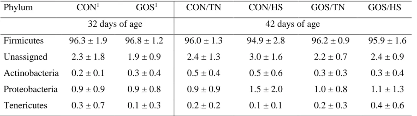
(bacteria per g/content) (Lu et al., 2003). In small intestine, digesta retention time takes only 2.5 hours, but in cecum can last 12-20 hours to favor longer digestion, fermentation and absorption. At the entrance of each cecum, long intertwining villi act as a sieve to leave out coarse particles, reason why ceca content is mainly liquid, with soluble particles (Sergeant et al., 2014). Compared to other gut sections, in cecum microbial population forms a separate cluster and, while in the precedent tracts Lactobacillaceae represented most of Firmicutes, here this phylum is characterized by the predominance of obligate anaerobic bacteria, with facultative anaerobes in less proportion and a very variable composition. The higher microbial diversity of ceca compared to the upper gut is supported by higher organic acid concentration and variation (Rehman et al., 2007; Ranjitkar et al., 2016; Xiao et al., 2017). Bacteria belonging to Clostridia class are reported to be dominant by most of studies on ceca microbial community profiling using 16S rRNA gene sequencing (Lu et al., 2003; Gong et al., 2007; Danzeisen et al., 2011; Shaufi et al., 2015; Ranjitkar et al., 2016). Inside this class, most reported abundant families are Ruminococcaceae, Lachnospiraceae (Danzeisen et al., 2011; Ranjitkar et al., 2016) and Clostridiaceae (Lu et al., 2003; Ranjitkar et al., 2016). A smaller proportion of Lactobacillus species (Lu et al., 2003; Gong et al., 2007; Ranjitkar et al., 2016), E. coli and other coliforms (Gong et al., 2007) are present. The second main phylum in cecum is that of Bacteroidetes (Lu et al., 2003; Shaufi et al., 2015; Ranjitkar et al., 2016), even if in a recent study of Xiao and colleagues (2017) Bacteroidetes resulted to be the main dominant phylum in cecum (Xiao et al., 2017), while Awad and colleagues reported Tenericutes as second most abundant phylum in cecum and found Bacteroidetes at very low level (Awad et al., 2016). According to these studies, also a recent study on cecal microbiota composition by whole DNA shotgun metagenomic sequencing in
13 adult broiler chickens reported Clostridia as most abundant class (70.5%) within Firmicutes (with Ruminococcaceae as most abundant family, 29.53%), followed by Bacilli class (20.7%) (De Cesare et al., 2017).
Beside known bacterial genera, cecal microbiota in chicken have been estimated in consisting of over 600 species from more than 100 genera with most of them still unknown and unclassified (Stanley et al., 2014).
Nevertheless, since many factors contribute to differences in gut microbial composition such as diet, environment and rearing conditions, it is not ever easy to compare with different studies. Regarding Actinobacteria, even if Bifidobacterium genus is known as often representing culturable anaerobe of this minor phylum in chickens, it seems to be reported as relatively low and rare in GIT in terms of Actinobacteria-related abundance from 16S rRNA gene amplicon sequences (Clavijo and Flórez, 2018), but also in cecal microbiota from a study based on TTGE identification (Zhu et al., 2002). Cecal microbiota has the main task to digest and ferment cellulose, starch and resistant polysaccharides, producing the highest SCFA concentration than elsewhere in the gut: after fermentation, protonate forms of SCFAs (like acetate, propionate and butyrate) can pass through the cecal epithelium and catabolized by the host, contributing in host nutrition. Furthermore, SCFAs improve mineral absorption, decrease ceca pH inhibiting some pathogens and, mainly butyrate, can be used as energy source from epithelial cells (Józefiak et al., 2011; Oakley et al., 2014b; Sergeant et al., 2014). Digestion of complex nutrients such as non-starch polysaccharides (NSP) occurs thanks to many microbial hydrolytic enzymes like those of butyrate-producing bacteria such as some species of Clostridium, Ruminococcus, Eubacterium, Fusobacterium, Roseburia and Faecalibacterium (Gong et al., 2002; Rinttilä and Apajalahti, 2013). In fact, metagenomic studies revealed thousands of different genes encoding NSP-degrading enzymes including sequences for glucanases (acting on oligosaccharides), xylanases and endoglucanases domains (xylan-degrading enzymes) in chicken cecum (Sergeant et al., 2014; Borda-Molina et al., 2018). Furthermore, SCFA production seems to be promoted also by the presence of hydrogen-consuming bacteria that uptake hydrogenases so avoiding hydrogen accumulation, which could inhibit fermentations (Sergeant et al., 2014). Also bacterial groups belonging to Bacteroidetes, such as Bacteroides, are involved in breaking down polysaccharides and, more, in anti-inflammatory cytokine production, another important cecum-related function (Xiao et al., 2017). The importance of butyrate-producing bacteria and resistant carbohydrate degraders has been also associated with good chickens performance (Stanley et al., 2014). Beside the carbohydrate fermentation, cecal bacteria take part in nitrogenous metabolism, with SCFA and ammonia production from dietary and urinary (e.g. uric acid) nitrogenous sources. Ammonia can be so integrated into glutamate, useful for bacterial protein and glucose synthesis
14 (Gabriel et al., 2006). Another important function of some cecal bacteria regards also fatty acid transformation: bacteria like some Roseburia species can form conjugated linoleic acid from linoleic acid, with control on fat metabolism (Danzeisen et al., 2011).
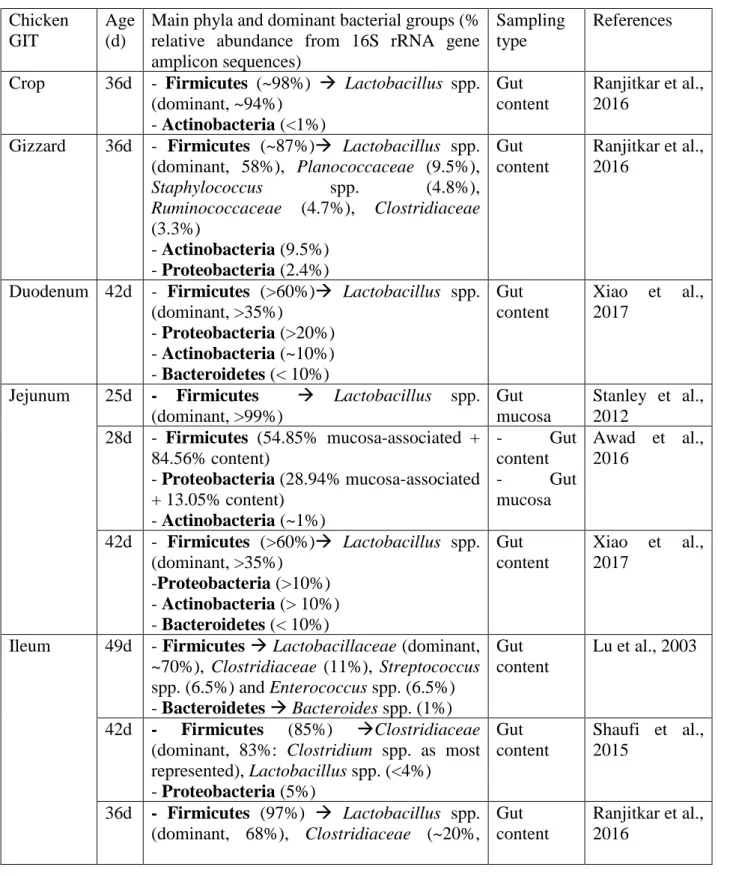
Table 2- Main bacterial composition in young chicken gastro-intestinal tract (GIT) in different 16S rRNA targeted amplicon sequencing-based studies.
Chicken GIT
Age (d)
Main phyla and dominant bacterial groups (% relative abundance from 16S rRNA gene amplicon sequences)
Sampling type
References
Crop 36d - Firmicutes (~98%) → Lactobacillus spp. (dominant, ~94%) - Actinobacteria (<1%) Gut content Ranjitkar et al., 2016
Gizzard 36d - Firmicutes (~87%)→ Lactobacillus spp. (dominant, 58%), Planococcaceae (9.5%), Staphylococcus spp. (4.8%), Ruminococcaceae (4.7%), Clostridiaceae (3.3%) - Actinobacteria (9.5%) - Proteobacteria (2.4%) Gut content Ranjitkar et al., 2016
Duodenum 42d - Firmicutes (>60%)→ Lactobacillus spp. (dominant, >35%) - Proteobacteria (>20%) - Actinobacteria (~10%) - Bacteroidetes (< 10%) Gut content Xiao et al., 2017
Jejunum 25d - Firmicutes → Lactobacillus spp. (dominant, >99%) Gut mucosa Stanley et al., 2012 28d - Firmicutes (54.85% mucosa-associated + 84.56% content) - Proteobacteria (28.94% mucosa-associated + 13.05% content) - Actinobacteria (~1%) - Gut content - Gut mucosa Awad et al., 2016 42d - Firmicutes (>60%)→ Lactobacillus spp. (dominant, >35%) -Proteobacteria (>10%) - Actinobacteria (> 10%) - Bacteroidetes (< 10%) Gut content Xiao et al., 2017
Ileum 49d - Firmicutes → Lactobacillaceae (dominant, ~70%), Clostridiaceae (11%), Streptococcus spp. (6.5%) and Enterococcus spp. (6.5%) - Bacteroidetes → Bacteroides spp. (1%) Gut content Lu et al., 2003 42d - Firmicutes (85%) →Clostridiaceae (dominant, 83%: Clostridium spp. as most represented), Lactobacillus spp. (<4%) - Proteobacteria (5%) Gut content Shaufi et al., 2015 36d - Firmicutes (97%) → Lactobacillus spp. (dominant, 68%), Clostridiaceae (~20%, Gut content Ranjitkar et al., 2016
15 Clostridium spp. as most represented),
Streptococcus spp. (5%) - Actinobacteria (1.7%) 42d - Firmicutes (>60%)→ Lactobacillus spp. (dominant, >35%) - Actinobacteria (> 10%) - Proteobacteria (< 10%) - Bacteroidetes (< 10%) Gut content Xiao et al., 2017
Cecum 49d - Firmicutes → Clostridia (dominant, ~65%: Clostridium spp. and Ruminococcus spp. as most represented), Fusobacterium spp. (14%), Lactobacillus spp. (8%) - Bacteroidetes → Bacteroides spp. (5%) - Proteobacteria (2.8%) Gut content Lu et al., 2003
35d - Firmicutes → Clostridia (dominant, 40%: Ruminococcaceae as most represented, with Faecalibacterium prausnitzii 14% and Ruminococcus spp. 6%), E. coli (11%), Lactobacilli (7%) Gut mucosa Gong et al., 2007 35d - Firmicutes (~80%) → Clostridia (dominant, ~65%: Lachnospiraceae and Ruminococcaceae as most represented)
Gut content
Danzeisen et al., 2011
42d - Firmicutes (49%) → Clostridia (dominant, ~45%)
- Bacteroidetes (21%) → Bacteroidia (dominant, 20%: Alistipes spp. and Bacteroides spp. as most represented)
Gut content
Shaufi et al., 2015
28d - Firmicutes (81.50% mucosa-associated + 70.86% content) → Clostridia (dominant) - Tenericutes (13.38% mucosa-associated + 22.61% content) - Proteobacteria (3.73% mucosa-associated + 22.61% content) - Gut content - Gut mucosa Awad et al., 2016 36d - Firmicutes (~75%) → Clostridia (dominant: Ruminococcaceae 36%, Lachnospiraceae 22.7%, Clostridiaceae 4.8%), Lactobacillus spp. (3.3%) - Bacteroidetes → Alistipes spp. (20%) Gut content Ranjitkar et al., 2016 42d - Bacteroidetes (>50%)→ Bacteroides spp. (dominant, ~40%) - Firmicutes (~40%) - Proteobacteria (<10%) Gut content Xiao et al., 2017
2.2.2 Factors affecting gut microbiota
Even if gut microbial community is quite defined in adult chickens, different factors can influence its composition. From a genetic point of view, intensive selection for high feed efficiency may have contributed in some distinguished characteristics between chickens. In laying hens differing for feed
16 efficiency has been observed higher biodiversity and different composition in cecal microbiota of better feed efficient hens, with higher abundance of Lactobacillus species. Higher Lactobacillus spp. abundance has been found also in duodenum of hens with better feed efficiency, even if in this tract, differently from cecum, a lower microbial diversity has been also reported (Yan et al., 2017). Stanley and colleagues (2012) found Bacteroides genus-related sequences differentially abundant in cecum of better feed efficient broilers, while Clostridia resulted to be more present in low feed efficient animals, considering a possible connection between higher feed efficiency and higher Bacteroides presence, maybe linked to undigestible carbohydrate degrading and propionate producing capacity (Stanley et al., 2012). However, it has also been reported that, while Firmicutes increase nutrient absorption, Bacteroidetes may decrease it (Choi et al., 2015). Different rearing systems also affect microbial composition. Indeed, it has been reported differences in cecal microbiota between commercial broiler chicken and free-range chicken grown in semi-wild conditions, with Firmicutes as dominant phylum in the former while in the latter Bacteroidetes increased reaching the same level of Firmicutes (Mancabelli et al., 2016). Being the main important site for NSP fermentation, cecum and its microbiota resulted also to be affected more than upper gut tracts both by dietary water-soluble NSP and by NSP-degrading enzyme enriched diets: the stimulation of butyrate-producing and cellulose and starch-degrading bacteria as well as SCFA production has been correlated with better performance (Gabriel et al., 2006; Józefiak et al., 2011; Stanley et al., 2014). Lastly, metabolic potential of cecal microbiota changes in response to antibiotics, with repercussion on butyrate-producing bacteria such as Roseburia spp. (Danzeisen et al., 2011)
2.2.3 Temporal microbial community development in growing chicken
Differentiation in microbial community along gut sections follows gut maturation, with general temporal shift and fluctuations in microbiota within 42 days of age. The microbial transient succession is high in chicks, then a more stable state corresponding to skeletal growth period seems to occur,with again fluctuations after 28 days of age and then stabilization (Lu et al., 2003; Shaufi et al., 2015). As the GIT grows, changes in bacterial community occur. Once reached a steady state development and stable environmental conditions, microbial community stabilizes and decreases in diversity (Lumpkins et al., 2010). Firmicutes phylum results predominant at all ages, with genus-relative abundance decreasing as chicken age increases due to the increase in heterogeneity and diversity of microbiota. In chicks post-hatched, lactobacilli appear as earlier colonizers along all GIT and after, throughout growth, they stay stable in upper gut while are mostly replaced in ceca. Indeed, in crop, Lactobacillaceae family is dominant and stable at all ages like in gizzard where
17 Lactobacillaceae are dominant and Actinobacteria phylum increases as chicken age increases (Ranjitkar et al., 2016). In small intestine, bacterial community establishment take less time than in cecum, where protracted variations in microbiota occur. Around first days post-hatching, small intestine harbors streptococci, enterococci and coliforms (with E. coli as most abundant inside Firmicutes phylum), but they undergo a decline and are replaced by lactobacilli, whose proportion increases with age becoming predominant already in 2-week older chickens. In cecum, a longer transient microbiota occurs during chicks development, with more presence of facultative anaerobes in chicks few days post-hatching, then replaced by anaerobes (Rehman et al., 2007; Awad et al., 2016; De Cesare et al., 2017). Indeed, during first days of life in the healthy chicks ceca, predominant bacterial class within Firmicutes seems to be that of Bacilli, followed by Clostridia class, while within Proteobacteria, Enterobacteriaceae family-members result as predominant (De Cesare et al., 2017). Enterococcaceae (e.g. Enterococcus spp.) and Lactobacillaceae (e.g. L. salivarius, L. acidophilus and L. fermentum) appear in highest proportion around 3-4 days of age, but then show a decline (Lu et al., 2003; Józefiak et al., 2011). At 7 days of age, Oakley et al. (2014a) reported Clostridiales order as dominant in cecum, and particularly Faecalibacterium genus as dominant at day 21 (Oakley et al., 2014a). Between day 15 and 22, a development of a more mature microbiota seems to occur especially in ileum and ceca, with a striking increase in relative abundance of Lactobacillus spp. and Clostridia class in the former and a decrease in LAB and increase in Bacteroidetes in the latter. Particularly, in cecum, lactic acid produced by LAB act as substrate for butyrate-producing bacteria development and indeed, it results lower in older chickens (Ranjitkar et al., 2016). Cecal SCFA concentration changes according to chicks growth, with higher acetate concentration during first days after hatching, while butyrate and propionate increase after (Józefiak et al., 2011). Around 40 days of age, cecal bacteria are in a steady state with total anaerobes (such as Clostridium spp. and Bifidobacterium spp.), enterococci, Bacteroidia and a less proportion of lactobacilli (Rehman et al., 2007). Inside Clostridia class, Oakley et al. (2014a) reported Faecalibacterium genus as dominant along with an increase in Roseburia genus at day 42 of age, both butyrate-producing bacteria (Oakley et al., 2014a). So, early cecum colonization from facultative anaerobes is a transitory period, where aerobes consume oxygen and its shortage modifies and drives cecum environment towards more reducing conditions, becoming favorable for the successive strictly anaerobic growth (Awad et al., 2016).
As mentioned before, different factors may influence microbiota composition and characteristics. Targeted selection for fast-growing birds seem to affect gut bacterial dynamics in new chick lines. Indeed, during the early age (first week post-hatching), in ileum of new broiler lines bacterial communities clustered with those of historical line, probably due to a still in progress development of intestine together with a not yet established microbiota (unstable environmental condition of GIT
18 environment), but along with GIT maturation, a clear separation of a more stable microbiota resulted between modern broilers and older lines (Lumpkins et al., 2010). More recently, differences in microbiota between post-hatched chicks of a modern or a heritage lineage have been highlighted. Modern lineage chicks showed a more diverse and differentiated microbiota and the authors hypothesized that genetic selection created a new habitat in GIT in order to obtain a line of chickens with high growth performance, so maybe selecting for a potentially more nutritionally efficient microbiota compared to heritage lines (Pedroso et al., 2016).
2.2.4 . Functions of gut microbial population
As already described before, the first role of microbiota certainly regards nutrient exchange with host’s gut (mutualism). On one hand, bacteria provide SCFAs, ammonia, AAs and vitamins, on the other, host also provides nutrients, such as those from dietary compounds but also mucins. In fact, presence of some mucin-degrading bacteria in gut has been associated with intestinal health. Moreover, gut digestive physiology is affected by microbiota since its development post-hatch and throughout animal life. Microbiota can influence digestive enzyme activities and furthermore, bacteria-derived SCFAs feed enterocytes. It has been observed that germ-free chicken have smaller small intestine and ceca and thinner gut walls compared to conventional ones (Clavijo and Flórez, 2018). Those host digestive and nutritional functions improved by and linked to commensal colonization are due to modulation of gut gene expression. Differences in bacterial composition induce different gene activation. Not only is different gene activation linked to nutrition, but it also regards immune functions (Schiffrin and Blum, 2002). In fact, other important roles of microbiota concern pathogens exclusion and modulation of immune system, contributing in gut health maintenance (Rodrigues and Choct, 2018).
Among main pathogen bacteria that can colonize poultry GIT, the most common are E. coli (usually at low abundance in gut throughout chicken life), species of genus Salmonella (as sporadic and transient bacteria in chicken intestine) and Clostridium (with Cl. perfringens as principal cause of necrotic enteritis). These pathogens impair gut health status and can produce toxins and induce diarrhea (Gabriel et al., 2006; Oakley et al., 2014b; Choi et al., 2015). Also Campylobacter can be found in chickens, but is usually non-pathogenic for avian species while it is for humans (Oakley et al., 2014b). Nevertheless, it has been reported that infection by Campylobacter jejuni increases Clostridium spp. presence, probably because Campylobacter jejuni acts as hydrogen sink and induces higher mucus production, both stimulating factors for Clostridium growth. So, higher abundance in both Campylobacter jejuni and Clostridium spp. may result in higher endotoxin production with
19 increase of gut permeability and consequent possible pathogen colonization and gut diseases (Awad et al., 2016). As mentioned, commensal gut bacteria have a protective role against pathogen colonization. Indeed, over mucosal surface, the dense and complex layer formed by microbial communities in healthy birds acts by blocking passage and adhesion of enteric pathogens (Pan and Yu, 2014). Furthermore, this competitive exclusion to limit pathogen invasion is also expressed through production of bacteriocins and antimicrobial metabolites such as lactic acid and other SCFAs, (Gabriel et al., 2006; Rinttilä and Apajalahti, 2013), along with competing for essential nutrients and epithelial binding sites and through gut immune response modulation (Gabriel et al., 2006; Burkholder et al., 2008). It has been reported that Salmonella colonization in ceca of chicks after hatching can be limited by treatment with cecal microbiota from healthy adults and that lactobacilli counteract coliform growth, demonstrating the importance of gut beneficial bacteria establishment in order to prevent or reduce pathogen invasion (Gabriel et al., 2006; Choi et al., 2015). In support of this, Schokker et al. (2015) compared two broiler lines with same GR but differing for bacterial infection susceptibility and observed that the more resistant broiler line had higher counts for enterococci and lactobacilli and lower Escherichia spp. counts (Schokker et al., 2015).
2.3 Gut homeostatis and determinant variables
Well-being is closely connected to gut health, based on complex functions and mechanisms that ensure a steady and regular intestinal homeostasis. These mechanisms include intestinal structure and mucosal integrity, efficiency of the immune system and relationship between host and gut commensal microorganisms.
Luminal commensal bacteria and pathogens are separated from internal environment by the intestinal barrier, composed by both physical, chemical, immunological and microbiological components (Yegani and Korver, 2008). Mucosal strategies of defense together with control of inflammatory reactivity are prerequisites for gut protection and mucosal integrity preservation (Schiffrin and Blum, 2002).
2.3.1 Gut barrier structure
- Mucus layer
With the task of protecting gut epithelium, mucus layer is a component of gut mucosal innate immune system, being the first line of defense against pathogen invasion of intestine. Between its functional properties, there are those of trapping bacteria and providing colonization site and nutrients for commensals. Released in mucus layer, antimicrobial peptides produced by Paneth cells (such as
20 defensins) and IgA immunoglobulins (Igs) contribute in gut barrier maintenance, slowing diffusing into the mucus. Mucin glycoproteins, as major mucus components, are produced by goblet cells and classified as membrane-bound (transmembrane mucins covering goblet cell apical surface forming the glycocalyx) or secreted (gel-forming) mucins, and are densely decorated with complex carbohydrates (Pan and Yu, 2014; Pelaseyed et al., 2014; Broom, 2018). Mucus thickness increases as moving towards the lower intestine. While, in physiological conditions, small intestinal mucus is not or weakly attached to the epithelium, forming a diffusion barrier with peptides with high antimicrobial activity (which create an antibacterial gradient), in lower intestine (most prominent in colon) microorganisms stay in an outer loose mucus layer, clearly separated from an inner denser and epithelium-adherent layer free of bacteria (Pelaseyed et al., 2014; Johansson and Hansson, 2016). The inner mucus layer is further converted by proteases to the expanded outer loose layer, which lubricate feces and where mucin glycans allows mucus-associated bacteria to make niches. In fact, mucin domain surfaces present glycans that interact with bacterial adhesins and supply energy, since some bacteria have enzymes to degrade mucin glycans: different carbohydrate chains determining mucin functional properties can influence also microbiota composition. While the inner adherent mucus layer requires proteases to be dissolved, the loose layer, even if penetrable, protects small intestine from bacteria invasion because of the continue flow towards the low intestine, which contrasts on its own bacteria mobility (Johansson and Hansson, 2016; Broom, 2018). Mucus aspects such as mucin differentiation (neutral, sialo- and sulfo-mucins), goblet cell density and mucus thickness are key factors playing in gut barrier mechanism. Among mucin types, acidic ones seem to more protect gut against bacteria translocation (Broom, 2018).
- Epithelium
Placed under the glycosylated mucin-rich layer, a single layer of epithelial cells bordering the intestinal lumen provides physical and biochemical obstructions to microbes thanks to brush border of actin-rich microvillar extensions, intercellular tight junctions (TJs) and antimicrobial peptides production (Artis, 2008). The structure of gut epithelium is based on a monolayer of columnar epithelial cells constituted of four major cell types: goblet cells (producing mucins), Paneth cells (producing antimicrobial peptides), endocrine cells and enterocytes, most abundant cell type responsible for final digestion and nutrient absorption. All these cells derive from stem cells localized in the crypts of small intestine. These cells maintain the intestinal tract integrity by creating a strength barrier, held steady by TJ protein complexes. Regulatory molecules concurring in gut barrier forming and in paracellular permeability regulation (Kurashima et al., 2013; Broom, 2018). Paracellular way represents the major path for passing gut epithelium and getting to submucosa. In healthy conditions, spaces between epithelial cells are well sealed by the apical junctional complex, composed of the TJs