Research report
Members of the NF-nB family expressed in zones of active
neurogenesis in the postnatal and adult mouse brain
Suzanne Denis-Donini
a,b,*, Andrea Caprini
c, Carolina Frassoni
d, Mariagrazia Grilli
ea
Department of Biology, Section of Zoology and Cytology, University of Milan, Milan, Italy
b
CNR Institute of Neuroscience, Milan, Italy
c
Italian Foundation for Cancer Research Institute of Molecular Oncology, Milan, Italy
d
Department of Experimental Neurophysiology, National Neurological Institute bC.BestaQ, Milan, Italy
eSchering Plough Research Institute, San Raffaele Science Park, Milan, Italy
Accepted 12 October 2004 Available online 10 November 2004
Abstract
The Rel/NF-nB family of transcription factors is implicated in cell proliferation, cell death, cell migration and cell interactions. Here, we examined by immunohistochemistry the expression pattern of various members of this family during postnatal telencephalon development and during adulthood, and we used neuronal and glial markers to identify the cells types where they are expressed. Distinct Rel/NF-nB proteins are highly expressed postnatally in the subventricular zone and in the rostral migratory stream. In particular, Rel A and p50 are expressed in radial glial cells, in migrating neuron precursors and in a population belonging to the astrocytic lineage. Rel B, on the other hand, is only expressed in migrating neuron precursors, whereas c-Rel is present in a few cells located at the edges of the rostral migratory stream. The expression of Rel A and p50 persists into adulthood, particularly in subventricular zone astrocyte-like cells and in migrating neuron precursors, respectively. The selective expression of NF-nB members in the postnatal subventricular zone and rostral migratory stream and their persistence into adulthood in regions of ongoing neurogenesis suggests possible mechanisms linking NF-nB expression with cell proliferation and migration. Their presence in actively proliferating progenitor cells, detected by BrdU staining, further suggests that NF-nB may be part of a signaling pathway that is important for neurogenesis.
D 2004 Elsevier B.V. All rights reserved.
Theme: Development and regeneration Topic: Genesis of neurons and glia
Keywords: Transcription factor; Subventricular zone; Radial glia; Stem cell; Postnatal neurogenesis
1. Introduction
It is now well documented that in rodents and primates, olfactory bulb interneurons continue to be generated after birth and throughout adult life in the subventricular zone (SVZ), which persists as a neurogenic region[2,1,17,22,29,26,43]. Multipotential neural stem cells have been isolated from this region and have been characterized both in vivo and in vitro
[11,26,38,39,45,56]; they are relatively quiescent and divide upon stimulation to give rise to a large population of rapidly dividing cells, which in turn are able to generate neuron precursors and astrocytes. From their site of birth, neuron precursors migrate from the wall of the lateral ventricle over a long distance without dispersion, following a highly stereo-typed pathway—the rostral migratory stream (RMS) up to the ipsilateral olfactory bulb where they differentiate into granule and periglomerular interneurons[1,27–29]. The orientation of migration is tangential, and the neuroblasts migrate by sliding along each other, forming chains held together through homotypic interactions that are mediated at least in part by polysialic residues on N-CAM[7,18,47,57]. Interestingly, the 0165-3806/$ - see front matterD 2004 Elsevier B.V. All rights reserved.
doi:10.1016/j.devbrainres.2004.10.010
* Corresponding author. Department of Biology, University of Milan, Via Celoria, 26, Milan 20133, Italy. Tel.: +39 2 5031 4783; fax: +39 2 5031 4781.
neuron precursors continue to divide while migrating to the olfactory bulb[29,34], and in contrast to most migrating cells, they do not seem to require astroglial guidance at least during the 1st postnatal week[21,24].
The cell types belonging to the lineage of olfactory bulb interneurons have been identified in the adult SVZ, and they present a unique spatial organization. Newborn neuroblasts (Type A cells) migrate from the site of birth in the SVZ to the RMS, closely intermingled with clusters of rapidly dividing cells (Type C cells), through interconnected channels formed by slowly proliferating GFAP+ astrocytes (Type B cells)
[10]. So in the adult SVZ and RMS, in contrast to the
neonate, the chains of migrating precursors are ensheathed by GFAP+astrocytes whose role in guidance is still unclear. Little is known about the control of SVZ cell proliferation and cell fate determination, but some evidence indicate that cell interactions and local environmental factors may be critical for regulating these processes (reviewed by Ref.[53]);[32].
In the present study, we searched for the expression of transcription factors in the SVZ and RMS in the mouse forebrain at early postnatal stages and in adults. We focused our attention on the Rel/NF-nB family of transcription factors which integrate directly extracellular stimuli in a rapid transcriptional response. Five mammalian proteins of the Rel/NF-nB family, NF-nB1 (p50, p105), NF-nB2 (p49/ 52, p100), Rel A (p65), Rel B and c-Rel have been described so far. Before stimulation, Rel/NF-nB is retained in the cytoplasm in an inactive form due to its binding to the inhibitor (InB) proteins. In response to a number of different stimuli, InB is phosphorylated and targeted to the protea-some for degradation. This allows Rel/NF-nB to translocate to the nucleus and activate a new program of gene expression (reviewed in Refs.[6,12,13,14,42,55].
During brain development, previous studies have dem-onstrated with different methodologies the presence not only of rel A/p50 heterodimers but also of other combinations of Rel family proteins both in an inducible and activated state
[5,15,19,48].
The existence of multiple combinations of NF-nB subunits raises the possibility that dimers of various composition may play a variety of roles in the developing and adult CNS. These predicted roles depend in part on the cell type localization and on their temporal window of expression. To determine the regional and cellular local-ization of NF-nB proteins during postnatal telencephalon development and into adulthood, we have used antibodies to various members of the family combined with lineage markers of neuronal and glial precursors.
2. Materials and methods 2.1. Animals
CD1 mice (Charles River) were used in all experiments. The mice were handled according to the standards of animal
experiments in our University in accordance with the guidelines established in the Principles of Laboratory Animal Care (NIH Publication no. 86-23, revised 1985). 2.2. Immunohistochemistry
A total of 20 neonate pups (postnatal days 4–7; P4–7) and six adult mice (2 months old, 30 g) were anesthetized with 4% chloral hydrate (1 ml/100 g i.p.) and intracardially perfused with a buffered solution of 4% paraformaldehyde. The brains were immediately dissected out and postfixed overnight in the same fixative at 4 8C. They were dehydrated in graded ethanol and embedded in paraffin. Sagittal sections (8 Am) were mounted on gelatin-coated slides. Alternatively, brains were embedded in 6% agarose, and 50-Am-thick serial sagittal or coronal sections were cut by means of a Vibratome VT1000S (Leica, Heidelberg, Germany).
For single immunohistochemistry, paraffin sections were incubated 10 min in 1% H2O2 in PBS to inactivate
endogenous peroxidase, permeabilized 30 min in PBS containing 2% normal goat serum and 0.3% Triton X-100 and immunodecorated overnight at 4 8C with primary antibodies (dilutions as described below in 1% normal goat serum and 0.03% Triton X-100), washed five times for 10 min in PBS and incubated in biotinylated secondary antibodies (Vector) for 45 min. Immunoreactivity was revealed with a conventional ABC kit Elite (Vectastain-Vector). Some sections were counterstained with thionin. For double-staining immunofluorescence, both free-float-ing Vibratome sections and paraffin sections were used. They were incubated overnight at 4 8C with primary antibodies (alone or in combination), washed five times for 10 min in PBS and incubated for 1 h with the appropriate fluorochrome-conjugated secondary antibodies. Fluores-cence images were captured with a confocal laser scanning microscope (Leica Lasertecknik GmbH, Heidelberg, Ger-many). Single optical sections of 1.5 Am were selected, or Z-series images were collected at 1-Am steps on a PC using a Leica Power Scan software. To show colabelling of two antigens, only sections in the same focal plane were used for producing merge figures except for projection data, as explained in the text. Primary antibodies were obtained from the following sources and used at the indicated dilutions: rabbit polyclonal anti-NF-nB p50 was raised by M. Grilli against a bacterially produced and affinity purified mouse p50 protein, as previously described [20] (dilution 1:100–1000); rabbit polyclonal anti-Rel A, Rel B and c-Rel (Santa Cruz Biotechnology, Santa Cruz, CA; 1:100–1000), mouse monoclonal
anti-PSA-CAM (kind gift from G. Rougon [46]; 1:200–1200),
rabbit polyclonal anti-GLAST (kind gift from G. Pietrini
[44]; 1:500), mouse monoclonal anti-GFAP (Boerhinger;
1:500), mouse monoclonal anti-RC2 (Developmental stud-ies Hybridoma bank, University of Iowa, 1:500), mouse monoclonal anti-type III h-tubulin (TuJ1; Sigma; 1:100),
mouse monoclonal anti-BrdU antibody (DAKO; 1:100). Secondary antibodies were conjugated to Alexa Green (Molecular Probes, Eugene, OR; 1:400) or TRITC (Jackson ImmunoResearch, West Grove, PA; 1:200). Controls were performed for the Santa Cruz antibodies by preincubation with blocking peptides and for the other antibodies by omitting the first antibody and incubating with secondary antibody alone. In all cases, the controls resulted in no detectable staining. The specificity of the anti-p50 antibody has also been checked by Western blot on brain extracts from p105/p50-deficient mice (B6, 129P-NF-nB1) and their respective controls B6/J129 obtained from Jackson Laboratories.
2.3. Western blotting
Brain extracts were obtained from minced pieces of telencephalon by lysis and sonication at 4 8C in RIPA (Tris 20 mM pH 7.5, NaCl 150 mM, EDTA 2 mM, DOC 1%, TritonX-100 1%, SDS 0.25% and 1 mM PMSF). Protein concentration was determined using a Pierce assay. Protein extracts (10 Ag) were fractionated on 7.5% SDS–polyacry-lamide gels and transferred to nitrocellulose membranes. The membranes were probed with primary p50 anti-bodies (1:1000), then incubated with a horseradish perox-idase-conjugated goat antirabbit secondary antibody (Sigma; 1:2500). Specific proteins were visualized by enhanced chemiluminescence (ECL, Amersham).
2.4. BrdU treatment and BrdU immunohistochemistry To label the rapidly dividing cells, two male mice (2 months old) were perfused 90 min after a single intra-peritoneal injection of a sterile solution of BrdU (Sigma; 10 mg/ml in PBS, 50 mg/kg of body weight).
Vibratome sections were treated for 30 min at 37 8C with HCl 2N in PBS containing 0.5% TritonX-100. They were rinsed in sodium tetraborate buffer (0.1 M, pH 8.5) and processed for immunohistochemistry as described above, using an anti-BrdU antibody.
3. Results
3.1. Expression of various members of the NF-jB family: p50, Rel A, Rel B, and c-Rel in the postnatal telencephalon (P4–7)
During postnatal telencephalon development, the highest signal intensities for all antibodies, except for c-Rel, are associated with the lateral ventricular and subventricular zone, the RMS and the olfactory bulb.
p50 immunoreactivity is mainly detected along the wall of the lateral ventricle, in the anterior subventricular zone and in the RMS up to the core of the olfactory bulb. In the olfactory bulb, immunoreactivity is also present in the
granular and periglomerular cell layer (Fig. 1A). In the SVZ/ RMS, p50 immunoreactivity reveals the presence of tightly packed cells (Fig. 1B). Radially oriented cells in the white matter (WM) originating from the dorsal border of the SVZ/ RMS are also labelled (Fig. 1B). The specificity of the anti-p50 antibody has been checked by Western blot on brain extracts from wild type and p50 null mice. The antibody recognizes two bands of MW 105 and 50 kD in wild-type extract and none in the p50 null extract (Fig. 1C).
The pattern of Rel A immunoreactivity is quite similar to that of p50. Immunoreactivity is concentrated along the border of the lateral ventricle and in the SVZ/ RMS where tightly packed immunoreactive cells are observed (Fig. 1D). Radially oriented cells in the WM are also labelled (Fig. 1E).
Rel B is also expressed predominantly in the SVZ/RMS, but the immunoreactive cells appear loosely organized in ropes or chains, suggesting that they are migrating precursors (Fig. 1F). c-Rel expression was not detectable at P4–5. However, at P7, it is highly expressed in a few cells located at the dorsal and the ventral border of the RMS (Fig. 1G).
To identify which cells express different NF-nB members, confocal image analysis was performed on sections double stained for p50, Rel A or Rel B and PSA-CAM, the highly polysialilated adhesion molecule which is expressed by migrating Type A neuroblasts[7,47]
or TuJ1 a marker of early neuronal differentiation recognizing class III h-tubulin [25]expressed in migrating neuroblasts in the RMS [33]. As shown in Fig. 2A and G, p50+ cells and Rel A+ cells appear to fill the entire SVZ/
RMS region. On the other hand, PSA-CAM+ cells
identified as Type A neuroblasts form a stream in the
core of the SVZ/RMS (Fig. 2 B, E, H). Merged images
clearly show that the postnatal SVZ/RMS harbors at least two distinct cell populations, one exclusively p50+ (Fig. 2C, F) or Rel A+ (Fig. 2I) and the other which is PSA-CAM+/p50+, as shown at higher magnification in Fig. 2F, or PSA-CAM+/Rel A+ (Fig. 2I). Double-staining immuno-histochemistry with Rel A and TuJ1 shows in the RMS that some Rel A+cells are TuJ1+, thus confirming that Rel A is expressed in migrating Type A neuroblasts (Fig. 2J– L). In these cells, Rel A appears to be particularly concentrated at sites of cell contacts (Fig. 2L).
Type A neuroblasts, in addition to p50 and Rel A, also express Rel B. Double-staining immunohistochemistry with Rel B and PSA-CAM shows coexpression of the two markers in the majority of cells (Fig. 2M–O). Occasionally, some cells that are exclusively Rel B+ are found amidst migrating Type A neuroblasts (Fig. 2O).
To identify the Rel A+PSA-CAM cells in the SVZ/RMS and the radially oriented cells in the WM, we stained adjacent sections with Rel A and GLAST, the astrocyte specific
glutamate transporter [8], which is also present in a
subpopulation of radial glial cells [16,49]. In the SVZ/ RMS, a stripe of GLAST+cells oriented parallel to the plane
of migration is observed (Fig. 3A). The close similarity to the pattern of Rel A immunoreactivity (Fig. 3B) suggests that the Rel A+PSA-CAM cells belong to the astrocytic lineage.
In the WM, as expected, cells with long radial processes extending from the ventricle wall to the pia are similarly marked by GLAST (Fig. 3C) and Rel A antibodies (Fig. 3D). To support the notion that Rel A and p50 are expressed in radial glial cells, we double-stained sections with Rel A or p50 and RC2, a specific marker of radial glial cells[36]. Colocalization of Rel A and RC2 (Fig. 3E) and p50 and RC2 (Fig. 3F) is observed mainly in the processes of radial glial cells, thus demonstrating that Rel A and p50 are expressed in radial glial cells.
In conclusion, during postnatal development, Rel A, p50 and Rel B are expressed in the SVZ/RMS in Type A migrating neuroblasts. In addition, Rel A and p50 are expressed in radial glial cells and in another population that appears to line the entire SVZ/RMS, most probably belonging to the astrocytic lineage.
The selective and partly overlapping expression of many members of the NF-nB family in the SVZ/RMS and in radial glial cells suggests that NF-nB dimers of various composition control a variety of events correlated with the generation of new neurons and glia and/or with oriented migration processes.
3.2. Expression of Rel A and p50 in the adult SVZ
The SVZ, which is a remnant of the embryonic germinal zone, becomes the predominant neurogenic region into adulthood [1,3,52]and SVZ astrocyte-like cells have been identified as the precursors of the newly generated neurons which continue to arrive in the olfactory bulb[11].
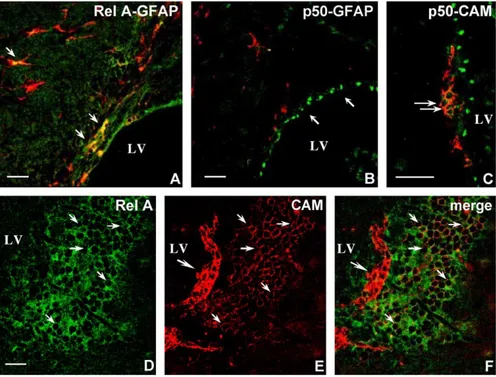
The expression of NF-nB members is more widespread in the adult forebrain than in the newborn. Nevertheless, Rel A and p50 staining are mainly detected along the wall of the lateral ventricle and in the SVZ. Confocal image analysis of double-stained sections with Rel A or p50 and GFAP shows Fig. 1. Distinct and partly overlapping expression of various NF-nB proteins in the postnatal telencephalon in sagittal paraffin sections (P4–7). (A and B) Pattern of p50 immunoreactivity at P4–5. (A) p50 is detected along the border of the lateral ventricle, in the SVZ/RMS (arrows) and in the olfactory bulb. (B) Detail of the SVZ/RMS showing densely packed immunoreactive cells. Radially oriented cells originating from the dorsal border of the SVZ/RMS are observed in the WM (arrows). (C) Specificity of the anti-p50 antibody. Western blot of brain extracts from wild-type (WT) and p50 / mice probed with the a-p50 antibody. Two bands of MW 105 and 50 kD corresponding to p105 and a-p50, respectively, are visible in wild-type extracts, whereas none are detected in p50 null extracts. (D and E) Pattern of Rel A immunoreactivity at P4–5. (D) Rel A staining is prominent in the SVZ/RMS where tightly packed immunoreactive cells are present. (E) Detail of the dorsal border of the SVZ/RMS showing radially oriented cells in the WM (arrows). (F) Pattern of Rel B immunoreactivity at P4–5 in the SVZ/RMS revealing a network of loosely organized immunoreactive cells. (G) Pattern of c-Rel immunoreactivity at P7. Only few cells located at the dorsal and the ventral border of the RMS are c-Rel positive. Section lightly counterstained with thionin. LV—lateral ventricle; OB—olfactory bulb; St— striatum; SVZ/RMS—subventricular zone/rostral migratory stream; WM—white matter. Scale bar, 300 Am (A), 50 Am (B), 100 Am (D) and 25 Am (E, F, G).
Fig. 2. Phenotypic characterization of p50, Rel A and Rel B expressing cells in the postnatal SVZ/RMS (P5). Confocal image analysis of Vibratome sagittal sections double stained with NF-nB proteins and PSA-CAM (CAM) or TuJ1. (A) p50 (green) is expressed through the SVZ/RMS. (B) Chains of PSA-CAM+ cells (red) identified as migrating Type A neuroblasts form a stream in the core of the SVZ/RMS. (C) Overlay of the two channels (merge) demonstrates the existence of two cell populations, one expressing exclusively p50 and the population of migrating neuroblasts where p50 and PSA-CAM are coexpressed (yellow). (D–F) Detail of the framed region demonstrating that the great majority of Type A neuroblasts express p50 (arrows). (G) Rel A (green) is expressed through the SVZ/RMS, whereas Type A neuroblasts (red) are located more centrally (H). (I) Overlay of the two channels (merge) identifies two distinct populations; one expressing exclusively Rel A and the other, PSA-CAM+, which also expresses Rel A (yellow). (J–L) In the RMS, Rel A (green) colocalizes with the neuron precursor marker TuJ1 (red). Overlay of the two channels (merge) clearly shows colocalization at sites of cell contacts (yellow, arrows). (M–O) Coincidence of the pattern of Rel B (green) and PSA-CAM (red) immunostaining. Overlay of the two channels (merge) show coexpression of both markers (yellow) in the great majority of cells. Amidst Type A neuroblasts, some cells express exclusively Rel B (arrows). Single optical sections. Scale bar, 50 Am (A– C, G–I and M–O), 25 Am (D–F and J–L).
that some SVZ astrocyte-like cells express Rel A (Fig. 4A). In contrast, p50 is expressed in the nucleus of some cells lining the ventricle, most probably ependymal cells, but is not detectable in SVZ astrocyte-like cells (Fig. 4B). In addition, p50 is expressed in the chains of Type A migrating neuroblasts (Fig. 4C). Rel A staining is also prominent along the most ventral portion of the lateral ventricle, identifying a patch of brightly fluorescent cells (Fig. 4D). Double-labelled sections with CAM show that the patch harbors PSA-CAM+cells, some of which, located closer to the ventricle, exhibit the typical organization in chains of migrating Type A neuroblasts, are brightly fluorescent but do not express Rel A. The others, located more centrally, exhibit a less intense
PSA-CAM immunoreactivity (Fig. 4E), but some of them
express Rel A (Fig. 4D, E, F).
The expression of Rel A in SVZ astrocyte-like cells in cells closely associated to neuron precursors and in some
neuron precursors suggests that it could be correlated with the generation of new neurons in the adult brain.
To test this hypothesis, we used the proliferation marker BrdU to label cells in the S-phase of mitosis and double-stained sections with Rel A.Fig. 5A which is a single optical section shows coexpression of Rel A and BrdU in cells located along the wall of the lateral ventricle. Fig. 5B represents a projection from sets of stacked optical sections of the lateral wall of the lateral ventricle subjacent the patch of Rel A+cells along the most ventral portion of the lateral ventricle, showing that the distribution of actively prolifer-ating cells largely coincides with that of Rel A expression.
4. Discussion
This study provides the first detailed description of the expression of Rel/NF-nB proteins in the mouse telencepha-lon during early postnatal stages and into adulthood. Rel/ NF-nB proteins are a pleiotropic family of transcription factors whose target genes are implicated in cell prolifer-ation, cell death, cell migration and cell interactions. We show that most NF-nB subunits are selectively expressed during postnatal development in the SVZ/RMS, and that their expression persists into adulthood in the SVZ, the predominant neurogenic region in the adult brain. The selective expression of NF-nB members in distinct cell types of the SVZ/RMS indicates a multiplicity of potential roles, particularly in the control of migration processes and in regulating the generation of new neurons.
At postnatal stages, p50, Rel A and Rel B are present in migrating Type A neuroblasts. In addition, Rel A and p50 are expressed in radial glial cells, and in another cell population, that appears to line the entire SVZ/RMS. The great similarity between the pattern of Rel A immunor-eactivity and that of GLAST strongly suggests that these cells belong to the astrocytic lineage. Up to the present time, the observation that the young SVZ/RMS lacks GFAP expressing cells had led to the assumption that astrocytes were not involved in cell migration during the peak of neurogenesis which occurs during the 1st postnatal weeks
[21,24]. More recently, radial glial processes have been
shown to form a scaffold along the SVZ/RMS [4], and
numerous RC2+cells have been observed in the RMS at P5
[16]. Our data on the expression of GLAST confirm the
notion that various populations of glial cells are present during the 1st postnatal week in the SVZ/RMS and suggest that they could provide the substrate for migration and/or at the same time confine the migrating neuroblasts to the RMS. In migrating Type A precursors, the transcription factors appear to be concentrated at sites of homotypic or heterotypic cell contacts. This suggests the existence of an abundant inducible pool, possibly activated by discrete alterations in cell interactions that occur during migration. In this regard, it is worthwhile to note that N-CAM binding to neurons and astrocytes rapidly activates NF-nB[23], and in Fig. 3. Phenotypic characterization of Rel A expressing cells in the SVZ/
RMS and in the WM. (A, B) Adjacent sagittal sections of the SVZ/RMS immunostained with GLAST (A) and Rel A (B) show that both antibodies decorate cells oriented parallel to the plane of migration. (C, D) Similarity of the staining pattern of GLAST (C) and Rel A (D) in adjacent sections of the WM. Both antibodies decorate radially oriented cells with the typical long processes of radial glial cells. (E, F) Coexpression of Rel A (green) or p50 (green) and the radial glial marker RC2 (red). Rel A (E) and p50 (F) colocalize with RC2 (yellow) in radial glia processes. SVZ/RMS— subventricular zone/rostral migratory stream; St—striatum; WM—white matter. Scale bar, 50 um (A, B) and 25 Am (C–F).
turn, N-CAM gene expression is regulated by NF-nB[51]. Rel A and p50 are present postnatally in radial glial cells, and in the adult brain, Rel A is present in SVZ astrocyte-like
cells, while p50 is expressed in ependymal cells. Several reports have demonstrated that in the embryonic cortical ventricular zone, radial glial cells are themselves neuronal progenitor cells [31,37,40,41], whereas in the adult, SVZ astrocyte-like cells act as neural stem cells[11]. Recently, it has been suggested that some radial glial cells might give rise to multiciliated ependymal cells, and that a subset of radial glia transforms into SVZ astrocyte-like cells[54]. The persistent expression of NF-nB members in putative stem cells from postnatal stages to adulthood strongly suggests that NF-nB complexes of various composition (hetero and homodimers) possibly act as transcriptional regulators of neurogenesis. Remarkably, Rel A immunostaining is prom-inent in zones of active neurogenesis identified by BrdU immunostaining, thus confirming the notion that cells expressing at least Rel A are coinvolved in the generation of new neurons. It remains to be determined whether NF-nB acts in a cell autonomous manner, directly regulating the cell cycle of stem cells or precursors cells, or dynamically controls the expression of cytokines and growth factors necessary for creating and maintaining the properties of the stem cell niche.
In the nervous system NF-nB is induced by several molecules that play key roles in neural function and development. For example, NF-nB is induced by glutamate
in cerebellar granule cells [15,19], and by NGF in
sympathetic and sensory neurons[30]. A number of growth factors and cytokines, like EGF, FGF2, IGF1 and TGF-a, all Fig. 5. Coexistence of Rel A and S-phase cells in the SVZ and in the lateral
wall of the lateral ventricle. BrdU labelling (red) in adult mice to identify S-phase cells and Rel A labelling (green). (A) Optical section through the Rel A expressing cells along the lateral wall of the lateral ventricle. Overlay of the two channels showing that S-phase cells express Rel A (yellow, arrows). (B) Projection data from Z stacks showing in depth the ventral portion of the lateral wall of the lateral ventricle (LWLW) and the patch of Rel A positive cells in the ventral SVZ, demonstrating that S-phase cells are confined to the region of high Rel A immunoreactivity. LV—lateral ventricle; SVZ—subventricular zone. Scale bar, 20 Am.
Fig. 4. Phenotypic characterization of Rel A and p50 expressing cells in the adult telencephalon. Confocal image analysis of sagittal paraffin sections of the SVZ double stained for Rel A (green) or p50 (green) and the lineage markers GFAP (red) and PSA-CAM (red) as indicated on the figures. (A) Rel A is expressed in SVZ astrocyte-like cells (arrows) and in cells lining the ventricle. (B) p50 is expressed in the nuclei of cells lining the ventricle (arrows) but not in SVZ astrocyte-like cells. (C) p50 is also present in chains of PSA-CAM+Type A neuroblasts (arrows). (D) Rel A expressing cells form a patch located in the
most ventral portion of the lateral ventricle. (E) PSA-CAM+cells are found in this patch. Overlay of the Rel A and PSA-CAM channels is shown in panel (F)
(merge), illustrating that some cells coexpress both markers (arrows), whereas the organized chains of Type A neuroblasts located at the edge of the patch do not express Rel A (big arrow). LV—lateral ventricle. Scale bar, 20 Am.
appear to play critical roles in supporting stem cell proliferation, whereas CNTF, BMP2 and PDGF appear to influence the relative proportion of neurons and astrocytes, as reviewed in Ref. [53]. Most of these cytokines/growth factors are NF-nB target genes or induce NF-nB activation
[9,35]. Interestingly, erythropoietin regulates the in vivo production of neuronal progenitors by forebrain neural stem cells, and this action is mediated by NF-nB[50].
Regarding the composition of NF-nB dimers in the SVZ/ RMS, p50, Rel A, c-Rel and Rel B are present in postnatal Type A migrating neuroblasts, suggesting that all possible combinations of subunits can potentially take place, whereas in radial glial cells where only p50 and Rel A have been detected, both the most common heterodimer, p50/Rel A and p50 homodimers may be recruited as well. This holds also true for the adult brain where only p50 and Rel A are found. Our results are in agreement with previous studies showing that p50/Rel A and Rel A/c-Rel heterodimers, as well as p50 homodimers, are present in the postnatal brain but not in adults where the most abundant complexes are
p50/Rel A and p50/c-Rel [5]. However, the analysis of
transgenic mice carrying nB-dependent LacZ reporter gene has suggested that in the developing brain, only the heterodimer p50/Rel A contributes to NF-nB transcriptional activity[48].
Understanding the relative contribution of single subunits to transcriptionally active dimers in regions where neuro-genesis occurs will be the next goal, as well as elucidation of the genes activated or silenced by NF-nB complexes in specific cell populations in the RMS/SVZ. Blocking function experiments and the careful analysis of knockout animals will certainly help working towards this goal.
Acknowledgments
We thank Drs. G. Rougon and G. Pietrini for the kind gift of PSA-CAM and GLAST antibodies, respectively, and Drs. B. Ortino and F. Inverardi from the National Neuro-logical Institute bC. BestaQ for the acquisition of confocal images. This work was in part supported by FIRB grant bStem cellsQ.
References
[1] J. Altman, Autoradiographic and histological studies of postnatal neurogenesis: IV. Cell proliferation and migration in the anterior forebrain, with special reference to persisting neurogenesis in the olfactory bulb, J. Comp. Neurol. 137 (1969) 433 – 458.
[2] J. Altman, G.D. Das, Autoradiographic and histological studies of postnatal neurogenesis: I. A longitudinal investigation of the kinetics, migration and transformation of cells incorporating tritiated thymidine in neonate rats, with special reference to postnatal neurogenesis in some brain regions, J. Comp. Neurol. 127 (1966) 337 – 390.
[3] A. Alvarez-Buylla, J.M. Garcia-Verdugo, Neurogenesis in adult subventricular zone, J. Neurosci. 22 (2002) 629 – 634.
[4] J.A.J. Alves, P. Barone, S. Engelender, M.M. Froes, J.R.L. Menezes, Initial stages of radial glia astrocytic transformation in the early postnatal anterior subventricular zone, J. Neurobiol. 52 (2002) 251 – 265.
[5] G.Y. Bakalkin, T. Yakovleva, L. Terenius, NF-nB-like factors in the murine brain. Developmentally-regulated and tissue-specific expres-sion, Mol. Brain Res. 20 (1993) 137 – 146.
[6] P.A. Bauerle, T. Henkel, Function and activation of NF-nB in the immune system, Annu. Rev. Immunol. 12 (1994) 141 – 179. [7] L. Bonfanti, D.T. Theodosis, Expression of polysialylated neural cell
adhesion molecule by proliferating cells in the subependymal layer of the adult rat, in its rostral extension and in the olfactory bulb, Neuroscience 62 (1994) 291 – 305.
[8] B.D. Carter, C. Kaltschmidt, B. Kaltschmidt, N. Offenhauser, R. Bohm-Matthaei, P.A. Bauerle, Y.A. Barde, Selective activation of NF-nB by nerve growth factor through the neurotrophin receptor p75, Science 272 (1996) 542 – 545.
[9] F.A. Chaudhry, K.P. Lehre, M. Van Lookeren Campagne, O.P. Ottersen, N.C. Danbolt, J. Storm-Mathisen, Glutamate transporters in glial plasma membranes: highly differentiated localization revealed by quantitative ultrastructural immunocytochemistry, Neuron 15 (1995) 711 – 720.
[10] F. Doetsch, J.M. Garcia-Verdugo, A. Alvarez-Buylla, Cellular composition and three-dimensional organization of the subventricular germinal zone in the adult mammalian brain, J. Neurosci. 17 (1997) 5046 – 5061.
[11] F. Doetsch, I. Caille, D.A. Lim, J.M. Garcia-Verdugo, A. Alvarez-Buylla, Subventricular zone astrocytes are neural stem cells in the adult mammalian brain, Cell 97 (1999) 703 – 716.
[12] S. Gerondakis, M. Grossman, Y. Nakamura, T. Pohl, R. Grumont, Genetic approaches in mice to understand Rel/NF-nB and InB function: transgenics and knockouts, Oncogene 18 (1999) 6888 – 6895. [13] S. Ghosh, M. May, E.B. Kopp, NF-nB and rel proteins: evolutionary conserved mediators of immune responses, Annu. Rev. Immunol. 16 (1998) 225 – 260.
[14] M. Grilli, J.J. Chiu, M.J. Lenardo, NF-nB and rel: participants in a multiform transcriptional regulatory system, Int. Rev. Cytol. 143 (1993) 1 – 59.
[15] L. Guerrini, F. Blasi, S. Denis-Donini, Synaptic activation of NF-nB by glutamate in cerebellar granule neurons in vitro, Proc. Natl. Acad. Sci. U. S. A. 92 (1995) 9618 – 9622.
[16] F. Hartfuss, R. Galli, N. Heins, M. Gotz, Characterization of CNS precursor subtypes and radial glia, Dev. Biol. 229 (2001) 15 – 30. [17] J.W. Hinds, Autoradiographic study of histogenesis in the mouse
olfactory bulb: II. Cell proliferation and migration, J. Comp. Neurol. 134 (1968) 305 – 322.
[18] H. Hu, H. Tomasiewicz, T. Magnuson, U. Rutishauser, The role of polysialic acid in migration of olfactory bulb interneuron precursors in the subventricular zone, Neuron 16 (1966) 735 – 743.
[19] C. Kaltschmidt, B. Kaltschmidt, P.A. Bauerle, Stimulation of ionotropic glutamate. Receptors activates transcription factor NF-nB in primary neurons, Proc. Natl. Acad. Sci. U. S. A. 92 (1995) 9618 – 9622.
[20] S.M. Kang, A.C. Tran, M. Grilli, M.J. Lenardo, NF-nB subunit regulation in non transformed CD4+lymphocytes, Science 256 (1992)
1452 – 1456.
[21] K. Kishi, J.Y. Peng, S. Kakuta, K. Murakami, M. Kuroda, S. Yokota, S. Hayakawa, T. Kuge, T. Asayama, Migration of bipolar subependy-mal cells, precursors of the granule cells of the rat olfactory bulb, with reference to the arrangement of the radial glial fibers, Arch. Histol. Cytol. 53 (1990) 219 – 226.
[22] D.R. Kornack, P. Rakic, The generation, migration and differentiation of olfactory neurons in the adult primate brain, Proc. Natl. Acad. Sci. U. S. A. 98 (2001) 4752 – 4757.
[23] L.A. Krushel, B.A. Cunningham, G.M. Edelman, K.L. Crossin, NF-nB activity is induced by neural cell adhesion molecules binding to neurons and astrocytes, J. Biol. Chem. 274 (1999) 2432 – 2439.
[24] A.K.T. Law, V. Pencea, C.R. Buck, M.B. Luskin, Neurogenesis and neuronal migration in the neonatal rat forebrain anterior subventricular zone do not require GFAP-positive astrocytes, Dev. Biol. 216 (1999) 622 – 634.
[25] M.K. Lee, J.B. Tuttle, L.I. Rebhun, D.W. Cleveland, A. Frankfurter, The expression and posttranslational modification of a neuron-specific B-tubulin isotype during chick embryogenesis, Cell Motil. Cytoskelet. 17 (1990) 118 – 132.
[26] C. Lois, A. Alvarez-Buylla, Proliferating subventricular zone cells in the adult mammalian forebrain can differentiate into neurons and glia, Proc. Natl. Acad. Sci. U. S. A. 90 (1993) 2074 – 2077.
[27] C. Lois, A. Alvarez-Buylla, Long distance neuronal migration in the adult mammalian brain, Science 264 (1994) 1145 – 1148.
[28] C. Lois, J.M. Garcia-Verdugo, A. Alvarez-Buylla, Chain migration of neuronal precursors, Science 271 (1996) 978 – 981.
[29] M.B. Luskin, Restricted proliferation and migration of postnatally generated neurons derived from the forebrain subventricular zone, Neuron 11 (1993) 173 – 189.
[30] S.B. Maggirwar, P.D. Sarmiere, S. Dewhurst, R.S. Freeman, Nerve growth factor dependent activation of NF-nB contributes to survival of sympathetic neurons, J. Neurosci. 18 (1998) 10356 – 10365. [31] P. Malatesta, E. Hartfuss, M. Gotz, Isolation of radial glial cells by
fluorescent activated sorting reveals a neuronal lineage, Development 127 (2000) 5253 – 5263.
[32] H.A. Mason, S. Ito, G. Corfas, Extracellular signals that regulate the tangential migration of olfactory bulb neuronal precursors: inducers, inhibitors, and repellents, J. Neurosci. 21 (2001) 7654 – 7663. [33] J.R.L. Menezes, M.B. Luskin, Expression of neuron-specific tubulin
defines a novel population in the proliferative layers of the developing telencephalon, J. Neurosci. 14 (1994) 5399 – 5416.
[34] J.R.L. Menezes, C.M. Smith, K.C. Nelson, M.B. Luskin, The division of neural progenitor cells during migration in the neonatal mammalian forebrain, Mol. Cell. Neurosci. 6 (1995) 496 – 508.
[35] G. Middleton, M. Hamanoue, Y. Enokido, S. Wyatt, D. Pennica, E. Jaffray, R.T. Hay, A.M. Davies, Cytokine-induced nuclear factor kappa B activation promotes the survival of developing neurons, J. Cell Biol. 148 (2000) 325 – 332.
[36] J.P. Misson, M.A. Edwards, M. Yamamoto, V.S. Cavines Jr, Identification of radial glial cells within the developing murine central nervous system: studies based on a new immunohistochemical marker, Brain Res. Dev. Brain Res. 44 (1988) 95 – 108.
[37] T. Miyata, A. Kawaguchi, H. Okano, M. Ogawa, Asymetric inheritance of radial glial fibers by cortical neurons, Neuron 31 (2001) 727 – 741.
[38] C.M. Morshead, D. van der Koy, Postmitotic death is the fate of constitutively proliferating cells in the subependymal layer of adult mouse brain, J. Neurosci. 12 (1992) 249 – 256.
[39] C.M. Morshead, B.A. Reynolds, C.G. Craig, M.W. McBurney, W.A. Staines, D. Morassutti, S. Weiss, D. van der Koy, Neural stem cells in the adult mammalian forebrain: a relatively quiescent subpopulation of subependymal cells, Neuron 13 (1994) 1071 – 1082.
[40] S.C. Noctor, A.C. Flint, T.A. Weissman, R.S. Dammerman, A.R. Kriegstein, Neurons derived from radial glial cells establish radial units in the neocortex, Nature 409 (2001) 714 – 720.
[41] S.C. Noctor, A.C. Flint, T.A. Weissman, W.S. Wong, B.K. Clinton, A.R. Kriegstein, Dividing precursor cells of the embryonic cortical
ventricular zone have morphological and molecular characteristics of radial glia, J. Neurosci. 22 (2002) 3161 – 3173.
[42] H.L. Pahl, Activators and target genes of Rel/NF-nB transcription factors, Oncogene 18 (1999) 6853 – 6866.
[43] V. Pencea, K.D. Bingaman, L.J. Freedman, M.B. Luskin, Neuro-genesis in the subventricular zone and rostral migratory stream of the neonatal and adult primate forebrain, Exp. Neurol. 172 (2001) 1 – 16. [44] C. Perego, C. Vanoni, M. Bossi, S. Massari, H. Basudev, R. Longhi, G. Pietrini, The GLT-1 and GLAST glutamate transporters are expressed on morphologically distinct astrocytes and regulated by neuronal activity in primary hippocampal cocultures, J. Neurochem. 75 (2000) 1076 – 1084.
[45] B. Reynolds, S. Weiss, Generation of neurons and astrocytes from isolated cells of the adult mammalian central nervous system, Science 255 (1992) 1707 – 1710.
[46] G. Rougon, C. Dubois, N. Buckley, J.L. Magnani, W. Zollinger, A monoclonal antibody against meningococcus group B polysaccarides distinguishes embryonic from adult N-CAM, J. Cell Biol. 103 (1986) 2429 – 2437.
[47] P. Rousselot, C. Lois, A. Alvarez-Buylla, Embryonic (PSA) N-CAM reveals chains of migrating neuroblasts between the lateral ventricle and the olfactory bulb of adult mice, J. Comp. Neurol. 351 (1995) 51 – 61.
[48] R. Schmidt-Ulrich, S. Memet, A. Lilienbaum, J. Feuillard, M. RaphaJl, A. IsraJl, NF-nB activity in transgenic mice: developmental regulation and tissue specificity, Development 122 (1996) 2117 – 2128. [49] T. Shibata, K. Yamada, M. Watanabe, K. Ikenada, K. Wada, K.
Tanaka, Y. Inoue, Glutamate transporter GLAST is expressed in the radial glia-astrocyte lineage of developing mouse spinal cord, J. Neurosci. 17 (1997) 9212 – 9219.
[50] T. Shingo, S.T. Sorokan, T. Shimazaki, S. Weiss, Erytropoietin regulates the in vitro and in vivo production of neuronal progenitors by mammalian forebrain neural stem cells, J. Neurosci. 21 (2001) 9733 – 9743.
[51] C.S. Simpson, B.J. Morris, Regulation of neuronal cell adhesion molecule expression by NF-nB, J. Biol. Chem. 275 (2000) 16879 – 16884.
[52] I. Smart, The subependymal layer of the mouse brain and its cell production as shown by autoradiography after thymidine H3injection,
J. Comp. Neurol. 116 (1961) 349 – 367.
[53] S. Temple, A. Alvarez-Buylla, Stem cells in the adult mammalian central nervous system, Curr. Opin. Neurobiol. 9 (1999) 135 – 141. [54] A.D. Tramontin, J.M. Garcia-Verdugo, D.A. Lim, A. Alvarez-Buylla,
Postnatal development of radial glia and the ventricular zone (VZ): a continuum of the neural stem cell compartment, Cereb. Cortex 13 (2003) 580 – 587.
[55] I.M. Verma, J.K. Stevenson, E.M. Schwartz, D. Van Antwerp, S. Miyamoto, Rel/NF-nB/InB family: intimate tales of association and dissociation, Genes Dev. 9 (1995) 2723 – 2735.
[56] S. Weiss, C. Dunne, J. Hewson, C. Wohl, M. Wheatley, A.C. Peterson, B.A. Reynolds, Multipotent CNS stem cells are present in the adult mammalian spinal cord and ventricular neuroaxis, J. Neurosci. 16 (1996) 7599 – 7609.
[57] H. Wichterle, J.M. Garcia-Verdugo, A. Alvarez-Buylla, Direct evidence for homotypic glia-independent neuronal migration, Neuron 18 (1997) 779 – 791.