Ph.D. PROGRAMME IN LIFE SCIENCES
FERRITIN NANOCAGES FOR
THERANOSTIC APPLICATIONS
Ph. D. candidate:
Lorenzo Calisti
Cycle XXX (2014-2017)
Tutor:
Coordinator:
2
Aknowledgements
The experimental work presented in this thesis was carried on at the Department of Biochemical Sciences A. Rossi Fanelli of University of Rome “Sapienza” and supported by Istituto Pasteur – Fondazione “Cenci-Bolognetti”.
First and foremost, I wish to express all my thankfulness to my supervisor Prof. Alberto Boffi for having accepted me within its group. I am so grateful for the constant supervision and the support he gave me over the entire PhD path. My deepest and sincere gratitude goes to Prof. Alessandra Bonamore and Dr. Alberto Macone, for all the care they spent in my personal growth and scientific training. Their support and guidance were way beyond of the academic supervision.
I would express all my affection to Dr. Paola Baiocco, Dr. Matilde Cardoso Trabuco, Dr. Irene Benni and to each of my colleagues in the Department for having showed me great friendship and shared funny moments during these years.
I would really thank Dr. Valeria de Turris, Prof. Amédée Des Georges, Dr. Tomasz Rygiel and Prof. Magdalena Krol for the precious collaboration and the invaluable contribution they gave me in my scientific research.
3
INDEX:
ACKNOWLEDGEMENTS pag. 2
1) INTRODUCTION: pag. 5
1.1 The Ferritin superfamily pag. 6
1.2 The Mammalian Ferritin pag. 9
1.2.1: Mammalian ferritin structure pag. 9 1.2.2: Iron oxidation and mineralization in Mammalian
ferritins pag. 15
1.2.3: Mammalian Ferritin biophysical properties pag. 21 1.3 Archaeal and bacterial ferritins pag. 23 1.4 Ferritin biotechnological applications: state of art and future
perspectives pag. 32
1.4.1: Therapeutic applications pag. 35
1.4.2: Bioimaging applications pag. 37
1.4.3: Other applications pag. 39
2) AIM OF THE WORK pag. 41
3) MATERIALS AND METHODS pag. 44
3.1 Assembly and molecular permeability of Archaeal ferritin
mutants pag. 45
3.2 A novel chimeric “Humanized” archaeal ferritin pag. 48 3.3 Engineered mammalian ferritin for lanthanide binding pag. 53
4) RESULTS AND DISCUSSIONS pag. 59
4.1 Assembly and molecular permeability of Archaeal ferritin
4
4.1.1: Molecular design of Archaeal ferritin mutants pag. 60 4.1.2: Structural and association properties of Archaeal
Ferritin mutants pag. 62
4.1.3: Molecular diffusion properties of Archaeal Ferritin
Ferritin mutants pag. 65
4.2: A novel chimeric “Humanized” archaeal ferritin pag. 72 4.2.1: Molecular design of chimeric HumAfFt pag. 72 4.2.2: Structural and biophysical properties of HumAfFt pag. 76 4.2.3: TfR-1 binding and cellular uptake of HumAfFt pag. 79 4.3 Engineered mammalian ferritin for lanthanide binding pag. 84 4.3.1: Molecular design of HFt-LBT pag. 84 4.3.2: Luminescent properties of HFt-LBT-Tb(III) complex pag. 86 4.3.3: Structural characterization of HFt-LBT pag. 89 4.3.4: HFt-LBT as fluorescent bioimaging probe pag. 93
5) CONCLUSIONS pag. 96
6) GLOSSARY pag. 100
7) BIBLIOGRAPHY pag. 102
APPENDIX I pag. 124
APPENDIX II pag. 132
APPENDIX III pag. 142
5
6
1.1 The Ferritin superfamily
Iron is virtually the most versatile and essential active metal for all forms of life: its unique electrochemical properties make it an ideal redox cofactor for many biologic processes that involve electron transfer, ranging from cellular respiration to nitrogen fixation and photosynthesis. Proteins can contain iron as part of different cofactors, such as iron-sulfur clusters (Fe-S) and heme groups. Moreover, it is used as a sensor of cellular redox status, acting as a switch to control protein activity in response to changes in cellular redox balance [Wayne Outten F et al., (2009)].
However, several problems are related to its employment in cellular biochemistry: iron is poorly available to the majority of life and its extremely low solubility in aqueous solution strongly reduces its bioavailability. Moreover, iron is potentially highly toxic in the presence of O2 due to its redox
activity, thus leading to ROS production and oxidative stress damages [Crichton R, (2016)].
To overcome these problems, sophisticated cellular mechanisms have been evolved to achieve iron storage, detoxification and metabolism. The Ferritin superfamily proteins play a vital role in this regard, acting as cellular repository of iron excess. Their biochemical properties allow to store iron in a safe but accessible form and then to release it again in a controlled way, thus avoiding toxicity [Andrews SC et al., (2010)].
The Ferritin superfamily comprehends three subfamilies of highly symmetrical and oligomeric proteins (see Fig. 1.1): bacterioferritins (Bfr), ‘DNA-binding protein from starved cells’ (Dps) and canonical Ferritins (Ft).
Bfr and Dps are homopolymers composed of 24 and 12 subunits, respectively, that are restricted to the archaeal domains of life [Andrews SC, (1998); Haikarainen T et al., (2010); Le Brun NE et al., (2010)]. Due to their different molecular geometry, these proteins diverge in their biological role: whereas
7
Bfr are mainly involved in iron storage, Dps handle the detoxification of free ferrous iron and hydrogen peroxide, thus preventing oxidative damages related to the Fenton chemistry [Chiancone E et al., (2010)].
The canonical Ferritin proteins are found in all three domains of life and represent the archetypical members of the Ferritin family. Although the sequence homology within the ferritin family is low, in some cases less than 20%, all ferritins share a common overall three-dimensional structure.
Ferritins are composed of 24 subunits that assemble to form a hollow and spherical nanocage, with internal and external diameters of about 8 and 12 nm respectively. Each subunit consists of a characteristic four-helical bundle (helices A-D), further divided into two homologous pairs of anti-parallel helices (A-B and C-D helices). A fifth short helix E points inside the protein cavity, roughly at 60° to the principal helix bundle. A long extended and unfolded loop traverses the full length of the helical bundle and connects the helix B being to helix C. [Andrews SC, (2010)]. Channels are formed where subunits meet, located at the six fourfold and eight threefold axes. Such pores connect the inner cavity to the outside and allow for the entry and the exit of iron ions and other small molecules [Watt RK et al., (2010)].
The main feature of Ferritins is their capability to catalyse the oxidation of ferrous iron to the ferric state. The catalytic site of this reaction, known as the “ferroxidase centres”, is located in the middle of the four-helical bundle of individual subunits [Bou-Abdallah F, (2010); Andrews SC, (1998)].
Ferritins can reversibly store up to 4500 iron atoms inside the cavity in the form of a hydrous ferric oxy-hydroxide mineral. The surrounding protein shell carries out the dual task of maintaining the iron core in a soluble state and preventing the formation of reactive oxygen species. Afterwards, depending on cellular conditions and needs, several signals and pathways may lead to the release of ferric ions from the mineral core to the cellular environment [Melman et al., (2013)]. Ferritins are thus able to perform a fine tuning of cellular iron recycling and availability. Besides iron storage and detoxification,
8
they play a crucial role in several other processes such as cellular differentiation and proliferation, response to oxygen and programmed cell death named ferroptosis [Arosio P et al., (2017)].
Fig. 1.1: three-dimensional structure and interior cavity comparison of (A)
human heavy-chain ferritin and (B) Listeria innocua Dps. [modified from Uchida M et al., (2010)].
9
1.2 The Mammalian Ferritin
1.2.1: Mammalian ferritin structure
In all vertebrates, ferritins are heteropolymers composed of two types of polypeptide chains: the light (L) chain and the heavy (H) chain, respectively of 19 kDa and 21 kDa molecular weight. Amphibians have an additional M-type ferritin that strongly resembles the H-M-type [Ha Y et al., (1999); Kwak Y et al., (2014)].
Despite a 53% sequence identity, the H-chain and the L-chain display a considerably high degree of structural homology in both subunit fold and inter-subunit interactions. Nearly three quarters of their residues are disposed within five -helical regions, named A, B, C, D and E helices from the N-terminal end (Fig. 1.2). In both H- and L-chain, the A-D helices are shaped into the characteristic four-helical bundle, while the short helix E lies at about 60° to the bundle. An unfolded loop of nineteen residues connects the opposite ends of B and C helices and short non-helical regions are present at the N- and C-termini. The two ends of the helical bundle are characterized by hydrophobic interactions while hydrophilic residues form a network of hydrogen bonds in the central core. X-ray crystallographic measurements pointed out that structural homology has been highly conserved across the evolutionary line: vertebrate amino acid sequences of either H- or L-chains share about 95% and 85% amino acid identity, respectively [Andrews SC et al., (1992)]. Several residues have been conservatively substituted during the evolutionary path, thus conferring different biological properties to H and L subunit. The residues involved in protein assembly are highly conserved and count for the formation of inter- and intra-species hybrids. These hybrid proteins are fully functional and correctly assembled into their three-dimensional structure [Otsuka S et al., (1981); Rucker P et al., (1996)].
10
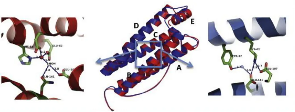
Fig. 1.2: Three-dimensional structure of ferritin monomer. Ferritin
subunit consists of a four-helix bundle composed by two couples of anti-parallel α-helices (A–B helices and C–D helices respectively) connected by a long loop and a short C-terminal α-helix (E) that lies approximatively at 60° to the bundle. In the central panel, the superimposition of human ferritin L-chain (blue) and H-chain (red) is reported. The two subunits display a different aminoacid arrangement in the central part of four-helix bundle, as highlighted in the blue box. More precisely, the H-chain is characterized by a set of iron coordinating residues that constitute the ferroxidase center (shown in detail on the left side of the image). Those residues are instead replaced by a salt bridge forming amino acids in the L chain (shown on the right). [modified from Levi S et al., (2015)].
H-chain and L-chain are isostructural and assemble in different proportions to make various tissue-specific isoferritins. Tissues that need to minimize iron toxicity or exhibit high ferroxidase activity (i.e. heart and brain) contain isoferritins with high proportion of H-chain, while tissues such liver and spleen that are involved in long-term storage of iron have an excess of L-chains [Drysdale J, (2008)]. The H and L subunits, in fact, play a complementary role within the protein functions. The H-chain contains a dinuclear ferroxidase site that catalyses the oxidation of ferrous ion by O2, producing H2O2. The L-chain,
11
instead, lacks catalytic activity but holds a microenvironment that facilitates iron nucleation and mineralization.
The ferroxidase site is located in the central hydrophilic region of H-chain and includes seven residues that are essential for maximal activity (shown in Fig. 1.2). More precisely, the amino acids Glu27, Tyr34, Glu61, Glu62, His65, Glu107, Gln141 (according to Human H-ferritin numbering) are involved in iron binding and oxidation. Such residues are highly conserved in vertebrates and are also present in the ferritins of plants and bacteria. In L-chains, these aminoacids are substituted with hydrophilic residues with the exception of Tyr34 and Glu61 that remain highly conserved. At variance, all mammalian L-chains contain two glutamate residues (Glu57 and Glu60 in human L-chain) that are placed inside the iron storage cavity and are reported to bind metal ions. They provide the L-chain with increased efficiency in promoting ferrihydrite nucleation and iron mineralisation [Hempstead PD et al., (1997); Chasteen ND et al., (1999)].
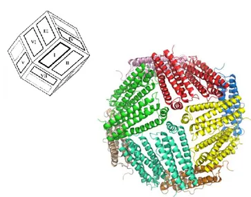
The three-dimensional structure of mammalian ferritin (shown in Fig. 1.3) is composed of 24 subunits assembled into a hollow spherical architecture. The protein shell is characterized by a 432-point symmetry and has the approximate geometry of a rhombic dodecahedron. Each of the twelve faces consists of a pair of subunits oriented with the E helices located at opposite ends of the dimer in an antiparallel topology. Although the oligomerization pathway is still unclear, these dimmers are very likely the first intermediates in the formation of the quaternary structure, in which each subunit interacts with other six subunits of the 24mer.
12
Fig. 1.3: Three-dimensional structure of mammalian ferritin.
Representation of mouse H-ferritin as model for canonical mammalian ferritin (PDB code: 3WNW). The molecule is shown down the 4-fold symmetry axis and each ferritin dimer is reported with the same color. As highlighted, the protein cage is shaped in a rhombic dodecahedron with each of the twelve faces consisting of a dimeric subunit.
The N-terminus, the BC loop and the A and C helices line the outer surface of the molecule, whereas the B and D helices are located inward. Subunits are tightly packed and a number of intersubunit interactions confer high stability to the protein structure. These interactions mainly occur at helix interfaces and involve hydrogen bonds, salt bridges and hydrophobic contacts between facing residues [Crichton RR et al., (2016); Crichton RR et al., (2010); Harrison P et al., (1998)]. Other stabilizing interactions consist in salt bridges between the highly conserved Asp84 and Lys86 (Human H-ferritin numbering) inside the BC loops of facing subunits. [Bernacchioni C et al., (2014)]. Point mutations, deletions and insertions have been extensively used to identify the residues involved in quaternary assembly. Such genetic engineering also allowed to
13
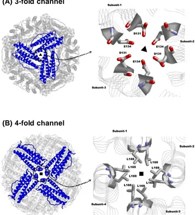
modify the structural properties to the ferritin scaffold [Chen H et al., (2016); Huard DJ et al., (2013); Tosha T et al., (2012)]. The protein assembly leads to the formation of channels, which connect the inner cavity to the external environment. These channels lie along the 4-fold and 3-fold axes of symmetry and allows for the entry and the exit of ions and little diffusants through the protein shell (Fig. 1.4). The eight 3-fold channels, located at the convergence of three contiguous subunits, are lined by the C-terminal end of helix C and the N-terminal end of helix D of each subunit. They are hydrophilic and negatively charged pores with a funnel shaped structure. The inner passage is narrow (3.4 Å diameter) and lined by three aspartates and three glutamates (Asp131 and Glu134, in human H-ferritin) from the D helices. These aminoacids are highly conserved in both H and L vertebrate ferritins and confer cations selectivity to the channels, that are functional to the transit of ferrous ions through the protein shell.
The six 4-fold channels, instead, are bordered by the E helices of four neighbouring subunits, whose helical axes point parallel to the inner cavity. Each E helix provides the channel with three leucine residues (Leu158, Leu165, Leu169) aligned along the inner surface of the pore, making a long hydrophobic channel. The fourfold channels are impermeant to all cations with the exception of protons. They are probably involved in protons transfer inside and outside ferritin, that is essential to maintain the electroneutrality of the protein during iron oxidation and mineralization [Crichton RR et al., (2010); Bernacchioni C et al., (2014); Takahashi T et al., (2003)].
14
Fig. 1.4: Schematic representation of ferritin 3-fold channels and 4-fold channels. The eight 3-4-fold channels (A) are formed at the
juxtaposition of three contiguous subunits. The six 4-fold channels (B), instead, are located at the convergence of four subunits. For both these pores, the lining aminoacids are shown. [modified from Bou-Abdallah F et al., (2010)].
(A) 3-fold channel
15
1.2.2: Iron oxidation and mineralization in
Mammalian ferritins
Despite the differences in the aminoacid sequence and kinetic parameters, all vertebrate ferritins share a common catalytic mechanism. Ferrous ions move through the protein shell along the 3-fold channels and once inside the cavity they migrate to the ferroxidase center. The ferroxidase center represents the catalytic site where iron oxidation occurs, yielding H2O2 as side product. Ferric
ions are then mineralized in the presence of phosphate and shape the mineral core [Theil EC et al., (2013)].
Iron uptake and movements inside the ferritin cavity are driven by electrostatic gradients between the actives sites of the protein, which are correlated with high charge density. The external surface of human H-ferritin is characterized by highly conserved acidic residues (Asp84, Asp89, Asp91, Asp92, and Glu94) and the inner passage of 3-fold channels is lined by conserved glutamates and aspartates from the three facing subunits (Glu131, Asp134 and Glu140). The ferroxidase center too displays a wide region of negative residues (Glu27, Glu61, Glu62, Glu107, Glu140 and Glu147) and six glutamates at the nucleation site (Glu61, Glu64 and Glu67 from two different monomeric subunits) form a ring of negative charges. Several asparagine, glutamine and lysine residues (Gln10, Asn11, Asn109, Gln112 and Asn125), instead, surround the external entrance of the 3-fold channels. Thus, the 3-fold channels, the ferroxidase site and the nucleation site all show negative values of the electrostatic potential, while the outer entrance to the three-fold channels is characterized by regions of positive potential. This arrangement creates an electrostatic field along the 3-fold channels directed towards the interior cavity that guides cation entrance into the protein cavity [Douglas T et al., (1998)]. Several other residues also contribute to the translocation of iron along the 3-fold channels and the accepted mechanism of iron entrance is reported in Fig. 1.5: the His118 and Cys130 residues are responsible for the initial contact of fully hydrated ferrous ions ([Fe(H2O)6]2+) at the wide entrance of the 3-fold
16
channels. The hexaaquo ferrous ions then migrate to the deeper triads of Glu134 and Asp131 residues and move along the narrow path of the pore gradually loosing their solvation sphere.
X-ray measurements of human H-ferritin confirmed two [Fe(H2O)6]2+ ions
coordinated by Asp131 and Glu134 and similar results were also obtained the crystal structure of frog M-ferritin treated with Fe+2 [Bradley JM et al., (2017); Pozzi C et al., (2015) (a); Pozzi C et al., (2015)(b)].
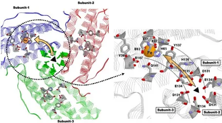
Fig. 1.5: Putative Fe+2 pathway from the 3-fold channel to the
ferroxidase center of ferritin. Residues responsible for iron initial
binding and migration are reported. Moreover, the aminoacids of the ferroxidase center are also shown [ modified from Bou-Abdallah F et al., (2010)].
Electrostatic factors appear to play a dominant role also in the migration of ferrous ions to the ferroxidase center. In fact, the ferroxidase site too has very negative values of the potential, which attract ferrous ions from the inner opening of the 3-fold channels. Bou-Abdallah and co-workers proposed that
17
the residue Thr135, His136, and Tyr137 could be involved in this pathway [Bou-Abdallah F et al., (2008)].
The ferroxidase center of H-chain catalyses the fast oxidation of Fe+2 to Fe+3. During the reaction, two Fe+2 ions are simultaneously oxidized from a single molecule of O2 that is reduced to H2O2, yielding a Fe+2/O2 ratio of 2:1.
The ferroxidase center comprises a dinuclear iron center, consisting of two metal binding-sites named site A and B. All the seven residues involved in the catalytic mechanism (shown in Fig. 1.6 (a)) are highly conserved along the evolutionary paths and are found also in prokaryotic and plant ferritins. The first Fe+2 ion is bound in the site A by a histidine (His65) and a monodentate
glutamate (Glu27), while the second Fe+2 in site B is ligated by a terminal glutamate (Glu107). Moreover, a bridging glutamate (Glu62) contributes to iron binding in both sites. An additional glutamate (Glu62) is found to adopt two conformations, one bound to the metal at the B site, the other projecting toward the cavity, suggesting its involvement in the transfer of ferric iron into the cavity. Besides these residues involved in metal binding, also Gln141 and Tyr34 residues are strictly conserved amongst eukaryotic and prokaryotic ferritins. The latter residue, in particular, seems to play a major role in the electron transfer between the cavity and the ferroxidase center that occurs within the catalytic mechanism [Bradley JM et al., (2014); Ebrahimi KH et al., (2013)].
The ferroxidase reaction (reported in eq.1) proceeds through several steps and leads to the production of different intermediate species. A general overview of this mechanism is reported in Fig. 1.6 (b) [Bou-Abdallah F, (2010)].
Eq.1: 2Fe2+ + O2 + 4H2O → [Fe2O2]2+ →
18
The resulting ferric ions then leave the catalytic center as oxo(hydroxo)-complexes and move to the inner cavity of ferritin, where become part of the stable nucleus of ferrihydrite mineral core. The nucleation site is correlated with the highest negative potential value in the ferritin structure, indicating that this region is very attractive to positively charged ions. A major role in this sense is played by the L-chains, that support the ferroxidase activity of the H-subunits by accelerating iron translocation from the di-iron center into the cavity. Moreover, the L- subunit assists the formation of polynuclear ferrihydrite crystals at the nucleation site and contributes to its electrostatic stabilization [Crichton RC et al., (2010)].
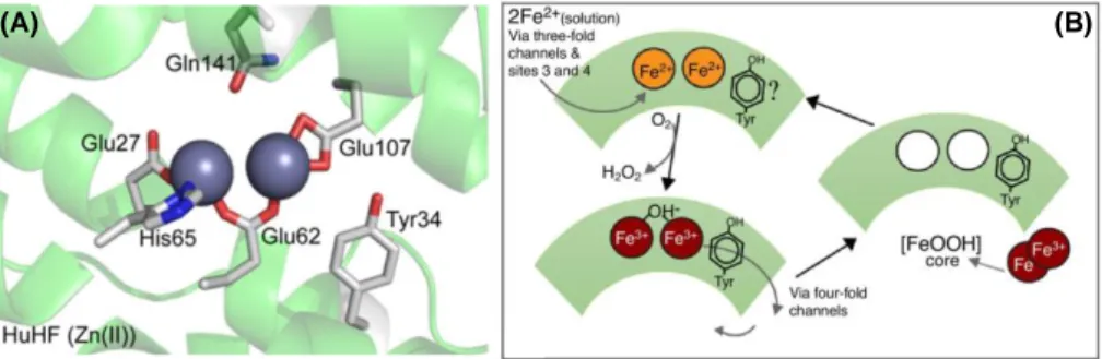
Fig. 1.6 (a): Structural features of the H-chain-type ferroxidase center.
(A): Structure of Human H-chain ferroxidase center with Zn+2 bound at the
center. The A and B binding sites are displayed [from PDB 2CEI; modified from Bradley JM et al., (2014)].
(B): Schematic representation of ferroxidase reaction [modified from Bradley JM et al., (2017)].
(B) (A)
19
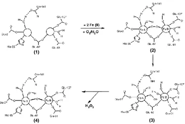
Fig. 1.6 (b): Accepted catalytic mechanism of the H-chain-type ferroxidase center. This scheme reports the accepted catalytic mechanism
and the overall intermediates produced within the ferroxidase reaction: two
Fe+2 ions stepwise bind to the sites A and B of Apoprotein (1). Dioxygen
subsequently binds to site B, leading to the production of the μ-1,2-peroxo di-iron(III) intermediate (2). This complex undergoes towards hydrolysis, thus producing the –hydroperoxo di-iron (III) complex (3). This intermediate quickly decays to the –oxo di-iron (III) complex (4), releasing an H2O2 molecule. Lastly, the resulting ferric ions leave the
catalytic center as oxo(hydroxo) iron (III) complexes to become part of the growing mineral core [modified from Bou-Abdallah F et al., (2010)].
Besides the ferroxidase mechanism, other two iron oxidation reactions occur within the protein shell, whose relevance is a function of the iron flux into the protein. They correspond to the mineral surface and the Fe+2 + H
2O2
detoxification reactions. The mineral core building in vertebrate ferritins thus results from the combination of these three pathways [Zhao GH et al., (2003); Yang XK et al., (1998); Xu B et al., (1991)].
At low iron flux (48-800 Fe+2/protein), iron oxidation is entirely carried out at the ferroxidase centre (see eq.1). At higher iron loadings (higher than 800
(1) (2)
(3) (4)
20
Fe+2/protein), instead, the dinuclear site is saturated and the dominant reaction switches to the mineral surface reaction (eq.2). Under such conditions, the oxidation and deposition of further iron directly occurs on the growing surface of the mineral in an autocatalytical way, in agreement with the crystal growth model. The process involves the transfer of four electrons and results in the complete reduction of dioxygen to water [Bradley JM et al., (2014)].
Eq.2: 4Fe+2 + O2 + 6H2O 4FeOOH(core) + 8H+
Lastly, at intermediate iron loadings (100-500 Fe+2/protein), part of the H2O2
produced during the ferroxidase mechanism is consumed in the detoxification reaction (eq.3). This reaction allows the ferritin to minimize the hydroxyl radical production during mineralization and to avoid damages related to ROS production [Bou-Abdallah F, (2010)].
Eq.3: 2Fe+2 + H2O2 + 2H2O 2FeOOH(core) + 4H+
Thus, once the core has formed, the mineral core growth is carried out by a complex interplay of multiple iron oxidation pathways.
21
1.2.3: Mammalian Ferritin biophysical properties
The high number of non-covalent interactions and the tight subunit packing provide the ferritin scaffold with an extremely high resistance to chemical denaturation, pH changes and heat stress.
Ferritins are endowed with an uncommon thermostability, that is generally exploited for the purification of both native heteropolymers and recombinant homopolymers. They easily withstand temperature higher than 70°C and the thermal stability improves as a function of the L-chain content within the protein shell [Stefanini S et al., (1996)].
However, the most attractive feature of ferritin nanocage is the ability to undergo towards a pseudoreversible disassembly-reassembly process controlled by pH. Nearly all ferritins disassemble into subunits under extreme acidic conditions (pH ~ 2) without denaturing. They then reconstitute the 24-mer shell when pH is adjusted back to ~7. Both pathways proceed in a stepwise manner, leading to the hierarchical formation of several oligomeric intermediates [Kim M et al., (2011)].
Gerl and co-workers proposed a general mechanism for ferritin assembly that has been further confirmed by several authors. Monomeric subunits are highly unstable in solution and rapidly dimerize, thus forming stable dimers that act as single structural units. The protein self-assembly then proceeds through a sequence of association steps during which dimers combine among themselves and produce a mixture of partially assembled subunits. These subunits include tetramers, examers and, in small amounts, dodecamers. Hence, tetraeicosameric cage would result from the progressive juxtaposition of dimeric units via tetramer and hexamer intermediates [Zhang Y et al., (2011); Gerl M et al., (1987); Gerl M et al., (1988)].
Measurements on mammalian ferritins pointed out that structural recovery upon acidic treatment occurs within one hour, being faster for the H/L-chain heteropolymers than for the H-homopolymers. The formation of heteropolymers is favourited respect to the corresponding homopolymers and
22
this is mainly due to the preferential formation of H/L heterodimers over homodimers during the self-assembly process [Carmona F et al., (2017)]. The self-assembly property encouraged the employment of recombinant ferritins as biomedical nanocarriers. Bioactive compounds can be encapsulated within the internal cavity but the extremely acidic pH required for the protein shell dissociation might represent a major concern for the stability of several molecules. Hence, several works have been carried out in order to engineer ferritin interfaces and allow for the dissociation under milder conditions. Modifications involving the Lys62-Glu107 salt bridge in human H-chain, for instance, increases its stability to low pH and other denaturants [Santambrogio P et al., (1992)]. Chen and co-workers recently developed a novel construct by cleaving the last 23 amino acids at the C-terminal end of human H-ferritin, which correspond to the DE turn and helix E and are involved in the 4-fold interactions. The deletion yielded a nanocages which disassemble at pH 4.0 and reassemble at pH 7.5 [Chen H et al., (2016)]. The engineering of ferritin surfaces even allowed to make the self-assembly of ferritin controllable by divalent copper binding [Huard DJ et al., (2013)].
A deeper understanding of the assembly mechanism and of the interactions that occur at the ferritin interfaces are therefore mandatory for the development of novel constructs suitable for biomedical applications.
23
1.3 Archaeal and bacterial ferritins
Archaeal and bacterial ferritins, as plant ferritins, are homopolymers composed of 24 identical subunits.
The ferritin from E. coli (FtnA) was discovered in the early ‘90s and is probably the one that has been most extensively characterized among the archetypal bacterial ferritins [Hudson AJ et al., (1993)]. Homologue proteins have been subsequently identified in several other bacteria, such as
Campylobacter jejuni, Porphyromonas gingivalis and Helicobacter pylori
[Clerte S et al, (1999); Ratnayake DB et al, (2000); Waidner B et al., (2002)]. More recently, two ferritins have been identified form Archaea Archaeoglobus
fulgidus (Af-Ft) and Pyrococcus furiosus (Pf-Ft), which bear an impressive
similarity to the bacterial ones [Johnson E et al., (2005); Tatur J et al., (2007)]. Although only distantly related to eukaryotic H-chain polypeptide (∼22% sequence identity), prokaryotic ferritins share most of their structural and functional features with vertebrate ones. Each subunit is shaped into the characteristic four-helical bundle (helices A–D), with the fifth short helix E arranged with a slope of 60°. Short unfolded regions are found at the N- and C- terminal ends, while the long unfolded BC loop traverses all the length of the bundle and connects the helices B and C. The protein shell consists of 24 subunits very similar to vertebrate H-chains, therefore all catalytically active. Subunits are arranged into the typical 4,3,2 octahedral symmetry and channels are formed at the juxtaposition of subunits (Fig. 1.7). The 3-fold channels are significantly less polar than their eukaryotic equivalents, being lined by both hydrophobic and hydrophilic residues. The 4-fold channels, instead, are polar at both inner and outer entrance and hydrophobic in the central part.
Several residues involved in inter-subunit interactions and in the three-dimensional assembly are unchanged with respect to vertebrate ferritins. Similarly to eukaryotic proteins, most of prokaryotic ferritins display the characteristic pH-dependent assembly, although important differences may
24
arise depending on each sequence. Besides, the tight packing of the helical-bundle and the high number of hydrogen bonds and salt bridges that occur within the subunit confer an overall thermostability to prokaryotic ferritins. Ferritin from hypertermophilic organisms such as Archaeoglobus fulgidus are endowed with an extreme thermal resistance and among these, the Pyrococcus
furiosus one is considered the most thermostable. It has been stated that the
preservation of the monomer fold, rather than the 24-mer assembly, protects the ferritin from inactivation by heat [Le Brun NE et al., (2010); Stillman TJ et al., (2001); Tatur J et al., (2007)].
1.7: Three-dimensional structure of prokaryotic ferritin. Cartoon
representation of E.coli FtnA subunit dimer (PDB 1EUM); (B) overall threedimensional structure of E.coli FtnA tetraeicosamer [modified from Le Brun NE et al., (2010)]. (C): sequence comparison of Pyrococcus
furiosus ferritin (PfFtn), Archaeoglobus fulgidus ferritin (AfFtn), E.coli
ferritin (EcFtn) and human H ferritin (HuHF) [modified from Tatur J et al., 2007].
(A) (B)
25
The major differences between prokaryotic and vertebrate ferritins are related to the structure of the catalytic site. The bacterial ferroxidase centre closely resembles that of mammalian H-chain, comprising the typical dinuclear iron centers (site A and B) and all the seven residues that has been reported to be involved in eukaryotic catalytic mechanism. However, prokaryotic ferritins have an additional and closely located third iron-binding site (site C) that is unique to these proteins and is ∼7 Å away from the B-site and ∼11 Å from the A-site.
The structure of the ferroxidase center of E. coli FtnA in its iron-bound form is shown in Fig. 1.8. Iron is coordinated in site A by a monodentate glutamate (Glu17), a histidine (His53) residue and a bridging glutamate (Glu50). Besides Glu50, iron is ligated at site B by a terminal glutamate (Glu94) and by another glutamate ligand (Glu130). Glu130 residue is actually a bridging ligand to the third iron-binding site, the site C, which comprises three additional glutamates (Glu49, Glu126 and Glu129) for iron coordination. Lastly, prokaryotic ferroxidase center includes two additional residues (Tyr24 and Gln127) also involved in the catalytic reaction [Bou-Abdallah F et al., (2014); Bradley JM et al., (2014); Treffry A et al., (1998)]. Sequence comparisons showed that site C ligand and ferroxidase center residues and are highly conserved among prokaryotic ferritins. These include even two archaeal proteins from
Archaeoglobus fulgidus (Af-Ft) and Pyrococcus furiosus (Pf-Ft) [Le Brun NE
26
Fig. 1.8: The prokaryotic-type ferroxidase center. Three-dimensional
structure of E.coli FtnA ferroxidase center. (A): representation of bridged A and B sites where residues Gln127, Glu130, Glu94, Tyr34, Glu50, His53 and Glu17 accounts for iron binding. The center is shown without the site C to highlight the similarity with mammalian ferroxidase center. (B): representation of the overall ferroxidase center with site C included. Residues Glu49, Glu129 and Glu126 of site C provide the prokaryotic ferroxidase center with an additional iron binding site [modified from Bradley JM et al., (2014)].
Iron mineralization occurs via a mechanism related to that of eukaryotic H-chains but complicated by the presence of the site C (Fig. 1.9). As for mammalian H-chain, the binding of ferrous ions and dioxygen at the ferroxidase center leads to iron oxidation. All iron oxidation intermediates shown in vertebrate mechanism are formed also in bacterial and archaeal proteins, although the relative rate of formation and decay can represent a limit to their detection [Honarmand Ebrahimi K et al., (2009)]. Moreover, isothermal titration calorimetry measurements demonstrated the existence of essentially two classes of binding sites for each subunit, with a stoichiometry of 2 Fe+2 for the site A and B of each dinuclear ferroxidase centers [Bou-Abdallah F et al., (2005)].
27
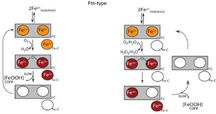
Fig. 1.9: catalytic mechanism of prokaryotic ferroxidase center and alternative roles proposed for site C. On the left, the site C acts as an
additional catalytic site contributing to Fe+2 oxidation. Upon oxidation of
Fe+2 atoms bound at the site C, the electrons produced are transferred to the
nearby dinuclear catalytic center and to the Fe+2 atoms bound to the A and
B sites. On the right, site C acts as an iron transit/holding site slowing down the iron flux to the nearby dinuclear catalytic center [modified from Bradley JM et al., (2014)].
The role of the site C has not yet been entirely clarified. Compared to site A and B, it exhibits just weak iron binding properties and seems rather involved in modulating the stoichiometric and kinetic properties of the protein. E.coli FtnA, for instance, differs from human H-chain in having an Fe2+/O2
stoichiometry for the initial oxidation reaction of ∼3 versus ∼2 for human one. Similarly, Pf-Ft has a stoichiometry of ∼3. The observation that the iron/dioxygen stoichiometry dropped to 2 when site C residues were substituted further confirmed this hypothesis [Yang X et al., (1998); Xu B et al., (1991); Treffry A et al., (1998)].
Moreover, the site C appears to be of variable importance for catalytic activity in ferritins from different organisms. In FtnA from E.coli, the site C appears to control the iron flux to the ferroxidase center and to modulate Fe+2 binding at the adjacent iron sites A and B, working both as a transit site and as gated iron
28
pore. A strong interplay exists between the site C and the dinuclear site, suggesting the presence of inter- and intrasubunit negative cooperativity. Site C seems to slow iron oxidation and turnover at the dinuclear ferroxidase center, as the iron flux through the ferroxidase centre of FtnA is much slower than the eukaryotic H-chain one. Moreover, the loss of site C makes the center became structurally and functionally similar to the H-chain-type center [Bou-Abdallah F et al., (2014); Bou-Abdallah F et al., (2005); Treffry A et al., (1998)]. In Pf-Ft from Pyrococcus furiosus, instead, the site C seems to play a different role and contributes to iron oxidation and mineralisation. An electron transfer occurs from the Fe+2 bound at the site C to the di-Fe+3–protein complex at the
dinuclear site. The ferroxidase center therefore acts as a stable catalytic center and accepts electrons derived from the oxidation of Fe2+ at nearby site C. The resulting Fe+3 then leaves the C site to the cavity and becomes part of the mineral core, while molecular oxygen is reduced to hydrogen peroxide to complete the cycle of the electron transfer chain [Tatur J et al., (2005); Honarmand Ebrahimi K et al., (2009); Bou-Abdallah F et al., (2010)].
Hence, even if the exact role of the site C in prokaryotic ferritins still remains undefined, it is generally accepted that it fulfils distinct tasks in different proteins, contributing to iron oxidation in some while acting as transit and holding site in others.
However, some major point regarding prokaryotic catalytic properties are still debated: a parallel mechanism for iron oxidation seems to occur, as a tyrosyl radical related to the highly conserved Tyr24 has been observed in both Ftna and Pf-Ft proteins. It has been proposed that the electron from this radical, along with three resulting from oxidation of Fe2+ ions at sites A, B and C,
would participate to the four-electron reduction of O2 to water [Bradley JM et
al., (2014)].
As for mammalian proteins, the mineral core building in prokaryotic ferritins results from multiple iron-oxidation pathways. At low iron flux, the catalytic reaction at ferroxidase center is dominant. At higher iron loading, iron
29
oxidation directly occurs on the mineral surface in an autocatalytic way. The hydrogen peroxide collaterally produced by the ferroxidase reaction further oxidises Fe+2 ions, contributing to the mineral core growth and to the radical detoxification [Bou-Abdallah F et al., (2014)].
Among the bacterial structures, the Archaeoglobus fulgidus ferritin (Af-Ft) has recently emerged due to its unprecedented structural and biophysical features. Af-Ft shares a high sequence identity with human H-ferritin, E. coli ferritin A and Pyrococcus furiosus ferritin (31%, 37% and 50% respectively). Moreover, it is closely related to archetypal bacterial and eukaryotic ferritins in its iron binding mode, secondary and tertiary structure. In spite of the high degree of structural similarity, the quaternary structure of Af-Ft strikingly differs from the other three. In fact, whereas the archetypal ferritins have the canonical octahedral (4-3-2) architecture, the Af-Ft tetracosamer exhibits a tetrahedral (2-3) symmetry, which is typical of smaller dodecamer ferritin-like proteins (Fig. 1.10). This unusual assembly does not display the 4-fold channels and constrains the quaternary structure, thus leading to the appearance of four large triangular openings (~ 45 Å wide) in the protein shell. These large pores display positive electrostatic surface potential at the apices and provide the ferritin molecule with unconventional route for the movement of ions and larger molecules through internal cavity. The “open” quaternary structure of Af-Ft contrasts the “closed” paradigm of a typical ferritin cage and to date is considered unique among all other known structures [Johnson E et al., (2005)].
30
1. 10: Three-dimensional structure of Archaeoglobus fulgidus ferritin.
On the left, three-dimensional representation of the unique “open” tetraeicosameric assembly of Archaeoglobus fulgidus ferritin (PDB code: 1S3Q). In detail, one of the four 45 Å-wide triangular pores at the 3-fold symmetry axis is shown. On the right, a closed archetypal tetraeicosameric ferritin (human H-ferritin, PDB code: 2FHA) is reported for comparison purposes [modified from Johnson E et al., (2005)].
Af-Ft also exhibits uncommon association-dissociation properties, as its self-assembly seems to be dependent on ionic strength instead of pH. Af-Ft nanocage largely exists as tetraeicosamer at high ionic strength, while the equilibrium shifts towards dimeric species at low ionic strength. Several hydrophobic contacts in fact occur at the subunit interfaces and play an important role in the stabilization of the assembled structure. Hence, salt-mediated association is driven by the improvement of hydrophobic interactions at high ionic strength [Swift J et al., (2009)].
The stability of the Af-Ft tetrahedral configuration is governed by two critical and non conserved residues in the helix E of the 4-helices bundle, namely Lys150 and Arg151 (Fig. 1.11). The side chain of Lys150 forms an hydrogen bond with the backbone O of Met111 that stabilizes the tetrahedral configuration. The Arg151 side chain, instead, sterically hinder the association
31
of helices E at the octahedral 4-fold interface, thus preventing the octahedral assembly [Johnson E et al., (2005)].
To unravel the structural role of these residues, a double mutant (namely Af-Ft-AA or Af-Ft K150A/R151A) has been recently developed by replacing Lys150 and Arg151 with two alanines. The replacement of these key residues produces an important rearrangement of the quaternary structure and flips a “symmetry switch” from the Af-Ft tetrahedral structure to the Af-Ft-AA octahedral symmetry. In Af-Ft-AA, the canonical “closed” architecture of the archetypal ferritins is restored due to the stabilization provided by the four mutant Ala151 residues at the 4-fold symmetry interfaces (Fig. 1.11). Although Af-Ft-AA retains salt-mediated assembly properties, its tetraeicosameric shell is much more stable compared to Af-Ft one, as a result of the improvement of the interface hydrophobic interactions provided by these mutations [Sana B et al., (2013)].
1.11: comparison of ferritin shell pores in Af-Ft and Af-Ft-AA proteins. (A) detailed view of the large triangular pore of wild type Af-Ft
ferritin (PDB code: 1S3Q). Residues Lys150 and Arg151 are drawn as red and blue sticks and their relative intermolecular distance in reported. (B) Detailed view of narrow pore of Af-Ft-AA mutant at the 4-fold simmetry axis (PDB code: 3KX9). Ala150 and Ala151 residues are shown with their relative intermolecular distance in evidence [modified from Sana B et al., (2013)].
32
1.4 Biotechnological applications of
ferritins: state of art and future
perspectives
The last few years have seen a growing interest in the development of biomedical nanodevices to improve the pharmacokinetic and toxicological profile of therapeutics or diagnostic probes. Several inorganic or polymeric nanoparticles have been designed, but just a few of them have been clinically approved due to their toxicity, non-selective biodistribution and general low clearance. Liposomes as well are suitable for improving drug bioavailability and reducing toxicity profile, but generally lack of method for targeting a specific tissue or cell type [Peer D et al., (2007); Mok H et al., (2013); Xing M et al., (2015)].
Protein nanodevices, instead, are particularly attractive as nanomedicine platforms: compared to polymeric materials, proteins are highly biocompatible and biodegradable materials, displaying as well lower toxicity although immunogenicity may be a significant drawback. Recombinant technologies allow to produce highly purified proteins at gram scale and several chemical or genetical manipulation can be employed to further modify their structure [Elzoghby AO et al., (2012); Maham A et al., (2009)].
In this context, ferritin is arising as an extremely promising scaffold for the development of smart nanocarriers. Both the inner and the outer surface of ferritin can be engineered, thus conferring several functionalities onto a single molecule. Its ability to assemble and disassemble in a pH-dependent manner allow to encapsulate a wide variety of molecules within the cavity, while the interactions with the inner surface prevent the leakage of the cargo. Figure 1.12 reports a general overview of currently employed strategies for drug loading and release using protein cages as smart nanocarriers.
33
Fig. 1.12: Biotechnological approaches for drug loading and release using protein cages [ modified from Molino et al., (2014)].
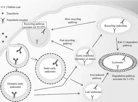
Moreover, ferritin is endowed with attractive targeting features to specific cell types. Li and co-workers pointed out that extracellular ferritin interacts with cells through the Transferrin Receptor-1 (TfR-1) and undergoes internalization, being found in early and recycling endosomes [Li L et al., (2010)]. The pathway for tranferrin recognition and internalization by TfR-1 receptor is shown in Fig. 1.13. Most likely, ferritin molecule follow the same route. As TfR1 is overexpressed in many types of cancer cells, ferritin can provide the selective homing of drugs or probes to tumors. The cellular specificity of this protein has been further extended by addition on the outer surface of chemical or peptide targeting moieties, such as the RGD peptide, the anti-melanocyte stimulating hormone peptide, the extracellular domain of
34
myelin oligodendrocyte glycoprotein and several antibodies [Truffi M et al., (2016)].
Figure 1. 13. Transferrin-1 receptor-mediated endocytosis. Upon
binding, transferrin undergoes internalization through a clathrin-coated endocytosis mechanism thus reaching the cytoplasm. Transferrin enters the endosomes where dissociation of iron from transferrin occurs. The complex is then recycled back to the surface (85-95%) or undergo lysosomal degradation (5-15%) [modified from Tortorella et al., (2014)].
35
1.4.1: Therapeutic applications
The major limit of conventional chemotherapy is the lack of selectivity in the mechanism action, which leads to harmful side effects, drug resistance and difficulty in dosage selection. A drug delivery system is thus required to prevent the exposure of healthy cells to the drug and to ensure the proper delivery to cancer cells. The employment of engineered ferritin constructs provided encouraging results in this sense [Truffi M et al., (2016)].
Cisplatin has been successfully entrapped in the ferritin shell and the pharmacological properties of the resulting nanoparticles have been studied on tumour lines [Yang Z et al., (2007); Xing R et al., (2009); Ji XT et al., (2012)]. In particular, Falvo and co-workers recently developed a novel antibody–drug conjugate by chemically functionalising the cisplatin-loaded apoferritin with an antibody against the melanoma antigen CSPG4 [Falvo E et al., (2013)]. All these studied provided strong evidence of a general improvement of the cisplatin antiblastic efficacy even in tumor such as melanoma, which is totally refractory to traditional chemotherapy at its advanced stages.
The inner cavity of ferritin is also suitable for the incorporation of non-metal-containing drugs, despite lower interactions occur with the protein cage. Doxorubicin is a widely used anticancer drug but is affected by major toxicities and poor pharmacokinetic properties. The encapsulation within the protein cage yielded in an improvement of Doxorubicin bioavability and, due to targeted delivery, the tumor growth inhibition was achieved at lower doses compared to the plain drug [Liang M et al., (2014); Kilic MA et al., (2012)]. Recent findings also highlighted that Doxorubicin-loaded ferritin translocates into the nucleus and releases the drug directly into the nuclear compartment. Although the mechanism is not fully understood, this outcome suggests that ferritin is intrinsically able to undergo nuclear translocation and can be exploited for the development of a novel class of smart drug delivery system with nuclear target [Zhang L et al., (2015); Bellini M et al., (2014)].
36
The ability of ferritin to sequester transition metals can been further exploited in radiotherapy cancer treatment to deliver radioisotopes to the tumor. Hainfeld demonstrated that ~800 235U atoms can be encapsulated into each nanocage. The resulting nanoparticles provide enough radiation to kill surrounding tumor cells [Hainfeld JF et al., (1992)]. Moreover, a novel construct of human ferritin has been recently developed for the targeted delivery of small interfering RNA (siRNA) to cancer cells [Lee EJ et al., (2015)].
Self-assembling protein nanoparticles are currently studied also as novel potential platforms for vaccination and immunization purposes. While the 19th and early 20th century vaccines were made of killed or inactivated pathogens, modern ones contain highly purified antigens obtained by recombinant technologies. Modern vaccines are thus safer, but tend to induce lower levels of protective immunity. By fusing the antigen onto the monomeric subunit, ferritin shell can present multiple copies of antigens with higher stability and in a well-ordered manner. Numerous binding events can occur simultaneously between the nanoparticle and the host immune cells, thus resulting in the improvement of the immunogenicity. Moreover, since ferritin is composed by subunits related by three-fold axis symmetry, it is perfectly suitable for the presentation of trimeric antigens [López-Sagaseta J et al., (2015)].
Kanekiyo and co-workers fused the influenza virus haemagglutinin (HA) at the interface of adjacent subunits of Helicobacter pylori ferritin, so that it spontaneously assembled into eight trimeric viral spikes on the external surface. Injected in mice, the particle elicited the production of neutralising antibodies against different HA variants with an higher potency compared to a commercially available influenza vaccine. Moreover, no autoimmune reaction against the Helicobacter pylori ferritin was observed, despite the high degree of divergence from mammalian ferritin sequences [Kanekiyo M et al., (2013)]. For similar purposes, other groups linked antigens from Epstein–Barr virus and Hepatitis C virus onto the outer surface of ferritins [Kanekiyo M et al., (2015); He L et al., (2015)]. The satisfying outcomes support the hypothesis that ferritin may represent a useful scaffold for vaccination and immunization aims.
37
1.4.2: Bioimaging applications
The improvement of diagnostic technologies required the development of more sensitive and versatile diagnostic tools, in order to enhance screening accuracy and allow the early detection of malignancy onset [He D et al., (2015)]. Ferritin nanoparticle can be labelled with a wide range of dye molecules and tagged with targeting moieties. The resulting constructs can be used as optical imaging probes for both in vivo and in vitro imaging applications [Li X et al., (2012)]. Moreover, the pH-mediated procedure allows to combine differently engineered monomers into hybrid nanoparticles endowed with multiple functionalities [Lin X et al., (2011)].
Most of the diagnostic applications of ferritins are related to the versatility of the in vitro mineralization, which allows to internalize labelled heavy atoms and sequestrate them within the mineral core.
Magnetic Resonance Imaging (MRI), for instance, is an highly sensitive and non-invasive technique in tumor imaging that relies on the detection of proton spin alignment under a magnetic field. Paramagnetic contrast agents (such as Gd+3) are required to increase the contrast between normal and pathological tissues, but most of them lack specificity for cancer cells. Their broad biodistribution results in the enhancement of all highly vascularized tissues and micrometastasis may not be detected. Hence, the development of novel targeted carrier for MRI contrast agents are still a major concern to overcome these problems.
The iron oxide nanoparticles synthesized inside the ferritin cavity can be employed as contrast agents due to their super-paramagnetic properties and the dark contrast they give in MRI images [Jin R et al., (2014); Uchida M et al., (2006)]. However, native iron containing ferritins often display unsatisfactory degree of cellular selectivity and provide just a poor MRI contrast [Vande VG et al., (2011)]. On this basis, novel recombinant ferritins have been tagged with homing peptides and loaded with elements with higher MRI contrast than iron,
38
such as gadolinium [Hooker JM et al., (2007)]. A five-nanometer gadolinium nanoparticle have been produced inside ferritin, whose relaxivity was up to 70 times higher than commercially available Gd-chelates [Sanchez P et al., (2009)]. Gadolinium-loaded ferritin has been further engineered with a peptide epitope specific for the neural cell adhesion molecule and successfully employed for the visualization of Endothelial tumour cells [Geninatti CS et al., (2006)]. Mn-loaded ferritin as well has been reported to be a good MRI contrast agent with optimal relaxivity properties [Kalman FK et al., (2010)].
The ferritin capability of internalizing and mineralizing heavy atoms has been also exploited for Positron Emission Tomography (PET) applications. Lin and co-workers managed to load the ferritin nanocage with 64Cu, a radioisotope commonly used for PET, and further functionalize the outer surface with a fluorescent dye and homing peptide. The obtained probe demonstrated to be suitable for both for PET and fluorescence dually functional imaging [Lin X et al., (2011)].
All these studies highlighted that engineered ferritins hold an impressive potential in targeted diagnostic applications. Moreover, their ability to internalize heavy atoms with broad selectivity suggests their employment as smart nanocarrier for the delivery of metal ions with relevant biomedical properties. Lanthanide ions, for example, are emerging as novel fluorescent probes due to their unique photophysical properties [Handl HL et al., (2005); Hemmila I et al., (2005); Bunzli J., (2006)]. Lanthanides display narrowband emission spectra, large Stokes shift (150-300 nm) and long fluorescence lifetimes, which have been exploited in time-resolved spectroscopy to overcome the short living background signals [Rajapakse HE et al., (2009)]. Unlike traditional organic dyes, they are unaffected by photobleaching or photochemical degradation, which results in prolonged detection [Pandya S et al., (2006)]. Moreover, their fluorescence can be greatly enhanced by Förster resonance energy transfer (FRET) from an appropriately placed sensitizer-fluorophore, a phenomenon often referred to as “antenna effect”. FRET requires spectral overlap between fluorophore emission and lanthanide
39
absorption spectra and its efficiency relies on the close proximity between the two species (~5-6 Ǻ distance) [Gudgin Dickson EF et al., (1995)]. Tb+3 and
Eu+3 have been widely exploited due to their more intense microsecond fluorescence in the visible region [Selvin PR, (2002); Bunzli JC, (2004)], although several other lanthanides are currently evaluated as candidates for cancer treatment, photodynamic therapy and radiation therapy [Teo RD et al., (2016)].
Proteins are widely known to act both as lanthanide-chelating agents and FRET-sensitizer, due to their acidic and aromatic moieties, and such behavior have been largely exploited to study protein structure, interaction and metal binding properties [Allen JE et al., (2006); Xu X et al., (2002); O'Neil JD et al., (1984)]. However, wide possibilities would arise in biomedical field by combining the optical properties of lanthanides with the biotechnological features of protein nanocarriers.
1.4.3: Other applications
Beyond biomedical field, the ferritin scaffold has been successfully employed for several other biotech applications. Just to mention some of the most interesting, the ferritin cage has been exploited as a reaction chamber for the mineralization of different non-physiological metals and metal complexes. Ferritin encapsulated metal nanoparticles display homogeneous shape and atomic composition and can be extracted from the protein envelop through chemical denaturation. The obtained metal particles can be used for the production of ordered semi-conductor arrays, quantum dots, and as anti-bacterial nanoparticles [Yoshimura H, (2006); He D et al., (2015)].
Recently, an industrial-scale process for the water treatment has been developed based on Pyrococcus furiosus ferritin [Jacobs JF et al., (2010)]. Ferritin sequesters phosphate within its core along with iron and this feature
40
can be applied to remove phosphate from water and prevent water pollution and biofouling. Compared to the current methods, the ferritin-based solution is cheaper, highly stable, suitable for recycling and endowed with an high capacity for phosphate binding.
41
42
Ferritins are emerging as novel biotech platforms for biomedical applications due to their ability to encapsulate cargo molecules, broad functionalization possibilities and selective targeting properties. In this framework, the present work has been focused on the development and characterization of engineered mammalian and archaeal ferritin constructs to expand the scope of their nanotechnological applications.
Two ferritins from Archaea have been chosen as model to further investigate the biological and biophysical properties of prokaryotic homopolymers. The molecular diffusion through the ferritin cavity appears to be a complex phenomenon that is only partially understood. Despite experimental observations demonstrated that the permeation of small molecule through the mammalian 3-fold channels is a charge-selective process [Yang X et al., (1996); Crichton RR et al., (2010); Douglas T et al (1998)], the molecular diffusion within archaeal ferritins have been little investigated. Differences in the shape, aminoacid composition and electrostatic properties of prokaryotic open pores within these ferritins suggest different routes for small molecules entry and expand the biotechnological relevance of these proteins. In the present work, the Pyrococcus furiosus ferritin (Pf-Ft), the “open”
Archaeoglobus fulgidus ferritin and the “closed” Archaeoglobus fulgidus
construct (Af-Ft K150A/R151A) have been chosen as model of prokaryotic ferritins. A set of engineered mutants have been obtained by placing a reactive cysteine residue per subunit in the same topological positions either inside or outside the internal cavity. The permeability of the prokaryotic protein shell toward diffusants has been characterized by studying within these mutants the cysteine reactivity toward the bulky and negatively charged DTNB molecule (5,5'-dithiobis-2-nitrobenzoic acid).
Moreover, Archaeoglobus fulgidus ferritin has been genetically engineered by changing the surface exposed loop connecting helices B and C to mimic the sequence of the analogous human H-chain ferritin loop. This novel “humanized” chimeric construct (named HumAf-Ft) thus combines the unique open structure and self-assembly properties of Af-Ft with the typical human
43
H-ferritin ability to bind the Transferrin Receptor TfR-1, which is overexpressed in several types of tumor cells. HumAfFt has been structurally and biophysically characterized and the improved cellular uptake has been demonstrated on HeLa cell line.
Among mammalian isoforms, instead, mouse H-ferritin has been chosen as favoured scaffold for the development of bioimaging tools with potential in
vivo and in vitro applications.
To exploit lanthanide fluorescence properties and develop an intrinsically fluorescent nanoparticle, a novel construct has been developed by genetically fusing at the C-terminal end of mouse H-ferritin a lanthanide binding tag (LBT). LBTs are short peptides (20 or fewer aminoacids) that have been recently designed on the basis of calcium binding loop and selectively bind lanthanide ions at low-nanomolar affinities, thanks to six coordinating aminoacid side chains. They also include a tryptophan residue in its close proximity that provides strong FRET sensitization [Martin LJ et al., (2007)]. Our construct (named HFt-LBT) has been designed by locating the tag inside the inner cavity, so that the lanthanide ions diffusing through the surface pores can eventually bind to the LBT sequence. HFt-LBT would thus act both as carrier targeted to TfR-1 receptor and as a FRET sensitizer. Fluorescence improvement and lanthanide binding properties have been investigated by spectrophotometric measurements using Tb+3 as lanthanide probe. The structural characterization has been carried out and cellular uptake by HeLa cell line has been assessed as well.
44
3 Materials and
methods
45
3.1 Assembly and molecular permeability
of Archaeal ferritin mutants
Point mutations and protein expression
The genes encoding for bacterial ferritin from Af-Ft and Pf-Ft were cloned into the expression vector pET22b (Novagen). Point mutants FtM54C, Af-FtM54C/K150A/R151A, Pf-FtG52C and Pf-FtP77C were obtained by PCR using QuickChange Mutagenesis kit (Stratagene). The recombinant plasmids were transformed into E. coli TOP 10 cells and the resulting colonies were screened by DNA sequencing. Plasmids bearing the desired mutations were transformed into BL21(DE3) E. coli strain for protein expression. For each mutant, protein overexpression was obtained as follows: 1 L LB broth medium was inoculated with 2 ml overnight culture of a single colony and the gene expression was induced with 1 mM IPTG when the absorbance at 600 nm reached 0.6. Cells were harvested by centrifugation after overnight induction at 37 °C and the cell pellets were stored at −20°C.
Protein purification
Harvested cells form 1 L colture over-expressing FtM54C and Af-FtM54C/K150A/R151A mutants were resuspended in 20 ml buffer A (25 mM HEPES pH 7.5, 20 mM MgCl2) containing a cOmplete™ MiniProtease
Inhibitor Cocktail Tablet (Roche) and disrupted by sonication. The soluble fraction was thermally purified by heating at 85 °C for 10 min followed by removal of denatured proteins by centrifugation at 14000 rpm for 30min at 4 °C. The supernatant was fractioned by ammonium sulfate precipitation. 70% ammonium sulfate pellet containing highly purified protein was resuspended in buffer A, dialysed versus the same buffer, sterile filtered and stored at 4 °C. Cells over-expressing Pf-FtG52C and Pf-FtP77C were sonicated in 25mMHEPES buffer at pH 7.5 containing 0.5 mM EDTA, 0.3 M NaCl and
46
cOmplete™ Mini Protease Inhibitor Cocktail Tablet. After sonication, the crude bacterial extract was digested with DNase for 1 h at 37 °C, heated at 55 °C for 10 min and then at 80 °C for 8min. Heat treatment was followed by centrifugation to remove insoluble material and ammonium sulfate precipitation.70% ammonium sulfate pellet was resuspended in 20mM HEPES pH 7.5 plus 150 mM NaCl, dialysed versus the same buffer and loaded onto a HiLoad 26/600 Superdex 200 pg column (GE Healthcare). Fractions containing highly purified protein were pooled, sterile filtered and store at 4 °C.
Preparation of ferritin-DTNB adducts
All mutants were reduced with 3 mM TCEP (tris(2-carboxyethyl)phosphine) in their storage buffers and then loaded onto a desalting column (GE Healthcare) to remove the reducing agent. Each mutant was reacted with 40-fold molar excess of Ellman's Reagent (DTNB) per cysteine for 3 h at room temperature. Stock DTNB solutions were prepared in ethanol. The excess (non-reacted) reagent was removed by ultra-filtration using 100 kDa Amicon Ultra-15centrifugal devices (Millipore Corporate). The Ferritin–DTNB samples were analyzed by mass spectrometry as described below.
Stopped flow experiments
Kinetic measurements were carried out on a thermostated Applied Photophysics stopped-flow apparatus (Leatherhead, UK) by mixing 8–10 μM protein solutions, previously reduced with TCEP, with solutions containing different concentrations of DTNB (from 0.2 to 0.7 mM after mixing) in 20 mM HEPES, 20 mM MgCl2 pH 7.5. In order to avoid interference of the instrument
phototube from the high concentrations of DTNB and the released chromophore 5-thio-2-nitro-benzoic acid(TNB), the reaction was followed at 430 nm and the extinction coefficient calculated to be 12205 mM−1 cm−1, as
47
determined from the extinction coefficient of 14150 mM−1 cm−1 at 412 nm [ Riddles PW et al., (1979)]. All fitting procedures were carried out by using the Matlab software (Mathworks,USA). Experimental traces were fitted by non-linear regression to either exponential of biexponential processes by using an Levenberg-Marquardt algorithm.
Self-assembly study
MgCl2 -mediated self-assembly of ferritin mutants was studied by incubating
aliquots of proteins (1 mg/ml) with different salt concentrations in 25 mM HEPES buffer, pH 7.5. Molecular sizes of Af-FtM54C, Af-FtM54C/K150A/R151A, Pf-FtG52C and Pf-FtP77C was determined by size exclusion chromatography (SEC) using HiPrep 16/60 Sephacryl S300 column (GE Healthcare). The column was equilibrated with 25 mM HEPES, pH 7.5, containing MgCl2 at the same concentration in which the protein was
pre-incubated and the same buffer was used as mobile phase. Molecular weight of each mutant was determined by comparing their elution volumes with the elution volumes of standard proteins in the same salt concentration. Dynamic light scattering measurements (DLS) were performed using an ALV-5000 logarithmic correlator in combination with a standard optical set-up based on a He–Ne (λ = 632.8 nm) 10 mW laser and a photomultiplier detector. The intensity autocorrelation functions were directly obtained as g2(q, t) = 〈I (q, t)I (q, 0)〉/〈I (q, 0)〉2, where q is the modulus of the scattering vector defined as q = (4πn/λ)sin(θ/2) (θ = 90° in the present experiment). Quantitative analysis of the measurements was obtained through a fit of the data with a single exponential expression:g2(q, t) = 1+be−t/τ where b is the coherence factor and τ is the relaxation time related to the motion of the particles, specifically to the diffusion coefficient [Berne BJ et al., (1976)].

![Fig. 1.12: Biotechnological approaches for drug loading and release using protein cages [ modified from Molino et al., (2014)]](https://thumb-eu.123doks.com/thumbv2/123dokorg/2885525.10795/33.892.260.630.138.501/biotechnological-approaches-loading-release-using-protein-modified-molino.webp)