Large symptomatic gastric diverticula: Two case reports
and a brief review of literature
Luigi Marano, Gianmarco Reda, Raffaele Porfidia, Michele Grassia, Marianna Petrillo, Giuseppe Esposito, Francesco Torelli, Angelo Cosenza, Giuseppe Izzo, Natale Di Martino
Luigi Marano, Gianmarco Reda, Raffaele Porfidia, Michele Grassia, Marianna Petrillo, Giuseppe Esposito, Francesco Torelli, Angelo Cosenza, Giuseppe Izzo, Natale Di Martino, 8th General and Gastrointestinal Surgery, Department of Internal Medicine, Surgical, Neurological Metabolic Disease and Geria-tric Medicine, Second University of Naples, 80138 Naples, Italy Author contributions: Marano L and Di Martino N contributed equally to this work; Marano L and Di Martino N designed the research; Marano L, Di Martino N, Torelli F, Cosenza A, Reda GM, Porfidia R and Izzo G performed the research; Marano L, Di Martino N, Grassia M, Petrillo M and Esposito G analyzed the data; Marano L and Di Martino N wrote the paper.
Correspondence to: Luigi Marano, MD, 8th General and Gas-trointestinal Surgery, Department of Internal Medicine, Surgical, Neurological Metabolic Disease and Geriatric Medicine, Second University of Naples, Piazza Miraglia 2, 80138 Naples,
Italy. [email protected]
Telephone: +39-81-5665058 Fax: +39-81-5665055 Received: March 12, 2013 Revised: June 29, 2013 Accepted: July 18, 2013
Published online: September 28, 2013
Abstract
Gastric diverticula are rare and uncommon conditions. Most gastric diverticula are asymptomatic. When symp-toms arise, they are most commonly upper abdominal pain, nausea and emesis, while dyspepsia and vomiting are less common. Occasionally, patients with gastric diverticula can have dramatic presentations related to massive bleeding or perforation. The diagnosis may be difficult, as symptoms can be caused by more common gastrointestinal pathologies and only aggravated by diverticula. The appropriate management of diverticula depends mainly on the symptom pattern and as well as diverticulum size. There is no specific therapeutic strat-egy for an asymptomatic diverticulum. Although some authors support conservative therapy with antacids, this provides only temporary symptom relief since it is
not able to resolve the underlying pathology. Surgical resection is the mainstay of treatment when the diver-ticulum is large, symptomatic or complicated by bleed-ing, perforation or malignancy, with over two-thirds of patients remaining symptom-free after surgery, while laparoscopic resection, combined with intraoperative endoscopy, is a safe and feasible approach with excel-lent outcomes. Here, we present two cases of uncom-mon large symptomatic gastric diverticula with a dis-cussion of the cornerstones in management and report a minimally invasive solution, with a brief review of the literature.
© 2013 Baishideng. All rights reserved.
Key words: Gastric diverticulum; Laparoscopic gastric
diverticulectomy; Abdominal pain; Dysphagia; Gastric; Diverticulum
Core tip: Gastric diverticula are infrequent anatomic
ab-normalities and are usually asymptomatic. They pres-ent with variable symptoms. Although most symptom-atic patients are diagnosed during evaluation of vague epigastric discomfort, severe complications, including perforation and hemorrhage, may occur. We report a successful laparoscopic approach as a minimally inva-sive solution to a symptomatic gastric diverticula with a brief literature review on this rare condition. Knowledge of the pitfalls in diagnosis and treatment of a gastric diverticulum are essential for successful and complete relief of symptoms.
Marano L, Reda G, Porfidia R, Grassia M, Petrillo M, Esposito G, Torelli F, Cosenza A, Izzo G, Di Martino N. Large symptomatic gastric diverticula: Two case reports and a brief review of litera-ture. World J Gastroenterol 2013; 19(36): 6114-6117 Available from: URL: http://www.wjgnet.com/1007-9327/full/v19/ i36/6114.htm DOI: http://dx.doi.org/10.3748/wjg.v19.i36.6114
INTRODUCTION
A gastric diverticulum is a pouch protruding from the gastric wall and has similar characteristics to duodenal, jejunal and colonic diverticula[1]. Generally it is a very rare
and uncommon condition with a prevalence of 0.04% in contrast radiographs and 0.01%-0.11% in upper gas-trointestinal endoscopies[2,3]. It occurs equally in men and
women, typically in the fifth and sixth decades. Although most patients are asymptomatic, occasionally abdominal symptoms occur, including vague pain, epigastric full-ness, bleeding or perforation[4-6]. Gastric diverticula are
usually 1-3 cm in diameter and can be divided into true diverticula comprising all gastrointestinal layers and pseu-dodiverticula which are often found in the antrum[7,8].
We present two cases of uncommon large symptomatic gastric diverticula with discussion of the cornerstones in management, and report a minimally invasive solution, with a brief review of literature.
CASE REPORT
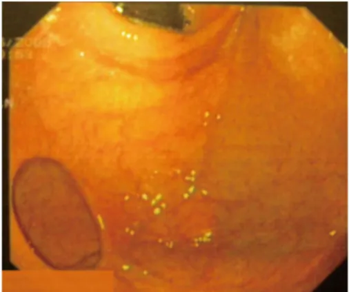
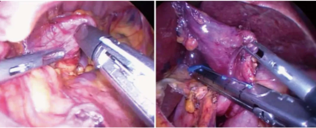
Two 51-year-old and 49-year-old women were referred to our Department for evaluation of symptoms of epigas-tralgy and upper abdomen tenderness, respectively. They denied weight loss, hematemesis and melena. The patients did not complain of vomiting or any abnormal bowel function. Abdominal examinations revealed no masses or other abnormalities. A barium esophagogastric study was performed, which showed an image of a protruding pouch in the upper gastric region (Figure 1). To confirm the diagnosis, an upper endoscopy was conducted, dem-onstrating diverticula directed posteriorly off the fundus of the stomach that measured 5 cm and 8 cm in diameter, respectively (Figure 2). The diverticula had a narrow neck but no ulcer was identified. Functional examination with esophageal stationary manometry and 24-h esophagogas-tric pH-multichannel intraluminal impedance monitoring showed normal values. The electrogastrography monitor-ing showed an increase in bradyarrhythmic gastric activity in both patients. Patients were presented for surgery and the operations were performed laparoscopically. The in-terventions were substantially the same for both patients. Because inspection of the stomach did not show a diver-ticulum at the anterior surface, the bursa omentalis was opened by dividing the gastrocolic omentum. Under en-doscopic control, the diverticula were resected at the neck with EndoGIA (Universal, US Surgical Corporation, Nor-walk, CO, United States) and then successfully retrieved with a laparoscopic pouch (Figure 3). Then, the staple lines were carefully examined and tested by submerging insufflated stomach under sterile irrigation to observe bubbles and there was no evidence of a leak. Histology confirmed a gastric diverticula of 5 cm and 8 cm, respec-tively, with normal mucosa in both cases. The procedures were uneventful. Patients were placed on a regular diet on postoperative day 3 and discharged 5 d later. Patients did not show any complications after surgery. On their first 6-mo follow-up visit, neither patient complained of any
symptoms, while normal radiographic and endoscopic ex-aminations, and regular values of esophageal manometry and 24-h esophagogastric pH-multichannel intraluminal impedance were found.
DISCUSSION
Moebius in 1661 and later Roax in 1774 first described gastric diverticula[9]. The literature suggests that most
symptomatic diverticula are found in patients between 20 and 60 years of age[10-12], while a unique study showed
that only 4% of gastric diverticula occurred in patients younger than 20 years[11] and no differences in incidence
between sexes have been described[4]. Two types of
gas-tric diverticula are recognized according to Akerlund[13]
and Schmidt et al[14]: congenital (true) and acquired (false)
diverticula, with congenital types being more com-mon[2,15-17]. True diverticula have all layers of the gastric
wall, and it is believed that these congenital diverticula oc-cur as a result of splitting of the longitudinal muscular fi-bers at the cardia level, leaving only circular muscle fifi-bers in the gastric wall and creating a weakening to allow a di-verticulum to form during the fetal period. This hypoth-esis is supported by Reich[18] who reported fetal gastric
diverticula and by Lewis, which described gastrointestinal diverticula in embryos in 1908[19]. False diverticula, also
classified as pulsation and traction diverticula, do not car-6115 September 28, 2013|Volume 19|Issue 36|
WJG|www.wjgnet.com
Marano L et al. Treatment of symptomatic gastric diverticula
Figure 1 A barium study reveals diverticula directed posteriorly off the fundus of the stomach that measured 5 cm and 8 cm in diameter in 51-year-old and 49-year-old women, respectively.
ry all layers of the gastric wall[20]. Pulsation diverticula are
those arising from increased intraluminal pressure, such as chronic coughing, obesity and pregnancy. Traction diverticula arise from perigastric adhesions from concur-rent diseases, such as peptic ulcer disease, pancreatitis, malignancy, gastroesophageal reflux and cholecystitis[11,12].
Gastric traction diverticula have been reported after sur-gical procedures on the stomach, including Roux-en-Y gastric bypass[12,21,22]. Even if gastric diverticula can arise
virtually anywhere along stomach, most congenital diver-ticula (70%) are typically located on the posterior wall of the stomach just below the gastroesophageal junction[2],
while acquired diverticula are usually situated near the gastric antrum. Typical diverticula are 1-3 cm in diameter but large diverticula as demonstrated above can occur[2,15].
Most gastric diverticula are asymptomatic[22],
how-ever when symptoms arise, depending on the size of the diverticular neck, they are most commonly upper abdominal pain, nausea, and emesis, and are described in 18%-30% of cases[2,21]. Wide-neck diverticula often
go unnoticed perhaps because food and digestive juices are less likely to become trapped. It has been suggested that food retention with subsequent distension of the gastric diverticulum may cause pain[23,24]. Dyspepsia and
vomiting are less common[21,25]. Occasionally, patients
with gastric diverticula can have dramatic presentations related to massive bleeding or perforation[26] due to food
retention with subsequent release of gastric juices within the mucosal sac, causing diverticulitis and possibly ul-ceration or hemorrhage. There are two reports of inva-sion with adenocarcinoma[27,28]. The diagnosis may be
difficult, as complaints can be caused by more common gastrointestinal pathologies and only be aggravated by diverticula. Methods of detection can fail, therefore, a combined approach should be used[8]. The presence of a
mucosal sac can be confirmed with upper gastrointestinal contrast studies and endoscopies. These are the most reliable diagnostic tests but reports in the literature con-firm that they can still miss the lesion if it has a narrow neck that precludes entry of the contrast or scope, giving false negative results[2,11,29]. In a large review, Palmer[11]
reported that 5% of gastric diverticula are missed during upper gastrointestinal investigation. Other reports rec-ommend the use of upper endoscopy[22,30] for diagnosis,
as this modality easily confirms the location and size of the diverticulum and provides the opportunity to biopsy any concurrent pathology. This diagnostic tool can rule out associated pathology and may be able to reproduce symptoms with distention of the diverticulum, indicating which patients would benefit from resection[15,22].
Com-puterized tomography scans are also used to diagnose diverticula; however, these have also mistaken diverticula for adrenal masses[31].
Appropriate management of a diverticulum depends mainly on the symptoms as well as on the diverticulum size. There is no specific therapeutic strategy for an asymptomatic diverticulum[5,18]. However, routine
sur-veillance with a periodic physical examination is recom-mended, given the potential onset of complications[20].
Diverticula exceeding 4 cm are more prone to produce complications and tend to respond less favorably to medications. Although some authors support conserva-tive therapy with antacids, this provides only temporary symptom relief and is not able to resolve the underlying pathology[20]. A necessity for successful treatment with
complete relief of symptoms is the association of the symptoms with the diverticulum. Palmer[11] found that in
30 of 49 symptomatic patients with a gastric diverticu-lum, symptoms were attributable to other gastrointesti-nal diseases, and 6 of 9 patients with symptoms caused by a gastric diverticulum who underwent open surgery showed excellent outcomes. Resection of gastric diver-ticula in all patients will lead to unsatisfactory results[22].
Surgical resection is the mainstay of treatment when the diverticulum is large, symptomatic or complicated by bleeding, perforation or malignancy, with over two-thirds of patients remaining symptom-free after sur-gery[11]. Several surgical approaches have been described
including invagination of the diverticulum as well as partial gastrectomy[32,33], however, since the first
success-ful laparoscopic resection of a gastric diverticulum in the late 1990s, this approach is now considered safe and feasible[34]. The most favorable approach providing better
exposure is by placing a, right upper quadrant port, and 2 left upper quadrant ports. Laparoscopic dissection has been performed by either releasing the gastrocolic liga-ment, thus allowing exposure of the superior posterior wall of the stomach[5,34-36]. Simple diverticulum resection
6117 September 28, 2013|Volume 19|Issue 36|
WJG|www.wjgnet.com
269-273
17 Love L, Meyers MA, Churchill RJ, Reynes CJ, Moncada R, Gibson D. Computed tomography of extraperitoneal spaces.
AJR Am J Roentgenol 1981; 136: 781-789 [PMID: 6784475]
18 Reich NE. Gastric Diverticula. Am J Dig Dis 1941; 8: 70 [DOI: 10.1007/BF03014635]
19 Lewis FT, Thyng FW. Regular occurrence of intestinal diver-ticula in embryos of pig, rabbit and man. Am J Anat 1908; 7: 505 [DOI: 10.1002/aja.1000070406]
20 DuBois B, Powell B, Voeller G. Gastric diverticulum: “a wayside house of ill fame” with a laparoscopic solution. JSLS 2012; 16: 473-477 [PMID: 23318077 DOI: 10.4293/108680812X 13462882736330]
21 Meeroff M, Gollán JR, Meeroff JC. Gastric diverticulum. Am
J Gastroenterol 1967; 47: 189-203 [PMID: 4960419]
22 Anaise D, Brand DL, Smith NL, Soroff HS. Pitfalls in the diagnosis and treatment of a symptomatic gastric diverticu-lum. Gastrointest Endosc 1984; 30: 28-30 [PMID: 6423437 DOI: 10.1016/S0016-5107(84)72291-7]
23 Kilkenny JW. Gastric diverticula: It’s time for an updated review. Gastroenterology 1995; 108: A1226 [DOI: 10.1016/0016-5085(95)29201-2]
24 Tillander H, Hesselsjö R. Juxtacardial gastric diverticula and their surgery. Acta Chir Scand 1968; 134: 255-263 [PMID: 4978307]
25 Simstein NL. Congenital gastric anomalies. Am Surg 1986;
52: 264-268 [PMID: 3085565]
26 Elliott S, Sandler AD, Meehan JJ, Lawrence JP. Surgical treatment of a gastric diverticulum in an adolescent. J Pediatr
Surg 2006; 41: 1467-1469 [PMID: 16863856 DOI: 10.1016/
j.jpedsurg.2006.04.010]
27 Fork FT, Tóth E, Lindström C. Early gastric cancer in a fundic diverticulum. Endoscopy 1998; 30: S2 [PMID: 9548048 DOI: 10.1055/s-2007-1001221]
28 Adachi Y, Mori M, Haraguchi Y, Sugimachi K. Gastric diver-ticulum invaded by gastric adenocarcinoma. Am J
Gastroen-terol 1987; 82: 807 [PMID: 3111246]
29 Seltzer MH, Koch AW. A huge gastric diverticulum. Am
J Dig Dis 1971; 16: 167-170 [PMID: 5542606 DOI: 10.1007/
BF02284458]
30 Velanovich V. Gastric diverticulum. Endoscopic and ra-diologic appearance. Surg Endosc 1994; 8: 1338-1339 [PMID: 7831610 DOI: 10.1007/BF00188296]
31 Schwartz AN, Goiney RC, Graney DO. Gastric diverticulum simulating an adrenal mass: CT appearance and embryogen-esis. AJR Am J Roentgenol 1986; 146: 553-554 [PMID: 3080856 DOI: 10.2214/ajr.146.3.553]
32 Brian JE, Stair JM. Noncolonic diverticular disease. Surg
Gy-necol Obstet 1985; 161: 189-195 [PMID: 3927497]
33 Cosman B, Kellum J, Kingsbury H. Gastric diverticula and massive gastrointestinal hemorrhage. Am J Surg 1957; 94: 144-148 [PMID: 13424891 DOI: 10.1016/0002-9610(57)90638-4] 34 Fine A. Laparoscopic resection of a large proximal gastric di-verticulum. Gastrointest Endosc 1998; 48: 93-95 [PMID: 9684678 DOI: 10.1016/S0016-5107(98)70142-7]
35 Kim SH, Lee SW, Choi WJ, Choi IS, Kim SJ, Koo BH. Lapa-roscopic resection of gastric diverticulum. J Laparoendosc Adv
Surg Tech A 1999; 9: 87-91 [PMID: 10194699 DOI: 10.1089/
lap.1999.9.87]
36 Vogt DM, Curet MJ, Zucker KA. Laparoscopic management of gastric diverticula. J Laparoendosc Adv Surg Tech A 1999; 9: 405-410 [PMID: 10522535 DOI: 10.1089/lap.1999.9.405] 37 Rashid F, Singh R, Cole A, Iftikhar SY. Troublesome belching
with fetor odour. Gut 2010; 59: 310, 324 [PMID: 20207635 DOI: 10.1136/gut.2009.177337]
P- Reviewers Aurello P, Marinis A S- Editor Zhai HH L- Editor Cant MR E- Editor Zhang DN with a laparoscopic cutting stapler has been reported to
be successful[37]. In some cases the surgical approach can
be challenging because the diverticulum is often collapsed and hidden in the splenic bed. Sometimes, a resection of the wrong part of the stomach has also been described[22].
For this reason the surgical procedure should be com-bined with intraoperative endoscopy to find an elusive diverticula by stretching the diverticular sac[4,34-36].
In conclusion, laparoscopic resection is a safe and feasible surgical approach with excellent outcomes and is strongly indicated for symptomatic gastric diverticula.
REFERENCES
1 Chen JH, Su WC, Chang CY, Lin H. Education and imaging.
Gastrointestinal: bleeding gastric diverticulum. J
Gastroen-terol Hepatol 2008; 23: 336 [PMID: 18289361 DOI: 10.1111/
j.1440-1746.2007.05301.x]
2 Rodeberg DA, Zaheer S, Moir CR, Ishitani MB. Gastric
diver-ticulum: a series of four pediatric patients. J Pediatr
Gastroen-terol Nutr 2002; 34: 564-567 [PMID: 12050587 DOI: 10.1097/01.
MPG.0000014963.68729.15]
3 Schiller AH, Roggendorf B, Delker-Wegener S, Richter
K, Kuthe A. [Laparoscopic resection of gastric diverticula: two case reports]. Zentralbl Chir 2007; 132: 251-255 [PMID: 17610199 DOI: 10.1055/s-2007-960753]
4 Donkervoort SC, Baak LC, Blaauwgeers JL, Gerhards MF.
Laparoscopic resection of a symptomatic gastric diverticu-lum: a minimally invasive solution. JSLS 2006; 10: 525-527 [PMID: 17575774]
5 Palmer ED. Gastric diverticulosis. Am Fam Physician 1973; 7:
114-117 [PMID: 4632747]
6 Raffin SB. Diverticula, rupture and volvulus. In: Sleigenger
MH, Fordtran JS. Gastrointestinal Disease, Pathophysiology Diagnosis, Management. 5th ed. Philadelphia: Saunders WB, 1989: 735-742
7 Lajoie A, Strum WB. Gastric diverticulum presenting as acute
hemorrhage. Gastrointest Endosc 2008; 67: 175-176 [PMID: 18028917 DOI: 10.1016/j.gie.2007.04.029]
8 Simon M, Zuber-Jerger I, Schölmerich J. True gastric
diver-ticulum. Dig Liver Dis 2009; 41: 370 [PMID: 18701360 DOI: 10.1016/j.dld.2008.06.016]
9 Moses WR. Diverticula of the stomach. Arch Surg 1946; 52:
59-65 [PMID: 21015447]
10 Gockel I, Thomschke D, Lorenz D. Gastrointestinal: Gastric diverticula. J Gastroenterol Hepatol 2004; 19: 227 [DOI: 10.1111/ j.1440-1746.2004.3339a.x]
11 Palmer ED. Gastric diverticula. Int Abstr Surg 1951; 92: 417-428 [PMID: 14840911]
12 Schweiger F, Noonan JS. An unusual case of gastric divertic-ulosis. Am J Gastroenterol 1991; 86: 1817-1819 [PMID: 1962629] 13 Akerlund D. Gastric diverticulum. Acta Radiol 1923; 2:
476-485
14 Schmidt HW, Walters W. Diverticula of stomach. Surg Gynec
Obst 1935; 60: 106 [DOI: 10.1016/S0002-9610(41)90528-7]
15 Wolters VM, Nikkels PG, Van Der Zee DC, Kramer PP, De Schryver JE, Reijnen IG, Houwen RH. A gastric diverticulum containing pancreatic tissue and presenting as congenital double pylorus: case report and review of the literature. J
Pe-diatr Gastroenterol Nutr 2001; 33: 89-91 [PMID: 11479415]
16 Vasilescu A, Dimofte G, Crumpei F, Moldovanu R, Mihăilescu A. Gastric diverticula on the greater curvature. J Chir Iasi 2007; 3: