681
Veterinarni Medicina, 62, 2017 (12): 681–684
Case Report
doi: 10.17221/104/2016-VETMED
A rare case of partial paraxial radial hemimelia
in a puppy: a case report
F. Macri
1*, S. Di Pietro
1, A. Palumbo Piccionello
2, G. Rapisarda
3,
G. Lanteri
1, V. Angileri
1, F. Marino
41
Department of Veterinary Sciences, University of Messina, Messina, Italy
2School of Veterinary Medical Science, University of Camerino, Camerino, Italy
3Department of Veterinary Prevention, Provincial Health Authority of Catania,
Gravina di Catania, Italy
4
Department of Chemical, Biological, Pharmaceutical and Environmental Sciences,
University of Messina, Messina, Italy
*Corresponding author: [email protected]
ABSTRACT: This article reports a case of a congenital unilateral forelimb defect in a two-month-old male puppy with clinical and radiographic findings. Congenital deformities are structural or functional congenital defects arising from errors during development, and they can affect an isolated portion of the body, the complete body or parts of several systems of the body. The congenital absence of a portion of the proximal epiphysis of the radius is an extremely rare condition in human and animal species. Although similar congenital limb deformities have been infrequently reported in dogs, the present case is, to the best of the authors’ knowledge, the first report of this type of partial forelimb paraxial radial hemimelia in a puppy, and it represents an addition to the scant literature on this topic.
Keywords: radial hemimelia; radiography; congenital defect; dog
Errors during foetal development may be followed
by structural or functional congenital
malforma-tions that are present at birth as defects (Noden
and De Lahunta 1985). Congenital malformations
can affect an isolated portion of the body, the entire
body or parts of several systems of the body (Dennis
and Leipold 1979).
Congenital limb malformations or partial
malfor-mations of limbs are varied in their manifestations,
ranging from the absence of a single structure to
partial or complete absence of the limbs (Lallo et
al. 2001).
The lack of a uniform and precise
nomencla-ture for limb malformations often complicates
their description. This clinical case was of a type
of hemimelia, a congenital abnormality with the
complete or partial absence of one or more bones
(Towle and Breur 2004). Longitudinal hemimelia
is the congenital absence of one or more bones
along the preaxial (medial) or postaxial (lateral)
side of a limb (Mo and Manske 2004; Towle and
Breur 2004). Hemimelia can also be intercalary,
in which case all or part of the middle bones of
a limb are absent, with the proximal and distal
portions being present.
Also, hemimelia can be classified as transverse
hemimelia, with a complete absence of the distal
portion of the limb, and paraxial hemimelia
char-acterized by aplasia of either the radius or ulna, or
tibia and fibula (Palmer 1993).
The objective of our study was to describe the
macroscopic and radiographic findings of a rare
case of a unilateral forelimb defect observed in a
puppy.
682
Case Report
Veterinarni Medicina, 62, 2017 (12): 681–684
doi: 10.17221/104/2016-VETMED
Case description
A two-month-old male Chihuahua was
present-ed to the Veterinary Teaching Hospital, Faculty of
Veterinary Medicine, University of Messina, for
an orthopaedic examination of a deformity of the
right forelimb. The malformation had been
pre-sent since birth and on physical examination a
non-functional right forelimb, markedly reduced
in size, with 90° varus deviation of the elbow, was
observed (Figure 1). There was no pain, crepitation
or evidence of fractures during physical
examina-tion of the deformed limbs. The paw was normal.
Radiographic views of the right forelimb were
tak-en using an analogic Radiographic/Fluoroscopic
Table System (Dedalus Mb 90/20 IMX-2A, Imago
Radiology S.r.l., Abbiategrasso (MI), 20081, Italy)
with a digital radiography system (Fujifilm Medical
Systems, Italy); X-ray settings of 55 kV, 10 mAs at
a film focus distance of 100 cm, without grid, were
used. The site of deformation was radiographed in
mediolateral and oblique views.
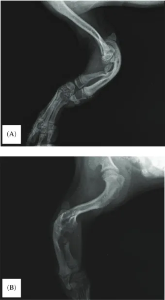
Radiographs showed a severe hypoplasia of the
radius, pronounced curvature of the ulna and poor
congruency of the humeroulnar joint (Figure 2). No
surgical treatment was performed.
Figure 1. Dorsal (A) and ventral (B) views. Deficient right forelimb characterized by the complete ankylosis of the elbow and short leg
Figure 2. Lateral (A) and oblique (B) views. The X-ray of the right forelimb shows severe hypoplasia of the radius, pronounced curvature of the ulna and poor congruency of the humeroulnar joint
(A)
(B)
(A)
683
Veterinarni Medicina, 62, 2017 (12): 681–684
Case Report
doi: 10.17221/104/2016-VETMED
The critical period for the development of the
entire limb in the canine embryo is between the 3
rdand 4
thweeks of gestation, when tissues are more
susceptible to external influences (Noden and De
Lahunta 1985).
The exact cause of the described malformations
could not be ascertained. Preaxial longitudinal
intercalary radial hemimelia is the most
com-mon type of hemimelia in dogs and cats (Alam
et al. 2006), while congenital partial paraxial
ra-dial hemimelia represents an extreme variant of
hemimelia.
Indeed, the present case is, to the authors’
knowledge, the first report of this type of forelimb
malformation in a dog and represents an
impor-tant addition to the literature on this topic.
REFERENCES
Ahalt BA, Bilbrey SA (1997): What is your diagnosis? Radial agenesis with secondary contractual deformity of the right forelimb. Journal of Small Animal Practice 38, 539– 571.
Alam MR, Heo SY, Lee HB, Kim JH, Park YJ, Lee KC, Choi IH, Kim NS (2006): Preaxial longitudinal intercalary ra-dial hemimelia in a dog: a case report. Veterinarni Me-dicina 51, 118–123.
Alonso RA, Hernandez A, Diaz P, Cantu JM (1982): An autosomal recessive form of hemimelia in dogs. Veteri-nary Record 110, 128–129.
Barrand KR, Cornillie PK (2008): Bilateral hindlimb adac-tyly in an adult cat. Journal of Small Animal Practice 49, 252–253.
Betts CW (1981): Agenesis of the radius. In: Bojrab MJ, Wingfield WE, Olds RB (eds): Pathophysiology in Small Animal Surgery. Lea and Febiger, Philadelphia. 650–653. Chiang C, Litingtung Y, Harris MP, Simandl BK, Li Y, Beachy PA, Fallon JF (2001): Manifestation of the limb prepattern: limb development in the absence of sonic hedgehog func-tion. Developmental Biology 236, 421–435.
Cornillie P, Van Lancker S, Simoens P (2004): Two cases of brachymelia in cats. Anatomia Histologia Embryologia 33, 115–118.
De Lima JA (1915): On the skeleton of an ectromelic goat. Journal of Anatomy and Physiology 49, 378–385. Dennis SM, Leipold HW (1979): Ovine congenital defects.
Veterinary Bulletin 49, 233–239.
Gemmill TJ, Clarke SP, Carmichael S (2004): Carpal agen-esis in a domestic short haired cat. Veterinary and Com-parative Orthopaedics and Traumatology 17, 163–166.
DiSCuSSioN AND CoNCluSioNS
Different patterns of congenital lower extremity
shortening have been reported in dogs and cats:
brachymelia (Cornillie et al. 2004), hemimelia
(Pedersen 1968; Alonso et al. 1982; Schultz and
Watson 1995; Ahalt and Bilbrey 1997; Lallo et al.
2001; Rahal et al. 2005; Alam et al. 2006; Pisoni et al.
2012), ectromelia (De Lima 1915; Macri et al. 2009),
adactyly (Barrand and Cornillie 2008; Macri et al.
2011), aphalangia (Macri et al. 2013a), radial
agen-esis (Swalley and Swalley 1978; Richardson 1979;
Betts 1981; Winterbotham et al. 1985; O’Brien et
al. 2002; Gemmill et al. 2004; Rahal et al. 2005;
Hildreth and Johnson 2007; McKee and Reynolds
2007), syndactyly (Macri et al. 2013b) and preaxial
terminal longitudinal hemimelia (Macri et al. 2014).
In our patient, the clinical signs were similar to
those described in previous reports of hemimelia
in dogs (Lallo et al. 2001; Rahal et al. 2005). Radial
hemimelia can be caused by environmental or
ge-netic factors, or by a combination of both.
Possible causes of hemimelia include
administra-tion of chemotherapeutics (tetracycline,
griseof-ulvin, parbendazole, etc.), malnutrition (lack of
riboflavin), intake of drugs such as thalidomide or
corticosteroids (in chick embryos), trans-placental
virus infections and X-rays, dietary mineral
defi-ciency (e.g., zinc, manganese, copper) and vaccines
(Johnson 1965; Karnofsky 1965; Warkany 1965;
Riddle and Leighton 1970). Other causes of distal
limb absence in young animals include
strangula-tion by restrictive bands, in utero accidents and
postnatal traumas (Johnson et al. 1995).
The heritability of radial hemimelia has been
sug-gested: Alonso et al. (1982) described an autosomal
recessive form of hemimelia in Chihuahuas, while
Hoskins (1995) suggested that hemimelia in Siamese
and domestic shorthair cats may be a hereditary trait.
Abnormalities in the function of the molecules
responsible for embryonic limb development along
the three main axes are responsible for
develop-mental malformations in embryo limbs. Especially
in human, chick and mouse embryos, mutation of
Wnt7a, En-1, FGF-2, Shh and Lmx-1 genes have
been linked to the development of hemimelia
(Chiang et al. 2001; Towle and Breur 2004; Woods
et al. 2006). Also, hemimelia results from a lack
of AER mesodermal interaction during limb
out-growth (Rantanen and Hegreberg 1982; Ogden and
Grogan 1987; Towle and Breur 2004).
684
Case Report
Veterinarni Medicina, 62, 2017 (12): 681–684
doi: 10.17221/104/2016-VETMED Hildreth BE, Johnson KA (2007): Ulnocarpal arthrodesis for
the treatment of radial agenesis in a dog. Veterinary Com-parative Orthopaedics and Traumatology 20, 231–235. Hoskins J (1995): Congenital defects of cats. Compendium
on Continuing Education for the Practicing Veterinarian 17, 385–405.
Johnson EM (1965): Nutritional factors in mammalian teratology. In: Wilson JG, Warkany J (eds): Teratology, Principles and Techniques. The University of Chicago Press, Chicago. 113–130.
Johnson KA, Watson ADJ, Page RL (1995): Skeletal diseases. In: Ettinger SJ, Feldman EC (eds): Textbook of Veterinary Internal Medicine. 4th edn. Saunders W.B., Philadelphia. 2077–2102.
Karnofsky DA (1965): Mechanism of action of certain growth inhibiting drugs. In: Wilson JG, Warkany J (eds): Teratology, Principles and Techniques. The University of Chicago Press, Chicago. 185–214.
Lallo MA, Bondan EF, Xavier JG, Fernandes TP, Kolber M, Zanco NA (2001): Bilateral anterior hemimelia in a dog: a case report. In: 26th World Small Animal Veterinary Association (WSAVA) World Congress. Vancouver, Brit-ish Columbia, Canada. August 8.–11., 2001.
Macri F, De Majo M, Rapisarda G, Mazzullo G (2009): Two cases of feline ectromelia: autopodium lia associated with humero-ulnar synostosis and zeugopodium ectromelia. Journal of Feline and Medicine Surgery 11, 731–734. Macri F, Marino F, Rapisarda G, Lanteri G, Mazzullo G
(2011): A case of unilateral pelvic limb adactyly in a puppy dog. Anatomia Histologia Embryologia 40, 104–106. Macri F, Ciotola F, Rapisarda G, Lanteri G, Albarella S, Aiudi
G, Liotta L, Marino F (2013a): A rare case of simple syndac-tyly in a puppy. Journal Small Animal Practice 55, 170–173. Macri F, Lanteri G, Rapisarda G, Marino F (2013b): Unilat-eral forelimb partial aphalangia in a kitten. Journal of Feline and Medicine Surgery 14, 272–275.
Macri F, Rapisarda G, Lanteri G, Di Pietro S, Auriemma E, Marino F (2014): Congenital absence of humerus with preaxial terminal longitudinal hemimelia and hypoplasia of the scapula in a dog: a case report. Veterinarni Me-dicina 59, 506–510.
McKee WM, Reynolds J (2007): Ulnocarpal arthrodesis and limb lengthening for the management of radial agenesis in a dog. Journal of Small Animal Practice 48, 591–595. Mo JH, Manske PR (2004): Surgical treatment of type 0
radial longitudinal deficiency. Journal of Hand Surgery 29, 1002–1009.
Noden DM, de Lahunta A (eds) (1985): The Embryology of Domestic Animals, Development Mechanisms and Mal-formations. Williams Wilkins, Baltimore. 367 pp.
O’Brien CR, Malik R, Nicoll RG (2002): Bilateral forelimb deformity and abnormal gait in a young Devon Rex. Jour-nal of Feline Medicine and Surgery 4, 112–113.
Ogden JA, Grogan DP (1987): Prenatal skeletal development and growth of the musculoskeletal system. In: Albright JA, Brand RA (eds): The Scientific Basis of Orthopedics. Appleton and Lange, Norwalk. 47–89.
Palmer N (ed.) (1993): Pathology of Domestic Animals. 4th edn. Academic Press, San Diego. 45–47.
Pedersen NC (1968): Surgical correction of a congenital de-fect of the radius and ulna of a dog. Journal of the Ameri-can Veterinary Medical Association 153, 1328–1331. Pisoni L, Cinti F, Del Magno S, Joechler M (2012): Bilateral
radial hemimelia and multiple malformations in a kitten. Journal of Feline and Medicine Surgery 14, 598–602. Rahal SC, Volpi RS, Ciani RB, Vulcano LC (2005): Use of
the Ilizarov method of distraction osteogenesis for the treatment of radial hemimelia in a dog. Journal of the American Veterinary Medical Association 226, 65–68. Rantanen NW, Hegreberg GA (1982): Naturally occurring
radial aplasia in mink. Veterinary Radiology 23, 27–29. Richardson DC (1979): Radial agenesis. Journal of
Veteri-nary Orthopaedics 1, 39–42.
Riddle Jr WE, Leighton RL (1970): Osteochondromatosis in a cat. Journal of American Veterinary Medical Asso-ciation 156, 1428–1430.
Schultz VA, Watson AG (1995): Lumbosacral transitional vertebra and thoracic limb malformations in a Chihuahua puppy. Journal of American Animal Hospital Association 31, 101–106.
Swalley J, Swalley M (1978): Agenesis of the radius in a kitten. Feline Practice 8, 25–26.
Towle HAM, Breur GJ (2004): Dysostosis of the canine and feline appendicular skeleton. Journal of the American Veterinary Medical Association 225, 1685–1692. Warkany J (1965): Development of experimental
mamma-lian teratology. In: Wilson JG, Warkany J (eds): Teratology, Principles and Techniques. The University of Chicago Press, Chicago. 1–12.
Winterbotham EJ, Johnson KA, Francis DJ (1985): Radial agen-esis in a cat. Journal of Small Animal Practice 26, 393–398. Woods CG, Stricker S, Seemann P, Stern R, Cox J, Sherridan E, Roberts E, Springell K, Scott S, Karbani G, Sharif SM, Toomes C, Bond J, Kumar D, Al-Gazali L, Mundlos S (2006): Mutations in WNT7A cause a range of limb mal-formations, including Fuhrmann syndrome and Al-Awadi/Raas-Rothschild/Schinzel Phocomelia syndrome. American Journal of Human Genetics 79, 402–408.
Received: June 27, 2016 Accepted after corrections: November 8, 2017