Università degli Studi di Ferrara
DOTTORATO DI RICERCA IN
"FARMACOLOGIA E ONCOLOGIA MOLECOLARE"
CICLO XXII
COORDINATORE Prof. Pier Andrea Borea
Elucidating the role of the tumor suppressor Protein
Phoshatase 2A in Chronic Myeloid Leukemia
Settore Scientifico Disciplinare BIO/17
Dottorando Tutore
Dott. Neviani Paolo Prof. Volinia Stefano
_______________________________ _____________________________
(firma) (firma)
ABSTRACT (ENGLISH VERSION) 2
ABSTRACT (ITALIAN VERSION) 4
INTRODUCTION 6
PROTEIN PHOSPHATASE 2A 7
PP2A AND CANCER 11
CHRONIC MYELOID LEUKEMIA 16
AIM OF THIS STUDY 21
MATERIAL AND METHODS 22
CELL CULTURES AND PRIMARY CELLS 22
PLASMIDS 24
RTPCR OF IMMUNOPRECIPITATED RNA (RIBONOMICS) 25
NESTED RTPCR 25
WESTERN BLOT ANALYSIS 26
ANALYSIS OF APOPTOSIS AND CASPASE ACTIVATION 26
PHOSPHATASE ASSAYS 27
CLONOGENIC ASSAYS 27
LEUKEMOGENESIS IN SCID MICE 27
INTRACELLULAR FLOW CYTOMETRY 28
LONGTERM CULTURE INITIATINGCELL ASSAYS 28
CFSE ASSAY 29
RESULTS 29
SET/HNRNP A1/PP2A INTERPLAY 29
RELEVANCE OF PP2A ACTIVITY SUPPRESSION IN CML 31
PRECLINICAL EVALUATION OF THE PP2A ACTIVATOR FTY720. 33
FTY720 RESTORES THE ACTIVITY OF PP2A AND INHIBITS THAT OF P210 AND P190 BCR/ABL 34
ANTILEUKEMIC ACTIVITY OF FTY720 IN PH1 LEUKEMIA CELLS IN VITRO 36
FTY720 SUPPRESSES IN VIVO BCR/ABL LEUKEMOGENESIS 39
FTY720 INDUCES APOPTOSIS OF THE CML LEUKEMIA INITIATING CELLS 41
DISCUSSION 44 REFERENCES 47 APPENDIX 62 FTY720, A NEW ALTERNATIVE FOR TREATING BLAST CRISIS CHRONIC MYELOGENOUS LEUKEMIA AND PHILADELPHIA CHROMOSOMEPOSITIVE ACUTE LYMPHOCYTIC LEUKEMIA
ABSTRACT (English Version)
Protein phosphatase-2A (PP2A) is one of the major cellular serine-threonine phosphatases and is involved in the regulation of cell homeostasis through the negative regulation of signaling pathways initiated by protein kinases. As several cancers are characterized by the aberrant activity of oncogenic kinases, it was not surprising that a phosphatase like PP2A has progressively been considered as a potential tumor suppressor. Indeed, multiple solid tumors (e.g. melanomas, colorectal carcinomas, lung and breast cancers) present with genetic and/ or functional inactivation of different PP2A subunits and, therefore, loss of PP2A phosphatase activity towards certain substrates. Likewise, impaired PP2A phosphatase activity has been linked to B-cell chronic lymphocytic leukemia, Philadelphia-chromosome positive acute lymphoblastic leukemia and blast crisis chronic myeloid leukemia (CML). In CML the deregulated tyrosine kinase activity of p210-BCR/ABL oncoproteins is sufficient to induce and sustain the leukemic phenotype, and contributes to disease progression. Imatinib mesylate, a BCR/ABL kinase inhibitor, is effective in most of chronic phase CML patients. However, a significant percentage of CML patients develop resistance to imatinib and/or still progresses to blast crisis, a disease stage that is often refractory to imatinib therapy. Furthermore, there is compelling evidence indicating that the CML leukemia stem cell is also resistant to imatinib. We have recently reported that the phosphatase activity of PP2A is markedly inhibited in blast crisis and, to a lesser extent, chronic phase CML patient cells and that pharmacologic re-activation of PP2A phosphatase activity by activating drugs such as Forskolin and FTY720 led to growth suppression, enhanced apoptosis impaired clonogenic potential and decreased in vivo leukemogenesis of imatinib-sensitive and
-resistant CML-BC and CML-CP patient. Furthermore we have evidence that PP2A inactivation is an event that occurs also at the level of the leukemic stem cell and that PP2A re-activation hampers the survival and self-renewal of CML stem cells and possibly overcome the resistance of CML quiescent stem cells to tyrosine kinase inhibitor monotherapy. Thus, inducing PP2A phosphatase activity has the potential to eradicate CML by efficiently targeting both leukemic stem and progenitor cells regardless of their degree of sensitivity to kinase. Therefore, the combination of PP2A phosphatase-activating and BCR/ABL kinase-inhibiting drugs may represent a powerful therapeutic strategy for CML patients.
ABSTRACT (Italian Version)
La protein phosphatase-2A (PP2A), una delle principali serin-treonin fosfatasi, è coinvolta nella regolazione dell’omeostasi cellulare, attraverso il controllo negativo di pathways protein-chinasi dipendenti. Molti tumori sono caratterizzati dall’espressione di oncogeni ad attività chinasica costitutiva, pertanto non sorprende che una fosfatasi come PP2A sia sempre più spesso descritta come onco-soppressore. In effetti, molti tumori solidi (melanoma, carcinomi del colon, del polmone e della mammella) presentano alterazioni genetiche e/o funzionali di diverse subunità di PP2A, con conseguente perdita della sua attività fosfatasica verso substrati specifici. Inoltre, l’alterazione dell’attivita’ di PP2A è stata collegata alla leucemia linfatica cronica di tipo B, alla leucemia linfoblastica acuta e alla leucemia mieloide cronica (LMC). Nella LMC l'attività costitutiva della tirosin-chinasi p210-BCR/ABL è sufficiente per indurre e sostenere il fenotipo leucemico e contribuisce alla progressione della neoplasia. L’Imatinib mesilato, un inibitore dell’attivita’ chinasica di BCR/ABL, è efficace nella maggior parte dei pazienti nella fase cronica della LMC. Tuttavia, una percentuale significativa dei pazienti con LMC sviluppa resistenza a Imatinib e/o progredisce verso la crisi blastica, fase della malattia che spesso risulta refrattaria alla terapia; inoltre dati recenti sembrano mettere in evidenza lo sviluppo di resistenza all’Imatinib da parte della cellula staminale leucemica della LMC. Recentemente abbiamo dimostrato che l’attività di PP2A è inibita sia nella fase di crisi blastica, sia, seppur in minor misura, nella fase cronica; inoltre, l’utilizzo di farmaci come Forskolin e FTY7220 porta alla riattivazione farmacologica di PP2A, provocando l’inibizione della proliferazione e del potenziale clonogenico, nonchè l’induzione dell’apoptosi in cellule di pazienti in fase cronica e in crisi blastica suscettibili o resistenti all’Imatinib. Dati preliminari dimostrano che l’inattivazione di PP2A avviene anche
a livello della cellula staminale leucemica e che la sua riattivazione ne inibisce la sopravvivenza e il self-renewal; tale approccio, potrebbe rappresentare una strategia per superare la farmaco-resistenza a inibitori dell’attività chinasica di BCR/ABL. Quindi, l’induzione dell’attivazione di PP2A attraverso il targeting sia dei progenitori che delle cellule staminali leucemiche ha il potenziale di eradicare la LMC. La combinazione di farmaci attivatori di PP2A e di inibitori di BCR/ABL potrebbe rappresentare una nuova strategia terapeutica per i pazienti affetti da LMC.
INTRODUCTION
The ability to balance a complex network of signal transduction pathways is a key element in the normal homeostasis of a cell. Survival, proliferation and differentiation signals must be tightly controlled in order for a eukaryotic cell to keep its place in a multicellular organism and accurately react to external stimuli whether by proliferating, differentiating or eventually dying. Genetic or functional alterations of specific cell components may lead to inadequate responses to the microenvironment and therefore aberrant activation of pathways that control cell proliferation, survival and differentiation, ultimately leading to tumorigenesis and cancer (1). Mutations, amplifications or rearrangements may lead to the uncontrolled activity of proteins encoded by so-called proto-oncogenes (2), which are frequently cell cycle-promoting and/or apoptosis-repressing factors. This is often associated with the genetic or functional loss of tumor suppressor proteins, which usually have cell cycle-repressing and/or apoptosis-promoting effects (3). Gain of oncogene function associated with loss of tumor suppressors is now widely recognized as a hallmark of cancer initiation and progression (4).
One primary mechanism by which a normal cell maintains the balance of tumor-inducing and tumor-suppressing signals and appropriately responds to extracellular stimuli is reversible protein phosphorylation (5). Signals from the microenvironment (i.e. hormones and growth factors) are detected by cell surface receptors and translated into the activation of protein kinases which, in turn, act by phosphorylating substrates, thereby inducing an intracellular signal transduction cascade that directly affects the physiological response of the cell to external stimuli. The reversal of protein phosphorylation by protein phosphatases is essential in order to preserve correct cell physiology and tightly regulate these important signal transduction pathways. Given this scenario, it comes as no surprise that the majority of oncogenes identified thus far encode protein kinases,
and that aberrant regulation of their activity plays a major role in the induction of a cancerous phenotype (6).
Some of the best known examples of oncoproteins with unrestrained kinase activity are products of the t(9;22) translocation: the BCR/ABL oncoproteins, which are the driving force in the induction of both chronic myeloid leukemia (CML) and Philadelphia-positive acute lymphoblastic leukemia (Ph-ALL) (7, 8). Conversely, it seems logical to assume that protein phosphatases may also play a key role in cancer progression by acting as important regulators of the activation status of proteins involved in signal transduction. Undeniably, the study of protein phosphatases and their regulation has become an expanding field of research aimed at shedding light on the importance of these proteins in cancer (9). In particular, protein phosphatase 2A (PP2A) has been the subject of recent investigations that have begun to unravel the central role of this enzyme in cancer. By negatively regulating many of the signals triggered by oncogenic kinases, PP2A can be truly described as a tumor suppressor (10, 11).
Protein phosphatase 2A
PP2A is one of the major serine-threonine phosphatases. Highly conserved and ubiquitously expressed, PP2A is directly involved in a multitude of cellular processes by dephosphorylating specific substrates, therefore providing a negative feedback to signals initiated by protein kinases. PP2A phosphatase activity has been linked to the regulation of the cell cycle, signal transduction, DNA replication, transcription and translation (12, 13); for that reason, a peculiarity of PP2A is that a single phosphatase is responsible for regulating a variety of processes triggered by an array of different protein kinases. The ability of PP2A to affect the phosphorylation status of a wide variety of substrates is a consequence of its complex structure and regulation (Figure 1). PP2A is a heterodimeric or
heterotrimeric complex which contains a constant core dimeric structure, a 36 kDa catalytic subunit (PP2Ac, α and β isoforms) and a 65 kDa structural subunit (PP2A/A or PR65, α and β isoforms); this dimeric structure (PP2A/AC) may or may not be associated with a variable third subunit, a family of regulatory B subunits that provide substrate/tissue specificity and which are also differentially expressed during development (13, 14) (Figure 1). The B subunits are classified into four different families of unrelated proteins: PR55/B (four isoforms), PR61/B′ (encoded by five genes with up to eleven isoforms), PR72/B′′ (four isoforms) and PR93/110/B′′′ (two unrelated proteins) (14). The ability of PP2A to function as a dimer and also associate with an array of more than twenty interchangeable B subunits partially explains the variety of functions carried by this enzyme and underlines the potential complexity of PP2A phosphatase activity and its regulation. Thus, it seems logical that specific heterotrimeric configurations are
Figure 1: Schematic representation of the quaternary structure of the PP2A holoenzyme. The figure shows the different levels at which the enzyme can be modulated either by changing the dimeric/trimeric composition, post-translational modifications and/or regulation by inhibitory molecules
responsible for different PP2A functions. As an example, it has been found that PR55/B may regulate cytokinesis in yeast (15), correct segregation of chromatids in Drosophila (16) and neuronal differentiation in humans (17); the B′ subunit family appears to be important for regulating the formation of cyclin-G/PP2A complexes, which regulate the phosphorylation status of mdm2, indirectly affecting the stability of p53 (18); PR59/B’’ targets PP2A to dephosphorylate the retinoblastoma-related p107 protein, suggesting a role in the regulation of the cell cycle (19). Such a variety of forms and activities places PP2A at the center of the regulation of most signal transduction pathways triggered and sustained by protein kinases. As an example, PP2A can dephosphorylate and inactivate MEK1 and ERK-family kinases, therefore inhibiting mitogenic signals (20). Moreover the small-t antigen, a viral protein that is involved in cellular transformation, is believed to cooperate with the large-T antigen of the SV40 virus in inducing transformation and immortalization of target cells in part by hyper-activating the MAPK pathway as a consequence of the displacement of the variable B subunit which results in PP2A inactivation (21). Furthermore, a direct involvement of PP2A in the regulation of the cell cycle has been reported at many different levels. Specifically, the PR55/Bα:PP2A complex dephosphorylates and inhibits CAK (Cdk-activating kinase) that, when active, phosphorylates and activates Cdc2 in the MPF complex (M-phase-promoting factor) therefore inducing the G2/M transition (22). In this instance PP2A is able to exert a negative effect on regulating entry into mitosis. On the other hand, PP2A has a positive regulatory function in apoptosis (23); this is mediated by dephosphorylation and inactivation of the anti-apoptotic Bcl-2 protein which occurs following the targeting of PP2A to the mitochondria and direct interaction with Bcl-2 via the PR61α/B’ subunit, and also by PP2A-mediated dephosphorylation and activation of the pro- apoptotic factor Bad. Another PP2A substrate involved in pro-survival signals is Rel A, an NF-κB transcription factor,
which has been found physically associated with the PR65/A subunit and whose transactivating activity is diminished upon direct dephosphorylation by PP2A (24). It has also been demonstrated that PP2A can negatively regulate the PI3K/Akt pathway by associating to and directly dephosphorylating Akt (25). Furthermore PP2A has a role downstream of Akt, specifically inhibiting the mTOR pathway; in fact PP2A directly inactivates S6 kinase, which is responsible for promoting cell growth by enhancing the translation of 5′TOP (containing a short polypyrimidine stretch at the 5′ terminus) mRNAs (26-28). PP2A also has an essential role in the regulation of Wnt signaling, a pathway linked to embryonic development, cell growth and stem cell self-renewal. In fact, a PP2A complex containing the B56γ subunit has been shown to be part of the β-catenin degradation complex, together with GSK3β and Axin where the PP2A catalytic subunit is involved in the process of inactivation and degradation of β-catenin, therefore shutting down the Wnt pathway (29). It becomes obvious that with such a diversity of functions, PP2A phosphatase activity needs to be tightly regulated. The primary level of regulation, as mentioned above, is the hetero-oligomeric composition of the PP2A complexes; however, PP2A can also undergo post-translational modifications and can be controlled by its association with inhibitory proteins. Until recently, the regulation of the quaternary structure of PP2A was poorly understood. However, the recent elucidation of the crystal structure of PP2A (30) led to the definition of a major role for the highly conserved C-terminal sequence of the C subunit (12, 31). It was already known that a C-terminal tyrosine residue (Tyr307) on the PP2A catalytic subunit could be phosphorylated, and that this event induces inactivation of the enzyme itself. In addition, PP2Ac can also be inactivated by phosphorylation on other, as yet unspecified, threonine residues, and in both cases PP2A can reactivate itself by auto-dephosphorylation (12, 31). The C-terminus of PP2AC is also reversibly carboxymethylated on Leu309 by a specific carboxyl
methyltransferase (LCMT1). This post-translational event appears to not directly affect PP2A phosphatase activity, but it is thought to affect holoenzyme composition. This idea has been re-investigated recently, and it appears that each B subunit family requires different levels of carboxymethylation on the C subunit to allow for assembly into the trimeric complex (31). Some subunits, such as PR55/B, require PP2Ac to be methylated, and others, such as PR61/B’ and PR72/B’’, associate only when the catalytic subunit is not methylated (31). These latest findings give new insights into the complexity of regulation of PP2A activity.
Another level of complexity in the regulation of PP2A is the occurrence of several endogenous PP2A inhibitors, called I1PP2A, I2PP2A and CIP2A (32, 33). The biological significance of I1PP2A is still for the most part unknown. However, recent work has suggested a role for this protein in mediating the abnormal phosphorylation of the tau protein seen in Alzheimer’s disease (34). The Cancerous Inhibitor of PP2A (CIP2A) enhances the stability of the c-Myc through a mechanism that inhibits PP2A activity toward Serine 62 of this proto-oncogene. At last, I2PP2A, better known as SET, is the most characterized endogenous inhibitor of PP2A. SET is phosphorylated on two serine residues, most likely by PKC, and exerts its potent PP2A inhibitory activity via its N-terminal sequence (12, 35). Most importantly, SET has been found fused to the nucleoporin 214 (NUP214, CAN) protein in a form of acute non-lymphocytic myeloid leukemia with t(6,9) (36), associated with the oncoprotein MLL (mixed lineage leukemia, also called ALL1, HRX) in acute myeloid leukemia (37).
PP2A and cancer
The ability of PP2A to negatively and positively regulate mitogenic and apoptotic signals, respectively, has raised the hypothesis that PP2A might be involved in cancer development and progression. However, given the complexity of
PP2A regulation and its involvement in a wide variety of cellular mechanisms, it has been difficult to unequivocally and formally define PP2A as a bona fide tumor suppressor. The first lines of evidence implicating PP2A in cell transformation came in the late 1980s and early 1990s and were based on two observations. Firstly, that okadaic acid, a naturally occurring toxin and potent tumor promoter, is an inhibitor of PP2A (38). Secondly, that the SV40 small-t antigen interacts with PP2A/A and inhibits PP2A phosphatase activity by displacing several B subunits and that such inhibition is required for its function in cell transformation (39). However, it is only recently that inhibition of PP2A has become more widely accepted as being an essential step which allows cells to undergo complete transformation in response to activated oncogenes (11, 33, 40). Different PP2A subunits have now been found either mutated or aberrantly expressed in a number of malignancies that show loss of PP2A activity in conjunction with increased stability/activation of protein kinases and/or of their downstream targets (i.e. MAPK and Akt).
The B subunit B56γ (of the PR61/B′ family) has been found overexpressed in metastatic melanoma cells compared to normal melanocytes (41); in these cells, PP2A failed to dephosphorylate the protein paxillin in focal adhesions, therefore enhancing the invasiveness of metastatic clones (42). Conversely, overexpression of B56γpartiallyreversed the tumorigenic activity of SV40-transformed cells and lung cancer cells (43). Accordingly, a truncated isoform of B56γ leading contributes to an enhanced metastatic potential of melanoma cells (44). This is in line with the notion that the major signaling pathways whose alterations are classically considered to trigger malignant transformation are regulated by the PP2A B56γ subunit. In fact, B56γ has been described as the regulatory subunit that targets PP2A to the Wnt/β-catenin and the MAPK signaling pathways and that elicits caspase-dependent apoptosis (29, 45, 46).
Although it seems that the PP2A:B56 form of the holoenzyme might represent a major player in cell transformation, the scaffolding PR65/A subunit has now been described as a tumor suppressor per se. Somatic mutations of both the α and β isoforms of PR65/A have been reported with high frequency in cancer. Interestingly, the Aβ isoform (PPP2R1B) gene has been found mutated in 15% of primary lung tumors, in 15% of colorectal carcinomas and in 13% of breast cancers (47-51). Moreover Aβ maps to chromosome 11q23, a region frequently deleted in various cancers and leukemias (52), and its loss-of-function cooperates with the expression of the large-T antigen, hTERT and H-RAS to transform human cells (40). Importantly, reintroduction of a wild-type Aβ partially reverses the tumorigenic phenotype of lung cancer cell lines (40). In this regard, a recent study (40) provided new insight into the role of PP2A/Aβ in cell transformation: PP2A/Aβ interacts with and modulates the activity of the small GTPase RalA, a known regulator of membrane transport, apoptosis, transcription, cell migration and cell proliferation (53). Sablina et al. (40) found RalA hyper-phosphorylated in cells lacking PP2A/Aβ expression and that regulation of RalA phosphorylation seems to be essential for the transformation associated with loss of PP2A/Aβ in cancer. In addition, the role of PP2A/Aβ as a tumor suppressor may likely be functionally explained by the disruption of the quaternary structure of PP2A complexes as consequence of the defective binding to B and/or C subunits and, therefore, resulting in impaired PP2A phosphatase activity towards specific substrates (48, 54). Supporting the importance of PP2A A subunit in tumor formation and, perhaps in metastatic progression, there is also evidence that mutation of the PP2A/Aα subunit impaired phosphatase activity, led to activation of the Akt-dependent survival signals and permitted cells to form tumors in mice, suggesting that cancer-associated mutations of PP2A/Aα contribute to cancer development by altering PP2A holoenzyme composition and function (55).
Inhibition of PP2A activity and, therefore, loss of its tumor suppressing potential, occurs at different levels, either within the structure of the multimeric enzyme itself (e.g. loss of expression/activity of one or more subunits), or through the activity of external effectors, whether endogenous (e.g. SET) or exogenous (e.g. okadaic acid, small-t antigen). For example, loss of Pml (promyelocytic leukemia gene) leads to tumorigenesis in the prostate through a mechanism that requires the recruitment of PP2A and phospho-Akt by Pml into the Pml nuclear bodies (56). Interestingly, Pml-null cells show suppression of PP2A phosphatase activity towards Akt that accumulates as nuclear phospho-Akt. As a consequence, the progressive reduction in Pml levels leads to inactivation of Foxo3a-mediated transcription of the proapoptotic protein Bim and the cell cycle inhibitor p27 (kip1) thus favoring cancer development (56). Likewise, a recently published report (33) identified a new endogenous inhibitor of PP2A, CIP2A (cancerous inhibitor of PP2A). This 90 kDa protein had already been found overexpressed in gastric cancers, but its function has remained elusive until recently (57). Junttila et al. (33) reported that siRNA-mediated downregulation of CIP2A in HeLa cells affected the expression of a variety known targets of c-Myc. Notably, the proto-oncogene c-Myc is a target of PP2A phosphatase activity (58-60), and the phosphorylation on Ser62 stabilizes this transcription factor and prevents its degradation (61). CIP2A strongly inhibits the phosphatase activity of PP2Ac, selectively targeting c-Myc-associated PP2A complexes to prevent Ser62 dephosphorylation and consequently allow sustained and unrestrained c-Myc activity (33). These data suggest that CIP2A inhibition may be a possible strategy to induce targeted degradation of c-Myc in cancer cells.
Recent studies, including from our group, have reported that the loss of PP2A phosphatase activity and, therefore its tumor suppressor ability, may play a central role in the development of different leukemias, such as
Philadelphia-chromosome positive leukemias like CML and Ph(+) B-ALL, and also B-cell chronic lymphocytic leukemia (B-CLL) (62-64). B-CLL is the most common leukemia in adults, and deletion of the chromosome region 11q22–q23 is the second most common genomic alteration in B-CLL, representing a cohort of patients characterized by an adverse prognosis (65); interestingly, this region harbors the PP2A/Aβ locus. In their 2007 study, Kalla et al. (64) screened the remaining copy of the PP2A/Aβ gene for mutations in a number of patients carrying monoallelic deletion of this chromosome region. No deleterious mutations were found, confirming a previous report (66). However, the basal transcription of the PP2A/Aβ gene was found to be significantly lower in patients independent of the 11q deletion. Moreover, they found three splicing variants of this PP2A subunit, characterized by skipping of exon 9, exon 3 or exons 2 and 3, and all were expected to interfere with the correct assembly of functional trimeric holoenzymes with consequent loss of domains necessary for interaction with C and B subunits (64). In agreement with the idea that disruption of the quaternary structure of PP2A most likely affects phosphatase activity, a significant reduction of PP2A activity was found in almost all of these B-CLL patients assayed, regardless of differences in the expression of the various PP2A/Aβ isoforms (64). As expected, an even greater reduction in PP2A phosphatase activity was observed in those patients expressing these splice variants. The potential role that PP2A may play in B-CLL was also investigated in a recent report by Liu et al. (67), in which the possibility of inducing PP2A activation as a possible therapeutic strategy for CLL was investigated in detail. The PP2A activator tested, FTY720 (Fingolimod; Novartis), is a known immunomodulator that is currently being evaluated in phase III clinical trials for multiple sclerosis (68, 69). FTY720 is a synthetic water-soluble myriocin analog that is structurally similar to sphingosine. It is a relatively non-toxic drug with high oral bioavailability and it reversibly arrests lymphocyte trafficking (70, 71).
FTY720 treatment of transformed B-cells induces apoptosis by a Bcl-2 independent mechanism, and it also reduces phospho-ERK1/2 levels (67). More importantly, FTY720 treatment prolonged animal survival in a xenograft SCID mouse model of B cell lymphoma/leukemia (67).
Chronic Myeloid Leukemia
Chronic Myeloid Leukemia is a clonal disorder of the pluripotent haematopoietic stem cells and is clinically characterized by two distinct stages, a protracted myeloproliferative disorder termed chronic phase (CML-CP) that progresses to a rapidly fatal blast crisis (CML-BC) (7). CML-CP, largely asymptomatic, is distinguished by the expansion and accumulation of terminally differentiated neutrophil granulocytes in the peripheral blood. If left untreated CML-CP inevitably progresses to the blast phase, characterized by accumulation of blast cells in the peripheral blood that are unable to differentiate; CML-BC is the terminal phase of this malignancy and behaves as an acute leukemia (7). CML has been the first cancer to be linked to a specific somatic genetic abnormality (7). In fact the main feature of CML is the presence of the Philadelphia chromosome (Ph1), product of the reciprocal translocation between chromosome 9 and chromosome 22, t(9;22)(q34;q11). The translocation results in the fusion of the BCR (9q22) to the c-ABL (22q11) gene. In CML, the BCR/ABL fusion gene encodes for a 210kDa protein called p210-BCR/ABL. Depending on the location of the breaking point on the BCR locus the product of the t(9;22) can also be a p190- or a p230-BCR/ABL, which are normally detected in Philadelphia-positive B-acute lymphoblastic leukemia (Ph1 ALL) and chronic neutrophilic leukemia (CNL) respectively (72).
It has been well established that the sole expression of p210-BCR/ABL is necessary and sufficient for the induction of chronic phase CML (7). In blast crisis
CML other frequent genetic and molecular abnormalities arise (e.g. double Ph1 chromosome, p53 inactivation) (73), but increased expression and activity of the BCR/ABL oncoprotein still appear to be playing a central role in the progression of the disease (74-76), in fact, sustained BCR/ABL expression in myeloid progenitor cell lines induces phenotypic changes (i.e. differentiation arrest) characteristic of CML-BC (77). Moreover high levels of BCR/ABL activity as seen in CML-BC trigger oxidative stress in the cells, which is likely responsible for the induction of DNA damage and secondary genetic abnormalities. The ability of BCR/ABL to induce and sustain the leukemic phenotype depends on its unrestrained tyrosine kinase activity (7). BCR/ABL constitutive kinase activity is essential for the recruitment and activation of multiple pathways that transduce oncogenic signals leading to growth factor-independent proliferation, increased survival and altered differentiation (78) of myeloid precursors. In fact, the effect of BCR/ABL transformation is mostly dependent on post-translational modifications (e.g. phosphorylation) of signaling molecules, like those involved in the RAS/MAPK, PI3K/Akt and STATs pathways, which control cell growth, survival and differentiation of haematopoietic cells by modulating the expression and/or activity of downstream effectors (79, 80) (Figure 2). In blast crisis, increased expression of the BCR/ABL oncoprotein accounts for the block of differentiation, inactivation of factors with tumour suppressor activity and decreased genomic stability of the Ph1 blasts (7, 78, 81, 82). Thus, dependence on BCR/ABL expression is not only a characteristic of CML-CP but also of CML-BC; however, BCR/ABL-independent mechanisms also seem to contribute to disease progression and treatment resistance in some CML cases (83, 84).
Since the deregulated BCR/ABL kinase activity is the recognized culprit of cause of CML, targeting its catalytic domain was the most rationale method for the development of small molecules that inhibit ABL kinase activity. Through a
combination of high-throughput screening and a structure-based drug design approach the Signal Transduction Inhibitor 571 (STI571) was developed in the late 90s. Now commercialized as Imatinib Mesylate (Gleevec, Novartis Basel, Switzerland), it is a c-Kit, c-Abl and PDGFR inhibitor, and induces apoptosis of the Ph1 CML progenitors by suppressing the ability of BCR/ABL to phosphorylate substrates through competitive inhibition at the BCR/ABL ATP binding site (85). The development of the BCR/ABL tyrosine kinase inhibitor Imatinib Mesylate as the treatment of choice for chronic phase CML has changed the natural history of the disease with up to 90% of the patients achieving a complete cytogenetic response in chronic phase. Its remarkable therapeutic effects suggest that blast crisis transition will be postponed for several years in the majority of CML patients (86, 87). However, the persistence of BCR/ABL transcripts in a cohort of patients with complete cytogenetic response (88) and the resistance of the primitive CML stem cell to Imatinib treatment (89) raises the possibility that treatment with Imatinib alone might delay but not prevent disease progression. Furthermore, most of the CML patients that start the treatment in the accelerated and blastic phases of the disease are either refractory or develop resistance to Imatinib monotherapy (86). In these CML-BC patients, Imatinib resistance often depends on reactivation of BCR/ABL tyrosine kinase activity via mechanisms involving BCR/ABL overexpression, gene amplification or mutations that suppress Imatinib-mediated kinase inhibition (i.e. E255V and G250E) or disrupt Imatinib binding (i.e. T315I) (90). Thus, development of Imatinib resistance appears to predispose to blastic transformation. Hence, second generation tyrosine kinase inhibitors have been developed in the past few years. Although new clinical trials with the dual Src/Abl inhibitor Dasatinib (BMS-354825) and the selective Abl inhibitor Nilotinib (AMN1070) show encouraging results (91), as they suppress the activity of most BCR/ABL mutants (except T315I) (91), in vitro evidence suggests that resistance
Figure 2. Top: Schematic representation of some of the pathways activated by p210BCR/ABL and involved in the
BCR/ABL-dependent induction of the transformed phenotype in CML. Bottom: Schematic representation of the BCR/ABL-induced and RNA binding protein-mediated pathways affecting proliferation survival and differentiation of CML-BC progenitors.
to these new compounds may develop through mechanisms involving the selection and expansion of BCR/ABL cell clones carrying the T315I BCR/ABL mutant (92). Additionally, Dasatinib and Nilotinib, like Imatinib, are not effective in the treatment of CML-BC patients (93).
The mechanisms responsible for transition of CML chronic phase into blast crisis remain poorly understood, although a reasonable assumption is that the unrestrained activity of BCR/ ABL in hematopoietic stem/progenitor cells is the primary determinant of disease progression. However, a plausible model of disease progression predicts that increased BCR/ABL expression promotes the secondary molecular and chromosomal changes essential for the expansion of cell clones with increasingly malignant characteristics, and remains crucial for the transformed phenotype even in advanced stages of the disease (7). According to this model, CML blast crisis would be expected to occur only in patients with an imatinib-resistant disease or in those developing resistance during treatment. Indeed, a recent study from the GIMEMA Working Party on CML reported that the early detection of BCR/ABL mutations in CML chronic phase patients is associated with a greater likelihood of disease progression (94). Interestingly, a direct correlation also seems to exist between levels of BCR/ABL activity and development of imatinib resistance (95, 96).
Another level of complexity in the treatment of CML is the fact that there is increasing evidence that the most primitive and quiescent CML stem cells, designated as leukemic stem cells or cancer initiating cells, appear to be intrinsically insensitive to the tyrosine kinase inhibitors(89, 97). Therefore, therapy with Imatinib may never be curative and indefinite treatment may be required for a sustained response; this may pose the risk of developing resistance or intolerance leaving the opportunity open for a possible relapse of CML given the persistence of a pool of leukemic stem cells. Interestingly the resistance of the quiescent stem
cells to BCR/ABL inhibition is raising the possibility that, paradoxically, the survival of LSCs may be to some degree, if not completely, independent of BCR/ABL.
Aim of This Study
The natural history of CML has been radically changed by the introduction of Imatinib and second-generation tyrosine kinase inhibitors (Dasatinib and Nilotinib), but relapse and progression into the blast phase of the disease, as a consequence of arising of BCR/ABL mutations and/or intolerance to the treatment, is a possibility that still exists and cannot be ignored. Moreover patients that may be diagnosed during the blast crisis do not have any feasible therapeutic option and to this day the only curative regimen for CML is bone marrow transplantation. Therefore, there is still a need for new drugs that, if used in combination with the available kinase inhibitors, may help decrease the rate of relapse, prevent blastic transformation and, perhaps, overcome Imatinib resistance. Therefore, a better understanding of the biology of CML-BC is necessary to identify molecular networks that, if appropriately modulated, will simultaneously affect the function of BCR/ABL and that of multiple signals aberrantly activated in CML-BC. Moreover, in light of the recent findings concerning the insensitivity of the leukemic stem cells to the tyrosine kinase inhibitors (97), many groups, including ours, are now poised to dissect the biology of the leukemic stem cells and to find alternative factors in charge of the survival and self-renewal of the cancer initiating cells, and that could be targeted in the hope of eradicating CML at a stem cell level, which could eventually result into a long-term cure. Therefore the main objective of these studies is to dissect the mechanisms responsible for disease progression in CML.
In BCR/ABL-expressing myeloid progenitor cell lines, high levels of BCR/ABL, as those observed in CML-BC, suppress differentiation, enhance survival and increase resistance to drug-induced apoptosis in part by enhancing
the expression and activity of specific RNA binding proteins (78, 82, 98, 99) (Figure 2). Such an increase in the expression of these mRNA-binding proteins correlates with the levels of BCR/ABL and is sensitive to the treatment with Imatinib (77, 82, 98, 100). In BCR/ABL-expressing hematopoietic cells, expression of these RNA-binding proteins results from enhanced gene transcription (eg hnRNP A2/B1, hnRNP K, JKTBP1, hnRNP D1, Tra2b, RNPS1, EWSh, SC-35, Pabp2 and hnRNP H1), or increased protein stability (ie TLS/FUS, hnRNP A1, hnRNP E2 and La/ SSB) (77, 78, 82, 99-101). In this study, we focused on the nucleocytoplasmic shuttling protein hnRNP A1, a previously described protein required for BCR/ABL leukemogenesis (100), and we identified SET (I2PP2A), a phosphoprotein already described as being overexpressed in solid tumors and leukemias (102, 103), as a novel target of BCR/ABL whose mRNA transport, from the nucleus to the translation machinery in the cytoplasm, is dependent on hnRNP A1(62). SET is a potent inhibitor of the tumor suppressor PP2A (35). This preliminary finding raised the possibility that PP2A could be involved in the leukemogenesis of CML, and that its inhibition, through SET, may be a mechanism that is put in place by BCR/ABL to suppress PP2A tumor suppressive ability and to allow for the progression of the disease.
MATERIAL AND METHODS
Cell cultures and primary cells
The Ph1 K562, the BCR/ABL-expressing 32Dcl3 and BaF3 cells, and their derivative lines were maintained in culture in IMDM/10% FBS/2 mM L-glutamine. The p210BCR/ABL, p210BCR/ABL (T315I), BaF3-p190BCR/ABL, 32D-BCR/ABL-pBABE-GFP, and 32D-BCR/ABL-GFP–small-t cells were generated by
retroviral infection followed by FACS-mediated sorting of the GFP-positive fraction (green fluorescent protein) or by antibiotic-mediated selection (78). Frozen samples of CD34+ NBM cells from different healthy donors were purchased from Cincinnati Children’s Hospital. Frozen samples of mononuclear hematopoietic cells from bone marrow or peripheral blood of unidentifiable CML and Ph1 ALL patients were Ficoll separated and used to isolate the CD34+ and/or CD19+ fraction by using the CD34 MultiSort kit and/or CD19 microbeads (Miltenyi Biotec). The same method was used to isolate the CD19+ fraction from the normal CD34+ marrow progenitors. Before being used in different assays, CD34+ and CD34+/CD19+ progenitors from healthy donors and leukemic patients were kept overnight in IMDM supplemented with 30% FBS, 2 mM glutamine, and human recombinant cytokines. Specifically, NBMCD34+, CML-CPCD34+ and CML-BCCD34+ cells were kept in IL-3 (20 ng/ml), IL-6 (20 ng/ml), Flt-3 ligand (100 ng/ml), and KL (100 ng/ml) (Stem Cell Technologies Inc.), whereas NBMCD34+/CD19+ and Ph1 ALLCD34+/CD19+
cells were cultured in IL-7 (100 ng/ml) (Peprotech Inc.), SCF, and Flt-3 ligand, respectively. All studies were performed with human specimens obtained from The Ohio State University Leukemia Tissue Bank; the Division of Hematology, Maisonneuve-Rosemont Hospital (Montreal, Canada), Division of Experimental Oncology, National Cancer Institute (Milan, Italy), and from the Department Hematology and Oncology, Oregon Health and Science University and were done with approval from The Ohio State University Institutional Review Board. Ph1 ALL patient samples were obtained from the Cancer and Leukemia Group B Leukemia Tissue Bank at The Ohio State University. The percentage of CML-CP Ph+ cells analyzed by FISH ranged from 91% to 100%. The CML-BC samples were all myeloid BC and mostly with complex karyotype, and no CML-BC samples had deletions of the der9q. Likewise, the Ph1 ALL bone marrow samples at diagnosis showed high blast counts and were mostly p190 BCR/ABL+ (2 of 12 expressed a
p210 BCR/ABL oncoprotein). Where indicated, cells were treated with the following: ALLN, okadaic acid, , sodium stibogluconate, and butyryl-forkolin (EMD Bioscience Inc.); forskolin, 1,9-dideoxy-forskolin (BioMol); Imatinib (Novartis); Dasatinib (Bristol-Myers Squibb). FTY720 was synthesized with subsequent HPLC purification as described in (104). The identity and purity were confirmed by nuclear magnetic resonance and mass spectrometry.
Plasmids
pHM6-HA-PP2Ac contains the HA-tagged PP2A catalytic subunit under the
control of the CMV promoter (P.B. Rothman, Columbia University, New York, NY).
MigRI-HA-PP2Ac. The HA-tagged PP2Ac cDNA was PCR amplified from
pHM6-HA-PP2Ac and subcloned into the HpaI/EcoRI sites of the bicistronic GFP-containing MigRI vector.
pSuper.retro-shSET. The shRNA SET construct was obtained by subcloning
the double-stranded 60 mer oligonucleotide containing the SET target sequence (5’-TGAAATAGACAGACTTAAT-3’) into the pSuper.retro.neo+GFP vector (OligoEngine).
MSCV-FLAG-SET. The human SET cDNA was obtained from K562 mRNA
by RT-PCR using an upstream primer containing a HpaI site, the FLAG epitope, and the first 16 nucleotides of SET cDNA, and a downstream primer containing the last 21 nucleotides of SET linked to an EcoRI restriction site. The HpaI/EcoRI-digested PCR product was subcloned into the MSCV-puro vector.
pBABE-GFP-sTAg. The retroviral vector pBABE-GFP and its derivative pBABE-GFP-sTAg carrying the SV40 small-t (Addgene plasmid 10673; provided by William C. Hahn, Dana-Farber Cancer Institute, Boston, Massachusetts, USA) have been described elsewhere (75).
RT‐PCR of immunoprecipitated RNA (Ribonomics)
K562 cells (108) were lysed in 2 pellet/vol of 10 mM HEPES (pH 7.0), 100 mM KCl, 1 mM MgCl2, 1 mM DTT, 0.5% Nonidet P-40, 100 U/ml RNase OUT (Invitrogen), 0.2 mM PMSF, 1 mg/ml pepstatin A, 5 mg/ml bestatin, and 20 mg/ml leupeptin, and clarified (100,000 × g, 2.5 hr). RNP-containing particles were purified by centrifugation (300,000 × g; 3 hr) and resuspended in 1 ml of 50 mM Tris (pH 7.4), 150 mM NaCl, 1 mM MgCl2, 0.05% NP-40, 100 U/ml RNase OUT, 0.2 mM PMSF, and 20 mM EDTA. Immunoprecipitation (IP) and extraction of the hnRNP A1 bound mRNA was performed as described (105). The presence of SET transcripts was assessed by RT-PCR performed on an equal amount of RNA, extracted from RNP-enriched lysates (input) as well as from the hnRNP-A1 or FLAG immunoprecipitates. To amplify SET mRNA, the following primers were used:
5’-GAGGTCAGAATTGATCGCCAAAATC-3’ 5’-TCAGATGAAATTCTTTGGAGAGAAC-3’
Nested RT‐PCR
To detect BCR/ABL transcripts, RNA was extracted (QIAamp RNA blood kit; Qiagen) from mouse peripheral blood. RNA from K562:32Dcl3 cells (ratio 1:106) served as positive control, whereas RNA from blood of mice that were not injected with BCR/ABL+ cells was used as negative control. Total RNA was reverse transcribed and cDNA was used to detect BCR/ABL transcripts by nested PCR using a first set of primers spanning the bcr exon 1/abl exon 3 and a second set amplifying a bcr exon 2/abl exon 3 fragment. The nested RT-PCR conditions were previously described (106). GAPDH levels were monitored as control for equal amplification.
Western blot analysis
Cells were lysed in RIPA buffer (150 mM NaCl, 1% NP-40, 0.1% SDS, 50 mM Tris [pH 8.0]) supplemented with protease and phosphatase inhibitors. After incubation on ice, lysates were clarified (12,000 × g, 15 min, 4°C), denatured, and subjected to SDS-PAGE and Western blot (100). The antibodies used were goat polyclonal anti-SET (I2PP2A) (Santa Cruz Biotechnology); monoclonal anti-Abl (Ab-3), (EMD); monoclonal anti-phosphotyrosine 4G10 monoclonal anti-PP2Ac, clone 1D6 (Upstate Inc.); monoclonal anti-hnRNP A1 (9H10) (G. Dreyfuss, University of Pennsylvania, Philadelphia, PA); monoclonal anti-GRB2 (BD Biosciences).
Analysis of apoptosis and caspase activation
Apoptosis was measured using the Annexin V–FITC apoptosis detection kit (BD Biosciences) according to the manufacturer’s instructions. Briefly, FTY720-treated and unFTY720-treated cells were Annexin V-FITC- and propidium iodide-stained for 15 minutes in binding buffer (10 mM HEPES, pH 7.4, 140 mM NaCl, 2.5 mM CaCl2) and analyzed by flow cytometry using a FACSCalibur (BD Biosciences). Likewise, caspase-3/7 activities were measured on untreated and FTY720-treated cells using the caspase Glo-3/7 assay kit (Promega Corp.). Briefly, 5×10³ cells were plated in a white-walled 96-well plate, and the Z-DEVD reagent, a luminogenic caspase 3/7 substrate, which contains the tetrapeptide Asp-Glu-Val-Asp, was added with a 1:1 ratio of reagent to cell solution. After 90 minutes at room temperature, the substrate cleavage by activated caspase-3 and -7, and the intensity of a luminescent signal was measured by a Fluoroskan Ascent FL luminometer (Thermo Electron Corp.). Differences in caspase-3/7 activity in FTY720-treated cells compared with untreated cells are expressed as fold-change in luminescence.
Phosphatase assays
PP2A phosphatase assays were carried out using the PP2A IP phosphatase assay kit (Upstate). Briefly, protein lysates in 100μl of 20 mM HEPES (pH 7.0), 100 mM NaCl, 5 μg of PP2Ac antibody (Upstate), and 25 μl of Protein A-agarose were added to 400 μl of 50 mM Tris (pH 7.0)/100 mM CaCl2, and IPs were
carried out at 4°C for 2 hr. IPs were washed and used in the phosphatase reaction according to the manufacturer’s protocol.
Clonogenic assays
Clonogenic assays were performed by plating 5×10⁴ cells from primary human NBM, CML, and Ph1 ALL progenitors in 0.9% MethoCult H4230 (StemCell Technologies Inc.). CML and NBMCD34+ cells were plated in the presence of
recombinant human IL-3 (100 ng/ml); Ph1 ALL and NBMCD34+/CD19+ cells were plated in the presence of recombinant human IL-7 (100 ng/ml). Colonies (>125 μm) were scored 15 days later.
Leukemogenesis in SCID mice
Four- to six-week-old ICR-SCID mice were intra-venously injected with 5×10⁵ 32D-p210BCR/ABL, 32D-p210BCR/ABL (T315I), or BaF3p190BCR/ABL cells (26 mice/group). After engraftment (presence of BCR/ABL+ cells in peripheral blood 7 days after cell injection), cell-injected mice (13 mice/group) were intra-peritoneally treated with 10 mg/kg/day of FTY720 in saline solution. Age-matched mice or mice injected with cells or drugs only (13 mice) served as controls. Four weeks after injection, 3 mice from each group were sacrificed and organs were analyzed for the presence of leukemia. The disease process was monitored by nested RT-PCR-mediated detection of p210 or p190 BCR/ABL transcripts using peripheral blood collected by lateral tail-vein incision. The remaining mice were used for survival
studies that were terminated 24 or 27 weeks after injection. For pathological examination, tissue sections from bone marrow, spleen, liver, kidney, heart, lung, and brain were fixed in formalin, embedded in paraffin, and H&E stained. Peripheral blood was collected by periorbital bleeding, depleted of red blood cells, cytospun onto a glass slide, and May-Grunwald/Giemsa stained.
Intracellular flow cytometry
CML-BCCD34+ and NBMCD34+ primary cells were stained with the following fluorochrome-conjugated monoclonal antibody directed to surface markers: anti-human CD34-FITC and CD45Rα-PE (BD Biosciences) and CD38-Alexa Fluor 700 (eBioscience). Cells were then fixed and permeabilized using the BD Cytofix/Cytoperm solution kit (BD Biosciences) according to manufacturer instructions. The permeabilized cells were then stained with one of the following unconjugated primary antibodies: rabbit monoclonal anti-I2PP2A (SET, GloboZymes), rabbit polyclonal phospho-Crkl or rabbit polyclonal anti-phospho-Abl (Cell signaling Technology). The detection of the intracellular marker was done by indirect staining with a secondary anti-rabbit f(ab)’ antibody fragment conjugated to Aleza Fluor 647 (Invitrogen). Samples were analyzed by acquisition on an LSRII flow cytometert (BD Biosciences).
Long‐term culture initiating‐cell assays
2x10⁶ bone marrow cells from CML-BC were cultured with irradiated
M2-10B4 fibroblasts in MyeloCult H5100 medium (StemCell Technologies) supplemented with hydrocortisone according to the manufacturer’s instructions. In the first week, the indicated drugs were added to the culture. The medium containing the compounds was then replaced after 7 days, and half of the medium was replaced every 7 days in the following weeks. After 5 weeks of culture, adherent and non-adherent cells were harvested and 5x10⁴cells were plated into
MethoCult medium containing recombinant human SCF, recombinant human GM-CSF, recombinant human IL-3, recombinant human IL-6, recombinant human G-CSF, recombinant human Erythropoietin (StemCell Technologies H4435). Numbers of colonies derived from long-term culture-initiating cells (LTC-ICs) were counted after 20 days.
CFSE Assay
CML-BCCD34+ and NBMCD34+ cells were stained with CarboxyFluorescein Succinimidyl Ester from the CellTrace CFSE Cell proliferation kit according to manufacture instruction (Invitrogen). A narrow gate of CFSE+ cells was then sorted with a FACS-AriaII (BD Biosciences) in order to allow for resolution of the peaks corresponding to the cell divisions. After nine days in culture in the presence of the indicated drugs the cells were harvested and stained with propidium iodide (PI, in order to exclude dead cells and debris from the analysis), Annexin-V V450 (BD Biosciences) and anti-CD34-Alexa Fluor 647 (eBioscience). The percentage of CFSEmax/CD34+ at nine days cells was representative of the number of cells that never underwent any division over the time in culture. Where indicated the CFSE-stained cells were utilized for the intracellular staining of phospho-Abl, as described above.
RESULTS
SET/hnRNP A1/PP2A interplay
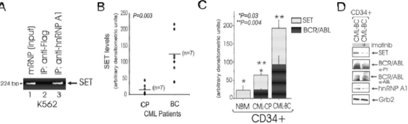
Using Ribonomics (105), SET mRNA was found specifically associated with cytoplasmic hnRNP A1 in the Philadelphia translocation positive K562 human cell line. To validate the SET mRNA-hnRNP A1 association, human SET-specific RT-PCR was carried out on anti-hnRNP A1- and anti-FLAG (unrelated antibody)-immunoprecipitated cytoplasmic mRNAs. High levels of SET mRNA transcripts,
similar to those present in the mRNP-enriched mRNA fraction, were clearly detectable in association with hnRNP A1 (Figure 3A). This suggested that the regulation of SET expression may be dependent on the nucleo-cytoplasmic shuttling of its mRNA bound to hnRNP A1 and, as a consequence, dependent BCR/ABL expression/activity. In fact, SET expression was higher in myeloid CML-BC than in CML-CP patient derived mononuclear marrow cells (Figure 3B); moreover SET protein levels correlated with levels of BCR/ABL, and were higher in myeloid CML-BCCD34+ than CMLCPCD34+ patient-derived bone marrow progenitor cells, and in CML-CPCD34+ patient cells than in CD34+ primary cells from healthy donors (NBM) (Figure 3C). Furthermore, treatment of CML-BCCD34+ cells with
Imatinib decreased SET and, as expected, hnRNP A1 levels (Figure 3D) further indicating that BCR/ABL activity is responsible for SET upregulation in CML-BC. In the BCR/ABL-transformed myeloid cell line 32Dcl3 (32D-BCR/ABL) and in
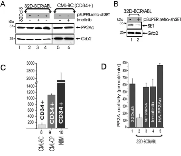
CML-BCCD34+ patient cells (Figure 4A) total expression of PP2A was not affected by
BCR/ABL expression and/or kinase activity (post-Imatinib treatment). By contrast, PP2A activity, was reduced by 94% and 52% in the CD34+ fraction of myeloid CML-BC and CML-CP cells, respectively, compared to the CD34+ fraction of NBM cells (Figure 4B), consistent with the notion that SET is an endogenous inhibitor of PP2A phosphatase activity. To determine whether SET upregulation is responsible
Figure 3. A: RT-PCR shows association of SET mRNA with hnRNP A1 in mRNP-enriched lysates, anti-FLAG-IP (negative
control), and antihnRNP A1IP from K562 cells. B: scatter plots show SET protein levels in BM cells from seven CMLCP and -BC patient specimens (p = 0.003; Wilcoxon rank sum test). C: graph shows SET (light bars) and -BCR/ABL (dark bars) protein expression (expressed as mean ± SE of densitometric units after normalization with Grb2 levels) in the CD34+ fraction from bone marrow of healthy donors (NBM) (n = 3), CML-CP (n = 3), and CML-BC (n = 3) patients (NBM versus CML-CP, p = 0.03; CML-CP versus CML-BC, p = 0.004; paired samples t test). D: SET, hnRNP A1, and BCR/ABL protein levels in untreated and imatinib-treated CML-BCCD34+ cells.
for suppression of PP2A activity, we interfered with SET expression by transducing 32D-BCR/ABL cells with a pSuper.retro-shSET construct carrying a short hairpin RNA (shRNA) targeting SET mRNA (Figure 4C). Expression of SET shRNA downregulated levels of SET and restored PP2A activity to levels similar to those of 32Dcl3 or imatinib-treated 32D-BCR/ABL cells (Figure 4B), without affecting PP2Ac expression (Figure 4A).
Relevance of PP2A activity suppression in CML
The suppression of the phosphatase activity of PP2A appears to be required by BCR/ABL to keep its tyrosine kinase in an active status, in fact
Figure 4. A: protein levels of PP2Ac and Grb2 in the following: 32Dcl3, untreated and imatinib-treated 32D-BCR/ABL and
primary CML-BCCD34+ marrow cells, and SET shRNA-expressing 32D-BCR/ABL. B: effect of the SET shRNA on SET
levels C and D: PP2A phosphatase assay in 32Dcl3, 32D-BCR/ABL, CML-BCCD34+, CML-CPCD34+, and NBMCD34+cells, and in BCR/ABL cells expressing the SET shRNA or HA-PP2Ac or treated with imatinib. Bars represent the PP2A activity (expressed as mean ± SE of three independent experiments performed with the indicated cell lines, and with NBMCD34+ (n = 3), CML-CPCD34+ (n = 3), and CML-BCCD34+ (n = 3) cells.
BCR/ABL itself is a target of PP2A. Expression of SET shRNA or HA-PP2A in 32D-BCR/ABL cells suppressed p210BCR/ABL tyrosine phosphorylation and expression (Figure 5A). This effect, was dependent on enhanced PP2A phosphatase activity because both SET overexpression and treatment with the phosphatase inhibitor okadaic acid used at concentrations that inhibit PP2A but not other phosphatases (107), restored BCR/ABL activity and expression in HA-PP2Ac-transduced cells (Figure 5A). Conversely, exposure of Imatinib-sensitive and -resistant (T315I) BCR/ABL+ cell lines and CML-BCCD34+ marrow blasts to the PP2A activators (108) forskolin (40μM), butyryl-forskolin (water soluble), or 1,9-dideoxy-forskolin (lacks adenylate cyclase-activating function) enhanced PP2A activity abolished BCR/ABL phosphorylation, and induced downregulation of BCR/ABL (Figure 5B). To determine whether PP2A-induced BCR/ABL dephosphorylation renders BCR/ABL more susceptible to proteolysis, we treated PP2A-expressing 32D-BCR/ABL cells with the proteasome inhibitor ALLN. As shown, ALLN restored BCR/ABL expression and, as expected, did not prevent PP2A-induced BCR/ABL tyrosine dephosphorylation (Figure 5C), which precedes and, likely, is required for its degradation. Interestingly, tyrosine dephosphorylation of BCR/ABL appeared to be dependent on the activity of another phosphatase, SHP-1, that had already been described as relevant in BCR/ABL expressing cells (109). In fact restored BCR/ABL phosphorylation was observed after treatment of SET shRNA- and HA-PP2A-expressing 32D-BCR/ABL cells with sodium stibogluconate (SS) (Figure 5D), a specific SHP-1 inhibitor (110). According to these observation it seemed plausible that PP2A inactivation by BCR/ABL in a kinase and dosage dependent manner may be an important step in the progression of CML to the blast phase and that inactivation of the tumor suppressor PP2A may be a necessary event required to keep BCR/ABL kinase activity unrestrained both in chronic phase and, to a greater extent, in blast crisis where BCR/ABL activity is at its highest and
PP2A activity, as a consequence, is at its lowest. In this scenario PP2A phosphatase activity induction could be envisioned as a new therapeutic strategy to be used in parallel with tyrosine kinase inhibitors to enhance BCR/ABL inhibition.
Pre‐clinical evaluation of the PP2A activator FTY720.
In light of the discovery of the role of PP2A in CML we have assessed the therapeutic potential of the PP2A activator FTY720 (111) (also known as Fingolimod, Novartis) in CML-BC and Philadelphia-positive Acute Lymphoblastic Leukemia (Ph1 ALL, expressing the isoform p190 of the BCR/ABL oncoproteins) on progenitors and in models of in vitro and in vivo p210 and p190 BCR/ABL
Figure 5. A: p210BCR/ABL activity (αPY) and expression (α-Abl) in 32D-BCR/ABL and in its derivative SET shRNA-,
HA-PP2A-, and HA-PP2A/FLAG-SET-expressing cell lines, and in HA-PP2A 32D-BCR/ABL cells treated with okadaic acid. B: Effect of the PP2A activators forskolin, butyryl-forskolin, and 1,9-dideoxy- forskolin on BCR/ABL activity and expression in primary CML-BC and ima- tinib-sensitive and -resistant BCR/ABL cell lines. C: p210BCR/ABL activity and expression in parental and untreated or ALLN-treated HA-PP2A-expressing 32D-BCR/ABL cells. D: Effect of the SHP-1 inhibitor sodium stibogluconate (SS) on BCR/ABL activity in parental and HA-PP2A- and SET shRNA-expressing 32D-BCR/ABL cells.
leukemogenesis. FTY720, a synthetic myriocin analog structurally similar to sphingosine, is a water-soluble drug with high oral bioavailability that reversibly arrests lymphocyte trafficking (mainly of CD4+T cells) (71, 112-114). It is currently used as an immunomodulator in Phase III trials for patients with multiple sclerosis. We have investigated and produced data that may support FTY720 as a novel therapeutic approach for patients with Imatinib/Dasatinib-sensitive and -resistant CML and also for Ph1-ALL.
FTY720 restores the activity of PP2A and inhibits that of p210 and
p190 BCR/ABL
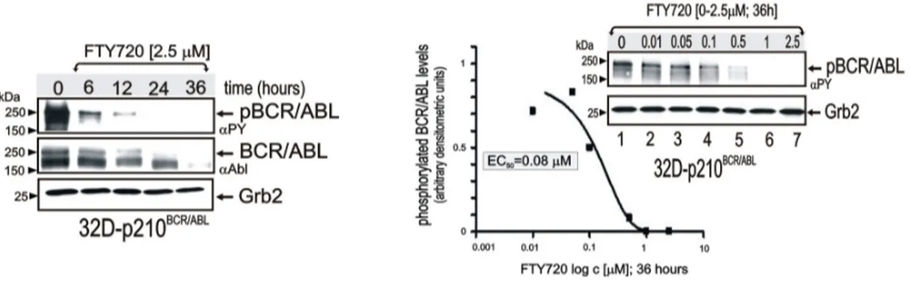
The dose/time-dependent effect of FTY720 on BCR/ABL-expression/activity was initially assessed in p210BCR/ABL-transformed myeloid precursor 32Dcl3 cells. Levels of tyrosine-phosphorylated (active) p210BCR/ABL were dramatically reduced within 6 hours, barely detectable at 12 hours, and absent at 24 hours of treatment with 2.5 ⎧M FTY720 (Figure 6). Similarly, BCR/ABL expression was downregulated by 2.5 ⎧M FTY720, although this occurred at later time points than dephosphorylation (Figure 6). BCR/ABL-transformed cells were also treated with escalating doses of FTY720 (concentrations of 0.01 to 2.5 ⎧M) (Figure 6). Indeed, FTY720 treatment at 36 hours suppressed BCR/ABL activity with a 50% effective
Figure 6: p210BCR/ABL tyrosine phosphorylation (αPY) and expression (αAbl) in 32D-BCR/ABL cells untreated and treated 6–36
hours with 2.5 μM FTY720 (left panel). Graph shows levels of phosphorylated BCR/ABL (expressed as arbitrary densitometric units normalized to Grb2 levels) in 32D-BCR/ABL cells treated for 36 hours with the indicated concentrations of FTY720. Right blot: p210BCR/ABL activity in 32D-p210BCR/ABL cells treated for 36 hours with FTY720.
concentration (EC50) of approximately 80 nM (Figure 6).
As already demonstrated, PP2A activity was reduced by 90% in myeloid
CML-BCCD34+compared to the CD34+ fraction from healthy bone marrow (NBM) donors,
and also reduced by 82% in Ph1 ALLCD34+/CD19+ lymphoid blasts compared to the CD34+/CD19+ fraction from NBM donors (Figure 2A). Consistently, treatment of CML-BCCD34+ and Ph1 ALLCD34+/CD19+ progenitors with FTY720 in the presence of myeloid or lymphoid cytokines reestablished PP2A activity to levels observed in normal CD34+ myeloid and CD34+/CD19+lymphoid progenitors (Figure 7A). By contrast, FTY720 did not over induce PP2A activity in primary NBM progenitors (Figure 7A).
As in myeloid CML-BC (62), suppression of PP2A activity in Ph1 ALL is also dependent on enhanced expression of the BCR/ ABL-regulated SET (62). In fact, SET protein levels were significantly higher in Ph1 ALLCD34+/CD19+ than in Ph1
NBMCD34+/CD19+progenitors (Figure 7A). Thus enhanced SET expression represents
a common mechanism used by both p210 (62) and p190 BCR/ABL oncoproteins
Figure 7. A: PP2A assay in untreated (black bars) and FTY720-treated (red bars) CML-BCCD34+ (n = 11), and Ph1
ALLCD34+/CD19+ (n = 12); Dot plot shows SET protein levels expressed as arbitrary densitometric units normalized to Grb2 protein
levels in NBMCD34+/CD19+ (n = 4) and Ph1 ALLCD34+/CD19+ cells (n = 12) (P < 0.001, Student t-test). B: Western blots show effect of small-t expression on BCR/ABL activity and expression in untreated and FTY720-treated 32D-p210BCR/ABL cells. C: p210BCR/ABL and p190BCR/ABL activity and expression in untreated and FTY720-, Imatinib-, and 1,9-dideoxyforskolin–
treated 32D-p210BCR/ABL (wild-type and T315I), K562, BaF3-p190BCR/ABL, CML-BCCD34+ (n = 3), and Ph1 ALLCD34+/CD19+ cells (n =
to suppress the phosphatase activity of PP2A.
To further demonstrate that the FTY720-induced suppression of BCR/ABL activity and expression depends on activation of PP2A, parental and BCR/ABL-expressing 32Dcl3 cells were transduced with a retrovirus (pBABE-GFP-sTAg) carrying the PP2A inhibitor SV40 small-t antigen. Expression of small-t (Figure 7B) abrogated the effects of FTY720; in fact, FTY720 treatment was unable to rescue to suppress BCR/ABL activity or expression (Figure 7B) in GFP+ small-t-expressing 32D-BCR/ABL cells.
While imatinib treatment suppresses wild-type BCR/ABL kinase activity only (Figure 7C, lanes 1 and 2), enhanced PP2A activity by FTY720 treatment abolished p210BCR/ABL phosphorylation and induced its downregulation in
imatinib-sensitive (32D-p210BCR/ABLand K562) and -resistant (T315I) cell lines (Figure 2B, lanes 3–8) and in primary CML-BCCD34+ cells (Figure 7C, lanes 12 and 13). The PP2A-mediated effects of FTY720 were also clearly evident in the lymphoid
BaF3-p190BCR/ABL cell line and Ph1 ALLCD34+/CD19+ patient cells. In fact, p190BCR/ABL
tyrosine phosphorylation and expression were strongly inhibited in
BaF3-p190BCR/ABLand IL-7-cultured Ph1 ALLCD34+/CD19+cells treated with FTY720 (Figure
7C, lanes 9-11, 14, and 15). Likewise, 1,9-dideoxyforskolin, a PP2A activator capable of impairing p210-BCR/ABL expression and leukemogenesis (62), efficiently suppressed p190-BCR/ABLactivity and expression in BaF3-p190BCR/ABL (Figure 7C, lane 10) and Ph1 ALLCD34+/CD19+cells.
Antileukemic activity of FTY720 in Ph1 leukemia cells in vitro
To assess the biologic effects and therapeutic relevance of FTY720 in Ph1 leukemias, primary bone marrow progenitors from imatinib-sensitive and -resistant CML-BC, imatinib-sensitive and -resistant CML-CP, and Ph1 ALL patients were treated with 2.5 ⎧M FTY720 for 48–96 hours and used to assess apoptosis and/or
clonogenic potential. For controls we used normal CD34+ and CD34+/CD19+ progenitors (NBM) treated with FTY720 at the same conditions. Consistent with its ability to strongly enhance PP2A activity in Ph1 but not in normal hematopoietic progenitors (Figure 7A), FTY720 markedly induced apoptosis (Figure 4) of IL-3/IL-6/Flt-3L/SCF-cultured and IL-7/Flt-3L/SCF-cultured CML-BCCD34+ and Ph1
ALLCD34+/CD19+ patient cells. A similar proapoptotic effect was observed in
CML-CPCD34+cells treated with FTY720 (Figure 8). In fact, the percentage of apoptosis
(annexin V+cells) in FTY720-treated primary CML-BCCD34+ and CML-CPCD34+ cells ranged between 86% and 97% (Figure 8). FTY720-treatedand Ph1 ALLCD34+/CD19+ cells also underwent marked cell death (98% annexin V+ cells) (Figure 8). By contrast, the percentages of apoptotic NBMCD34+, and NBMCD34+/CD19+ cells were not significantly altered in FTY720-treated cells compared with untreated cells (Figure 8). Because inhibition of the BCR/ABL-dependent PI3K/Akt pathway triggers caspase-dependent apoptosis (7), strong activation of caspase-3/7 was
Figure 8. Caspase-3/7 (graphs) and annexin-V/propidium iodide (annexin-V/PI) (flow cytometry dot plots) assays in untreated
and FTY720- treated (2.5 μM) NBMCD34+ (n = 3), CML-CPCD34+ (n = 3), CML-BCCD34+ (n = 3) and NBMCD34+/CD19+ (n = 3), Ph1
observed in FTY720-treated CML-BCCD34+, CML-CPCD34+, and Ph1 ALLCD34+/CD19+ cells (an average of 6-fold increase) but not in normal hematopoietic progenitors (Figure 8).
Similar to the inhibitory effect of forskolin or ectopic PP2Ac expression on colony formation of CML-BCCD34+ patient cells (62), a single dose of FTY720
strongly abolished (80%–95% inhibition) the ability of primary imatinib-sensitive and Imatinib- and Dasatinib-resistant CML-BCCD34+ and CML-CPCD34+ marrow cells to form IL-3-derived colonies in semisolid medium (Figure 9A). Also, IL-7-driven colony formation of Ph1 ALLCD34+/CD19+ lymphoid progenitors was dramatically suppressed (an average of 80% inhibition) by exposure to 2.5 ⎧M FTY720. Moreover, the presence of malignant cells bearing the Imatinib and Dasatinib-resistant T315I BCR/ABL mutant did not influence the responsiveness of bone marrow CD34+ CML-CP or CML-BC cells to FTY720 (Figure 9A). Conversely,
Figure 9. A: IL-3- (for myeloid progenitors) and IL-7-dependent (for lym- phoid progenitors) colony-forming ability (mean ± SD)
of untreated and FTY720-treated NBMCD34+ (n = 4), CML-CPCD34+ (n = 3), CML-BCCD34+ (n = 11), CML-CPCD34+ (T315I) (n = 1),
CML-BCCD34+ (T315I) (n = 3), NBMCD34+/CD19+ (n = 4), and Ph1 ALLCD34+/CD19+ (n = 12) cells. As controls, 100% clonogenic
potential was attributed to the IL-3- and IL-7-driven colony formation of NBMCD34+ and NBMCD34+/CD19+, respectively. B: Inhibition
of PP2A activity by okadaic acid (0.25 nM) rescues the clonogenic potential of FTY720-treated CML-CPCD34+, CML-BCCD34+,
FTY720 did not affect the IL-3- or IL-7-driven clonogenic potential of NBMCD34+ or
NBMCD34+/CD19+ cells, respectively. Consistent with the important role of PP2A as
mediator of the in vitro cytotoxic effect of FTY720, co-treatment with 2.5 ⎧M FTY720 and 0.25 nM okadaic acid significantly rescued cytokine-dependent colony formation of CML-CPCD34+, CML-BCCD34+, and Ph1 ALLCD34+/CD19+ bone marrow progenitors (Figure 9B).
FTY720 suppresses in vivo BCR/ABL leukemogenesis
To assess the effect of FTY720 on an in vivo model of CML and Ph1 ALL we used SCID mice that were intravenously injected with 32D-p210BCR/ABL,
BaF3-p190BCR/ABL,or 32D-p210BCR/ABL(T315I) cells (5⋅105 cells/mouse). Seven days
post-injection, cell engraftment was assessed by the presence of circulating BCR/ABL+ cells via nested RT-PCR–mediated detection of p210 or p190 BCR/ABL transcripts in peripheral blood (PB) (Figure 10A). Intra-peritoneal treatment with FTY720 (10 mg/kg/d; LD50 [50% lethal dose] = 300 mg/kg) was then initiated in 13 mice per
group (n = 39). As controls, 13 untransplanted mice received daily treatment with FTY720, whereas an identical number of cell-injected mice were left untreated. After 4 weeks of FTY720 treatment, all treated leukemic mice were alive and BCR/ABL negative (Figure 10A), whereas only 3-4 untreated leukemic mice were alive but appeared lethargic and were BCR/ABL positive (Figure 10A). Thus, 3 mice per group were sacrificed and organs were evaluated by visual inspection and light microscopy. Mice injected with wild-type or T315I p210 and p190 BCR/ABL+ cells showed massive splenomegaly, whereas, the morphology of spleens from FTY720-treated BCR/ABL+ cell-injected mice resembled that of control age-matched or FTY720-only treated mice (Figure 10A). Peripheral blood cytospins and hematoxylin and eosin-stained sections of spleen, bone marrow, and liver of 32D-p210BCR/ABL, 32D-p210BCR/ABL(T315I), and BaF3-p190BCR/ABLmice
Figure 10. Pharmacologic doses of FTY720 impair in vivo imatinib/dasatinib-sensitive and -resistant p210 and p190 BCR/ABL leukemogenesis. (A) Left: Nested RT-PCR for p210 and p190 BCR/ABL indicates the presence of BCR/ABL-expressing cells in the PB of untreated and FTY720-treated mice. Nested RT-PCR performed with RNA from PB of mice treated with FTY720 only (far right lane) and with RNA from a 1:106 dilution of K562 or BaF3p190 (BCR/ABL+ cells) with 32Dcl3 cells were used, respectively as negative and positive controls. GAPDH mRNA levels were used as a control. Right panel: Visual analysis of spleens from age-matched and FTY720-treated mice and untreated and FTY720-treated mice injected with the indicated cell lines. (B) May-Grumwald/Giemsa staining of PB and H&E staining of sections from bone marrow, spleen, and liver of untreated and FTY720-treated (4 weeks) control and cell-injected mice. Original magnification, ×400 (PB and bone marrow); ×250 (liver and spleen). (C) Effect of 10 mg/kg/d FTY720 on survival of SCID mice i.v. injected with the indicated cells lines (n = 13; orange lines). Mice injected with cells only (n = 13; blue lines) or drug only (n = 13; green lines) were used as controls. The red lines below each graph indicate the treatment regimen. Survival was calculated by the Kaplan-Meier method, and the log-rank test evaluated the differences among survival distributions. Overall, P < 0.0001; 32D-p210BCR/ABL untreated versus FTY720-treated, P < 0.0001; 32D-p210BCR/ABL (T315I)