UNIVERSITÀ DEGLI STUDI DELLA TUSCIA DI VITERBO
DIPARTIMENTO DI SCIENZE ECOLOGICHE E BIOLOGICHE
Corso di Dottorato di Ricerca in GENETICA E BIOLOGIA CELLULARE – XXVIII
“Analysis of the role of Werner Helicase Interacting Protein 1 in
response to replication stress”
(s.s.d. BIO / 11)Tesi di dottorato di: Dott. Giuseppe Leuzzi
Coordinatore del corso Tutore
Prof. (Giorgio Prantera) Dott.ssa (Annapaola Franchitto)
! ! Data della discussione
Dedicated to
my grandparents
INDEX
SUMMARY ... 1
1. INTRODUCTION ... 3
THE BASIC OF EUKARYOTIC DNA REPLICATION ... 4
Initiation of DNA Replication ... 5
Origins firing and chain elongation ... 5
Termination of DNA Replication ... 7
DNA REPLICATION STRESS ... 8
Causes of Replication Stress ... 8
Consequences of Replication Stress ... 11
RESPONSE TO REPLICATION STRESS ... 12
DNA Damage Checkpoint ... 12
The Replication Checkpoint ... 14
ATR signaling ... 16
RESTARTING MECHANISMS OF STALLED FORKS ... 18
Fork Repriming and Translesion Synthesis ... 20
Fork Reversal ... 21
Regulation of replication fork restart mechanisms ... 22
STABILIZATION AND PROTECTION OF STALLED FORKS ... 24
Fork Protection Factors ... 24
Cellular nucleases involved in fork degradation ... 27
Physiological function of fork protection ... 28
THE WERNER HELICASE INTERACTING PROTEIN 1 (WRNIP1/WHIP1) ... 29
Evidences supporting WRNIP1 role during Replication Stress ... 30
2. AIM ... 33
3. RESULTS ... 34
WRNIP1 IS REQUIRED FOR PROTECTION AND RESTART OF STALLED FORKS UPON REPLICATION STRESS ... 34
MRE11 DEGRADATES NASCENT DNA STRAND AT STALLED FORKS IN ABSENCE OF WRNIP1 ... 41
WRNIP1 DEPLETION PRODUCES PARENTAL-STRAND ssDNA
ACCUMULATION AND RAD51 DESTABILIZATION AFTER FORK STALLING 43 RAD51 AND MRE11 ARE DIFFERENTLY RECRUITED TO STALLED
REPLICATION FORKS IN WRNIP1-DEFICIENT CELLS ... 49
RAD51 PROTECTS NASCENT DNA STRAND FROM DEGRADATION AFTER REPLICATION STALLING IN WRNIP1-DEFICIENT CELLS ... 51
WRNIP1 STABILIZES RAD51 ON STALLED FORKS ... 54
LOSS OF WRNIP1 OR ITS ATPase ACTIVITY LEADS TO DNA DAMAGE ACCUMULATION AND CELL DEATH AFTER REPLICATION STALLING ... 58
UNPROTECTED STALLED FORKS LEAD TO CHROMOSOMAL INSTABILITY IN WRNIP1-DEFICIENT CELLS ... 61
4. DISCUSSION ... 66
5. MATERIALS AND METHODS ... 72
CELL LINES AND CULTURE CONDITIONS ... 72
CHEMICALS ... 72
SITE-DIRECT MUTAGENESIS AND CLONING ... 72
PLASMIDS AND RNA INTERFERENCE ... 73
DNA FIBER ANALYSIS ... 73
IN SITU PLA ASSAY ... 74
CO-IMMUNOPRECIPITATION, CELL FRACTIONATION AND WESTERN BLOT ... 74
CldU CO-IMMUNOPRECIPITATION OF PROTEINS AT STALLED FORKS... 76
NEUTRAL AND ALKALINE COMET ASSAY... 77
IMMUNOFLUORESCENCE ... 77
LIVE/DEAD STAINING ... 78
CHROMOSOMAL ABERRATION ANALYSIS ... 78
STATISTICAL ANALYSIS ... 79
6. REFERENCES ... 82
SUMMARY
Genome instability is a common feature of cancer cells. Most of the chromosomal abnormalities arising in tumours come from defective DNA replication (Abbas et al., 2013; Aguilera and Gómez-González, 2008; Branzei and Foiani, 2010; Zeman and Cimprich, 2014). For this reason, accurate handling of stalled replication forks is of paramount importance for the maintenance of genome stability. Recently, it has been demonstrate that RAD51 recombinase is involved in protecting stalled replication forks from nucleolytic attack by MRE11, which otherwise can seriously threaten genome stability (Hashimoto et al., 2010; Schlacher et al., 2011). However, the identity of other factors that can collaborate with RAD51 in this task and how this pathway operates are still poorly elucidated.
In this study, we have identified a previously uncharacterized function of the human Werner helicase interacting protein 1 (WRNIP1) as a factor working in conjunction with the RAD51 recombinase in response to replication stress.
We show that WRNIP1 is directly involved in protection and restart of stalled replication forks following replication stress. We also demonstrated that WRNIP1 is required for preventing uncontrolled MRE11-mediated degradation of nascent DNA strand at stalled replication forks. WRNIP1 depletion results in a large enhancement of parental-strand ssDNA accumulation produced by the action of MRE11 nuclease activity, but it does not lead to a greater amount of RAD51 bound to chromatin. Thus, WRNIP1-deficient cells show an overt RAD51 destabilization after fork stalling.
We establish that WRNIP1 is directly recruited to stalled replication forks and cooperates with RAD51 to safeguard fork integrity, by promoting RAD51 stabilization on ssDNA. We further demonstrate that replication fork protection does not require the ATPase activity of WRNIP1 that is however essential to achieve the recovery of perturbed replication forks. Loss of WRNIP1 or its catalytic activity exhibit high sensitivity to HU-induced fork stalling, leading to DNA damage accumulation and cell death, and that unprotected stalled forks is responsible for chromosomal instability arising, after fork stalling, specifically in WRNIP1-deficient cells. Interestingly, downregulation of the anti-recombinase FBH1, which promotes the removal of RAD51 from chromatin, can compensate for loss of WRNIP1 activity. Indeed, attenuation of replication fork degradation and chromosomal aberrations have been observed in WRNIP1-deficient cells after FBH1 depletion, due to
enhancement of the amount of RAD51 chromatin-bound. Consistently, over-expression of RAD51 in WRNIP1-deficient cells counteracts stalled replication fork degradation. Therefore, our results clearly indicate that WRNIP1 plays a crucial role in stabilizing RAD51 to stalled forks, protecting them from the MRE11-dependent degradation. Furthermore, we establish that WRNIP1 is implicated in the stalled fork resumption through its ATPase activity.
Altogether, our work suggests a molecular basis for the role of human WRNIP1 in safeguarding genome stability in response to replication stress. In particular, they unveil a unique role for WRNIP1 as a replication fork-protective factor in maintaining genome stability.
Highlights:
- WRNIP1 protects stalled replication forks from degradation;
- ATPase activity of WRNIP1promotes stalled replication forks restart;
- WRNIP1 contributes to the stabilization of RAD51 on stalled replication forks; - FBH1 downregulation compensates for loss of WRNIP1 activity.
1. INTRODUCTION
DNA replication is a highly complex cellular process by which eukaryotic cells accurately and efficiently duplicated their genome, generating identical sets of chromosomes, thereby transmitting genetic information to daughter cells. Efficient and error-free DNA replication is the key for faithful duplication of chromosomes before their segregation. Moreover, DNA replication is tightly monitored to ensure that the genome is replicated just once per cell cycle, before mitosis begins (Branzei and Foiani, 2010).
DNA replication is not only crucial to cellular division but also plays a crucial role in the maintenance of genomic integrity (Watanabe and Maekawa, 2010). Both exogenous and endogenous damaging agents constantly urge DNA, so that DNA lesions frequently occur. Thus, cells need to deal with DNA lesions during replication by activating an adequate cellular response. If not properly repaired, these lesions may hinder replication fork progression leading to fork arrest (fork stalling), which causes alterations of DNA replication dynamic known as “replication stress” (Zeman and Cimprich, 2014).
Since replication stress has considered the primary source of genome instability, cells must monitor fork integrity and they need to match DNA replication with other cellular processes, such as chromatin reassembly and the establishment of cohesion between sister chromatids. The success of all these processes is crucial to avoid DNA breaks, chromosomal rearrangements, and mutations that can cause not only the loss of cell viability, but also a large number of human syndromes, including premature aging, various cancer predispositions and genetic abnormalities (Branzei and Foiani, 2010). Therefore, it seems logical that studying DNA replication and the pathways that suppress the instability of replication fork is directly relevant to understanding the mechanisms by which cancers and other pathological disorders arise.
THE BASIC OF EUKARYOTIC DNA REPLICATION
DNA replication is the mechanism by which DNA polymerases synthesize a DNA strand complementary to the original template strand. This process allows the cell to duplicate a single DNA double helix into two DNA helices, so that the high-fidelity passage of genetic information from parental cell to daughter cells is assured. For this reason, DNA replication has to occur without errors. In G1 phase of the cell cycle, many of the DNA
replication regulatory processes are initiated. In eukaryotes, the vast majority of DNA synthesis occurs during S phase of the cell cycle, and the entire genome must be unwound and duplicated to form two daughter copies. During G2 phase, any damaged DNA or
replication errors are corrected. Finally, one copy of the genomes is segregated to each daughter cell at mitosis (M) phase (Leman and Noguchi, 2013). Each of these daughter copies contains one strand from the parental duplex DNA and one nascent antiparallel strand. This mechanism, conserved from prokaryotes to eukaryotes, is known as semiconservative DNA replication (Meselson and Stahl, 1958) and it is organized into three distinct phases: initiation, elongation and termination (Fig. 1).
Figure 1. Schematic representation of
different steps in eukaryotic DNA replication. Origin recognition and
formation of the preRC complex comprises binding of ORC to DNA and subsequent recruitment of the Mcm2-7 helicase complex by Cdc6 and Cdt1. Firing of origins is brought about by the loading of Cdc45 and other firing factors and requires CDK and DDK activity and (see text). Subsequently, ORC, Cdc6 and Cdt1 dissociate from DNA and replication chain elongation occurs by the coordinated action of polymerases and other components of the replication complex. Modified from (Claus Storgaard Sørensen, 2011).
Initiation of DNA Replication
In eukaryotes, replication initiates from multiple regions distributed along chromosomes, know as replication origins. In budding yeast, replication origins have been efficiently mapped owing to clear consensus sequences (Wyrick et al., 2001). However, this is more difficult in higher eukaryotes. Although origin sequences are not clearly defined in mammals, they have been associated with certain features, which help to predict potential replication initiation sites, such as AT rich sequences (Altman and Fanning, 2004; Paixão et al., 2004) and matrix attachment regions.
Replication initiation begins when the origins are marked by the formation of a pre-replicative complex (preRC) in G1, before DNA replication, through the binding of the origin recognition complex (ORC). After ORC, additional replication factors, such as Cdc6, Cdt1 and the hexameric MCM2-7 complex, are loaded onto chromatin (Masai et al., 2010). Because the preRC cannot be assembled later in the cell cycle, the maximum number of origins available for a single S phase is determined during the licensing state, which occurs in G1 when the preRC is formed. Thus, preRCs mark “potential” origins, but only a subset is licensed for use during each round of replication (Edwards et al., 2002; Hyrien et al., 2003).
Origins firing and chain elongation
The replisome complex, which is a massive complex coordinating many proteins, assembles at replication origins and subsequently initiates bidirectional DNA synthesis in S phase in a process called origins firing (Leman and Noguchi, 2013).
The critical step for DNA replication is the conversion of pre-RC into initiation complex (IC). This step requires phosphorylation activity of CDK and CDC7 (Hoang et al., 2007; Sclafani, 2000), which mediate activation of preRC allowing the loading of RPA, MCM10 and CDC45 onto chromatin to carry out origins firing (Zhu et al., 2007). Importantly, not all licensed origins are fired. Indeed, most of them, defined “dormant origins”, remain quiescents during normal replication although they have a crucial role during replication stress (Chen et al., 2015).
At the molecular level, the geometry of DNA replicating site is a fork-like DNA structure, where the DNA double helix is open or unwound, exposing unpaired DNA nucleotides for
recognition and base pairing for the incorporation of free nucleotides into double-stranded DNA (Leman and Noguchi, 2013).
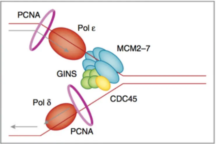
Parental DNA is unwound by the CMG helicase complex. CMG is composed of the CDC45 protein, the mini-chromosome maintenance 2-7 complex (MCM2–7) and the tetrameric GINS complex (Ilves et al., 2010). CMG moving in the 3’-5’ direction translocates along the leading strand while physically displacing the complementary strand by steric exclusion (Costa et al., 2014). DNA synthesis on the leading and lagging strands is performed by the Pol and Pol polymerases respectively, which make contact with CMG and PCNA (proliferating cell nuclear antigen). Polymerase ε (epsilon) synthesizes DNA in continuous manner, as it follows the same direction of DNA unwinding. This strand is known as “leading strand”. Polymerase δ (delta) synthesizes DNA on the opposite template strand in a discontinuous fashion and this strand is termed “lagging strand”. Because DNA polymerases require a primer on which to begin DNA synthesis, first, polymerase (Polα) acts as a replicative primase. Polα is associated with an RNA primase, and this complex accomplishes the priming task by synthesizing a primer. Importantly, this priming action occurs at replication initiation to begin leading and lagging strands synthesis (Georgescu et al., 2015) (Fig. 2).
Figure 2. Illustration of replication fork and key components of replisome. Parental DNA is unwound by
the CMG helicase complex. CMG is composed of the CDC45 protein (yellow), the minichromosome maintenance 2–7 complex (MCM2–7, blue) and the tetrameric GINS complex (green). CMG encircles the ssDNA on the leading strand and unwinds DNA by steric exclusion, moving in the 3′-to-5′ direction. DNA synthesis on the leading and lagging strands is performed by the Pol ε and Pol δ (orange) polymerases, respectively, which make contact with CMG. The sliding clamp PCNA (pink circle) acts as a processivity factor for the polymerases. From (Berti and Vindigni, 2016)
Termination of DNA Replication
Owing to stochastic origin firing and variable rates of replisome progression, the location and timing of eukaryotic termination is variable, making this process difficult to study. Little is known about termination process of DNA synthesis and most of our knowledge comes from studies on plasmid replication in Xenopus egg extracts or yeast chromosomal replication. Eukaryotic DNA replication terminates when converging replication forks meet (Santamaria, 2000). This process involves local execution of DNA synthesis, decatenation of daughter molecules and replisome break up, not necessarily in the following order. A large amount of evidence from simian virus 40 (SV40) and yeast systems suggests that sister chromatids become intertwined (catenated) at replication termination sites, and that the resolution of these structures, for successful completion of termination requires DNA topoisomerase II (TOPII) (Dewar et al., 2015).
Given the complexity of replication process, cells need to balance accuracy, speed, and the consumption of relevant resources, such as nucleotides and replication factors, to complete replication and to limit replicative stress events, which are considered the primary source of genome instability.
DNA REPLICATION STRESS
A wide variety of factors, such as DNA replication errors, spontaneous chemical reactions, reactive metabolic products, exogenous environmental agents or some anticancer therapeutics can cause DNA damage. It is estimated that DNA damage occurs at a rate of 1.000 to 1.000.000 molecular lesions per cell per day (Hoeijmakers, 2009). Furthermore, cells are particularly vulnerable to DNA damage during DNA replication, because virtually all forms of DNA damage block DNA replication, causing replication stress (Allen et al., 2011), which can compromise genome integrity if not properly processed.
Replication stress is defined as a phenomenon that arises when genetic or environmental conditions lead to the replicative polymerase to move slowly and/or stall, potentially leading to fork collapse and generating genome instability. It can be generated by a wide range of physical obstacles, and usually results in physical structures, namely stretches of single-stranded DNA (ssDNA), which represents a hallmark of replication stress (Zeman and Cimprich, 2014).
Causes of Replication Stress
Although, replication stress arises from many different sources, one of the most commonly recognized sources of replication stress is down-regulation of limiting replication factors . Indeed, faithful DNA replication requires numerous factors, and their limitation can result in the slowing of replication fork progression and, ultimately, in stalling of replication fork. These replication factors include components of the replication machinery, histones, histone chaperones that package replicated DNA and the pool of nucleotides (dNTPs) (Aguilera and García-Muse, 2013). Nucleotides are the building blocks for DNA synthesis and their titration is one of the key aspects during replication. Indeed, the reduced level of dNTPs slows down the progression of the forks and increases the chance of fork stalling per se (Anglana et al., 2003; Poli et al., 2012). Hydroxyurea (HU), a drug used to treat resistant chronic myelocytic leukemias and other tumors (Madaan et al., 2012; Patnaik and Tefferi, 2016), inhibits ribonucleotide reductase (RNR) and creates imbalances in the cellular dNTPs, which affect DNA polymerases and contribute to replication stress (Yarbro, 1992).
An excess of replication origin firing can also be a source of replication stress, through the exhaustion of factors essential for DNA synthesis and for the maintenance of fork integrity,
including RPA protein, which protects single-strand DNA (ssDNA). Indeed, the level of RPA becomes limiting when the number of replication origins increases. As a result, new ssDNA stretches cannot be protected by RPA, and therefore, the replication forks become more susceptible to collapse and breakage (Toledo et al., 2013).
In addition to limiting replication factors, a wide variety of obstacles can hamper replication fork progression leading to replication stress. These obstacles include DNA lesions, DNA-protein complexes and DNA sequences that can form secondary structures (Gelot et al., 2015). Some DNA sequences are intrinsically challenging for the replication machinery. For instance, common fragile sites (CFS) are normal components of human genome, unusually prone to breakage. These human genomic regions are difficult-to-replicate and display frequent events of fork stalling (Debatisse et al., 2012; Franchitto and Pichierri, 2011, 2014). In addition, trinucleotide repeats can form secondary DNA structures (hairpins, triplexes, etc) that are thought to block replication fork progression (Kim and Mirkin, 2015; McMurray, 2010). Recently, G-quadruplexes, secondary structures which form in GC-rich DNA, have also been highlighted as a significant source of DNA damage (Bochman et al., 2012; Paeschke et al., 2013).
In response to base damage, such as abasic site, stretches of ssDNA can be exposed at replication forks as a consequence of replicative helicases continuing to unwind the parental DNA while the replicative DNA polymerases are stalled. This uncoupling between helicase and polymerase activities is probably not the sole cause of accumulation of ssDNA at stalled forks. Indeed, agents that create physical blocks to helicase movement, such as inter-strand cross-links (ICLs) or torsional stress induced by the DNA topoisomerase I cleavage complex, are not expected to promote uncoupling. However, ssDNA can be detected in presence of these agents, suggesting a degradation process of newly-synthesized DNA, through the combined action of nucleases and DNA helicases, thereby creating ssDNA at fork junction (Berti and Vindigni, 2016).
Some studies have suggested that replication stress events can occur when replication forks and transcription complexes collide. Indeed, replication and transcription machineries both operate on DNA, so that, it is not unusual that the two processes interfere with each other, and that collision between replication and transcription complex occurs (Bermejo et al., 2012; Helmrich et al., 2013). Moreover, it has been proposed that replication stress can be induced by nucleotide misincorporation. Indeed, rNTPs stall the replicative polymerases,
and bypass of these rNTPs requires the DNA damage tolerance (DDT) pathways (Nick McElhinny et al., 2010) discussed above.
In the context of tumorigenesis, the oncogene-induced replication stress is an important matter (Gorgoulis and Halazonetis, 2010; Negrini et al., 2010). Oncogene activation alters DNA replication dynamic leading to increased replication stress and DNA breaks. There are several proposed mechanisms for the induction of replication stress upon oncogene activation. Two of the proposed mechanisms are related to an inappropriate/insufficient or excessive-usage of replication origins (Hills and Diffley, 2014). Overexpression of cyclin E reduces the number of replication origins that are licensed during G1 (Ekholm-Reed et al., 2004). As a consequence, replication stress increases in S-phase due to the shortage of back-up origins to cope with stalled forks. In contrast, the overexpression of certain oncogenes, such as MYC and RAS, has the opposite effect since it increases the origins firing (Dominguez-Sola et al., 2007) leading to a depletion of the cellular dNTPs (Fig. 3).
Figure 3. Schematic representation replication stress causes. Replicative stress results from endogenous
or exogenous obstacles to DNA replication. These include the incorporation of incorrect nucleotides or defects in DNA unwinding, each of which results in a structural hindrance to fork progression; other similar obstacles include lesions in the template DNA or the presence of protein complexes that are involved in transcription. A shortage of nucleotides or replication factors can also impair the progression of ongoing DNA replication. Modified from (Dobbelstein and Sørensen, 2015)
Consequences of Replication Stress
Genome instability is a common feature of cancer cell. Most of the chromosomal abnormalities founded in tumors arise from defective DNA replication, pointing out the role of replication stress in cellular transformation process (Gaillard et al., 2015).
In silico analysis of homozygous and heterozygous focal deletions in cancer samples and
cell lines revealed that most of the heterozygous deletions in transformed cells are found in already defined CFS or in large genes (Rajaram et al., 2013). Similar deletions can be induced by the treatment of cells with aphidicolin (Aph), an inhibitor of replicative DNA polymerases alfa, delta and epsilon, which induces replication stress (Baranovskiy et al., 2014; Krokan et al., 1981). These data indicate that replication stress is likely the source of most of the passenger deletions during transformation, suggesting a major role for replication stress in cancer genome development.
Another evidence supporting replication stress as driving force of malignant transformation is oncogene activation, which suggests that mild levels of replication stress allow the accumulation of genome instability that help to develop tumorigenesis. In addition, the connection between replication stress and tumorigenesis is further strengthened by the findings that the treatment of mice with hydroxyurea promotes leukemogenesis.
Lastly, replication stress might also lead to chromosomal instability (CIN) through an increase in defects on chromosome segregation. Consistently, the analysis of CIN+ versus CIN- colon adenocarcinoma cells reveals the presence of replication stress only in CIN+ cells, along with corresponding chromosome segregation defects (Burrell et al., 2013). Since replication stress has deleterious effects on genome stability, cells replicating their DNA must be able to initiate an adequate replication stress response to minimize the risk of chromosomal rearrangement accumulation.
RESPONSE TO REPLICATION STRESS
To reduce replication stress, eukaryotic cells are well equipped of a genomic maintenance apparatus. This sophisticated apparatus allows the replication stress response, and it includes a set of DNA surveillance mechanisms called the DNA damage checkpoints (Jossen and Bermejo, 2013). The importance of the cellular response to replication stress is highlighted by the array of genetic diseases, as well as increased cancer predisposition, associated with alterations in the genes that participate in the response (Zeman and Cimprich, 2014).
Given the complexity of the DNA replication stress response, here, a brief overview of the network will be provided, and only proteins and pathways immediately relevant to the present study will be described in detail.
DNA Damage Checkpoint
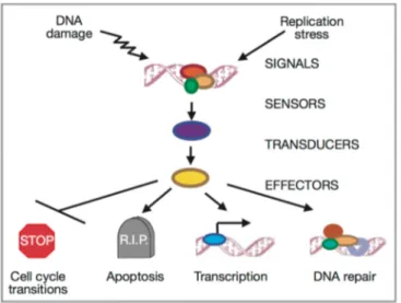
The DNA damage checkpoint network is considered a signal transduction cascade consisting of three major groups of proteins (sensors, transducers and effectors) that act in concert to promote cell-cycle arrest, DNA repair, transcription and apoptosis (Friedberg et al., 2006) (Fig. 4).
Figure 4. Conceptual organization of the signal transduction of checkpoint responses. DNA damages are recognized by sensor proteins. The signals are transmitted to transducers (mainly kinases) and the regulated transducer molecules activate effector kinases, which in turn promote cell-cycle arrest, DNA repair, transcription and apoptosis. (Zhou and Elledge, 2000)
The DNA damage checkpoint network is under control of members of the phosphoinositide 3-kinase-related kinase (PIKK) family. In mammals, signals initiated by the sensor very rapidly transduce to ATM and ATR kinases, which, in turn, phosphorylate a great number of substrates. ATM is 350 kDa oligomeric protein that exhibits significant homology to the PIKK. In humans, mutations in ATM cause ataxia telangiectasia, a rare autosomal recessive human disorder, characterized by genome instability, immunodeficiency and cancer predisposition (Shiloh, 1997). Cells lacking ATM are viable and patients and mice survive, suggesting that ATM is not essential for normal cell-cycle progression and cell differentiation. Activated ATM phosphorylates many proteins, including BRCA1, NBS1, CHK2 and p53, including itself (Shiloh and Kastan, 2001). ATR was discovered in the human genome database as a gene with sequence homology to ATM and Rad3, hence the name ATR. The gene encodes a protein of 303 kDa with a C-terminal kinase domain and regions of homology to other PIKK family members. Unlike ATM, ATR null mice are embryonic lethal and mutations causing a partial loss of its activity have been reported to be associated with the human autosomal recessive disorder Seckel syndrome (O’Driscoll et al., 2003). As ATM, ATR is capable of phosphorylating serine or threonine residues in SQ/TQ sites (Abraham, 2001). Once the active ATR is translocated to replication foci, it can phosphorylate and activate CHK1. This model is consistent with the observation that CHK1 is also essential for embryonic cell viability (Liu et al., 2000).
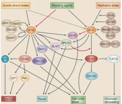
In response to DNA damage, the PIKK family kinases ATM and ATR phosphorylate target proteins on serine and threonine residues, thereby activating the DNA damage checkpoint. The ATM pathway responds to the presence of double-strand breaks (DSBs), and acts during all phases of the cell cycle. The ATR pathway can respond to agents that interfere with the function of DNA replication forks, such as ultraviolet light (UV) and HU. DNA-alkylating agents might activate both pathways, although these types of DNA damage impose stress on progressing replication forks, they clearly also could elicit strand breaks under some circumstances (Zhou and Bartek, 2004) (Fig. 5).
Figure 5. DNA damage response signal-transduction network (Zhou and Bartek, 2004).
The Replication Checkpoint
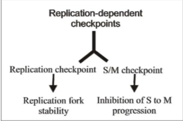
In eukaryotic cells there are multiple checkpoints that operate along the entire cell cycle. G1 and G2 phases of the cell cycle are under the control of a single checkpoint pathway, while at least three checkpoint activities can be founded associated with S-phase (Bartek et al., 2004). This difference can be explained with the high susceptibility of DNA during replication as well as the intrinsic difficulties to duplicate, in efficient manner, complex genome as that of human cells. So, the S-phase is under the control of multiple and, in some ways, redundant checkpoints pathways. There is a replication-independent checkpoint pathway that is activated by the presence of DSBs and the replication-dependent checkpoint, which can be divided in two different sub-pathways: replication checkpoint and S/M checkpoint. These pathways respond to replication stress, thus, to stalling of replication machinery that can be caused by several factors as discussed below (Branzei and Foiani, 2005).
The replication checkpoint is required to preserve stability of stalled forks until the causes of replication stress have been removed/resolved; whereas the S/M checkpoint is needed to prevent premature entry in mitosis with unreplicated or damaged chromosomes. To slow-down S-phase progression, the replication checkpoint targets kinases responsible for origin firing, while the S/M checkpoint inhibits the Cyclin1/CDK1 activity.
Since these two replication-dependent checkpoints are triggered by the same signal at stalled fork, they can be considered as two facets of a common pathway, so that in this work they will be collectively referred to as replication checkpoint (Fig. 6).
Figure 6. Schematic representation of the replication-dependent checkpoint operating in s-phase of cell cycle.
The two main functions of the replication checkpoint are maintenance of the integrity of the stalled forks and inhibition of the S/M transition. The first activity is not only important for prompt resumption of DNA synthesis once the arresting stimuli are relieved, but also to restrict access to recombination enzymes that could process replication intermediates at the fork after replisome disassembly, i.e. after replication fork collapse. On the other hand, the control of the S to M progression is essential to prevent mitotic entry with incompletely replicated DNA, a condition resulting in mitotic catastrophe and loss of cell viability. In addition, the replication checkpoint prevents firing of late origins, thus reinforcing the S/M blockage (Branzei and Foiani, 2005).
ATR signaling
The replication checkpoint is entirely controlled by the ATR kinase (Ataxia telangiectasia and Rad3-related), which “senses” replication blockage and propagates the checkpoint signal to replication, repair and cell cycle proteins, either directly or through the checkpoint kinase CHK1 (Cimprich and Cortez, 2008; Zou et al., 2003).
ATR responds to a broad spectrum of genotoxic agents that include UV, topoisomerase inhibitors, alkylating and cross-linking agents, as well as chemicals that interfere with DNA polymerization, such as Aph and HU (Byun et al., 2005; Cortez et al., 2001; Zhou and Elledge, 2000).
A lot of data obtained in different model systems indicates that when the polymerase encounters a lesion its progression is blocked, while the helicase keeps unwinding the DNA. The uncoupling between the stalled polymerase and the helicase generates a segment of ssDNA (Byun et al., 2005; Tercero et al., 2003).
ATR and its regulatory subunit ATRIP can sense fork stalling through their direct interaction with the ssDNA binding protein RPA (Ball et al., 2005; Zou et al., 2003). Association of ATR/ATRIP complex with chromatin leads to phosphorylation of several downstream targets, most notably the RAD9/RAD1/HUS1 complex (9.1.1 complex) that is recruited to stalled forks independently from ATR/ATRIP, but through another protein factor, RAD17, which binds directly to RPA (Abraham, 2001; Zou and Elledge, 2003). Recruitment of the RAD17/9.1.1 complex cooperates with ATR/ATRIP to determine full replication checkpoint activation and ATR-dependent phosphorylation of other targets (Zou et al., 2002). The 9.1.1 complex provides a docking station to other factors implicated in the replication checkpoint, facilitating subsequent phosphorylation by ATR. In other words, the 9-1-1 complex recognizes a DNA end that is adjacent to RPA-ssDNA stretch. Phosphorylation of several of the ATR targets also requires additional “mediators”, such as Claspin and BRCA1. Claspin is necessary for CHK1 phosphorylation by ATR (Gottifredi and Prives, 2005). CHK1 is the downstream checkpoint kinase in response to fork stalling or DNA damage originated at the replication fork, and directly contributes to maintain integrity and competence of stalled forks (Feijoo et al., 2001; Lopes et al., 2001). Indeed, CHK1 induces cell cycle arrest by its inhibitory phosphorylation of CDC25, the phosphatase activating CDK1 and CDK2 . ATR activation has both a positive and negative effect on replication-origin firing in response to replication stress: it prevents new origin
firing by inhibiting replication initiation, through CHK1-mediated inhibition of the CDC7 kinase activity, but it also promotes firing of dormant origins within preexisting replication factories, thus allowing completion of DNA synthesis in the vicinity of perturbed replication forks (Fig. 7) (Costanzo and Gautier, 2003; Shechter et al., 2004).
Figure 7. Schematic representation of ATR signaling activation. The ATR-ATRIP complex and the 9-1-1 complex are recruited to the ssDNA-5′ primer junction independently. RPA binds ATRIP and directs the Rad17-RFC complex to load the 9-1-1 checkpoint clamp at the 5′ primer junction. Loading of 9-1-1 brings the ATR activator TopBP1 to the damage site through an interaction involving two BRCT domains of TopBP1 and the phosphorylated C-terminal tail of Rad9 (see text). TopBP1 binds and activates ATR in an ATRIP-dependent manner, leading to phosphorylation of the downstream kinase Chk1 and other ATR effectors. In response to DNA damage or replication stress, ATR and its effectors ultimately slow origin firing and induce cell cycle arrest as well as stabilize and restart stalled replication forks. (Cimprich and Cortez, 2008)
Studies using Xenopus egg-extracts indicate that ATR/ATRIP and the 9.1.1. complexes are loaded to active origins also during unperturbed S phase, probably to supervise accurate timing of origin firing (Hekmat-Nejad et al., 2000). Likewise, the CHK1 kinase seems instrumental to normal S-phase progression. Consistently, biochemical data support in vivo evidence of an essential role of ATR and CHK1 during unperturbed cell growth, as shown by the embryonic lethal phenotype of ATR and CHK1-null mice (Brown, 2004; Sørensen et al., 2003).
If the large amount of work performed unveiled a lot of the mechanisms at the basis of the checkpoint activation and induction of cell cycle arrest, little is known about the branch of the checkpoint involved in maintaining stalled fork stability and restart. Indeed, how stalled forks are handled by eukaryotic cells and how DNA synthesis is recovered under different conditions is basically unclear.
RESTARTING MECHANISMS OF STALLED FORKS
Replication forks are vulnerable to stalling or collapse as they encounter obstacles on the DNA template, which can be unrepaired DNA damage, DNA-bound proteins or secondary structures. Similarly, chemical agents, like HU and Aph, inhibit replication elongation, leading to fork stalling or collapse (Kotsantis et al., 2015).
Because of redundancy in number of potential replication origins, higher eukaryotes including mammals, could easily overcome replication fork arrest by passive replication from a convergent fork (Kawabata et al., 2011). However, cells also possess several independent mechanisms that allow restart of replication from stalled forks, which are particularly important whenever passive replication is not possible (Yeeles et al., 2013). In recent years, single-molecule analyses of replication, by using DNA combing or the DNA fibre technique and electron microscopy, have led to a better understanding of mammalian replication fork restart (Berti and Vindigni, 2016; Petermann and Helleday, 2010). Various proteins that are not part of the core replication machinery promote efficient replication fork restart through different modes. In mammalian cells, fork repriming, translesion synthesis (TLS) and fork reversal seem to response a wide range of stimuli that cause replication fork arrest allowing fork restart (Fig. 8) (Berti and Vindigni, 2016; Petermann and Helleday, 2010).
Figure 8. Mechanisms of replication-fork processing and restart. Different mechanisms may resume
DNA synthesis when replication forks are stalled. (A-B) Replication-fork uncoupling leads to ssDNA accumulation at the fork junction through functional dissociation of the MCM helices and the stalled polymerase. Alternatively, fork uncoupling may result from nuclease-mediated resection of stalled forks. ssDNA is rapidly coated by the ssDNA-binding protein RPA (yellow spheres). (C) Fork repriming. DNA synthesis can be reprimed (green arrow) and reinitiated ahead of a lesion or block. The resulting gaps are repaired post replicatively by a recombination-based mechanism or by specific translesion synthesis (TLS) polymerases. TLS polymerases may also function at stalled replication forks to ensure continued DNA synthesis through damaged templates (not shown). (D) Fork reversal. A controlled resection and uncoupling event at stalled forks promotes loading of RAD51 (orange spheres) and primes fork reversal (E). The exact location of RAD51 binding within forks is not known. Fork reversal prevents collisions between the moving fork and a block or lesion, allowing the lesion to be repaired by the DNA repair machinery. Modified from (Berti and Vindigni, 2016)
Fork Repriming and Translesion Synthesis
Base modifications limited to one strand of the DNA template do not produce a physical block for the moving replicative helicase, but can stall polymerases and uncouple helicase unwinding from DNA synthesis. In contrast, lagging-strand DNA lesions are well tolerated because of the inherently discontinuous nature of Okazaki-fragment synthesis and maturation, leading strand lesions represent a major obstacle for processive DNA synthesis (Yeeles et al., 2013). In these cases, DNA-damage tolerance (DDT) mechanisms ensure that replication continues with a minimal effect on fork elongation, either by using specialized DNA polymerases or by postponing repair. Fork progression may be facilitated by specialized polymerases called TLS polymerases, which have the ability to replicate through a damaged template, albeit with lower fidelity (Sale et al., 2012). Alternatively, the replisome may skip the damaged DNA, thus leaving an unreplicated ssDNA gap to be repaired after replication. The bacterial replisome is able to reinitiate DNA synthesis downstream of a leading-strand lesion by de novo priming and recycling or exchange of stalled replicative polymerases (Heller and Marians, 2006; Yeeles et al., 2013). This mechanism also appears to efficiently restart replication in eukaryotes, and proteins capable of ‘repriming’ DNA synthesis beyond a lesion have recently been identified (Elvers et al., 2011; Lopes et al., 2006). The human primase PrimPol ensures resumption of DNA synthesis after UV irradiation and under conditions of dNTPs shortage. Interestingly, PrimPol has also TLS activity, although it is currently uncertain whether its fork-repriming or lesion-bypass activity is important for fork restart (Mourón et al., 2013). Thus, it is clear that define the mechanisms that orchestrate the choice between repriming and TLS is an important matter for future investigation.
After repriming, the replisome resumes DNA synthesis, leaving an ssDNA gap behind it. This gap is usually filled by an error-free, homology-directed repair (HDR)-mediated process or by specialized TLS polymerases (Ghosal and Chen, 2013). In this context, an important role is played by Proliferating cell nuclear antigen (PCNA) mono-ubiquitination and poly-ubiquitination that may coordinate the repair of these ssDNA gaps by TLS synthesis or HDR, respectively (see below). Post-replicative gap repair is crucial for genome stability because unrepaired ssDNA gaps may be converted to DSBs (Toledo et al., 2013).
Recent studies suggest that unreplicated regions lead to aberrant mitotic structures, probably due to an excess of ssDNA gaps, which might overpower the repair and filling mechanisms operating in G2, thus leading to chromosomal aberrations and breaks during mitosis or during the following replicative round (Chan et al., 2007; Harrigan et al., 2011).
Fork Reversal
Fork reversal is an alternative DDT mechanism in which stalled replication forks reverse their course to support DNA damaged repair through remodeling of replication forks. In particular, a typical replication fork (three-way junction) is converted into a four-way junction by the coordinated annealing of the two newly synthesized strands and the re-annealing of the parental strands, to form a fourth ‘regressed’ arm at the fork elongation point (Neelsen and Lopes, 2015). Recent findings in higher eukaryotic systems established fork reversal as an evolutionarily conserved response to various types of DNA replication stress, including topological constraints, DNA lesions, DNA secondary structures, template discontinuity, deregulated initiation of replication and imbalance in the dNTPs (Follonier et al., 2013; Ray Chaudhuri et al., 2012; Zellweger et al., 2015).
Currently, the knowledge in the formation of fork-reversal mechanism is very limited. In
vitro studies have demonstrated that several DNA translocases, including RAD54,
SMARCAL1, FANCM, ZRANB3, can promote fork reversal (Bétous et al., 2012; Blastyák et al., 2007; Bugreev et al., 2011; Ciccia et al., 2012; Gari et al., 2008). However, the same in vitro reaction can be catalyzed by different helicases, including human F-box DNA helicase protein 1 (FBH1), the RecQ helicase family members BLM and WRN. However, the in vivo function of these helicases has thus far been confirmed only for FBH1 in conditions of low nucleotide availability, in which its function is presumably the unwinding of the lagging strand (Fugger et al., 2015). The recombinase RAD51 seems to be important for converting uncoupled forks (forks with extended ssDNA stretches) into reversed forks following nucleotide depletion and topoisomerase inhibition. Thus, fork reversal may be primed by RAD51 loading at the extended ssDNA regions, which promotes the re-annealing of parental strands (Zellweger et al., 2015).
The restart of reversed-fork has been elucidated in more detail. Notably, the human RECQ1 helicase drives the restart of reversed replication forks, and its function is regulated by the poly (ADP-ribose) polymerase 1 (PARP1), which suppresses RECQ1 activity until the damage is repaired. For this reason, PARP1 is considered a molecular
switch to control transient fork reversal and replication fork restart following different sources of genotoxic stress (Berti et al., 2013). Recently, a second human DNA2 and WRN-dependent mechanism of reversed-fork processing and restart has been identified. The DNA2 nuclease and WRN helicase cooperate in resecting reversed replication forks with a 5′-to-3′ polarity and mediating fork restart (Thangavel et al., 2015). In particular, it has been postulated that the 3’ tail generated by resection may be specifically recognized by a protein that drives a branch migration to reestablish functional replication fork. A good candidate for this reaction is the DNA translocase SMARCAL1, which efficiently converts four-way junctions into functional replication fork when 3′-ssDNA tail is coated by RPA (Bétous et al., 2013). Alternatively, partially single-stranded DNA structures may activate an HDR-like mechanism of reversed-fork restart. In this scenario, the 3′ overhang on the regressed arm might be coated by RAD51, which would mediate invasion of the duplex ahead of the fork, thus resulting in a Holliday junction structure that could be resolved by specific resolvases (Bizard and Hickson, 2014; Issaeva et al., 2010).
Fork reversal has been proposed as a mechanism for DNA damage bypass in human cells, during which one newly synthesized strand serves as a transient alternative template for continued DNA synthesis in the face of lesions on the template DNA. It can be considered to be an ‘emergency brake’ that provides time and the correct DNA template, to allow the DNA repair machinery to repair damage before replication resumes (Neelsen and Lopes, 2015). However, it should keep in mind that fork reversal could also have pathological consequences. Indeed, under specific circumstances, such as checkpoint defects (Couch et al., 2013; Neelsen et al., 2013), the nucleolytic cleavage of reversed forks could contribute to genome instability in neurodegenerative syndromes and cancer.
Regulation of replication fork restart mechanisms
As above mentioned, fork repriming, translesion synthesis and fork reversal are the main mechanisms that allow the restart of stalled fork, in response to a wide range of stimuli that cause replication stress. However, it is not entirely clear how cells choose between these molecular mechanisms. Interestingly, repriming mechanisms at stalled forks limit extensive fork uncoupling and ssDNA gap formation, which instead are necessary to trigger fork reversal, thus suggesting that these mechanisms are mutually exclusive (Fumasoni et al., 2015).
On the basis of emerging evidence, it has been suggest that PCNA post-translational modifications may be a key regulator of pathway choice. For example, PCNA poly-ubiquitination might promote fork reversal through the recruitment of translocases with reported fork-regression activity, such as ZRANB3 (Ciccia et al., 2012). Alternatively, PCNA mono-ubiquitination may promote TLS by recruiting specific TLS polymerases to stalled forks (Mailand et al., 2013; Moldovan et al., 2007). In addition, RAD51 supports both the TLS activation via PCNA mono-ubiquitination (Chen et al., 2016) and the early stages of fork reversal (Zellweger et al., 2015), indicating that RAD51 may also act as a switch to balance fork reversal and TLS or repriming events. For this reason, a key objective for future research will be to identify specific RAD51 partner, mediators or signaling processes that promote one pathway versus the other.
STABILIZATION AND PROTECTION OF STALLED FORKS
As described above, multiple pathways work in the restarting of stalled replication forks to allow DNA replication completion. Beyond the importance of restarting stalled forks, replication fork protection seems to be equally important to assure genomic stability, as it is underscored by the increasing number of proteins identified as being part of this process. Recent studies have underlined a crucial role for proteins involved in the Fanconi Anaemia (FA)/homologous recombination (HR) pathway in maintaining genome stability during replication stress (Costanzo, 2011). Components of this pathway have traditionally been associated with the HR-dependent repair of inter-strand crosslinks (ICLs), and mutations in these genes give rise to Fanconi Anaemia, a rare human disorder characterized by severe developmental abnormalities and tumor predisposition (Lord and Ashworth, 2007; Wang and Gautier, 2010). In addition to their importance in promoting the repair of ICLs, it is now apparent that several FA/HR proteins also play a role in protection and stabilization of stalled replication forks from uncontrolled nucleolysis (Hashimoto et al., 2010; Schlacher et al., 2011, 2012). If left unprotected, excessive nucleolytic processing (fork degradation) renders such forks unrecoverable, and may perturb replication to such an extent that stretches of under-replicated DNA accumulate. Therefore, these fork protection factors represent important barriers to prevent genome instability.
Fork Protection Factors
Several FA/HR proteins are important to avoid replication fork over-processing by cellular nucleases, even if the DNA recombinase RAD51 seems to play a central role (Hashimoto et al., 2010; Schlacher et al., 2011, 2012). RAD51 is found over-expressed in many cancers, and mutations or polymorphysm in the RAD51 gene have been identified in several human tumors, including breast cancer and head and neck squamous cell carcinoma (Richardson, 2005). The most well-characterized function of RAD51 is to promote homologous DNA pairing and strand exchange in an ATP-dependent reaction, by displacing the single-stranded DNA binding protein RPA to form helical nucleoprotein filament preferentially assembling in the 3’-to-5’ direction (Baumann et al., 1996; Kowalczykowski, 2015). This RAD51 function play a central role in HR, which, in turn, is critical to recovery from double strand breaks (DSBs), one of the most deleterious lesions.
However, recent findings, obtained by using different model systems, have highlighted that the loading of RAD51 to replication forks also functions to assist continuous DNA synthesis by stabilizing replication fork intermediates and preventing deleterious nucleolytic over-processing. Moreover, it seems that this protective function of RAD51 depends on its ability to form nucleofilaments at stalled replication forks (Hashimoto et al., 2010; Schlacher et al., 2011).
In addition to RAD51, Breast cancer type 2 susceptibility protein (BRCA2), one of the two genes frequently found mutated in hereditary breast cancers (Petrucelli et al., 2013), also suppresses genomic instability upon replication fork stalling, by preventing the degradation of nascent DNA (Schlacher et al., 2011). Human BRCA2 has eight conserved RAD51 interaction motifs termed BRC repeats, which are essential for HR. The importance of HR for survival is reflected in the observation that truncations of BRCA2, including the BRC repeats, are lethal in mice during embryogenesis. In addition to the BRC repeats, a RAD51 interaction site has been identified in the C-terminal (C-ter) of BRCA2, which is distinct in sequence from the BRC repeats. Although BRCA2 truncations involving only the BRCA2 C-ter region appear developmentally normal, however, they confer shorter life spans, increased tumorigenesis, and hematopoietic dysfunction (Donoho et al., 2003; Lord and Ashworth, 2007; McAllister et al., 2002; Navarro et al., 2006). These RAD51 interaction domains have been shown to promote localization of RAD51 to DSBs, and stabilization of RAD51 oligomers bound to DNA. Since BRCA2 is required to load RAD51 onto single-strand DNA (ssDNA) at stalled replication forks, it is natural to assume that its ability to protect replication forks from degradation is due to its role in this process. Schlacher and colleagues (2011) found that a BRCA2 mutant, lacking the C-ter RAD51 binding domain, is defective for its stalled fork-protective function, but retains intact HR repair of DSBs. Therefore, after its loading onto nascent ssDNA, the stabilization of RAD51 by BRCA2 is crucial in preventing excessive nucleolytic processing. Indeed, the stabilization of RAD51 nucleofilaments on ssDNA by preventing its ATP-dependent dissociation protects against the over-processing of forks when BRCA2 is truncated for C-ter. These observations, together with other experiments reported in the same work, suggest that BRCA2 has a ‘stalled fork-protective’ function mediated by its stabilizing effect on the RAD51 nucleoprotein filament, but distinct from its role in traditional DSBs repair by HR (Schlacher et al., 2011).
Other factors seem to be involved in stalled fork protection. For instance, FANCD2 protein stabilizes and protects damaged forks from nucleolytic attack, even if this does not seem to be connected to the recruitment/stabilization of RAD51 to stalled forks (Schlacher et al., 2012).
On the contrary, the helicase/nuclease WRN and the TLS polymerase REV1 are deemed to prevent fork resection at nascent DNA by stabilizing RAD51 nucleoprotein filaments (Iannascoli et al., 2015; Su et al., 2014; Yang et al., 2015). In this scenario, the regulation of a RAD51 nucleoprotein filaments formation would be an important matter. Indeed, since RAD51 is a key player in replication-associated HR and in fork protection pathway, controlling the binding of RAD51 to its potential DNA substrates or to chromatin more generally, is an effective manner to preserve genome stability.
Several DNA helicases have been identified that can control the stability of a RAD51 nucleoprotein filaments (Bugreev et al., 2007; Hu et al., 2007). Many of these belong to the UvrD family of proteins, such as Srs2 in yeast and PARI in mammals (Moldovan et al., 2012; Veaute et al., 2003). Mammalian cells express an UvrD family member, FBH1, which combines a helicase function with the ability to ubiquitylate target proteins and it is considered the mammalian functional counterpart of Srs2 (Chiolo et al., 2007). FBH1 has been reported to suppress RAD51 nucleoprotein filaments formation and consistent with this, the level of RAD51 nuclear foci is greatly increased in FBH1-deficient cells (Simandlova et al., 2013). Recently, it has been proposed a mechanism of action according to which FBH1 translocates along DNA, where a physical interaction with RAD51 causes the dissociation of RAD51 from the developing nucleofilaments. Following this displacement of RAD51, the SCFFBH1 targets RAD51 for ubiquitination, preventing its
re-association with the DNA (Chu et al., 2015).
In certain contexts, RAD51 filament dissolution may also be crucial in maintaining replication fork stability. Indeed, the loss of RECQL5 helicase leads to fork degradation, although it works to dissolve RAD51 nucleofilaments (Hu et al., 2007), pointing out that both destabilization and over-stabilization of RAD51 nucleofilaments are deleterious to replication fork integrity and chromosomal stability.
Cellular nucleases involved in fork degradation
Nucleases have key roles in restarting stalled forks after genotoxic stress. However, excessive and uncontrolled nucleolytic activity is clearly detrimental to genome stability. Thus, it is important to distinguish the limited degradation of nascent DNA strands required for efficient fork restart from the extensive degradation of stalled replication intermediates, which underlies the pathological effects observed in FA/HR-deficient cells. Over the past decade much progress has been made in understanding the role of different nucleases involved in the replication stress response. MRE11, CtIP, DNA2, and EXO1 have been implicated in processing DSBs and in DNA end resection. It is thought that MRE11 and CtIP act together to perform short-range resection, whilst EXO1 and DNA2/BLM act independently to execute 5’-3’ long-range processing (Nimonkar et al., 2011).
The MRE11 possesses the 3' to 5' exonuclease activity and endonuclease activity, and it is involved in homologous recombination, telomere length maintenance, and DNA double-strand break repair (Liao et al., 2012; Paull and Gellert, 1998). Recently, MRE11 has been implicated in the uncontrolled resection, which leads to fork degradation, observed in the absence of FA/HR protection factors (Hashimoto et al., 2010; Schlacher et al., 2011, 2012). Indeed, MRE11-dependent fork resection underlies the increased chromosome breakage exhibited by BRCA2 null cells. Moreover, a direct inhibition of MRE11 nuclease, with the chemical inhibitor mirin, suppresses the over-processing of stalled fork and genomic instability (Dupré et al., 2008a; Schlacher et al., 2011). Thus, it seems that these FA/HR factors specifically restrict the activity of MRE11 at stalled replication forks to prevent over-processing. However, since MRE11 has limited nucleolytic processing activity, other nucleases acting downstream of MRE11 might promote the extensive degradation observed in these studies. In addition to MRE11, DNA2 and EXO1 also play important roles in fork processing. DNA2 knockdown, but not depletion of EXO1 or MRE11, has been shown to alleviate fork processing after HU treatment (Thangavel et al., 2015). Furthermore, RNAi-mediated depletion of DNA2 in FANCD2-deficient cells rescues their hypersensitivity to ICLs (Costanzo, 2011). Finally, EXO1 has also been directly implicated in fork over-resection (Iannascoli et al., 2015). Therefore, it seems that different fork protection factors act to antagonize the actions of different nucleases.
Physiological function of fork protection
The pathological consequences arising from the inability to protect stalled replication forks underline a pivotal physiological role of fork protection pathway during replication stress. Firstly, during oncogene-induced replication stress, fork protection factors would be crucial to prevent chromosomal aberrations, which would otherwise promote cellular transformation. Since most fork protection factors also promote DNA replication and/or HR repair, which are important tumor suppressor functions, it is difficult to assess the impact of fork protection on tumorigenesis directly. Nevertheless, mutations affecting a CDK phosphorylation site in the C-terminus of BRCA2, which is important for regulating fork protection (but not HR), are found in individuals affected with breast cancer (Esashi et al., 2005; Schlacher et al., 2011), suggesting that fork degradation-dependent mechanism may contribute to tumorigenesis.
Secondly, it is likely that the presence and function of these protective proteins influence an individual’s response to chemotherapeutic treatment, particularly in response to agents, such as HU that induces high levels of replication stress. In this case, loss of fork protection likely contributes to tumor progression by permitting wide-ranging genomic rearrangements. Moreover, in cells lacking these components, transient treatment with chemotherapeutics, which induce replication stress, would likely lead to further mutagenesis and genome instability.
Lastly, given that defects in replication stress response genes give rise to developmental abnormalities and microcephaly, it is likely that loss of the fork protection function contributes to the development of some of these clinical defects (Zeman and Cimprich, 2014).
Although many fork protection factors have been identified, it is possible that more novel factors remain to be discovered. In addition, it is currently unclear how these factors suppress deleterious nucleolytic over-processing, so that further studies are necessary to define a deeper understanding of the molecular mechanisms underlying fork protection pathway. Investigation of this mechanism could reveal an exciting area of research as it may provide new therapeutic approaches for diseases associated with an aberrant replication stress response.
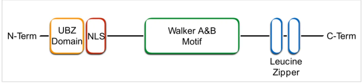
THE WERNER HELICASE INTERACTING PROTEIN 1 (WRNIP1/WHIP1) Among proteins participating in the maintenance of genome stability, whose function is still poorly characterized, is the human Werner helicase interacting protein 1 (WRNIP1), previously called WHIP1. The WRNIP1 protein was originally identified, through the yeast two-hybrid method, as mouse protein that physically interacts with the WRN protein (WRN) (Kawabe Yi et al., 2001), a member of the RecQ family of DNA helicases that plays a crucial role in response to replication stress, and significantly contributes to the recovery of stalled replication forks (Franchitto and Pichierri, 2011; Petermann and Helleday, 2010). WRNIP1 belongs to the AAA+ class of ATPase family proteins that is evolutionary conserved and whose central region is similar to Escherichia coli RuvB, a Holliday junction branch migration motor protein (Hishida et al., 2001; Kawabe et al., 2001). The human amino acid sequence of WRNIP1 has homology with the replication factor C (RFC) family of clamp loader proteins and possesses an ATPase domain containing a Walker A and B motif for ATPase activity in the middle of the molecule (Kawabe Yi et al., 2001). For WRNIP1 ATPase activity is required threonine on amino acid position 294. Indeed, a mutant protein of human WRNIP1 in which threonine 294, a conserved residue in the nucleotide-binding motif of AAA+ family proteins, was substituted with alanine led to suppression of ATPase activity (Tsurimoto et al., 2005). In addition to ATPase domain, WRNIP1 contains a nuclear localization signal, leucine zipper DNA binding domains, and an ubiquitin-binding zinc finger domain (UBZ domain-RAD18 type) that can bind ubiquitin (Fig. 9).
Figure 9. Schematic representation of human WRNIP1 structure. From left to right: UBZ
domain-RAD18 type; Nuclear Localization Signal (NLS); Walker A & B motif; Leucine zippers.
A conserved aspartate residue in the zinc finger domain is essential to bind ubiquitin and
polyubiquitin. Consistently, a single point mutation of aspartate on amino acid position 37
in alanine completely abolished ubiquitin binding in vitro (Bish and Myers, 2007). In
Walker A&B Motif UBZ Domain NLS N-Term C-Term Leucine Zipper
addition, WRNIP1 is one of many proteins whose ubiquitin-binding domain directs its own ubiquitination. WRNIP1 is heavily ubiquitinated, with 12 sites identified and several of these ubiquitination sites lie near critical conserved motifs within the ATPase domain, suggesting that ubiquitination may regulate WRNIP1 ATPase activity by directly interfering with nucleotide binding or hydrolysis (Bish and Myers, 2007).
Evidences supporting WRNIP1 role during Replication Stress
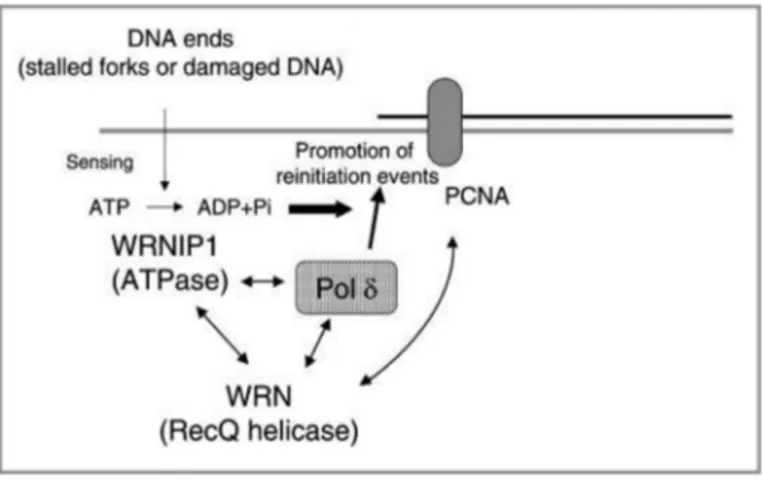
Little is known about the WRNIP1 function in human cells. On the contrary the Maintenance of Genome Stability 1 protein (MGS1), the budding yeast homolog of WRNIP1, has been extensively studied. MGS1 is involved in the maintenance of DNA topology and in the post-replication repair (Branzei et al., 2002; Hishida and Ohno, 2002). Mutations of the MGS1 can enhance aging processes in budding yeast (Hishida et al., 2002; Kim et al., 2005). Furthermore, genetic analysis using MGS1 mutants reveals that it is required for preventing the genome instability caused by replication arrest, but it is not involved in DNA lesions repair (Hishida et al., 2002). Over-expression of MGS1 is lethal or very toxic in combination with mutations in genes that encode proteins involved in DNA replication, such as DNA polymerase δ (Polδ), RFC, PCNA and RPA (Branzei et al., 2002). MGS1 physically and functionally interacts in vivo with budding yeast Pol31, the second subunit of Polδ (Vijeh Motlagh et al., 2006). Consistently, in vitro studies have demonstrated that human WRNIP1 forms homo-oligomeric complex that physically interacts with DNA Polδ. This interaction stimulates Polδ DNA synthesis activity, mainly increasing the frequency of DNA replication initiation events (Tsurimoto et al., 2005). These findings provide the first biochemical evidence that WRNIP1 is involved in a eukaryotic replication fork complex, and that it modulates Polδ activity. However, the exact function and regulation of WRNIP1 in human cell remains to be elucidated. WRN protein interacts with Polδ subunits p66 and p50 (Szekely et al., 2000), and human WRNIP1 interacts with three Polδ subunits except the p66. This means that they can interact simultaneously with Polδ through their common target p50. Therefore, taking into account their functional interaction and their possible simultaneous association with Polδ, it has been proposed a model in which WRN, WRNIP1 and Polδ form a ternary complex in functional situations. This model proposes that WRNIP1 may be a modulator for initiation or re-initiation events of DNA Polδ-mediated DNA synthesis. In particular, WRNIP1, through its ATPase activity, may function as a sensor of DNA damage or arrested
replication fork regulating the extent of DNA synthesis (Tsurimoto et al., 2005). According to this model, the WRNIP1 protein might play a crucial role during perturbed replication to avoid replication stress accumulation (Fig. 10).
Figure 10. Model of a ternary complex containing WRNIP1, WRN and polδ at an arrested replication fork. WRNIP1, WRN and polδ may form a ternary complex. WRN and polδ also interact with PCNA.
This complex may function to regulate pol δ-mediated DNA synthesis when the replication fork complex is stalled by DNA damage or structural stress. The ATPase activity of WRNIP1 functions as a sensor of DNA ends, and ATP hydrolysis regulates the stimulation of polδ. Thus, complex formation plays a crucial role in the re-initiation of stalled replication forks. (Tsurimoto et al., 2005)
Accordingly, in vitro investigations reveal that WRNIP1 binds in an ATP-dependent manner to forked DNA that mimics stalled replication forks (Yoshimura et al., 2009). A further study in human cells has demonstrated that WRNIP1 resides in DNA replication factories, since it localizes either with RPA and PCNA (Crosetto et al., 2008). Interestingly, this localization seems to specifically require its UBZ domain (Crosetto et al., 2008). Upon treatments with UVC light-induced stalled fork, the amount of chromatin-bound WRNIP1 significantly increases (Crosetto et al., 2008). In addition to the amount of chromatin-bound WRNIP1, also the percentage of WRNIP1 foci co-localizing with replication factories increases, suggesting that human WRNIP1 may deal with stalled forks, as inferred from earlier yeast studies.
Very recently, human WRNIP1 protein has been implicated in the activation of ATM-mediated checkpoint after replication stress induced by low-dose Aph, further supporting the hypothesis that WRNIP1 may be directly involved in response to mild replication stress (Kanu et al., 2015). Furthermore, WRNIP1 has been found de-regulated in a subset of
human tumours (Lukk et al., 2010), underscoring its possible role in maintenance of genome stability.
As discussed above, replication stress response is an intricate multi-step pathway, and we are only beginning to understand how the different steps integrate and how key proteins of the response are controlled. Although several evidences indicate the involvement of WRNIP1 during replication stress response, the exact molecular function that WRNIP1 accomplishes is not yet determined. Thus, a more detailed knowledge of the WRNIP1 function could help us to better understanding of the complex network of replication stress response, and it could also provide further insights into the molecular mechanisms underlying the chromosome instability phenotype of human WRNIP1-deficient cancer cells.