UNIVERSITY OF SASSARI
D
EPARTMENT OF
B
IOMEDICAL
S
CIENCES
Ph.D. Course in Life Sciences and Biotechnologies
Curriculum Biochemistry, Physiology and Molecular Biology
XXIX cycle
CORDINATOR: PROF. LEONARDO ANTONIO SECHI
LRRK2 effect on dopamine receptor trafficking –
implication in Parkinson’s disease
Tutor:
Dr. Ciro Iaccarino
Ph.D. student:
Mauro Rassu
Index
P.
Abstract
1Chapter 1|Introduction
31.1 Parkinson’s disease 3
1.2 Dopamine and dopamine receptors 6
Dopamine synthesis and metabolism 6
Dopaminergic system and dopamine receptors in the brain 8
Dopamine receptors and locomotor activity 10
Dopamine receptors signalling and trafficking 11
Receptor–Receptor Interactions:
Homomeric and heteromeric dopamine receptors
13
1.3 Current treatments of Parkinson disease 16
1.4 Etiology 18
Environmental factors 18
Genetic factors 21
General characteristic and structural domains of LRRK2 26
Mutations in LRRK2 gene and their implication in PD 31
LRRK2 functions in physiological and pathological conditions 32
Chapter 2 | Materials and methods
38Chapter 3 | Results
433.1 Characterization of SHSY-5Y Cells Stably Expressing dopamine receptor D1 or D2.
43
SH-SY5Y cells stably expressing dopamine receptor D1 or D2 43
Adenoviral delivery of LRRK2 43
3.2 LRRK2 affects DRD1 trafficking in cellular models. 45
3.3 LRRK2 alters the dopamine D2 receptor trafficking. 50
3.4 LRRK2 expression alters both DRD1 and DRD2 signalling 54
3.5 Analysis of DRD1 trafficking in LRRK2 animal models 55
Chapter 4 | Discussion
57Publications
62Abstract
Parkinson disease (PD) is the second most common neurodegenerative disorder affecting 4 million people worldwide. It is characterized by the loss of dopaminergic neurons in the Substantia Nigra pars compacta (SNpc) and by the presence of cytoplasmic inclusion bodies (Lewy bodies, LB). Cell death leads to a profound depletion of dopamine neurotransmitter involved mainly in the control of the movement. Mutations in LRRK2 (leucine-rich repeat kinase 2) gene (PARK8; OMIM #609007) are responsible for one of the autosomal-dominant forms of Parkinson’s disease. LRRK2 is a protein of 2527 amino acids composed by different functional domains: ankyrin, leucine-rich repeat (LRR), Roc (Ras in complex proteins), COR domain (C-terminal of Roc), protein kinase catalytic domain and a WD40 domain. LRRK2 mutations associated with PD have been identified in different protein domains. These observations, along with the lack of deletion or truncation mutants with dominant inheritance, suggest a gain of function mechanism. Up to date, the LRRK2 biological function is largely unknown. LRRK2 appears to be localized in different intracellular districts that play a critical role in the control of vesicular trafficking: ER, Golgi apparatus and associated vesicles, cytoskeleton, lipid raft and lysosomes. Although, some discrepancies between different experimental approaches, the involvement of LRRK2 in the regulation of vesicle trafficking appears quite consistent both in animal and cellular models. In neurons, vesicle trafficking is a complex process regulating multiple different cellular functions, in addition to the neurotransmitter release or re-uptake, such as the localization and levels of membrane receptors, changes in plasma membrane composition at the cell surface and, not least, organelle biogenesis.
This research focuses on LRRK2 role in the regulation of D1 and D2 dopamine receptors trafficking. Considering the relevance of dopamine receptor trafficking in DA neuronal physiology, this research may have a strong implication in the discovery of the pathological mechanisms underlying the PD onset and development. This work indicates that PD-associated mutant G2019S LRRK2 impairs dopamine receptor D1 internalization, leading to an alteration in signal transduction. Moreover, the mutant forms of LRRK2 affect dopamine receptor D2 turnover by decreasing the rate of the receptor trafficking from the Golgi complex to the cell membrane. Collectively, these findings are consistent with the conclusion that LRRK2 influences the motility of neuronal vesicles and the neuronal receptor trafficking. These findings have important implications to clarify the complex role that LRRK2 plays in neuronal physiology and the possible pathological mechanisms that may lead to neuronal death in PD.
Chapter 1
Introduction
1.1 Parkinson’s disease
Parkinson’s disease (PD) is a chronic, neurodegenerative, progressive disease, described for the first time in 1817 in the Essay on the Shaking Palsy by the English medical doctor James Parkinson. PD is the second most common neurodegenerative disorder after Alzheimer disease and affect approximately 20/100000 cases per year in the population over 50 years, up to 120/100000 new cases per year among the over 70 years old1. In about 95% of PD cases, no an apparent genetic cause (sporadic PD) in the
pathogenesis of PD can be detected, but in the remaining 5% it seems hereditary. Stronger differences in incidence are observed among different ethnic groups, probably due to the disease etiology linked to environmental risk factors or genetic susceptibility. The average age onset is around 60 years, even if the 4% of the patients show an early disease development (before 50 years). Unfortunately, since the clinical symptoms appear only when more than 50-60% of the neurons of the Substantia Nigra pars compacta (SNpc) are damaged and the striatal levels of dopamine are strongly reduced1, it is currently possible that the disease arises before the symptomatology
therefore there is a high percentage of people, apparently healthy, that are developing PD.
The cardinal motor features that describe PD are resting tremor, bradykinesia, postural instability and muscle rigidity.
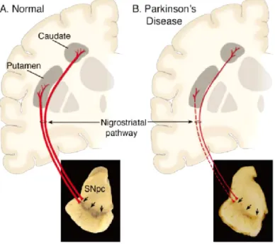
The PD clinical symptoms are due by a progressive and profound loss of dopaminergic neurons in the Substantia Nigra pars compacta (SNpc). These neurons project mainly to the striatum (i.e., putamen and caudate nucleus). The loss of the nigrostriatal dopaminergic neurons causes a profound depletion of dopamine in the striatum that is responsible of the trigger and control of movement (figure 1).
Figura 1. Schematic representation of the Nigrostriatal pathway. In red the Nigrostriatal pathway. It is composed
of dopaminergic neurons whose cell bodies are located in the SNpc. These neurons projections to striatum (A) in physiological conditions and (B) in PD conditions; Down is shown the (A) SNpc in normal conditions and (B) the depigmentation of SNpc caused by the loss of dopaminergic neurons in PD patients1.
PD is the most common form of Parkinsonism. In some cases the differential diagnosis among PD and other Parkinsonism forms is easy, in other cases more difficult. The main forms of Parkinsonism syndromes are described in table 1.
Up to date, the 80% of patients have a PD diagnosis “possible or probable”. The final diagnosis need the post mortem analysis to highlight the loss of dopaminergic neurons, the depigmentation of SNpc and presence of Lewy Body (spherical eosinophilic cytoplasmic protein aggregates composed of numerous proteins, including α-synuclein, parkin, ubiquitin, and neurofilaments2, 3) in the surviving neurons. LBs are
not specific for PD, they are also found in Alzheimer disease, in a condition called “dementia with LB disease” and as an incidental pathologic finding in people of
advanced age at a greater frequency than the prevalence of PD4. In life, the diagnosis
of PD is made through the analysis of the clinical features, but definite diagnosis requires the identification of both LB and SNpc dopaminergic neuron loss. This permit to distinguish PD to others parkinsonism forms1, 5.
Parkinsonism syndromes Primary Parkinsonism
Parkinson disease (sporadic, familial)
Secondary Parkinsonism
Drug-induced: dopamine antagonists and depletors Hemiatrophy-hemiparkinsonism
Hydrocephalus: normal pressure hydrocephalus Hypoxia
Infectious: postencephalitic
Metabolic: parathyroid dysfunction Toxin: Mn, CO, MPTP, cyanide Trauma
Tumor
Vascular: multiinfarct state
Parkinson-plus Syndromes
Cortical-basal ganglionic degeneration
Dementia syndromes: Alzheimer disease, diffuse Lewy body disease, frontotemporal dementia
Lytico-Bodig (Guamanian Parkinsonism-dementia-ALS)
Multiple system atrophy syndromes: striatonigral degeneration, Shy-Drager syndrome, sporadic olivopontocerebellar degeneration (OPCA), motor neuron disease-parkinsonism
Progressive pallidal atrophy Progressive supranuclear palsy
Familial Neurodegenerative Diseases
Hallervorden-Spatz disease Huntington disease
Lubag (X-linked dystonia-parkinsonism)
Mitochondrial cytopathies with striatal necrosis Neuroacanthocytosis
Wilson disease
MPTP, 1-methyl-4-phenyl-1,2,3,6-tetrahydropyridine; ALS, amytrophic lateral sclerosis. Table 1. The main forms of Parkinsonism syndromes1.
1.2 Dopamine and dopamine receptors
Dopamine synthesis and metabolism
Dopamine (DA) with adrenaline and noradrenaline, are the catecholamine neurotransmitter class. Even though DA is an important brain neurotransmitter, a significant part of the DA is not produced in the brain but by the mesenteric organs6.
Dates back 60 years ago the discovery of the physiological functions of 3-hydroxytyramine (DA)7. The two-step of DA synthesis begin in the cytosol of
catecholaminergic neurons. The first step is the hydroxylation of L-tyrosine at the phenol ring by tyrosine hydroxylase (TH) to generate DOPA. DOPA is subsequently decarboxylated to DA by aromatic amino acid decarboxylase (AADC, also known as DOPA decarboxylase). Moreover, a cytochrome P450-mediated pathway has been shown to exist in rat in vivo8, 9. In this pathway decarboxylation forerun hydroxylation;
tyrosine is decarboxylated to tyramine and then is hydroxylated by Cyp2D proteins (figure 2)9. Another pathway of DA synthesis is operated by tyrosinase via
hydroxylation. Eumelanins and phaeomelanins are normally synthetize by tyrosinase, but it has been shown that tyrosinase have a central role in TH-negative mice for catecholamine synthesis10. After synthesis, in catecholaminergic neurons, the vesicular
monoamine transporter 2 (VMAT2), by secondary active transport, operates the DA accumulation into synaptic vesicles11 (figure 2). In vesicles, the oxidation of DA is
stabilized by the acidic pH12 and this prevents oxidative stress in the cytosol13.
Upon excitation of dopaminergic neurons, the membrane of the synaptic vesicles melts with the presynaptic element cell membrane. In this way dopamine is released into the synaptic cleft to bind the postsynaptic DA receptors or the regulatory presynaptic DA autoreceptors14, 15. Successively, extracellular DA has to be removed
from the synaptic cleft to stop the signaling pathway. DA can be recycled after reuptake by dopaminergic neurons or be degraded after uptake by glial cells. The neuronal reuptake is operated by the dopamine transporter (DAT) at level of the presynaptic element and accumulates into synaptic vesicles by VMAT216. The DA that
ROS formation. MAO enzyme catalyzes the oxidative deamination of DA to produce hydrogen peroxide and 3,4 -dihydroxyphenylacetaldehyde (DOPAL). DOPAL can be inactivated in two major pathways, by reduction to the alcohol 3,4-dihydroxyphenylethanol (DOPET) via alcohol dehydrogenase (ADH) or by oxidation to the carboxylic acid 3,4-dihydroxyphenylacetic acid (DOPAC) via aldehyde dehydrogenase (ALDH). Under normal conditions, the majority of DOPAL is oxidized to the carboxylic acid DOPAC17.
In the synaptic cleft DA can be recovered by glial cells. Glial cells quickly degrade DA by MAO and also by catechol-O methyl transferase (COMT). COMT catalyze the metalation reaction by the transfer of the methyl groups from S-adenosylmethionine (SAM) to hydroxyl groups of various catecholic compounds. MAO reaction produces DOPAC that in subsequently converted in homovanilic acid (HVA), one of the most important products of the degradation of DA (figure 2). It has been shown that there is COMT activity in glial cells but no COMT activity in the dopaminergic nigro-striatal neurons18, 19.
Dopaminergic system and dopamine receptors in the brain
Three major dopaminergic pathways have been identified in the mammalian brain; the nigrostriatal (originating in the A9 area), mesolimbic-mesocortical (originating in the A10 area) and tuberoinfundibular (originating in the A8 area)20, 21. These different
pathways are known to be important for various vital functions regulated by the central nervous system. The nigrostriatal pathway is primary involved in the control of the motor functions. The mesolimbic-mesocortical pathway is mainly involved in the control of the behavior. The tuberoinfundibular is mainly involved in the control of the endocrine system22-25.
Based on their structural, pharmacological, and biochemical properties, dopamine receptors have been classified as D1-like dopamine receptors D1 and D5 or D2-like dopamine receptors D2, D3, and D426-28. It is know that the D1-like dopamine
receptors (D1 and D5) activate the Gαs/olf family of G proteins to produce cAMP by
stimulation of the adenylate cyclase. D1-like dopamine receptor are located exclusively on the plasma membrane of the postsynaptic neurons in the dopaminergic transmission. The D2-like dopamine receptors (D2, D3, and D4) interact with the Gαi/o
family of G proteins to induce inhibition of adenylate cyclase. In contrast to the D1-like dopamine receptors, D2 and D3 dopamine receptors are postsynaptic and presynaptic in the dopaminergic synaptic transmission29-31. The D1- and D2-like dopamine
receptors are also different at the level of genetic structure, presence of splice variants, G protein coupling agonists, selective agonists and antagonists and their brain distribution (table2).
D1-like D2-like Dopamine receptor subtype D1 D5 D2 D3 D4 Gene symbol DRD1 DRD5 DRD2 DRD3 DRD4 Chromosome gene map locus 5q35.1 4p16.1 11q23.1 3q13.3 11p15.5 Numbers of introns in the coding region None None 6 5 3 Pseudogenes None DRD5P1,
DRD5P2 None None None
Presence of
splice variants None None D2S, D2L Yes Yes
Number of aminoacids 446 477 D2S, 414; D2L, 443 400 387 Molecular weight 49,300 52,951 D2S, 47,347; D2L, 50,619 44,225 41,487 G protein coupling agonists
Gαs, Gαolf Gαs, Gαq Gαi, Gαo Gαi, Gαo Gαi, Gαo
Effector
pathway ↑cAMP ↑cAMP
↓cAMP, ↑K+ channel, ↓Ca2+ channel ↓cAMP ↓cAMP Selective agonists Fenoldopam, SKF-38393, SKF-81297 None Bromocriptine, pergolide, cabergoline, ropinirole 7-OH-DPAT, pramipexole, rotigotine, (+)-PD-128907 A-412997, ABT-670, PD-168,077 Selective antagonist 23390, SCH-39166, SKF-83566 None Haloperidol, spiperone, raclopride, sulpiride, risperidone Nafadotride, GR 103,691, GR 218,231, SB-277011A A-381393, FAUC 213, L-745,870, L-750,667 mRNA distribution in the brain Caudate-putamen, nucleus accumbens, olfactory tubercle Hippocampus, hypothalamus Caudate-putamen, nucleus accumbens, olfactory tubercle Olfactory tubercle, hypothalamus, nucleus accumbens Frontal cortex, medulla, midbrain
Dopamine receptors have different expression patterns in the brain. D1 dopamine receptors have been found at a high level of density in the nigrostriatal, mesolimbic, and mesocortical areas, as well as the striatum, nucleus accumbens, substantia nigra, olfactory bulb, amygdala, and frontal cortex. On the contrary dopamine receptor D1 is expressed at lower levels in the hippocampus, cerebellum, thalamic areas and hypothalamic areas. D5 dopamine receptors have been found at low levels of density in different brain regions, such as pyramidal neurons of the prefrontal cortex, the premotor cortex, the cingulated cortex, the entorhinal cortex, substantia nigra, hypothalamus and the hippocampus. A very low level of expression has also been observed in the MSNs of the caudate nucleus and nucleus accumbens23, 29, 33,34. At the
cellular level, the large spiny neurons of neostriatum in primates, that are typically cholinergic interneurons, only express D5 receptors35. Highest levels of dopamine
receptors D2 have been detected in the striatum, nucleus accumbens and in the olfactory tubercle D2 receptors are also expressed at detectable levels in the substantia nigra, ventral tegmental area, hypothalamus, cortical areas, septum, amygdala, and hippocampus23, 28, 36, 37. The dopamine receptors D3 are more limited in
distribution, the highest level of expression have been found in the limbic areas, such as in the shell of the nucleus accumbens, the olfactory tubercle, and the islands of Calleja23, 38. At lower levels, the dopamine receptors D3 have been found in the
striatum, the substantia nigra pars compacta, the ventral tegmental area, the hippocampus, the septal area, and in various cortical areas. Dopamine receptors D4 have been found a lower level of density in the frontal cortex, amygdala, hippocampus, hypothalamus, globus pallidus, substantia nigra pars reticulata, and thalamus23, 31.
Dopamine receptors and locomotor activity
Different experimental evidence shows that the dopaminergic system is involved in the control of locomotor activity across species39. Locomotor activity is at least regulated
by dopamine receptors D1, D2 and D323, 40. The activation of dopamine receptors D1
has a moderate stimulatory effect on locomotor activity. The roles of the dopamine receptors D2 and D3 are much more complex than D1 dopamine receptors due to their presynaptic and postsynaptic localization23, 40. Presynaptic autoreceptors are important
for the negative feedback mechanism involved in neuronal synthesis and release of the neurotransmitter in response to synaptic cleft neurotransmitter levels23, 40, 41.
Presynaptic D2-like autoreceptors stimulation gives rise to a decrease in dopamine release and consequently a decrease in locomotor activity. On the contrary, an activation of postsynaptic receptors stimulates locomotor activity. Since D2-like autoreceptors are more sensitive to dopamine than D2-like postsynaptic receptors, dopamine can induce a biphasic effect that is resumed in a decrease in locomotor activity at low dopamine doses and locomotor activation at high dopamine doses. Moreover, dopamine receptor D2 has two splice variants D2L and D2S, with different synaptic distributions. D2S is mainly presynaptic and D2L is mainly postsynaptic. Consequently, the effects of the postsynaptic and presynaptic D2 dopamine receptors are probably determined by the different contributions of these isoforms42, 43.
Different pharmacological44, 45 and genetic studies in dopamine receptor D3 knockout
mice40, 46 show that D3 autoreceptors can also play a role in the regulation of tonically
released dopamine, in a synergic manner with D2S autoreceptor in regulating the neuronal firing rate, synthesis and release of dopamine43. Dopamine receptor D3
probably has a moderate inhibitory function on locomotor activity due to autoreceptors and postsynaptic receptors activity40, 46. The roles played by dopamine
receptors D4 and D5, that are expressed at lower level in the primary motor regions of the brain, appear to be not very important in the control of locomotor activity23, 31, 40. It is obvious that both the postsynaptic D1- and D2-like dopamine receptors are
essential for the completely manifestation of locomotor activity47.
Dopamine receptors signaling and trafficking
G protein-coupled receptors (GPCRs) are seven transmembrane (TM) proteins representing the largest and most expressed cell surface receptors. They play important roles in a broad array of cellular functions and in diseases and represent the targets for a large fraction of existing drugs48-50. GPCRs, upon ligand binding, induce
dissociation of G proteins into their Gα and Gβγ components and ultimately modulate the activity of enzyme or ion channel effectors51-53. In the canonical view of G
the extracellular or transmembrane regions of the receptor. Subsequently the conformation of the GPCR receptor changes and operates as a guanine nucleotide exchange factor, catalyzing the exchange of the GDP in GTP on the Gα subunit. Than the Gα and Gβγ subunits dissociate from each other and from the GPCR receptor54.
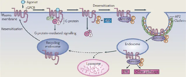
Subsequently, signal transduction cascades are activated directly or by generating second messengers (such as cyclic AMP, diacylglycerol (DAG) and inositol-1,4,5-trisphosphate (Ins(1,4,5)P3) that modulate downstream effectors, such as protein kinase A (PKA) and protein kinase C (PKC). The Gβγ subunits, after their release from the heterotrimeric G protein complex, can bind and regulate other downstream effectors, such as ion channels and PLCβ. G protein-mediated signaling can be terminated by GPCR phosphorylation by GPCR kinases (GRKs) and concomitant association with arrestins. Subsequently, close to the GPCR-arrestin complex is assembled an AP2-clathrin complex to drive GPCR internalization into endosomes and receptor desensitization. The mitogen-activated protein kinase extracellular signal-regulated kinase (ERK) pathway can also be activated not only via the main signal transduction cascades but also through the interaction of the GPCR-arrestin complex. Following internalization, the endosomes containing GPCR receptors can melt with lysosome to be ultimately degraded, or can be recycled in the recycling endosomes pool and go back to the cell surface in the functional process of resensitization53, 55, 56.
This general model can be applied to dopamine receptors (table 2 and figure 3).
Dopaminergic modulation of ATP in cAMP and cAMP in AMP conversion, operated by the adenylyl cyclase and phosphodiesterase respectively, results in the regulation of protein kinase A (PKA) and potentially other exchange proteins activated by cAMP25, 57.
PKA substrates such as Protein phosphatase 1 regulatory subunit 1B (DARPP-32/PPP1R1B) have been extensively studied over the last 30 years. When phosphorylated on Thr34 by PKA, DARPP-32 is a negative regulator of protein phosphatase 1 (PP1). In contraposition, phosphorylation of DARPP-32 on Thr75 by cyclin-dependent kinase 5 (CDK5), in response to dopamine receptor D1 activation, results in PKA inhibition55, 58.
Figura 3. Canonical mechanisms for GPRC’s signaling and trafficking53.
Furthermore, other important dopamine signaling pathways have also been discovered, including the modulation of the Akt-GSK3 signaling pathway59 and the
activation of the PAR4 signaling pathway60.
Receptor–Receptor Interactions: Homomeric and heteromeric dopamine
receptors
Dopamine receptors can physically interacts with its own type or other receptors in the plasma membrane of neurons, to form homomers or heteromers respectively, or high-order receptor oligomers61-64. In a receptor mosaic model view, each receptor
represents a single tile inside the mosaic; furthermore, receptors in the mosaic have different features and properties compared to each single receptor that compose the mosaic64-66.
In the brain, the dopamine receptors D1 and D2 are the most abundant dopamine receptor expressed. For reason of brevity in this paragraph it will be explained only the interaction D1-D2, and D1-D3 heteromers for their implication in movement disorders. See table 3 for more information about dopamine receptors heteromer interactions. The physiological relevance of dopamine receptor D1–D2 heterodimers is supported by the co-expression of D1 and D2 receptors in small populations of medium spiny neurons (MSNs) of the nucleus accumbens (NAc) in the mouse67 and in other regions
of the basal ganglia68. Dopamine receptors D1 and D2 can form heteromeric receptor
complexes through electrostatic interactions among a specific glutamic acid residues in the carboxyl-tail of the dopamine receptor D1 and an arginine residues in the third intracellular loop of the dopamine receptor D269. The expression of dopamine
receptors D1-D2 heteromers has been reported to exist at presynaptic but not at postsynaptic terminals of MSNs. Up to now, different data suggest that neurons expressing dopamine receptors D1-D2 heteromers may have a unique physiological function compared to the neurons expressing dopamine receptor monomers at local level and distal level70, 71. Dopamine receptors D1-D2 heteromers can stimulate
calcium signaling, by Gq/11 and phospholipase C (PLC) activation, resulting in the activation of calcium calmodulin kinase IIa (CaMKII)72-74 and increased expression of
brain-derived neurotrophic factor (BDNF) in NAc and ventral tegmental area (VTA)70, 73, 75. Evidence suggests an implication of dopamine receptors D1-D2 heteromers in drugs
addiction and schizophrenia70, 76.
Dopamine receptors D1-D3 heteromers have been found by different techniques in the striatonigral pathway of rat striatum77, 78. Data show that upon DA denervation and
intermittent L-Dopa therapy, dopamine receptor D3 is overexpressed in the dopamine receptor D1-GABA pathway. The dopamine receptor D3 overexpression can consequently contribute to the dopamine receptor D1 sensitization and development of L-Dopa-induced dyskinesias79, 80. The dopamine receptor D1-D3 interaction has been
reported to determine a receptor D3 stimulation and an increase of dopamine receptor D1 response77, 78. It was proposed that dopamine receptor D1-D3 heteromer
can operate a reinforcing of the dopamine receptor D1 signaling in turning affecting motor functions and can contribute to dyskinesia in PD patients78, 81.
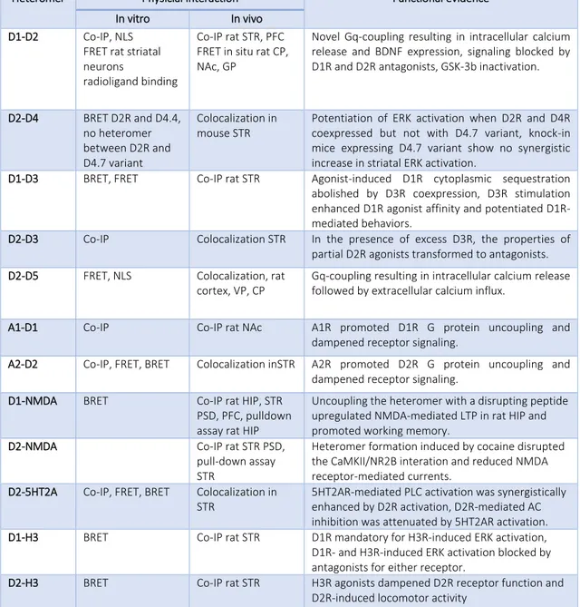
Heteromer Physicial interaction Functional evidence
In vitro In vivo
D1-D2 Co-IP, NLS
FRET rat striatal neurons
radioligand binding
Co-IP rat STR, PFC FRET in situ rat CP, NAc, GP
Novel Gq-coupling resulting in intracellular calcium release and BDNF expression, signaling blocked by D1R and D2R antagonists, GSK-3b inactivation.
D2-D4 BRET D2R and D4.4, no heteromer between D2R and D4.7 variant Colocalization in mouse STR
Potentiation of ERK activation when D2R and D4R coexpressed but not with D4.7 variant, knock-in mice expressing D4.7 variant show no synergistic increase in striatal ERK activation.
D1-D3 BRET, FRET Co-IP rat STR Agonist-induced D1R cytoplasmic sequestration
abolished by D3R coexpression, D3R stimulation enhanced D1R agonist affinity and potentiated D1R-mediated behaviors.
D2-D3 Co-IP Colocalization STR In the presence of excess D3R, the properties of
partial D2R agonists transformed to antagonists.
D2-D5 FRET, NLS Colocalization, rat
cortex, VP, CP
Gq-coupling resulting in intracellular calcium release followed by extracellular calcium influx.
A1-D1 Co-IP Co-IP rat NAc A1R promoted D1R G protein uncoupling and
dampened receptor signaling.
A2-D2 Co-IP, FRET, BRET Colocalization inSTR A2R promoted D2R G protein uncoupling and
dampened receptor signaling.
D1-NMDA BRET Co-IP rat HIP, STR
PSD, PFC, pulldown assay rat HIP
Uncoupling the heteromer with a disrupting peptide upregulated NMDA-mediated LTP in rat HIP and promoted working memory.
D2-NMDA Co-IP rat STR PSD,
pull-down assay STR
Heteromer formation induced by cocaine disrupted the CaMKII/NR2B interation and reduced NMDA receptor-mediated currents.
D2-5HT2A Co-IP, FRET, BRET Colocalization in
STR
5HT2AR-mediated PLC activation was synergistically enhanced by D2R activation, D2R-mediated AC inhibition was attenuated by 5HT2AR activation.
D1-H3 BRET Co-IP rat STR D1R mandatory for H3R-induced ERK activation,
D1R- and H3R-induced ERK activation blocked by antagonists for either receptor.
D2-H3 BRET Co-IP rat STR H3R agonists dampened D2R receptor function and
D2R-induced locomotor activity
Table 3. Physical and Functional Evidence for Dopamine Receptor Heteromers. Abbreviations: 5HT2AR, 5HT2A
receptor; A1R, adenosine A1 receptor; A2R, adenosine A2 receptor; AC, adenylyl cyclase; ADHD, attention-deficit hyperactivity disorder; BDNF, brain-derived neurotrophic factor; BRET, bioluminescent resonance energy transfer; CaMKII, calcium calmodulin kinase II; Co-IP, coimmunoprecipitation; CP, caudate putamen; D1R, dopamine D1 receptor; D2R, dopamine D2 receptor; D3R, dopamine D3 receptor; D4R, dopamine D4 receptor; ERK, extracellular signal-related kinase; FRET, fluorescent resonance energy transfer; GP, globus pallidus; GSK-3b, glycogen synthase kinase 3b; H3R, histamine H3 receptor; HIP, hippocampus; LTP, long-term potentiation; mGlu5, metabotropic glutamate receptor 5; NAc, nucleus accumbens; NR2B, NMDA receptor subunit 2B; PLC, phospholipase C; PFC, prefrontal cortex; PSD, postsynaptic density; STR, striatum; VP, ventral pallidum70.
1.3 Current treatments of Parkinson disease
In these last years, PD treatment has become articulated due to the presence of new drugs and treatments (figure 4). Treatments for PD include pharmacotherapy, functional stereotaxic neurosurgery (deep brain stimulation), and supportive therapy such as physiotherapy, speech therapy, and dietary measures. All treatments available until 2016 are of symptomatic nature. No therapy is currently available that slows down the progression of PD or even prevents its manifestation82.
L-Dopa
The first and the most efficient treatment established to recover the dopaminergic deficit was L-Dopa always in fixed combination with a decarboxylase inhibitor. L-Dopa is the physiological precursor of dopamine and unlike dopamine it is permeable at the blood brain barrier. According to present knowledge, L-Dopa does not influence the progression of the disease. The effects of L-Dopa are dose-dependent. L-Dopa adverse reactions depend mostly on its oxidative metabolism and its potential capability to produce reactive oxygen species. Treatment with L-Dopa is recommended in all stages of the disease. The uptake of L-Dopa starts from the duodenum into the blood and from the blood to the brain competing with the uptake mechanism for neutral amino acids. Once in the CNS, L-Dopa is converted in dopamine by a neuronal L-Dopa decarboxylase in the dopaminergic neurons. Therefore, dopamine is transported in the synaptic vesicles. When the impulse reach the dopaminergic neurons, vesicles fuse with plasma membrane and dopamine is released into synaptic cleft leading to its physiological effect83-86.
Dopamine agonists
Ten dopamine agonists (five ergot- and five non-ergotderivates) are usable for the treatment of PD. Ergot dopamine agonists include bromocriptine, cabergoline, adihydroergocriptine, lisuride, and pergolide. The non-ergot derivates include piribedil, pramipexole, ropinirole, apomorphine and rotigotine. Usually, dopamine agonists are provided in the advanced PD stages since they may increase the risk of cognitive
impairment or dementia development. However the anti-Parkinson efficacy is limited and motor complications are not completely prevent87, 88.
MAO and COMT inhibitors
The duration of the effect of L-Dopa can be prolonged blocking degrading enzymes of L-Dopa and dopamine. Usually, COMT inhibition (entacapone, opicapone or tolcapone) is associated with an increased stability of levodopa plasma level, suppressing peaks of levodopa concentrations associated with motor complications82. MAO-B inhibitors
(rasagiline, safinamide, selegiline) can amplify the effect of L-Dopa. However, clinical studies concerning the efficacy of this therapeutic drugs have produced controversial results.
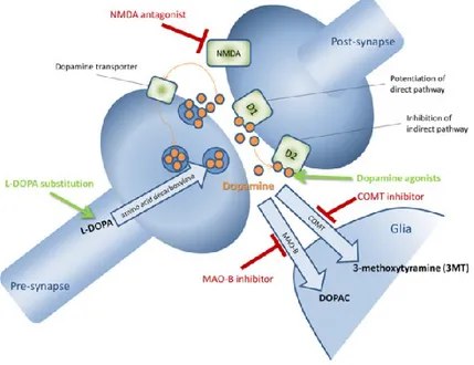
Figura 4. Pharmacology of dopaminergic transmission. Dopamine is released into the synaptic cleft, where it can
bind to post-synaptic D1-like (D1 and D5) and D2-like (D2, D3, and D4) receptors. Dopamine can be metabolized in glia cells to 3-methoxytyramine (3MT) by the catechol-O-methyl transferase (COMT) or to 3,4-dihydroxyphenylacetic acid (DOPAC) by monoamine oxidase B (MAO-B). Dopamine is also reuptaken into the pre-synapse by dopamine transporters. Drugs are L-Dopa, D1-, and D2-like dopamine agonists, MAO-B- and COMT-inhibitors82.
1.4 Etiology
To date the etiology of PD is unknown. PD has a complex and multi-factorial etiology, involving genetic and environmental factors. However, the molecular details of the degeneration are unknown. Nevertheless, the discovery of PD related genes has lit a new light on the cellular mechanisms related to PD neurodegeneration.
Environmental factors
Epidemiological analyses show that pesticide exposition (Paraquat, Rotenone), lifestyle (diet, habit, occupation), metal exposure (Al, Cu, Fe, Hg, Mn, Pb, Zn), chemical and industrial products (MPTP, organic solvents), trauma and viral infections can increase the risk of PD development89.
An example of how an exogenous toxin can mimic the clinical and pathological symptoms of PD is the MPTP (1-methyl-4-phenyl-1,2,3,6,-tetrahydropyridine or meperidine) intoxication90. In 1982 MPTP was discovered as dopaminergic neurotoxin
when drug users developed a rapidly progressive parkinsonian syndrome as a result of intravenous self-administration of a street preparation of 1-methyl-4-phenyl-4-propionoxypiperidine (MPPP), an analog of the narcotic Demerol contaminated by MPTP90. In humans and monkeys, MPTP produces a severe parkinsonian syndrome,
L-Dopa responsive, characterized by all the PD symptoms including the cardinal symptoms of PD.
In the brain, by crossing the blood brain barrier, MPTP is converted in to 1-methyl-4-fhenyl-2,3-didropiridine (MPTP+) by monoamine oxidase B (MAO-B) in glial cells, serotoninergic neurons and in each neuron that contain MAO-B enzyme. Subsequently, MPTP+ is converted in 1-metil-4-fenilpiridinio (MPP+), probably by spontaneous oxidation, and released in the extracellular space. MPP+ is transported in dopaminergic neurons through its high affinity for the dopamine transporter (DAT). Inside neurons MPP+ can follow at least three routes: (i) it can bind the vesicular monoamine transporter-2 (VMAT2 )91, which translocates MPP+ into synaptosomal
vesicles; (ii) it can be concentrated within the mitochondria92 and (iii) it can remain in
is toxic mechanism binding and blocking the complex I, which interrupts the transfer of electrons to ubiquinone (figure 5). This perturbation enhances reactive oxygen species (ROS) production and decreases ATP synthesis leading to a neuronal dead via apoptosis1, 90. The MPTP related Parkinsonism supports the environmental hypothesis
of PD etiology and the MPTP-treated animals can be an interesting models for PD studies. Other substances like rotenone, 6-hydroxydopamine (6-OHDA) and paraquat were identified PD causative and currently used in toxin-based animal models of PD94.
In addition to toxins, other environmental factors, which can be considered as risk factors rather than causative elements, are lifestyle and habits. Repetitive or simple violent traumas can provoke a Parkinson-like progressive syndrome as the one frequently observed among boxers that take the name of “Pugilistic Parkinson Syndrome”. Another two risk factors are age and sex, males are more affected than women (in a 1,5:1 ratio). In humans, neuronal and SNpc neuromelanin loss increases around 60 years, coinciding with the average age of PD onset. Since neuromelanin has a protective effect in neurons against free radicals and toxins, the decrease in this pigment may predispose the brain of aged people to Parkinson95.
Finally, an infective etiology has been hypothesized; clinical manifestations of PD-like syndromes have been observed in patients affected by a viral encephalitis propagated in 192096, 97.
Genetic factors
Until the end of the 19th century, PD has been considered a sporadic disease, even
though the 10-30% of PD patients presented a first line relative affected by the disease. Leroux in 1880 identified recurring PD cases in the same family, corroborating the hypothesis that hereditary factors could be related to pathology98, 99. However, in
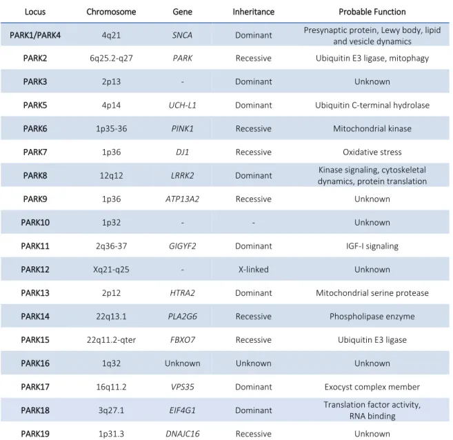
the last 20 years, the identification of gene mutations related to PD and risk factors, has proved the importance of genetic factors in the pathogenesis of the disease, even if less than 10% of the cases are familiar. Until now, 19 mendelian transmission forms and several risk factors have been related to PD (table 4)100.
Locus Chromosome Gene Inheritance Probable Function
PARK1/PARK4 4q21 SNCA Dominant Presynaptic protein, Lewy body, lipid
and vesicle dynamics
PARK2 6q25.2-q27 PARK Recessive Ubiquitin E3 ligase, mitophagy
PARK3 2p13 - Dominant Unknown
PARK5 4p14 UCH-L1 Dominant Ubiquitin C-terminal hydrolase
PARK6 1p35-36 PINK1 Recessive Mitochondrial kinase
PARK7 1p36 DJ1 Recessive Oxidative stress
PARK8 12q12 LRRK2 Dominant Kinase signaling, cytoskeletal
dynamics, protein translation
PARK9 1p36 ATP13A2 Recessive Unknown
PARK10 1p32 - - Unknown
PARK11 2q36-37 GIGYF2 Dominant IGF-I signaling
PARK12 Xq21-q25 - X-linked Unknown
PARK13 2p12 HTRA2 Dominant Mitochondrial serine protease
PARK14 22q13.1 PLA2G6 Recessive Phospholipase enzyme
PARK15 22q11.2-qter FBXO7 Recessive Ubiquitin E3 ligase
PARK16 1q32 Unknown Unknown Unknown
PARK17 16q11.2 VPS35 Dominant Exocyst complex member
PARK18 3q27.1 EIF4G1 Dominant Translation factor activity,
RNA binding
PARK19 1p31.3 DNAJC16 Recessive Unknown
Alpha synuclein (PARK1/PARK4)
Using traditional linkage mapping, the point mutation A53T in the SNCA gene was discovered in the large Italian-American family Contursi and subsequently identified in three Greek families with familial PD101. A decade later, the same mutation was
discovered in two Korean and one Swedish family102, 103. Subsequently to the discovery
that SNCA mutations are involved in a rare familial form of PD, Spillantini and colleagues demonstrated that α-synuclein is a major constituent of Lewy bodies104.
Mutations in SNCA are rare. Until now, five point mutations are discovered (A30P, E46K, H50Q, G51D, A53T) and related to autosomal dominant inheritance forms of PD. Moreover, triplications of the complete gene were discovered105. Triplication of the
SNCA gene was discovered in 2003 and reported in several PD families106-108. The gene
SNCA is located on the chromosome 4q21 consists of six exons109 and encodes a small
140 amino acid protein110 organized in three distinct domains: an N-terminal
amphipathic region (AA 1-60) consisting in six repeats of eleven amino acid with consensus sequence KTKEGV111. A central hydrophobic NAC domain (non-amiloid-β
component of plaque, AA 60-95) necessary for homomeric interactions112. Finally an
acidic unfolded C-terminal region (AA 96-140)113. Monomeric α-synuclein can form
conformers, including oligomers, protofibrils and fibrils, and they have been found in Lewy bodies. All the five point mutations are located in the amphipathic domain and can modify the homomeric interaction kinetic114. Until now the normal function of
α-synuclein remains poorly understood. The α-α-synuclein is mainly found in the cytosol where can bind the lipid rafts in an interaction that is required for its association with the synapse115. Data show that α-synuclein interacts with members of the Rab and
SNARE families, suggesting a role in vesicular trafficking100, 116-118. Parkin (PARK2)
The year following the discovery of the SNCA gene as PD causative, in a Japanese PD patient group was discovered a protein associated to an early-onset form of PD (AR-JP). PARK gene is one of the largest genes in the human genome, mapping on the 6q25.2-q27 chromosome, with a length of 1.38 Mb119. Numerous point mutations and
groups100, 120. Parkin protein is an E3 ubiquitin ligase of 465 amino acids organized in
different domains: an N-terminal ubiquitin like domain, a central linker region and C-terminal RING domain consisting of two RING finger motifs separated by a RING domain98, 121. The ubiquitin-like domain of E3 ubiquitin ligases can bind unfolded
proteins to be degraded by the ubiquitin-proteasome system122. A serious neuronal
loss in the substantia nigra is related to PARK2 PD form, with occasional tau pathology and rare Lewy Bodies in postmortem brains123-125 probably due to the young age of
Parkin disease onset126. The loss of the ubiquitin ligases activity, due to mutations in
the RING domain, can cause an accumulation of unubiquitinated proteins. It has been proposed that the overexpression of Parkin could protect from toxic forms of α-synuclein by Lewy Body formations98. The E2 enzyme associated with Parkin is UbcH7
and the mutation T240R in the first RING domain disrupt this interaction127. It has been
demonstrated that when mitochondria are damaged, PINK1 (another gene associated with AR-JP), phosphorylates Parkin. Subsequently, Parkin translocates to the surface of demented mitochondria to ubiquitinate mitochondrial membrane proteins128, 129. The
majority of Parkin mutations are linked to dysfunctions in the mitochondrial quality control, suggesting a role of the mitochondria in the pathogenesis of PD100.
PTEN-induced kinase 1, PINK-1 (PARK6)
After different studies, the locus PARK6 was mapped on the chromosome 1p35-p36 in a large Italian family presenting a mendelian form of PD with recessive inheritance130.
In 2004, Valente and colleagues identified two mutations in the PTEN-induced putative kinase 1 (PINK1) gene: the G309D missense mutation and a W437X truncating mutation131. The PINK1 gene is assembled in height exons and encode for a 581 amino
acid protein consisting of an N-terminal 34 amino acid mitochondrial targeting motif, a conserved serine–threonine kinase domain and a C-terminal auto regulatory domain. The majority of the mutations in the PINK1 gene are located in the region encoding for the kinase domain, suggesting the importance of PINK1 enzymatic activity in PD pathogenesis121. PINK1 related PD is a fast onset and slow progression Parkinsonism
form responsive to L-Dopa treatment132. It has been demonstrated that PINK1 can
mentioned PINK1 phosphorylates Parkin to regulate mitophagy129, 134. In a general
view, when mitochondria are damaged, mitochondrial membrane potential is reduced and PINK1 is recruited and binds the mitochondrial membrane. Parkin is then recruited to induce mitophagy via ubiquitination135-137. It is controversial the function of TRAP1
in this pathway138. Dj-1 (PARK7)
Mutations in the Dj-1 gene were first reported in two European family and associated with an autosomal recessive form of PD139. Dj-1 gene is composed of height exons and
encodes for a 189 amino acid ubiquitary protein first identified as oncogene140.
DJ-1-linked PDs are rare, L-Dopa responsive, and indistinguishable from Parkin or PINK1 linked PD 141. However, about 10 different point mutations and exonic deletions have
been described142. Dj-1 has been reported against oxidative stress like a chaperone
oxidative stress sensor143, 144. The DJ-1 protein forms dimeric structures145, and it
appears that most of Dj-1 PD linked mutants (L166P, E64D, M26I, and D149A) heterodimerize with wild-type DJ-1146. However, the mutated proteins are not
correctly folded and unstable, affecting Dj-1 neuroprotective function and antioxidant activity147, 148. DJ-1 also can function as redox-dependent chaperone to inhibit
α-synuclein aggregation and subsequent Lewy Body formation. Recently, DJ-1 has been linked to the Parkin/PINK1 pathway by the transcriptional regulation of PINK1149. Vacuolar protein sorting 35-VPS-35 (PARK17)
In 2011, Zimprich and colleagues using next generation sequencing identified the VPS35 gene in an Austrian family with 16 members affected by late-onset autosomal dominantly inherited parkinsonism. Later, the mutation D620N, was validated by Sanger100, 150. Simultaneously, Vilarino-Guell and colleagues, using next generation
sequencing, identified the D620N mutation as a cause of PD in a large Swiss family151.
VPS35 mutations are a rare cause of PD concerning 1% of familial parkinsonism and 0.2% of sporadic PD150, 152. VPS35 linked PD is characterized by bradykinesia, resting
tremor and is responsive to levodopa therapy comparable to idiopathic disease153.
amino acid protein. The yeast homologous of Vps35 has been well characterized and it has been shown to compose the retromer complex with other 4 Vps proteins (Vps5, Vps17, Vps26, Vps29). The retromer complex is involved in retrograde transport of proteins to the trans-Golgi network154. VPS35 is a helical solenoid like protein, such as
other proteins involved in coated vesicle trafficking155. The VPS35 human homolog
have been found to be involved in the same endosome–trans-Golgi network pathway as the yeast Vps35156. A recent study links the D620N mutation to dysfunction of the
retromer complex, generating a redistribution of the retromer endosomes to the perinuclear region in cell lines and PD patient-derived fibroblasts. Moreover, the D620N mutant alters the cathepsin D trafficking, a protein implicated in the α-synuclein degradation100.
1.5 Leucine rich repeat kinase 2 – (LRRK2)
PARK8 locus have been associated for the first time to PD by Funayama and colleague in the 2002157 in a large Japanese family presenting a dominant autosomal
Parkinsonism without Lewy Body. After two years, two groups independently have identified in PARK8 locus the LRRK2 gene (Leucine-rich repeat kinase 2)158, 159;
subsequently many pathological mutations have been identified and associated with PD. Mutations in LRRK2 gene are the most frequently cause of familiar PD. In 2014 Nalls and colleagues associated LRRK2 as a risk locus implied in sporadic PD160.
General characteristic and structural domains of LRRK2
The gene encoding for LRRK2 is located on chromosome 12.q12 and is composed by 51 exons. LRRK2 is a 286-kDa multidomain protein exhibiting both GTPase and kinase activities that belongs to the Roco protein family of G proteins protein. LRRK2 has 7 functional conserved domains (figure 6)161-163:
An “armadillo repeats” domain; An “ankyrin repeats” domain;
A leucine-rich repeats (LRR) domain; A Roc domain with GTPase activity; A carboxy terminal of Roc domain (COR);
A Kinase domain, homologous with MAP-kinase-kinase-kinase (MAPKKK); A C-terminal WD-40 repeat domain.
The Armadillo Repeats domain is a motif of 42 amino acid, composed by three α-helixes and was firstly identified in the “Armadillo” protein of Drosophila, whose human homologue is the β-catenin. The Armadillo domain forms a versatile molecular interaction domain like a platform available for different protein164.
The Ankyrin Repeats domain is a 7 ankyrin repeats motif, which forms helix-loop-helix structures that ends in a loop or hairpin (figure 7). This motif is common in a high number of prokaryotic and eukaryotic proteins like cytoskeletal proteins, transcription factors, signaling proteins and cell cycle regulators165.
LRR domain is made of an 11 amino acid conserved motif LxxLxLxxNxL (Leucines can be replaced by isoleucine, valine or phenylalanine). These repeats are formed by a β-strand followed by an α-helix that line up side-by side to form an arch-like structure (figure 7). LRR domains participate in the interactions with different proteins through binding to their extended solvent-accessible surface166, 167.
Figura 7. Homology models of (A) the ankyrin repeat domain and (B) the leucine-rich repeat (LRR) domain of LRRK2166.
The WD40 repeat domain (known as β-transducin repeat) has a high conserved tridimensional structure and form a seven-blade propeller-like structure. Each repeat is composed by four antiparallel β-sheet and together these repeats form a circular bladed propeller-like structure. The predicted WD40 domain of LRRK2 contains seven WD40 repeats (figure 8), to form an interaction platform for protein-protein reversible interactions. Proteins containing WD40 domains have been found in all eukaryote proteins with different functions including the Gβ subunit of heterotrimeric G proteins,
transcriptional regulators, protein phosphatase subunits, RNA processing complexes, cytoskeletal assembly proteins, and proteins involved in vesicle formation and trafficking. Different experimental results suggest an important role for the WD40 domain in the intramolecular regulation of LRRK2 activity166, 168.
Figure 8. Homology models of the WD40 repeat domain166.
LRRK2 and Roco proteins are serine/threonine specific kinases. Gilsbach colleagues proposed a structure of the LRRK2 kinase domain based on Dictyostelium discoideum Roco4 (figure 9) in its active and inactive state169. Dictyostelium Roco4 has the same
domain architecture of LRRK2, but is biochemically and structurally more easily worked than LRRK2. The Roco4 kinase structure consists of a canonical, two lobed kinase structure. The N-terminal lobe is composed of anti-parallel β sheets and contains the conserved αC-helix. The C-terminal lobe consists of α-helices and contains the activation loop with the conserved N-terminal DFG motif. The ATP binding site is formed by a cleft between those lobes and forms the catalytic site of the kinase. The formation of a polar contact between Roco4 K1055 from the β3-strandand E1078 from the αC-helix is essential for correct positioning of the αC-helix. The DFG motif is essential for catalysis: the Aspartic acid is essential to interact with ATP directly or via coordination of a magnesium ion; the Phenylalanine takes hydrophobic contacts to the αC-helix and the HxD motif and is responsible for the correct positioning of the DFG motif169, 170.
The putative GTPase domain of LRRK2 is a member of the ROCO family. In the ROCO proteins predicted GTPase (Roc) domain is always in tandem with the COR domain. This Roc–COR structure is conserved throughout evolution, suggesting the functional interdependence of the two domains170. Deng and colleagues suggested that the
structure of the LRRK2 Roc domain (figure 9) displays a homodimer with extensive domain-swapping. Each monomer contains five α-helices and six β-strands with loops in between, showing three subdomains: head, neck, and body. The head domain is composed by β1, α1, β2, and β3. The loop between α2 and β2, such as that between β2 and β3, are not folded. The neck domain consist of a bent helix α2. The body domain is structured with β4, α3, β5, α4, β6, and α5 with loops in between. In the same paper, Deng and colleagues, suggest that LRRK2 Roc domain structure revealed an inverted dimer or rather the N-terminal of one domain interacts with the C-terminal of the other171. In contrast, the C. tepidum Roc-COR domain structure showed that the
COR domain is the dimerization domain and that Roco proteins that are not able to dimerize are not able to hydrolyze GTP172.
Using size-exclusion column chromatography and native gel analysis, Greggio and colleagues and Sen and colleagues proved that recombinant LRRK2 expressed in HEK293FT cells exists as monomer, dimer and higher order oligomers, but only the Figure 9. Models of the (A) kinase domain of Dictyostelium discoideum ROCO4 and (B) Roc domain of LRRK2170, 171.
dimeric form has significant kinase activity173, 174. The existence of LRRK2 dimer in cells
was also confirmed by several other groups of researchers175-177.
Guaitoli and colleagues proposed a high-resolution structure model for LRRK2 (figure 10). This model suggest that there are close contacts of the kinase domain with the N-terminal ankyrin and LRR repeat domains. In the model, the kinase-ankyrin module localizes in a position close to the LRR domain and the α0-helix connecting the LRR and the Roc G-domain. In this model the dimerization domain is represented by the COR domain163.
Figure 10. LRRK2 high-resolution homodimer structure model163.
This LRRK2 structure is in contrast with the Deng model in which the dimerization domain is displayed by the Roc domain. In a general view there is the possibility that LRRK2 has a differential conformational structure and there are different possible conformations of a more dynamic molecule178.
It has been shown that the G-domain of LRRK2 is a GTP-binding protein and that GTP binding is essential for the regulation of kinase activity176, 179. Until now, the activation
mechanism of LRRK2 is unknown. In a general model (figure 11), LRRK2 activity is regulated by at least three different ways: dimerization, intramolecular activation, and binding of the substrates. After dimerization, LRRK2 can switch from an inactive state (GDP binding) to an active state (GTP binding). Subsequently, LRRK2 conformation changes; this change is imparted to the others domains of the protein. Subsequently the activation loops of the two-kinase protomers are auto phosphorylated and
activated. The GTPase reaction is also critically dependent on dimerization. In this way, the intramolecular GTPase reaction functions as a stopwatch for the activation and function of Roco proteins. The N- and C-terminal segments of LRRK2 regulate the intramolecular signaling cascade and are important for kinase activity, oligomerization, localization and determine the specificity of the Roco proteins170, 180.
Mutations in LRRK2 gene and their implication in PD
Up to now, more than 100 distinct missense and non-sense mutations have been reported in LRRK2100, 181; however, only a small number of these mutation are related
with PD (R1441C/G/H, Y1699C, S1761R, I2012T, G2019S and I2020T)182-186. These
pathogenic modifications are located in exons encoding the Roc domain, COR domain, or kinase domains of the protein. Phenotypically, LRRK2 mutation patients are fundamentally indistinguishable from sporadic PD presenting a late onset around 60 years of age, with a slow progression and responsive to levodopa therapy187.
The most studied mutation, G2019S, affects the kinase domain and is common in various populations: it has been identified in up to 42% of familial cases, depending on the ethnic group182, 188. It is frequent in North African, Middle Eastern, Ashkenazi
Jewish PD patients and North African Berber populations (35–40% of PD patients are G2019S carriers). G2019S mutation was found in sporadic cases of PD in approximately
2% in Northern European and US populations and up to 10% of sporadic cases worldwide. The penetrance of the G2019S mutation depends on the age from 28% at 59 years to 74% at 79 years of age100. As mentioned, the G2019S mutation is located in
the kinase domain of LRRK2, determining an approximatively twofold increase in its kinase activity189-191. Another mutation, the I2020T, was found in the kinase domain
and isolated in a Japanese family192, but the effect of this mutation on LRRK2 kinase
activity and cellular toxicity remains poorly understood193-196. The R1441C, R1441G and
R1441H mutations are located in the GTPase domain of LRRK2; the R1441G mutation are founded in > 40% of familial PD cases in the Basque population197, 198. The Y1699C
mutation is located between the GTPase and kinase domains, and have been reported 25 affected subjects in a large PD affected family in UK199. The R1441C/G/H and
Y1699C mutations show a decrease in GTP hydrolysis200-202, but the impact on the
kinase function of LRRK2 remains controversial198.
LRRK2 functions in physiological and pathological conditions
Up to now,although the extensive studies performed, the physiological role of LRRK2 remains elusive. LRRK2 is ubiquitously expressed in various tissues. LRRK2 is expressed throughout the brain, including the olfactory bulb, striatum, cortex, hippocampus, midbrain, brainstem, and cerebellum203, 204. Gene expression analysis suggests that
LRRK2 is expressed in peripheral organs such as kidney, lung, spleen and peripheral blood mononuclear cells and immune cells205-208. The subcellular localization of LRRK2
is fundamentally cytosolic with an important component associated with the membranes, Golgi apparatus, mitochondria, lysosomes, endoplasmic reticulum, microtubules and cytoskeleton structures203. LRRK2 has been implicated in different
cellular pathways, including cytoskeletal dynamics, autophagy, mitochondrial homeostasis, vesicular trafficking, protein aggregation and protein translation (figure 12).
Figure 12. Implication of LRRK2 in cellular functions198.
LRRK2 and cytoskeleton dynamics
Microtubules are cytoskeletal structures that are involved in different cellular functions such as neuronal polarity, neuronal morphology and transporting cargo proteins209. One of the most significant effects of LRRK2 mutants on the cytoskeleton is
the impairment in neurite outgrowth210-212. However, it is not clear if this is due to
LRRK2 mediated changes in tubulin phosphorylation, tubulin acetylation, MAP phosphorylation and/or microtubule associated protein kinase regulation. In vitro experiments in HEK293 cells revealed that LRRK2 colocalize with β-tubulin193. It has
been reported that LRRK2 can phosphorylates bovine brain β-tubulin and phosphorylation was significantly increased G2019S pathological mutant213.
Experimental evidence shows a functional interaction between LRRK2 and the microtubule-associated protein tau214. Kawakami and colleagues proved that LRRK2
LRRK2 and autophagy
It has been reported that blocking macroautophagy the toxicity of overexpressed LRRK2 G2019S was reduced216. Although there is experimental evidence on the
involvement of LRRK2 in autophagy, the current data are not sufficient and sometimes controversial for a LRRK2 accurate implication in this pathway, suggesting the possibility that LRRK2 may have different roles in different cell types or different physiological conditions. An age dependent biphasic alteration in macroautophagy has been observed in kidneys of LRRK2 knockout animal models, in which autophagy was enhanced at young age and reduced at old age. In this study there was an increased ratio of LC3-II/LC3-I in young animals and an opposite ratio was reported in older animals198, 217. On the contrary, another study reported a generalized increase in LC3-II
in 12 to 20 months animals218. Recently Manzoni and colleagues reported that in H4
glioma cell line and in primary astrocytes the LRRK2 kinase activity is involved in the non-canonical control of macroautophagy, working in parallel with the mTOR/ULK1 pathway and dependent on PI3P and Beclin-1 activity198, 219.
LRRK2 and mitochondrial dysfunctions
Mitochondria have essential functions for cellular homeostasis. Fibroblasts from LRRK2 G2019S PD patients display abnormal mitochondrial morphology220. It has been
demonstrate that an increase in mitophagy and altered calcium levels were observed in primary mouse cortical neurons expressing LRRK2 G2019S or LRRK2 R1441C mutants198, 221. It has been shown that endogenous LRRK2 directly interacts with the
fission regulator dynamin-related protein (Drp1) at the mitochondrial membrane, increases Drp1 phosphorylation and activation leading to mitochondrial fission222, 223.
This LRRK2-Drp1 dependent mitochondrial fragmentation is enhanced by overexpressing wild type LRRK2 or G2019S mutant form222, 224. LRRK2 also interacts
with the mitochondrial fusion regulators mitofusin protein 1/2 (Mfn1/2) and the dynamin-like 120 kDa protein (OPA1) modulating their activities; PD patients carrying the G2019S mutation have reduced levels of mature OPA1225, 226. Iaccarino and
colleagues have demonstrated that mutant LRRK2 toxicity in human (SH-SY5Y) and murine (ETNA embryonic neuronal precursors) neuronal cells is mediated by the
mitochondria-dependent apoptotic pathway227. These observations suggest that LRRK2
might be involved in the control of mitochondria homeostasis.
LRRK2 and vesicular trafficking
LRRK2 is associated with membranous structures and vesicles as well as Golgi complex in mammalian brain and Golgi fragmentation was observed in mice overexpressing LRRK2 wild-type or G2019S210, 228, 229, 228. As mentioned a subcellular localization study
performed in primary cortical neurons and rodent brains showed that LRRK2 co-localizes to Golgi apparatus and Golgi-associated vesicles, endoplasmic reticulum (ER), lysosomes and mitochondria, and in a lesser extent, to vesicle markers like synaptotagmin203. Different experimental evidence suggests that LRRK2 has a
functional role in the vesicle trafficking control, and alteration in synaptic vesicle trafficking seems a common pathological mechanism in PD230, 231. LRRK2 has been
implicated in the regulation of synaptic endocytosis with Rab5b; LRRK2 overexpression remarkably reduced synaptic vesicle endocytosis, and this phenotype was rescued by the introduction of Rab5b232. In mammalian cells, has been demonstrated that LRRK2
interacts with members of the dynamin GTPase superfamily Dnm1, Dnm2 and Dnm3, which play an important in clathrin-mediated endocytosis225. In Drosophila, Matta and
colleagues demonstrated that LRRK2 phosphorylates EndophilinA (EndoA), resulting in an EndoA decreasing affinity for membranes. LRRK2 G2019S mutant prevented synaptic endocytosis and this phenotype was rescued by pharmacological inhibition of LRRK2 kinase activity233. Recently, the same group validated these data in mammalian
cells, in which LRRK2 phosphorylate EndoA1, the neuron-specific EndoA isoform234. To
clarify the physiological role of LRRK2 in synaptic vesicular trafficking, Piccoli and colleagues analysed, at presynaptic and postsynaptic levels, the cortical neurons in which LRRK2 was silenced by RNA interference. Electrophysiological analyses were performed and revealed that LRRK2 silencing modifies synaptic transmission. Moreover, LRRK2 silencing perturbs vesicle dynamics and distribution in the recycling pool, determining a significant decrease in docked vesicles, but an increase in the amount of recycling vesicle235. LRRK2 can regulate synaptic vesicle exocytosis by
receptor (SNARE) complex activity and late endosomal transport236. Migheli and
colleagues demonstrated that the expression of disease-associated LRRK2 mutants lead to alteration of dopamine receptor D1 trafficking both in animal and cellular models. In particular, expression of G2019S LRRK2 determines an increase of dopamine receptor D1 on membrane that parallels a decrease in the vesicle pool237.
Two independent studies showed a genetic interaction between LRRK2 and Rab7L1238, 239; a genetic risk factor for sporadic PD. The expression of LRRK2 G2019S in primary
neurons cause lysosomal swelling and accumulation of cation-independent mannose-6-phosphate receptor (C6-MPR), a component of the retromer complex198, 238. The
C6-MPR is in general recycled between endolysosomes and the Golgi apparatus240. The
C6-MPR accumulation was rescued either by the overexpression ofVPS35 (another PD causative gene) component of the retromer or by the overexpression of Rab7L1238.
Later, Rab7 was found in complex with LRRK2 to further the clearance of Golgi vesicles to degradation. In vivo models of LRRK2, while do not show significant typical PD-like alterations or relevant signs of neurodegeneration, some of them present different synaptic alterations241 such as decreased DA release and re-uptake242, impairment of
dopamine D2 receptor signaling243, impaired of dopamine reuptake244, impaired
synaptic vesicles endocytosis234 and decreased extracellular dopamine levels, storage
and uptake198, 245. Even though some results are quite contradictory or difficult to
interpret, it is obvious a prominent role for LRRK2 in the tangled network of vesicular trafficking (figure 13). To date, LRRK2 appear to be involved in anterograde and/or retrograde vesicle trafficking between the ER and Golgi apparatus and/or Golgi and cell membrane and/or Golgi and lysosomes and/or endosome and endosome and/or endosome and lysosome trafficking in a dynamic process233, 238, 246-249.
Figure 13. The localization of LRRK2 in cells supports a role in vesicular trafficking. The distribution of LRRK2 is
represented as a shadow across multiple organelle compartments and associated with microtubules and the centrosome (in yellow)178.