Fabio Arturo Grieco,
1Guido Sebastiani,
2,3Jonas Juan-Mateu,
1Olatz Villate,
1Laura Marroqui,
1Laurence Ladrière,
1Ksenya Tugay,
4Romano Regazzi,
4Marco Bugliani,
5Piero Marchetti,
5Francesco Dotta,
2,3and Décio L. Eizirik
1MicroRNAs miR-23a-3p, miR-23b-3p,
and miR-149-5p Regulate the
Expression of Proapoptotic BH3-Only
Proteins DP5 and PUMA in Human
Pancreatic
b-Cells
Diabetes 2017;66:100–112 | DOI: 10.2337/db16-0592
Type 1 diabetes (T1D) is an autoimmune disease leading tob-cell destruction. MicroRNAs (miRNAs) are small non-coding RNAs that control gene expression and organ for-mation. They participate in the pathogenesis of several autoimmune diseases, but the nature of miRNAs contrib-uting tob-cell death in T1D and their target genes remain to be clarified. We performed an miRNA expression profile on human islet preparations exposed to the cytokines IL-1b plus IFN-g. Confirmation of miRNA and target gene modification in human b-cells was performed by real-time quantitative PCR. Single-stranded miRNAs inhibitors were used to block selected endogenous miRNAs. Cell death was measured by Hoechst/propidium iodide staining and activation of caspase-3. Fifty-seven miRNAs were de-tected as modulated by cytokines. Three of them, namely miR-23a-3p, miR-23b-3p, and miR-149-5p, were downreg-ulated by cytokines and selected for further studies. These miRNAs were found to regulate the expression of the proapoptotic Bcl-2 proteins DP5 and PUMA and con-sequent humanb-cell apoptosis. These results identify a novel cross talk between a key family of miRNAs and proapoptotic Bcl-2 proteins in human pancreaticb-cells, broadening our understanding of cytokine-inducedb-cell apoptosis in early T1D.
Type 1 diabetes (T1D) is a multifactorial autoimmune disease characterized by selective pancreatic b-cell destruction in the
course of islet inflammation (insulitis), which is triggered by a complex dialogue between the immune system and the target b-cells (1). Many of the key steps of this dialog are regulated by candidate genes for T1D (2–4), in cross talk with environmental cues such as viral infections (5–7). The inflammatory process is mediated by T cells (mostly CD8+and, to a lesser extent, CD4+lymphocytes) and mac-rophages (8–10). These invading immune cells contribute to selective b-cells destruction via both cell-to-cell contact and through the local release of proinflammatory cytokines such as IL-1b, IFN-g, tumor necrosis factor-a (TNF-a), and IL-17A (1,11,12).
MicroRNAs (miRNAs) are a family of endogenous small noncoding RNAs with ;22 nucleotides in length. They bind to the 39 untranslated region (UTR) of target genes and inhibit gene expression by degrading and/or preventing translation of their target messenger RNAs (13). miRNAs play a crucial role in organ formation during embryogenesis, including pancreas development and b-cell differentiation (14). Moreover, they display an important role in maintain-ing functional b-cell mass (15–17) and endocrine cell iden-tity (18,19) during adult life.
Several recent studies have indicated a role for miRNAs in the regulation of autoimmunity progres-sion and diabetes development (20–23), including the regu-lation of inflammatory cytokine-mediated b-cell dysfunction
1ULB Center for Diabetes Research, Medical Faculty, Université Libre de Bruxelles, Brussels, Belgium
2Diabetes Unit, Department of Medicine, Surgery and Neurosciences, University of Siena, Siena, Italy
3Umberto Di Mario ONLUS Foundation–Toscana Life Sciences Foundation, Siena, Italy
4Department of Fundamental Neurosciences, University of Lausanne, Lausanne, Switzerland
5Islet Cell Laboratory, Department of Clinical and Experimental Medicine, University of Pisa, Pisa, Italy
Corresponding authors: Fabio Arturo Grieco, [email protected], and Décio L. Eizirik, [email protected].
Received 6 May 2016 and accepted 8 October 2016.
This article contains Supplementary Data online at http://diabetes .diabetesjournals.org/lookup/suppl/doi:10.2337/db16-0592/-/DC1.
© 2017 by the American Diabetes Association. Readers may use this article as long as the work is properly cited, the use is educational and not for profit, and the work is not altered. More information is available at http://www.diabetesjournals .org/content/license.
ISLET
and death (24–26). Additionally, there may be a link between miRNAs and regulation of T1D candidate genes (27) and b-cell responses to viral infection (28). The ultimate mechanisms by which these miRNAs and their target genes regulate human b-cell dysfunction and death remain, however, to be clarified. Particularly, it remains unclear whether miRNAs, individually or as families, regu-late the activity of the proapoptotic Bcl-2 family mem-bers that execute pancreatic b-cell death (1,7). Against this background, we presently aimed to identify novel cytokine-modulated miRNAs in human pancreatic islets and, departing from thesefindings, to elucidate the proapoptotic pathways regulated by these miRNAs in the human b-cells. Our find-ings identified a novel family of miRNAs that regulate two key proteins involved in human b-cell apoptosis, namely DP5 and PUMA.
RESEARCH DESIGN AND METHODS
Culture of Human Islet Cells and the Humanb-Cell Line EndoC-bH1
Human islets from 13 donors without diabetes were isolated in Pisa using collagenase digestion and density gradient purification (29). The donors (seven men and six women) were 71 6 3 years old and had a BMI of 256 1 kg/m2(Supplementary Table 1). Islet b-cell percentual content, as evaluated by immunofluorescence for insulin using a specific anti-insulin antibody (Supplementary Table 2) was 54 6 3%. The islets were cultured at 6.1 mmol/L glucose as previously described (2,30). The human b-cell line EndoC-bH1 (provided by Dr. R. Scharfmann, University of Paris, Paris, France) (31) was cultured as previously de-scribed (12,32).
Cell Treatment
Both human islet cells and the EndoC-bH1 cells were exposed to the following cytokine concentrations, based on previous dose-response experiments performed by our group (30,32,33): recombinant human IL-1b (R&D Sys-tems, Abingdon, U.K.), 50 U/mL; and recombinant human IFN-g (PeproTech, London, U.K.), 1,000 U/mL.
TaqMan miRNA Array Profiling
Total RNA was isolated with the miRNeasy micro kit (Qiagen, Venlo, the Netherlands). DNase digestion was per-formed using RNase-Free DNase kit (Qiagen) following the manufacturer’s instructions. The quality of the extracted RNA was evaluated using a Bio Drop instrument (Isogen Life Science, Temse, Belgium). miRNA expression profiling was performed using TaqMan Array Human MicroRNA Cards Panel A v2.1 (Life Technologies, Paisley, U.K.), which allowed us to evaluate the expression of 384 miRNAs. miRNAs were reverse-transcribed using Megaplex RT pri-mers Human Pool A v2.1 (Thermo Fisher Scientific). A total of 500 ng RNA was used for each reaction, which included 1.33 mL of 103 Megaplex RT primers, 0.33 mL of 100 mmol/L deoxynucleotide nucleoside triphosphate, 1.33 mL of 103 RT buffer, 1.50 mL of 25 mmol MgCl2, 0.17 mL of 20 U/mL RNAse Inhibitor, 2.50 mL of 50 U/mL
Multiscribe Reverse Transcriptase, and 0.33 mL H2O (all from Life Technologies). The product of this reaction was then incubated for 40 cycles at 16°C for 2 min, 42°C for 1 min, 50°C for 1 s, and then at 85°C for 5 min. Then, 9 mL synthesized cDNA was loaded in TaqMan Array Human MicroRNA Cards following the manufacturer’s instructions (Life Technologies). A ViiA7 Real Time PCR instrument (Applied Biosystems-Life Technologies) was used to perform TaqMan Array Human MicroRNA Cards reaction runs.
RNA Interference
The short interfering RNAs (siRNAs) and single-stranded miRNA inhibitors used in this study are described in Sup-plementary Table 3. The siRNAs targeting DP5 and PUMA have been previously validated, including comparison against a second siRNA causing similar biological effects (34). The optimal concentration of siRNAs used for cell transfections (30 nmol/L) was previously established by our group (30,35). Single-stranded miRCURY LNA in-hibitors (Exiqon, Vedbaek, Denmark) that specifically block endogenous miRNAs were used at a concentration of 120 nmol/L based our own dose-response experiments (data not shown) and as described (25). Allstar Negative Control siRNA (siCTRL; Qiagen) was used as negative con-trol in all experiments. This siCTRL does not affect b-cell gene expression or insulin release as compared with non-transfected cells (35). Transient transfection was performed with Lipofectamine RNAiMAX lipid reagent (Invitrogen-Life Technologies) following the manufacturer’s instruction. After an 8-h transfection, cells were cultured for a 48-h recovery period before exposure to cytokines.
Assessment of Cell Viability
The percentage of viable, apoptotic, and necrotic cells was determined by staining with the DNA-binding dyes propidium iodide (5 mg/mL; Sigma-Aldrich, Bornem, Bel-gium) and Hoechst dye 33342 (5 mg/mL; Sigma-Aldrich) as described (30,36). A minimum of 600 cells were counted for each experimental condition by two inde-pendent observers, with one of them unaware of sample identity. The agreement between findings obtained by the two observers was always.90%. In some cases, apo-ptosis was also confirmed by caspase-3 cleavage by Western blot (see below).
Measurement of miRNA and mRNA Expression
A total of 40 and 150 ng total RNA was reverse-transcribed for miRNA and mRNA detection, respectively. cDNA was generated with the miScript II RT Kit (Qiagen) following the manufacturer’s instruction for miRNA detection or as previously described (9) for mRNA expression. A total of 1 ng cDNA was used as a template for Real-Time PCR reaction using the miScript SYBR Green PCR kit and ap-propriate miScript Primer Assay for individual miRNAs of interest (Qiagen), following the manufacturer’s instruc-tions. A Rotor-Gene Q (Qiagen) was used for both human islets and human EndoC-bH1 cell samples. All samples were run in duplicate, and miRNA expression was nor-malized to the expression level of two small nucleolar
RNAs (RNU6 and SNORD61) or miR-375-3p (an islet-specific miRNA for which expression is not modified by cytokine treatment; see RESULTS below). miRNA
expres-sion was determined using relative quantification (RQ) method (RQ = 22DCt). A total of 15 ng cDNA was used as a template for real-time PCR for mRNA amplification using iQ SYBR Green Supermix on a LightCycler instru-ment (Roche-Diagnostic, Vilvoorde, Belgium) and on a Rotor-Gene Q (Qiagen), respectively, for human islets and human EndoC-bH1 cells. The concentration of the gene of interest was calculated as copies per microliter using the standard curve method (37), and gene expres-sion values were corrected by the housekeeping gene b-actin, for which expression in human islets is not af-fected by cytokine treatment (30,38). The primers used in this study are provided in Supplementary Table 4.
Western Blot
For Western blot, cells were washed with cold PBS and lysed using Laemmli sample buffer. Total proteins were extracted and resolved by 8–14% SDS-PAGE, trans-ferred to a nitrocellulose membrane, and immunoblotted with the specific antibodies for the protein of interest (Supplementary Table 1) as described (30). The densito-metric values were normalized by the housekeeping pro-teins b-actin or a-tubulin.
Immunofluorescence
Double immunofluorescence for insulin and cleaved caspase-3 was performed on human dispersed islets after miR-23a-3p inhibition and cytokine treatment as previously described (39).
Luciferase Assay
HeLa cells were transfected with dual luciferase reporter plasmids pEZXMT04-DP5–39 UTR (HmiT088427-MT06) together with a precursor expressing vector pEZX-miR-23a (HmiR0298MR04; Genecopoeia, Rockville, MD). pEZX-miR scramble vector (CmiR-0001-MR04) was used as control. A total of 125 ng total DNA was transfected at a ratio of 1:50 (39 UTR/miRNA). HeLa cells were harvested 48 h posttransfection and assayed using Dual Luciferase Re-porter assay (Promega, Fitchburg, WI). Luciferase activ-ity was measured using a GLOMAX 20/20 luminometer (Promega).
Ethics Statement
Human islet collection and handling were approved by the local ethics committee in Pisa, Italy.
Statistical Analysis
Data are presented as mean values6 SEM or plotted as box plots, indicating lower quartile, median, and higher quartile, with whiskers representing the range of the remaining data points when the number of experiments is$4 for each conditions. Alternatively, when the num-ber of experiments is,4, data are represented as points indicating individual experiments. Comparisons were per-formed by two-tailed paired Studentt test or ANOVA fol-lowed by paired Studentt test with Bonferroni correction,
as indicated. A P value ,0.05 was considered as statisti-cally significant.
RESULTS
Cytokine Treatment Modifies miRNA Expression in Humanb-Cells
To identify relevant miRNAs involved in cytokine-induced b-cell apoptosis and dysfunction, three independent human islets preparations were left untreated or treated for 48 h in the presence of proinflammatory cytokines IL-1b plus IFN-g. A global miRNA expression profile was determined by TaqMan Human MicroRNA Array Cards (Fig. 1A). Data were exported using VIIA7 RUO software and then analyzed using Expression Suite software v1.0.3 (Life Technologies). Only miRNAs with a Ct,35, in which the Ct is the thresh-old cycle to detect fluorescence for specific miRNAs, and a high efficiency amplification plot were taken into consider-ation for subsequent analysis. A total of 231 of 384 miRNAs analyzed were considered as present in our samples. All raw data were normalized using three small nucleolar RNAs in-cluded in the card (RNU6, RNU44, and RNU48). Twenty-two miRNAs were found upregulated in cytokine-exposed human islets (considering a cutoff value of fold change $2 versus untreated islets), whereas 35 miRNAs were found downregu-lated after cytokine treatment (considering a cutoff value of fold change#0.75 versus untreated islets) (Supplementary Table 5). Comparison of this list of cytokine-modulated miRNAs against available data on miRNA expression profile performed in purified human a and b-cells (19) indicates that 12 modified miRNAs, namely miR-101-5p, miR-129-5p, miR-145-miR-129-5p, miR-149-miR-129-5p, miR-154-miR-129-5p, miR-193b-3p, 23b-3p, 27a-3p, 27b-3p, 361-5p, miR-494-5p, and miR-654-3p, are enriched in b-cells, whereas 3 modified miRNAs, namely miR-146a-5p, miR-221-3p, and miR-708-5p, are more expressed in a cells. Using the miRWalk database (40) and the miRecords target prediction program (41) and based on the available literature (42), we initially selected for subsequent studies on two miRNAs, namely miR-23a-3p and miR-23b-3p, targeting key genes potentially involved in b-cell death. Cytokine-induced down-regulation of miR-23a-3p and miR-23b-3p was confirmed in the same set of samples by real-time quantitative PCR (qPCR), using custom-designed primers (Fig. 1B and C). Moreover, downregulation of additional miRNAs poten-tially involved in cell death (i.e., miR-149-5p, miR-221-3p, and miR-27a-3p) was also evaluated (Supplementary Fig. 1B–D). Of note, there was no modification in the expression of miR-375-3p, a well-established islet-specific miRNA involved in pancreas development and mainte-nance of b-cell identity (17) (Supplementary Fig. 1A), whereas miR-146a-5p and miR-155-5p (Supplementary Fig. 1E and F), two well-established proinflammatory miRNAs (24,43), were upregulated. Similar results were observed in additional human islet preparations (inde-pendent experiments from the array screening) (Supple-mentary Fig. 2) and insulin-producing human EndoC-bH1 cells (Supplementary Fig. 3). There were, however, some
differences between human islets and human EndoC-bH1 cells regarding miRNA modulation. Specifically, the observed downregulation of miR-27a-3p in human islets (Supplementary Figs. 1D and 2D) was not con-firmed in EndoC-bH1 cells (Supplementary Fig. 3D), and there was a very low expression (Ct.35 or unde-tectable) of miR-155-5p in EndoC-bH1 cells. These small differences observed between human islets and human EndoC-bH1 cells may be due at least in part to the cell heterogeneity within the pancreatic islet. Thus, compar-ison between the miRNA expression profiles of whole human islets against the FACS-enriched b-cell popula-tion (44) indicates a higher miR-27a-3p and miR-155-5p expression in human islets as compared with enriched b-cells (44), whereas there is a good similarity for most of the other miRNAs.
miR-23a-3p and miR-23b-3p Regulate the Expression of Proapoptotic Bcl-2 Family Members in Humanb-Cells
We next investigated whether predicted target genes involved in b-cell death were regulated by cytokine
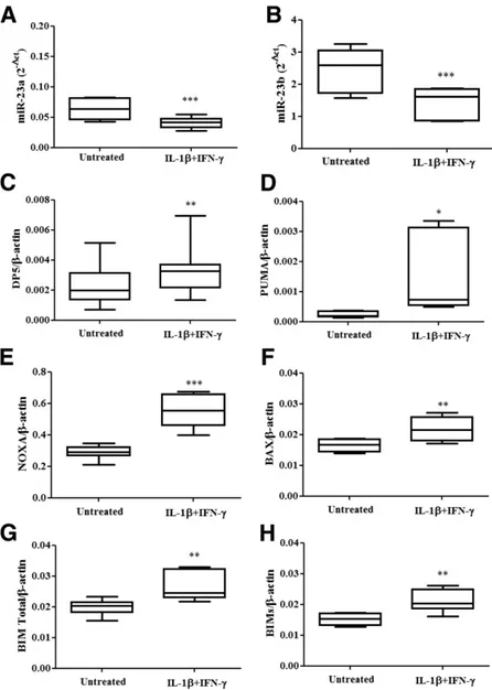
treatment, in an opposite direction as compared with miR-23a-3p and miR-23b-3p. The Ct values of expression of miR-23 family members under basal condition in hu-man islets and huhu-man EndoC-bH1 cells were, respec-tively, 18.3 6 0.1 and 24.1 6 0.1 for miR-23a-3p and 18.36 0.1 and 19 6 0.1 for miR-23b-3p (n = 7). A de-crease in miR-23a-3p and miR-23b-3p expression in hu-man islets (Fig. 2A and B) and huhu-man EndoC-bH1 cells (Fig. 3A and B) was paralleled by increased mRNA expres-sion of PUMA and NOXA (Fig. 2D and E) in human islets and DP5, PUMA, NOXA, BAX, BIM-total, and BIM-small (Fig. 3C–H) in human EndoC-bH1 cells. These results support the hypothesis that proapoptotic Bcl-2 family members are regulated, directly or indirectly, by miR-23a-3p and miR-23b-3p in b-cells.
To elucidate the molecular mechanisms by which miR-23a-3p and miR-23b-3p regulate b-cell survival, single-stranded miRNAs inhibitors were used to block these endogenous miRNAs. An 80 and 60% decrease of miR-23a-3p expression was, respectively, observed in human
Figure 1—miRNA profile of human islets exposed or not to proinflammatory cytokines. Human islets (A–C) were left untreated or treated with IL-1b plus IFN-g for 48 h. miRNAs were then isolated, and miRNA expression profiling was performed using TaqMan Array Human MicroRNA Cards Panel A v2.1 as described inRESEARCH DESIGN AND METHODS. A: Dendrogram of the global miRNA expression profile.
Columns correspond to individual samples exposed or not to cytokines, whereas rows correspond to the individual miRNAs analyzed. The scale color from blue to red corresponds to the abundance of miRNA expression reported as normalized delta cycle threshold (dCT; blue: dCT value,22.18; red: dCT value, 18). All raw data were normalized using three small nucleolar RNAs included in the card (RNU6, RNU44, and RNU48). Expression of miR-23a (B) and miR-23b (C) was assayed in the same samples by RT-PCR and normalized by two different small nucleolar RNAs (RNU6 and RND61). The results are from three independent human islet preparations (A–C).
islets and in human EndoC-bH1 cells (Fig. 4A and C) using an miR-23a-3p inhibitor, followed or not by cytokine ex-posure. A .90% decrease of miR-23b-3p expression was observed in human EndoC-bH1 cells using an miR-23b-3p inhibitor in both untreated and IL-1b plus IFN-g–treated cells (Fig. 4D). Because of the close similarity between miR-23a-3p and miR-23b-3p, we observed an important cross-reaction between the inhibitors, which decreased expression of both miRNAs in parallel in human EndoC-bH1 cells (Fig. 4C and D). Thus, findings with the indi-vidual inhibitors should be interpreted as mediated by inhibition of both miR-23a-3p and miR-23b-3p.
The miR-23a-3p inhibitor exacerbated basal apoptosis, as evaluated by nuclear dyes, in both human islets and human EndoC-bH1 cells (Fig. 4B and E and Supplemen-tary Fig. 4A and B, respectively). A similar increase in apoptosis was observed using the miR-23b-3p inhibitor in human EndoC-bH1 cells (Fig. 4E and Supplementary Fig. 4A and B). Cytokine-induced apoptosis was also augmented in both human islets and human EndoC-bH1 cells using miR-23a-3p inhibitor (Fig. 4B and E and Supplementary Fig. 4A and B) and in EndoC-bH1 cells after exposure to the miR-23b-3p inhibitor (Fig. 4E and Supplementary Fig. 4A and B). In line with these
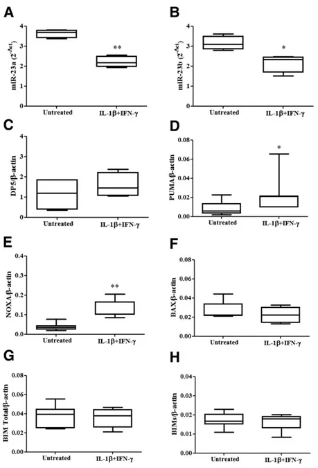
Figure 2—Cytokines coregulate the expression of miR-23a-3p, miR-23b-3p, NOXA, and PUMA in human pancreatic islets. Human islets were left untreated or treated with IL-1b plus IFN-g for 48 h. Expression of miR-23a-3p (A) and miR-23b-3p (B) was assayed by RT-PCR and normalized by two different small nucleolar RNAs (RNU6 and RND61). Expression of DP5 (C), PUMA (D), NOXA (E), BAX (F), BIM-total (G), and BIM-small (BIMs; H) was assayed by RT-PCR and normalized by the housekeeping gene b-actin. Results are represented as box plots, indicating lower quartile, median, and higher quartile, with whiskers representing the range of the remaining data points (A–H). *P < 0.05, **P < 0.01 vs. untreated cells, paired Student t test. Data are shown as mean 6 SEM of four to seven independent experiments.
observations, the inhibitors for miR-23a-3p and miR-23b-3p increased expression of cleaved caspase-3 in human EndoC-bH1 cells (Fig. 4F and G), confirming apoptosis activation. Similarfindings were observed by double immu-nofluorescence for insulin and cleaved caspase-3 in human islets after use of the miR-23a-3p inhibitor (Fig. 5), indi-cating that at least part of the observed cell death in hu-man islet cells takes place at the b-cell level, which is in line with the observed increase in cell death in the human b-cell line EndoC-bH1 cells (Fig. 4E and Supplementary Fig. 4).
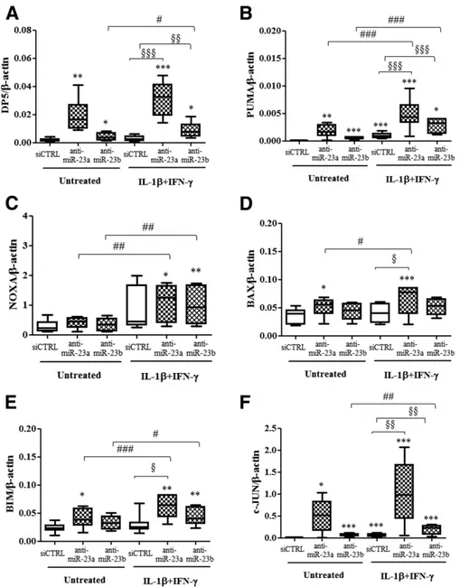
Exposure of cells to the miR-23a-3p inhibitor increased mRNA expression of the proapoptotic Bcl-2 family mem-bers DP5, PUMA, BAX, and BIM (Fig. 6A, B, D, and E,
respectively), whereas inhibition of miR-23b-3p increased expression of DP5 and PUMA (Fig. 6A and B) in human EndoC-bH1 cells following or not exposure to IL-1b plus IFN-g. No increase in NOXA was observed after inhibition of miR-23a-3p or miR-23b-3p (Fig. 6C). A mild increase of protein expression of DP5 and PUMA was observed after the use of miR-23a-3p and miR-23b-3p inhibitors in hu-man EndoC-bH1 cells (Supplementary Fig. 5A, B, C, and D, respectively), whereas no modification in BAX and BIM protein expression after miR-23 family inhibition was detected (data not shown). In contrast, there was no change in expression of the antiapoptotic Bcl-2 family members Mcl-1 and BCL-XL or in APAf-1 after the use of
Figure 3—Cytokines coregulate miR-23a-3p, miR-23b-3p, and proapoptotic Bcl-2 family members in human EndoC-bH1 cells. Human EndoC-bH1 cells were left untreated or treated with IL-1b plus IFN-g for 48 h. Expression of miR-23a-3p (A) and miR-23b-3p (B) was assayed by RT-PCR and normalized by two different small nucleolar RNAs (RNU6 and RND61). Expression of DP5 (C), PUMA (D), NOXA (E), BAX (F), BIM-total (G), and BIM-small (BIMs; H) was assayed by RT-PCR and normalized by the housekeeping gene b-actin. Results are represented as box plots, indicating lower quartile, median, and higher quartile, with whiskers representing the range of the remaining data points (A–H). *P < 0.05, **P < 0.01, ***P < 0.001 vs. untreated cells; paired Student t test. Data are shown as mean 6 SEM of seven independent experiments.
Figure 4—Inhibition of miR-23a-3p and miR-23b-3p exacerbates apoptosis in human islets and human EndoC-bH1 cells. Dispersed human islets (A and B) and human EndoC-bH1 cells (C–G) were transfected with siCTRL or anti–miRNAs targeting miR-23a-3p (human islets and EndoC-bH1 cells) or miR-23b-3p (EndoC-bH1 cells) for 8 h. After a 48-h recovery, cells were left untreated or treated with IL-1b plus IFN-g for 48 h as indicated. Expression of miR-23a-3p (A and C) and miR-23b-3p (D) was assayed by RT-PCR and normalized by two different small nucleolar RNAs (RNU6 and RND61) (A) or by miR-375-3p expression (C and D). Apoptosis was evaluated by Hoechst/ propidium iodide staining after 48 h of cytokine treatment (B and E). Cleaved caspase-3 and b-actin protein expression were evaluated by Western blot after 48 h of cytokine treatment; one representative blot offive independent experiments is shown (F). The optical density quantification of cleaved caspase-3 (as shown in F) was corrected by b-actin expression (G). Results are represented as box plots, indicating lower quartile, median, and higher quartile, with whiskers representing the range of the remaining data points (A–E). In G, results were normalized against the highest value in each independent experiment, considered as 1. *P < 0.05, **P < 0.01, ***P < 0.001 vs. siCTRL untreated cells; §P < 0.05, §§§P < 0.001, #P < 0.05, ##P < 0.01 as indicated by bars; ANOVA followed by paired Student t test with Bonferroni correction. Data are shown as mean 6 SEM of five to seven independent experiments.
miR-23a-3p or miR-23b-3p inhibitors in human EndoC-bH1 cells following or not exposure to IL-1b plus IFN-g (data not shown). Taken together, these results suggest that miR-23a-3p and miR-23b-3p regulate b-cells apoptosis by modulating expression of key proapoptotic Bcl-2 protein family members.
To evaluate if DP5 is a direct target of miR-23a-3p, HeLa cells were transfected with a luciferase construct containing the 39 UTR sequence of DP5 (Supplementary Fig. 6A). miR-23a-3p did not decrease the luciferase activ-ity in cells transfected with the construct containing 39 UTR of DP5, suggesting an indirect effect of miR-23a-3p on DP5 modulation. We next evaluated whether depletion of the miR-23 family upregulated expression of c-JUN, a key regulator of DP5 expression in b-cells (45). Inhibition of miR-23a-3p and miR-23b-3p increased by .16-fold c-JUN mRNA expression in human EndoC-bH1 cells un-der basal condition or following exposure to IL-1b plus
IFN-g (Fig. 6F). Interestingly, analysis on whether miR-23 family members target negative regulators of c-JUN mRNA expression, identified ARNT, HDAC4, MEF2C, NCOA2, TNFAIP3, ZNF382, and ZNF384 as predicted targets of miR-23 family.
Knockdown of PUMA or DP5 Protects Human EndoC-bH1 Cells From Cytokine-Induced Apoptosis in the Context of miR-23a-3p Inhibition
To determine whether the observed effects of miR-23a-3p on proapoptotic Bcl-2 proteins is causally related to b-cell death, we next knocked down PUMA or DP5 in combina-tion with the 23a-3p inhibitor. This abrogated miR-23a-3p inhibitor–dependent induction of PUMA and DP5 (Fig. 7A and B, respectively) in both untreated and cytokine-treated human EndoC-bH1 cells; the inhibition of miR-23a-3p was confirmed by real-time qPCR (Fig. 7C and D). Importantly, blocking PUMA and DP5 induction in parallel to miR-23a-3p inhibition also prevented the increase in
Figure 5—Inhibition of miR-23a-3p augments cleaved caspase-3 expression in human b-cells. Human dispersed islets were transfected with siCTRL (A–D and I–L) or an anti-miRNA targeting miR-23a-3p (E–H and M–P) for 8 h. After a 48-h recovery, cells were left untreated (A–H) or treated with IL-1b plus IFN-g (I–P) for 48 h. After cytokine treatment, cells were fixed and used for histological studies. Fluorescent microscopy analysis of insulin (A, E, I, and M in green) and cleaved caspase-3 (F, J, and N in red) shows the presence of double-positive cells for insulin and cleaved caspase-3 (H, L, and P, merged in yellow). Hoechst staining (C, G, K, and O in blue) shows the presence of nuclear condensation in the apoptotic cleaved caspase-3–positive cells (G, K, and O, arrows). Double-positive cells for insulin and cleaved caspase-3 are indicated by the arrows (E, F, H–J, L–N, and P).
apoptosis secondary to depletion of this miRNA (Fig. 7E and F), indicating that upregulation of these two BH3-only proteins is part of the mechanisms by which a cytokine-induced miR-23a-3p decrease contributes to apoptosis of human pancreatic b-cells. This, and the above-described activation of caspase-3, indicates that miR-23a-3p regulates the intrinsic or mitochondrial apo-ptotic pathway in human b-cells.
To investigate whether additional miRNAs contribute to cytokine-induced apoptosis mediated by Bcl-2 family members, we studied miR-149-5p, another miRNA observed as downregulated by cytokines in our initial screening
(Supplementary Fig. 1B and Supplementary Table 5). miRNA target prediction programs indicated miR-149-5p as a possible modulator of the Bcl-2 family members PUMA and BIM. Using an miR-149-5p inhibitor, there was a 60–80% decrease of miR-149-5p expression in hu-man EndoC-bH1 cells exposed or not to cytokines (Supple-mentary Fig. 7A). miR-149-5p depletion in both untreated and cytokine-treated human EndoC-bH1 cells increased apoptosis, as evaluated by nuclear dyes (Supplementary Fig. 7B). Furthermore, miR-149-5p inhibition led to upreg-ulation of DP5, PUMA, and BAX (Supplementary Fig. 7C, D, and F) and a mild modulation of BIM (Supplementary
Figure 6—Inhibition of miR-23a-3p and miR-23b-3p increases expression of proapoptotic Bcl-2 family members in human EndoC-bH1 cells. EndoC-bH1 cells were transfected with siCTRL or with anti-miRNAs targeting miR-23a-3p and miR-23b-3p for 8 h. After a 48-h recovery, cells were left untreated or treated with IL-1b plus IFN-g for 48 h as indicated. Expression of DP5 (A), PUMA (B), NOXA (C), BAX (D), BIM (E), and c-JUN (F). Results are represented as box plots, indicating lower quartile, median, and higher quartile, with whiskers representing the range of the remaining data points (A–F). *P < 0.05, **P < 0.01, ***P < 0.001 vs. siCTRL untreated cells; §P < 0.05, §§P < 0.01, §§§P < 0.001, #P < 0.05, ##P < 0.01, ###P < 0.001 as indicated by bars; ANOVA followed by paired Student t test with Bonferroni correction. Data are shown as mean6 SEM of six to seven independent experiments.
Fig. 7G), whereas no modification in Mcl-1 and BCL-XL was observed (data not shown). This supports the concept that individual genes can be regulated by different miRNAs and that several miRNAs can participate in the regulation of key genes (13), such as the ones involved in b-cell death. We next knocked down PUMA or DP5 in combination with the
miR-149-5p inhibitor. This abrogated miR-149-5p inhibitor– dependent induction of PUMA and DP5 (Supplementary Fig. 8A and B, respectively) in both untreated and cytokine-treated human EndoC-bH1 cells; the inhibition of miR-149-5p was confirmed by real-time qPCR (Supplementary Fig. 8C and D). Importantly, blocking PUMA and DP5
Figure 7—Knockdown (KD) of PUMA or DP5 protects human EndoC-bH1 cells against cytokine-induced apoptosis in the context of miR-23a-3p inhibition. EndoC-bH1 cells were transfected with siCTRL, siPUMA, or anti–miR-23a-3p or cotransfected with siPUMA plus anti– miR-23a-3p (A, C, and E) for 8 h. After a 48-h recovery, cells were left untreated or treated with IL-1b plus IFN-g for 48 h as indicated. The KD of PUMA (A) was confirmed by RT-PCR and normalized by the housekeeping gene b-actin. Expression of miR-23a-3p (C) was assayed by RT-PCR and normalized by miR-375-3p expression. Apoptosis was evaluated by propidium iodide/Hoechst staining after 48 h of cytokine treatment (E). EndoC-bH1 cells were transfected with siCTRL, siDP5, or anti–miR-23a-3p or cotransfected with siDP5 plus anti– miR-23a-3p (B, D, and F) for 8 h. After a 48-h recovery, cells were left untreated or treated with IL-1b plus IFN-g for 48 h as indicated. The KD of DP5 (B) was confirmed by RT-PCR and normalized by the housekeeping gene b-actin. Expression of miR-23a-3p (D) was assayed by RT-PCR and normalized by miR-375-3p expression. Apoptosis was evaluated by propidium iodide/Hoechst staining after 48 h of cytokine treatment (F). Results (A and B) were normalized against the highest value in each independent experiment, considered as 1. *P < 0.05, **P < 0.01, ***P < 0.001 vs. siCTRL untreated cells; §P < 0.05, §§P < 0.01, §§§P < 0.001 vs. siCTRL cytokine-treated cells; #P < 0.05, ##P < 0.01, ###P < 0.001, @@@P < 0.001 as indicated by bars; ANOVA followed by paired Student t test with Bonferroni correction. Data are shown as mean6 SEM of five independent experiments.
induction in parallel to miR-149-5p inhibition also pre-vented the increase in apoptosis secondary to depletion of this miRNA (Supplementary Fig. 8E and F), as we pre-viously observed for miR-23a-3p (Fig. 7E and F). DISCUSSION
A significant reduction of functional b-cell mass because of increased b-cell death is the key feature of T1D. During the early stages of the disease, pancreatic islets are ex-posed to proinflammatory mediators released by the in-vading immune cells. Available information, mostly from animal models and in vitro experiments, suggests that this results into changes in the expression of key b-cell gene networks involved in b-cell function and phenotype, induction of endoplasmic reticulum (ER) stress, and in-flammation (1,46,47). Locally produced proinflammatory mediators recruit further immune cells that enhance release of chemokines and cytokines, leading to ampli fi-cation of the inflammatory process. Prolonged cytokine exposure and the consequent persistent ER stress, possi-bly in combination with environmental factors such as viral infections, may activate downstream pathways such as JNK phosphorylation and the transcription fac-tors nuclear factor-kB and STAT1, eventually triggering expression of proapoptotic Bcl-2 proteins, cleavage, and activation of caspases and cell death (1,7,48). The fine regulation of this process, particularly in human b-cells, remains to be clarified.
The Bcl-2 family members can be subdivided into three different groups: antiapoptotic (i.e., Bcl-2, Bcl-XL, Mcl-1, and A1), proapoptotic (i.e., BAX, BAK, and BOK) Bcl-2 proteins, and proapoptotic BH3-only proteins (48). Addi-tionally, the BH3-only proteins can be divided into two subgroups: the sensitizers (i.e., DP5, BAD, and NOXA) and the activators (i.e., BID, BIM, and PUMA) of apopto-sis (48,49). It has been previously shown that PUMA and DP5, two of the most important regulators of the intrin-sic apoptotic pathway, are induced in b-cells by exposure to the cytokines IL-1b plus IFN-g or to chemical ER stres-sors, leading to BAX translocation to the mitochondria, caspase-3 activation, and b-cell death (45,50).
miRNAs are key regulators of gene expression, and some have been shown to sensitize b-cells to cytokine-induced apoptosis (24,25). Thus, prolonged exposure of mouse islets to IL-1b or to TNF-a results in upregulation of miR-146a-5p, miR-34a-5p, and miR-21-5p (24). Fur-thermore, cytokine-induced upregulation of miR-101a-3p and miR-30b-5p decreases expression of antiapoptotic Bcl-2 proteins in the mouse insulin- producing cell line MIN6 (26), whereas suppression of Bcl-2 upon miR-34a-5p induction contributes to palmitate-induced apoptosis in MIN6 cells (51). In line with these observations, cyto-kines upregulate miR-29-5p, which exacerbates cytokine-induced apoptosis via inhibition of the antiapoptotic Bcl-2 protein Mcl-1 (25). In contrast, the possible contribution of miRNAs to the cytokine-induced upregulation of pro-apoptotic BH3-only proteins in b-cells remains to be clarified.
Against this background, we presently performed a global miRNA expression profile on three independent human islets preparations treated or not with the cytokines IL-1b plus IFN-g. Twenty-two and 35 miRNAs were found up- and downregulated, respectively, in human islets after IL-1b plus IFN-g treatment. Of particular interest, these cytokines induced a significant downregulation of miR-23a-3p, miR-23b-miR-23a-3p, and miR-27a-miR-23a-3p, all belonging to the same miRNA family. miR-23b-3p is also downregulated in islets from 4- and 8-week NOD mice (25). Downregulation of miR-23a-3p and miR-27a-3p in posttraumatic brain in-jury contributes to neuronal cell death by directly upreg-ulating the proapoptotic Bcl-2 family members BAX, NOXA, and PUMA (42), suggesting that this may be an important mechanism by which cytokines upregulate these key proapoptotic Bcl-2 family members. To test this hypothesis, miR-23a-3p and miR-23b-3p were inhibited in
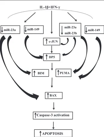
Figure 8—Proposed model for the cytokine-induced cross talk be-tween miRNAs and proapoptotic Bcl-2 proteins in b-cells. The proinflammatory cytokines IL-1b plus IFN-g downregulate expres-sion of miR-23a-3p, miR-23b-3p, and miR-149-5p in b-cells. De-creased expression of these inhibitory miRNAs leads to inDe-creased mRNA expression of key proapoptotic Bcl-2 proteins. Specifically, miR-23a-3p and miR-23b-3p modulate the BH3-only sensitizer DP5 (at least in part via upregulation of c-JUN) and the BH3-only acti-vator PUMA, whereas miR-23a-3p regulates both the BH3-only sensitizer BIM and the proapoptotic Bcl-2 protein BAX. Additionally, inhibition of miR-149-5p expressions results in increased mRNA expression of both DP5 and PUMA. This complex interaction be-tween miRNAs, transcription factors, and downstream proapoptotic proteins leads to cleavage and activation of caspase-3 and, sub-sequently, to b-cell apoptosis.
human b-cells. Because miR-23a-3p and miR-23b-3p are very similar and differ by only one nucleotide in their 39 ends, which is less important for miRNA action than the 59 end, the inhibitors of miR-23a-3p and miR-23b-3p used led to inhibition of both miRNAs. Thus, all data obtained based on these inhibitors should be interpreted as indicating broad effects of the miR-23 family. miR-23 inhibition increased apoptosis under both basal condition and following cytokine treatment. This was paralleled by upregulation of several BH3-only proteins, with the most consistent findings observed for DP5 and PUMA. Impor-tantly, knocking down PUMA or DP5 reverted the proapop-totic effects of miR-23a-3p inhibition, suggesting that these two BH3-only proteins are regulated in an indirect way by proinflammatory cytokines (i.e., via the inhibition of miRNAs of the miR-23 family). Using HeLa cells, we did not observe a direct interaction between the miR-23 family and DP5 (for technical reasons, these experiments could not be performed in human EndoC-bH1 cells). This sug-gests the possibility of an indirect effect of these miRNAs on DP5 via upregulation of c-JUN (current study), a tran-scription factor previously shown by us to regulate DP5 expression (45). It remains to be determined how miR-23 cross talk with PUMA. Of note, the results described above show additive effects of PUMA/DP5 and miR-23 in that they both affect apoptosis, and the BH3-only proteins seem to be regulated downstream of the miRNAs. It re-mains to be proven, however, that these signals interact and the ultimate mechanisms involved. The current study should therefore be interpreted with caution.
The fact that these novel observations were obtained in human islets and in a human b-cell line increases their potential relevance to human T1D. Additional miRNAs targeting Bcl-2 family members, such as miR-221-3p and miR-149-5p, were also found downregulated in our screening, suggesting a more complex cross talk between miRNAs and proapoptotic Bcl-2 family members in b-cells. In line with this, inhibition of miR-149-5p also led to upregulation of DP5, PUMA, and BAX. An overview of this complex cross talk is provided in Fig. 8.
In conclusion, the results of this study identify a potential novel cross talk between a family of miRNAs and key proapoptotic Bcl-2 proteins in human pancreatic b-cells, providing a new mechanistic understanding of in-flammation-induced b-cell apoptosis.
Acknowledgments.The authors thank the personnel of the ULB Center for Diabetes Research, including I. Millard, A. Musuaya, M. Pangerl, and N. Pachera, for excellent technical support.
Funding.This work was supported by grants from European Union (Seventh Framework Project of the European Union, Project NAMIT, and the European Union’s Horizon 2020 research and innovation programme, Project T2DSystems; grant 667191), the Fonds National de la Recherche Scientifique (Belgium), and the National Institutes of Health National Institute of Diabetes and Digestive and Kidney Diseases–Human Islet Research Network Consortium (grant 1UC4-DK-104166-01) to D.L.E. P.M., F.D., and D.L.E. have received funding from the Innovative Medicines Initiative 2 Joint Undertaking (grant 115797; INNODIA).
This Joint Undertaking receives support from the Union’s Horizon 2020 research and innovation programme, the European Federation of Pharmaceutical Indus-tries and Associations, JDRF, and The Leona M. and Harry B. Helmsley Charitable Trust.
Duality of Interest.No potential conflicts of interest relevant to this article were reported.
Author Contributions. F.A.G., G.S., R.R., P.M., F.D., and D.L.E. conceived and designed the experiments. F.A.G., G.S., J.J.-M., O.V., L.M., L.L., K.T., and M.B. acquired data. P.M., F.D., and D.L.E. supervised the study and contributed to reagent. F.A.G. and D.L.E. wrote the manuscript. All authors revised the manuscript. D.L.E. is the guarantor of this work and, as such, had full access to all the data in the study and takes responsibility for the integrity of the data and the accuracy of the data analysis.
References
1. Eizirik DL, Colli ML, Ortis F. The role of inflammation in insulitis and beta-cell loss in type 1 diabetes. Nat Rev Endocrinol 2009;5:219–226
2. Eizirik DL, Sammeth M, Bouckenooghe T, et al. The human pancreatic islet transcriptome: expression of candidate genes for type 1 diabetes and the impact of pro-inflammatory cytokines. PLoS Genet 2012;8:e1002552
3. Santin I, Eizirik DL. Candidate genes for type 1 diabetes modulate pan-creatic islet inflammation and b-cell apoptosis. Diabetes Obes Metab 2013;15 (Suppl. 3):71–81
4. Fløyel T, Kaur S, Pociot F. Genes affectingb-cell function in type 1 diabetes. Curr Diab Rep 2015;15:97
5. Richardson SJ, Morgan NG, Foulis AK. Pancreatic pathology in type 1 di-abetes mellitus. Endocr Pathol 2014;25:80–92
6. Grieco FA, Sebastiani G, Spagnuolo I, Patti A, Dotta F. Immunology in the clinic review series; focus on type 1 diabetes and viruses: how viral infections modulate beta cell function. Clin Exp Immunol 2012;168:24–29
7. de Beeck AO, Eizirik DL. Viral infections in type 1 diabetes mellitus–why the b cells? Nat Rev Endocrinol 2016;12:263–273
8. Roep BO, Peakman M. Diabetogenic T lymphocytes in human type 1 di-abetes. Curr Opin Immunol 2011;23:746–753
9. Chen MC, Proost P, Gysemans C, Mathieu C, Eizirik DL. Monocyte che-moattractant protein-1 is expressed in pancreatic islets from prediabetic NOD mice and in interleukin-1 beta-exposed human and rat islet cells. Diabetologia 2001;44:325–332
10. Coppieters KT, Dotta F, Amirian N, et al. Demonstration of islet-autoreactive CD8 T cells in insulitic lesions from recent onset and long-term type 1 diabetes patients. J Exp Med 2012;209:51–60
11. Cardozo AK, Proost P, Gysemans C, Chen MC, Mathieu C, Eizirik DL. IL-1beta and IFN-gamma induce the expression of diverse chemokines and IL-15 in human and rat pancreatic islet cells, and in islets from pre-diabetic NOD mice. Diabetologia 2003;46:255–266
12. Grieco FA, Moore F, Vigneron F, et al. IL-17A increases the expression of proinflammatory chemokines in human pancreatic islets. Diabetologia 2014;57: 502–511
13. Fabian MR, Sonenberg N, Filipowicz W. Regulation of mRNA translation and stability by microRNAs. Annu Rev Biochem 2010;79:351–379
14. Dumortier O, Van Obberghen E. MicroRNAs in pancreas development. Di-abetes Obes Metab 2012;14(Suppl. 3):22–28
15. Esguerra JL, Mollet IG, Salunkhe VA, Wendt A, Eliasson L. Regulation of pancreatic beta cell stimulus-secretion coupling by microRNAs. Genes (Basel) 2014;5:1018–1031
16. Guay C, Regazzi R. MicroRNAs and the functionalb cell mass: For better or worse. Diabetes Metab 2015;41:369–377
17. Poy MN, Hausser J, Trajkovski M, et al. miR-375 maintains normal pancreatic alpha- and beta-cell mass. Proc Natl Acad Sci U S A 2009;106:5813–5818 18. Mohan R, Mao Y, Zhang S, et al. Differentially expressed microRNA-483 confers distinct functions in pancreatic beta- and alpha-cells. J Biol Chem 2015; 290:19955–19966
19. Klein D, Misawa R, Bravo-Egana V, et al. MicroRNA expression in alpha and beta cells of human pancreatic islets. PLoS One 2013;8:e55064
20. Guay C, Roggli E, Nesca V, Jacovetti C, Regazzi R. Diabetes mellitus, a microRNA-related disease? Transl Res 2011;157:253–264
21. Guay C, Regazzi R. Role of islet microRNAs in diabetes: which model for which question? Diabetologia 2015;58:456–463
22. Ventriglia G, Nigi L, Sebastiani G, Dotta F. MicroRNAs: Novel players in the dialogue between pancreatic islets and immune system in autoimmune diabetes. Biomed Res Int 2015;2015:749734.
23. Sebastiani G, Grieco FA, Spagnuolo I, Galleri L, Cataldo D, Dotta F. Increased expression of microRNA miR-326 in type 1 diabetic patients with ongoing islet autoimmunity. Diabetes Metab Res Rev 2011;27:862–866
24. Roggli E, Britan A, Gattesco S, et al. Involvement of microRNAs in the cy-totoxic effects exerted by proinflammatory cytokines on pancreatic beta-cells. Diabetes 2010;59:978–986
25. Roggli E, Gattesco S, Caille D, et al. Changes in microRNA expression contribute to pancreaticb-cell dysfunction in prediabetic NOD mice. Diabetes 2012;61:1742–1751
26. Zheng Y, Wang Z, Tu Y, et al. miR-101a and miR-30b contribute to in-flammatory cytokine-mediated b-cell dysfunction. Lab Invest 2015;95:1387–1397 27. Takahashi P, Xavier DJ, Evangelista AF, et al. MicroRNA expression profiling and functional annotation analysis of their targets in patients with type 1 diabetes mellitus. Gene 2014;539:213–223
28. Kim KW, Ho A, Alshabee-Akil A, et al. Coxsackievirus B5 infection induces dysregulation of microRNAs predicted to target known type 1 diabetes risk genes in human pancreatic islets. Diabetes 2016;65:996–1003
29. Marchetti P, Bugliani M, Lupi R, et al. The endoplasmic reticulum in pan-creatic beta cells of type 2 diabetes patients. Diabetologia 2007;50:2486–2494 30. Moore F, Colli ML, Cnop M, et al. PTPN2, a candidate gene for type 1 di-abetes, modulates interferon-gamma-induced pancreatic beta-cell apoptosis. Diabetes 2009;58:1283–1291
31. Ravassard P, Hazhouz Y, Pechberty S, et al. A genetically engineered human pancreaticb cell line exhibiting glucose-inducible insulin secretion. J Clin Invest 2011;121:3589–3597
32. Brozzi F, Nardelli TR, Lopes M, et al. Cytokines induce endoplasmic re-ticulum stress in human, rat and mouse beta cells via different mechanisms. Diabetologia 2015;58:2307–2316
33. Eizirik DL, Pipeleers DG, Ling Z, Welsh N, Hellerström C, Andersson A. Major species differences between humans and rodents in the susceptibility to pan-creatic beta-cell injury. Proc Natl Acad Sci U S A 1994;91:9253–9256 34. Cunha DA, Igoillo-Esteve M, Gurzov EN, et al. Death protein 5 and p53-upregulated modulator of apoptosis mediate the endoplasmic reticulum stress-mitochondrial dialog triggering lipotoxic rodent and human b-cell apoptosis. Diabetes 2012;61:2763–2775
35. Moore F, Cunha DA, Mulder H, Eizirik DL. Use of RNA interference to in-vestigate cytokine signal transduction in pancreatic beta cells. Methods Mol Biol 2012;820:179–194
36. Hoorens A, Van de Casteele M, Klöppel G, Pipeleers D. Glucose pro-motes survival of rat pancreatic beta cells by activating synthesis of proteins which suppress a constitutive apoptotic program. J Clin Invest 1996;98: 1568–1574
37. Overbergh L, Valckx D, Waer M, Mathieu C. Quantification of murine cy-tokine mRNAs using real time quantitative reverse transcriptase PCR. Cycy-tokine 1999;11:305–312
38. Cardozo AK, Kruhøffer M, Leeman R, Orntoft T, Eizirik DL. Identification of novel cytokine-induced genes in pancreatic beta-cells by high-density oligonu-cleotide arrays. Diabetes 2001;50:909–920
39. Marroqui L, Dos Santos RS, Fløyel T, et al. TYK2, a candidate gene for type 1 diabetes, modulates apoptosis and the innate immune response in human pancreatic beta-cells. Diabetes 2015;64:3808–3817
40. Dweep H, Gretz N, Sticht C. miRWalk database for miRNA-target interac-tions. Methods Mol Biol 2014;1182:289–305
41. Xiao F, Zuo Z, Cai G, Kang S, Gao X, Li T. miRecords: an integrated resource for microRNA-target interactions. Nucleic Acids Res 2009;37:D105–D110 42. Sabirzhanov B, Zhao Z, Stoica BA, et al. Downregulation of miR-23a and miR-27a following experimental traumatic brain injury induces neuronal cell death through activation of proapoptotic Bcl-2 proteins. J Neurosci 2014;34: 10055–10071
43. Duan Q, Mao X, Xiao Y, et al. Super enhancers at the 146a and miR-155 genes contribute to self-regulation of inflammation. Biochim Biophys Acta 2016;1859:564–571.
44. van de Bunt M, Gaulton KJ, Parts L, et al. The miRNA profile of human pancreatic islets and beta-cells and relationship to type 2 diabetes pathogenesis. PLoS One 2013;8:e55272
45. Gurzov EN, Ortis F, Cunha DA, et al. Signaling by IL-1beta+IFN-gamma and ER stress converge on DP5/Hrk activation: a novel mechanism for pancreatic beta-cell apoptosis. Cell Death Differ 2009;16:1539–1550
46. Brozzi F, Eizirik DL. ER stress and the decline and fall of pancreatic beta cells in type 1 diabetes. Ups J Med Sci 2016;121:133–139
47. Eizirik DL, Miani M, Cardozo AK. Signalling danger: endoplasmic reticulum stress and the unfolded protein response in pancreatic islet inflammation. Dia-betologia 2013;56:234–241
48. Gurzov EN, Eizirik DL. Bcl-2 proteins in diabetes: mitochondrial pathways of b-cell death and dysfunction. Trends Cell Biol 2011;21:424–431
49. Kim H, Rafiuddin-Shah M, Tu HC, et al. Hierarchical regulation of mito-chondrion-dependent apoptosis by BCL-2 subfamilies. Nat Cell Biol 2006;8: 1348–1358
50. Gurzov EN, Germano CM, Cunha DA, et al. p53 up-regulated modulator of apoptosis (PUMA) activation contributes to pancreatic beta-cell apoptosis induced by proinflammatory cytokines and endoplasmic reticulum stress. J Biol Chem 2010;285:19910–19920
51. Lin X, Guan H, Huang Z, et al. Downregulation of Bcl-2 expression by miR-34a mediates palmitate-induced Min6 cells apoptosis. J Diabetes Res 2014; 2014:258695.