Scuola Dottorale in Biologia
Sezione “Biologia Applicata alla Salute dell’Uomo” Ciclo XXIII
Characterization of cord-blood derived T cells immune response after stimulation with aminobisphosphonate compounds: their hypothetical role
in the control of neonatal viral infections transmitted during pregnancy. Caratterizzazione fenotipica e funzionale dei linfociti T isolati dal sangue di cordone ombelicale dopo stimolazione con aminobifosfonati: il loro ipotetico ruolo nel controllo delle infezioni virali neonatali trasmessi
durante la gravidanza.
DOTTORANDA Ida Gabriele
Tutor: Dott. Giorgio Mancino
Co-Tutor: Prof.ssa Maria Marino Coordinatore: Prof. Paolo Visca
INDEX
ABSTRACT page 3
INTRODUCTION page 8
RESULTS page 23
DISCUSSION page 44
CONCLUSION page 48
REFERENCES page 50
Abstract
The innate immunity is the first defence line against microbes and pathogens, and exerts its protective activity by killing the infected host-cells, inducing an effective adaptive immunity. In microbial infections, adaptive and innate immune reactions cooperate to protect the host and, whenever it is possible, to eradicate or control the infection. Among its cellular components are mainly polymorphonuclear phagocytes, mononuclear phagocytes, Natural Killer (NK) cells, characterized by natural cytotoxic activity, and a minority population of T lymphocytes, that exhibit T cell receptor.
T lymphocytes shown an high reactivity against bacteria, protozoa and viruses, and their percentage peripheral blood was significantly higher than normal in individuals with infections such as tuberculosis, salmonellosis, listeriosis, brucellosis, leishmaniasis, malaria, toxoplasmosis, infection by viruses such as Herpes Simplex (HSV-1)[Born WK et al. 2006; Roger Sciammas, Jeffrey A Bluestone. 1999]. The neonatal HSV diseases are often the result of infections developed in the last quarter of pregnancy with a risk of transmission between 30% and 60%. The immune system of the newborn in comparison with the adult is functionally immature, and it is different quantitatively and phenotypicaly in host defence [Bradley MB and Cairo MS 2005]. T cells isolated from cord blood (CB) compared to adult peripheral blood cells, show a reduced production of cytokines, a poor response to mitogenic stimulation, and reduced cytotoxic activity [Cairo C et al. 2008]. The immaturity of the neonatal immune system has been associated with increased susceptibility to various pathogens, resulting in higher mortality [Paula AV et al. 2006]. For this reason, aim of our project is to investigate the possibility to immunomodulate the innate immunity response as new therapeutical approach of neonatal pathologies.
In cord blood T cells, which represent less than 1% of T lymphocytes, recognize non-peptide phospho-antigens expressed in microbial pathogens. These antigens were identified in bioactive fractions of M. tuberculosis and belong to isoprenoid pyrophosphates, a class of Pyrophosphomonoesters, such as isopentenil-pyrophosphate (IPP). Also synthetic molecules, known as Aminobisphosphonates (ABs), have been also identified as cell antigens. We have demonstrated that ABs, such as Pamidronate (PAM) and Zoledronate (ZOL), are able of stimulating T lymphocytes isolated from CB and differentiate into a regulatory phenotype. Initially we found that T cells isolated from CB respond significantly in terms of expansion to stimulation with PAM and ZOL, while stimulation with IPP does not involve any expansion. The response to these compounds is demonstrated by the
membrane expression of activation markers such as CD25 and CD69, which are more present after stimulation. We also observed that treatments with PAM and ZOL induce the expansion of an effector population of T cells (CD62L-/CD27-) with regulatory phenotype (CD45RA-/CD16-), confirmed by their ability to produce specific cytokines (IFN and TNF ). By flow cytometric analysis we observed that IL-2 or IL-2+ABs treatment induced V 2
+
T cells to differentiate into a non-cytotoxic phenotype that was confirmed by absence of intracellular perforin expression [Placido R et al, accepted]. On the other hand, IL-2 or IL-2+ABs stimulation reduced significantly the non-cytotoxic phenotype of CB V 2- T cells, suggesting their probable differentiation toward a cytotoxic subset. To characterize the CB cell-mediated cytotoxicity, we used cytotoxicity assay in which effector cells (CB) were incubated with targets cells (K562, human erythromyeloblastoid leukemia cell line) and cell death was evaluated at 6h after co-culture. IL-2 treated cells induced a significantly increase of tumor cell death (apoptosis). Since T lymphocytes are not able to acquire a cytotoxic phenotype and only IL2 stimulation induced cytotoxicity activity of CB cells, it was plausible that other cellular subsets could be involved in their cytolytic activity, as Natural Killer (NK). In order to confirm this hypothesis, we studied the production of cytotoxic effectors, such as perforin and granzymes, in CB NK cells. By flow cytometry analysis we observed that at 24h after treatment with IL-2, CB CD56+ cells (NK) showed a clear reduction of intracellular perforin and granzymes, as consequence of their release in the extracellular environment. To complete the analysis of cytotoxic profile of lymphocytes, we studied the production of an important mediator of cytotoxicity, the interferon- IFN IFNγ is a critical cytokine for innate and adaptive immunity against viral and bacterial infections and for tumor control. Flow cytometric analysis showed that expression of IFN was present in T lymphocytes and, moreover, different stimuli, such as IL-2, increased its expression. By contrast, NK cells were resulted IFNγ negative and unresponsive to IL-2 treatment (data contained in CD, Fig. A).
lymphocytes and NK cells are two cell subsets involved mainly in innate immunity response against several pathogens, as bacteria and viruses. For this reason, we considered interesting to study the effect of viral infections, as HSV-1, on these two cell populations.
Thus, aim of our project was to evaluate the activity of CB-derived T and NK lymphocytes in co-culture with HSV-1 infected cells. CB cells, that were previously stimulated with IL-2 and/or PAM for 14 days and then characterized phenotypically/quantitatively, were co-cultured with mock or HSV-1-infected human fibroblasts. At 24h after co-culture (48h after virus
infection) we observed that the percentage of T lymphocytes did not change in the presence of uninfected and infected cells. Furthermore, T cells showed to have a non-effector phenotype (CD16-), independently co-culture conditions, suggesting that they did not exhibit a HSV-1 mediated response. Contextually, a significant reduction of non-cytotoxic phenotype of CB-derived V - T lymphocytes was observed after 24h of treatment with IL-2 or IL-IL-2+PAM. These data confirmed our previous findings of increased cytotoxic activity of NK cells. Considering that CD16 is a marker of citotoxic activity of NK cells, we evaluated their response against HSV-1 infection, as cell expansion and cytokines production. Results showed that NK cell proliferation was induced by co-culture with human fibroblasts and greatly with HSV-1 infected human fibroblasts. By the contrast, IFN synthesized by NK cells was not modified by co-culture conditions, as well as the treatment with IL-2 or ABs. Differently, the co-culture with fibroblasts increased INF production by T lymphocytes. In order to verify if increased activation of CB immune system was able to contrast viral infection, we titrate the viral particles by cytopatic effect evaluation (TCID50). After 24h co-culture (48h after virus infection) we observed an increase in viral titer in co-culture with CB-derived cells IL-2 treated. This result could be explained as a consequence of viral particles released from fibroblasts lysed by cytolitic activity of NK cells. By the contrast, after 48h co-culture, the viral titer was reduced dramatically, as a result of lacked viral replication caused by cell death triggered by CB cells occured at 24h (preliminary data).
Our conclusions have been suggested that T lymphocytes and NK cells cooperates to inhibit viral replication. T cells activated by IL-2 and/or ABs acquire a regulatory phenotype that allows them to produce immunoregulatory cytokines, such as INF , that consequently inhibits viral replication directly and promotes cytolytic activity of NK cells as confirmed by their ability to produce and release of perforin and granzymes, in response to a pathogen stimulus.
Abstract
L'immunità innata è la prima linea di difesa contro microbi e patogeni, esercita la sua attività di protezione uccidendo le cellule infette, inducendo così la risposta dell’immunità adattativa. In caso di infezioni microbiche, la reazione immunitaria innata e quella adattativa cooperano per tutelare l'ospite e, quando è possibile, per controllare o eliminare l'infezione. I suoi principali componenti cellulari sono: fagociti polimorfonucleati, fagociti mononucleari, cellule Natural Killer (NK), caratterizzati da un’attività citotossica naturale,
ed una popolazione minoritaria dei linfociti T, che presentano un recettore delle cellule T (TCR) di tipo . I linfociti T mostrano un’ elevata reattività contro batteri, protozoi e virus, è possibile osservare, infatti, come la loro percentuale nel sangue periferico risulti significativamente più alta del normale in individui con infezioni come tubercolosi, salmonellosi, listeriosi, brucellosi, leishmaniosi, malaria, toxoplasmosi, infezione da virus come Herpes Simplex (HSV) [Born WK et al. 2006; Roger Sciammas, Jeffrey A Bluestone,1999]. HSV è il principale agente eziologico di molte patologie del neonato e del bambino. Tali malattie sono spesso il risultato di infezioni sviluppate nell'ultimo trimestre di gravidanza, con un rischio di trasmissione tra il 30% e 60%. Il sistema immunitario del neonato, se confrontato con quello dell'adulto, è funzionalmente immaturo, ed è quantitativamente e fenotipicamente diverso nei meccanismi di difesa dell’ospite [Bradley MB and Cairo MS 2005]. Linfociti T isolati da sangue di cordone ombelicale (CB), rispetto alle cellule del sangue periferico in un adulto, mostrano una ridotta produzione di citochine, una scarsa risposta alla stimolazione mitogenica, e una ridotta attività citotossica [Cairo C et al. 2008]. L'immaturità del sistema immunitario neonatale è stata associata ad un’aumentata suscettibilità a vari agenti patogeni, con conseguente aumento della mortalità [Paula AV et al. 2006]. Per questo motivo, scopo del progetto è stato quello di indagare la possibilità di immunomodulare la risposta dell'immunità innata come nuovo approccio terapeutico delle patologie neonatali.
Nel CB i linfociti T , che rappresentano meno dell'1% dei linfociti T circolanti, sono in grado di riconoscere antigeni fosforilati di natura non-peptidica espressi da vari patogeni microbici. Tali antigeni sono stati identificati in frazioni bioattive di M. tuberculosis e risultano appartenere principalmente ai pirofosfati isoprenoidi, una classe di Pirofosfomonoesteri, come isopentenil-pirofosfato (IPP). Anche altre molecole, note come Aminobisfosfonati (ABs), sono state identificate come antigeni delle cellule . Abbiamo dimostrato che gli ABs, come Pamidronato (PAM) e Zoledronato (ZOL), sono in grado di stimolare i linfociti T isolati da CB e di indurne l’attivazione. Inizialmente abbiamo riscontrato la capacità di tali cellule T di rispondere significativamente, in termini di espansione, alla stimolazione con Interleuchina-2 (IL-2) + PAM e/o ZOL, mentre la stimolazione con IPP non comporta alcuna espansione della popolazione in esame. La risposta a tali composti si traduce anche in un aumento dell’espressione sulla loro membrana di markers d’attivazione, quali CD25 e CD69. I dati ottenuti ci hanno permesso di affermare che il trattamento con IL-2+ABs induce l’espansione di una popolazione effettrice di linfociti T
(CD62L-/CD27-), caratterizzata da un fenotipo regolatorio (CD45RA-/CD16 -), ma non in grado di sviluppare un fenotipo citotossico. Questi dati sono stati confermati dalla loro capacità di produrre citochine pro-infiammatorie specifiche (IFN e TNF ), e dalla mancata espressione di perforina intracellulare [Placido R et al, accepted]. D'altra parte, il trattamento con sola IL-2 o IL-2+ABs riduce significativamente le cellule T V 2- isolate dal CB caratterizzate da fenotipo non citotossico, suggerendo una probabile attivazione dei linfociti T verso una popolazione effettrice. Per caratterizzare la citotossicità cellulo-mediata riscontrata nel CB, abbiamo utilizzato test di citotossicità in cui sono state incubate cellule effettrici (CB) con cellule target (K562, linea cellulare umana di eritroleucemia), dopo 6 ore dalla co-coltura è stata valutata la morte cellulare (apoptosi). Le cellule trattate con IL-2 hanno indotto un significativo aumento della mortalità delle cellule tumorali. Considerando che i linfociti T non sono capaci di acquisire un fenotipo citotossico e che l’unica attività citotossicità risultava dalle cellule trattate con IL2, abbiamo ipotizzato il coinvolgimento di altre popolazioni linfocitarie nell’attività citolitica, come le Natural Killer (NK). Al fine di confermare questa teoria, abbiamo studiato la produzione di alcune molecole effettrici della citotossicità, come perforina e granzima, da parte delle NK isolate da CB, confrontadola con la percentuale prodotta da cellule CD3+. Mediante analisi al citofluorimetro, abbiamo osservato che le cellule CD56+ (NK) mostravano una evidente riduzione della perforina e del granzima intracellulare, a conferma del rilascio di tali mediatori nell'ambiente extracellulare. Abbiamo voluto approfondire questo aspetto analizzando la produzione di un altro importante mediatore della citotossicità,
l'Interferone-IFN IFNγ è una citochina fondamentale nei meccanismi dell'immunità innata e adattativa contro le infezioni virali e batteriche e nel controllo dei tumori. Pertanto tramite analisi citofluorimetrica abbiamo dimostrato come l’espressione di IFN da parte dei linfociti T risulti ulteriormente indotta dalla stimolazione con IL-2, contrariamente a quanto osservato per le cellule NK, che pare non rispondano al trattamento con la stessa Interleuchina. E’ noto il coinvolgimento dei linfociti T e delle cellule NK nella risposta immunitaria innata contro numerosi patogeni, come batteri e virus. Per questo motivo, abbiamo ritenuto interessante studiare l'effetto di infezioni virali, come HSV-1, su queste due popolazioni. Le cellule, precedentemente stimolate con IL-2 e/o PAM per 14 giorni, e caratterizzate quantitativamente e fenotipicamente, sono state messe in co-coltura con una linea di fibroblasti umani infettatati e non con HSV-1. Dopo 24 ore di co-coltura abbiamo osservato che i linfociti T non subiscono variazioni né in percentuale né del fenotipo, mantenendo le caratteristiche di una popolazione non effettrice
(CD16-), indipendentemente dalle condizioni di co-coltura e dalla presenza del virus, suggerendo che essi non presentano una risposta HSV-1 mediata. Di contro il trattamento con IL-2 o IL-2 PAM induce una significativa riduzione del fenotipo non citotossico dei linfociti T V -. Il mancato coinvolgimento dei linfociti T nella risposta immunitaria contro HSV-1 ci ha permesso di rivolgere la nostra attenzione sulla popolazione delle NK, precedentemente risultata capace di sviluppare un fenotipo citotossico dopo la stimolazione con IL-2. Inoltre, considerando che il CD16 è un marker specifico dell’attività citotossica delle NK, abbiamo potuto valutare la reazione di tali cellule contro l'infezione da HSV-1, analizzandone l'espansione e la loro capacità di produrre di citochine. I risultati hanno dimostrato che la co-coltura con la linea di fibroblasti umani e principalmente nel caso dei fibroblasti infettati, induce la proliferazione delle NK, mentre la quantità di IFN sintetizzato dalle stesse non risulta modificata dalla co-cultura, così come dal trattamento con IL-2 e/o ABs. Al contrario è possibile osservare un aumento della produzione della suddetta citochina, indotto dalla co-coltura, nei linfociti T . Per verificare se tale attivazione del sistema immunitario è stata capace di contrastare l'infezione herpetica, abbiamo titolato le particelle virali mediante valutazione dell'effetto citopatico (TCID50). Dopo 24 ore dalla co-coltura (48 ore dopo l'infezione), abbiamo osservato un aumento del titolo virale nelle condizioni di co-coltura con le cellule stimolate con IL-2. Questo risultato potrebbe essere la conseguenza del rilascio di particelle virali da parte dei fibroblasti lisati in seguito all’attività citolitica delle cellule NK. Di contro, dopo 48 ore dalla co-coltura, il titolo virale si riduce drasticamente, probabilmente a causa della mancata replicazione del virus provocata dalla morte cellulare indotta dalle cellule attivate del CB, attivazione verificatasi a 24 ore (dati preliminari).
I risultati ottenuti ci permettono di ipotizzare che i linfociti T e le cellule NK cooperano nell’inibizione della replicazione virale.
Cellule T attivate da IL-2 e/o ABs acquisiscono un fenotipo regolatorio che consente loro di produrre citochine immunomodulanti (INF ) in grado di inibire la replicazione virale, e successivamente promuovere l’attività citolitica delle cellule NK, come confermato dalla loro capacità di produrre e rilasciare perforina e granzima, in risposta ad uno stimolo patogeno.
Introduction
Innate immunity and cord blood
The immune system is the set of tissues, cells and molecules responsible for maintaining the integrity of the individual. These elements are necessary to protect the body from attack extraneous agents, called antigens, and cellular components include (granulocytes, monocytes, macrophages and lymphocytes) and soluble molecules (antibodies, complement system, cytokines and chemokines), that acting in a defensive role. The term immunity was originally know as resistance an organism to microbial infection.
The immune system is able to implement multiple defense strategies through a network of interactive cells and soluble molecules that communicate through messengers and receptors. The immune system protects organisms from external attacks through two different process: innate immunity and adaptative responses. Innate immunity triggers proinflammatory reactions and is involved in the initial clearance of pathogens that can control their proliferation. Conseguently, the innate immune response orchestrates the successive adaptive responses through cytokines and chemokines
.
The innate immune system comprises a number of elements including: I. Barriers to physical/chemical body (epithelia and the antimicrobial
substances produced by them, such as sebum, mucus, sweat, tears, etc.)
II. Cells with phagocytic activity (neutrophilis and monocytes/macrophages), those with cytotoxic activity Natural (Natural Killer or NK), dendritic cells, the lymphokine-activated killer cells (LAK) and γδ T cells.
III. Humoral factors (the system of interferons, the cytokines and the complement system).
The innate immune system is the first barrier against the invading pathogens at the mucosal cell surface. The innate (or non specific) immunity is a mechanism existing defence encounter with an antigen and capable of responding rapidly and indiscriminately against all microorganisms (hence the name of “non-specific”). The innate immune system is thought to be hard-wire and unable to establish immunological memory.
From this primitive immune response, a sophisticated adaptive immune system is derived. The acquired immune response is characterized by the generation of a large effector cell population from a small pool of precursors.
The subsequent secondary response is usually not only faster, but also more potent than the primary response, and better limits the pathogenic effects of the invading agent on the host. The secondary response that is specific because of acquired immunity cells express membrane receptors able to discriminate the subtle differences in chemical composition of different antigens. The acquired immunity is characterized by a power and defensive capabilities that are enhanced in each successive exposure to the same antigen. This property is called immunological memory. It is made possible thanks to the fact that any exposure to a particular antigen expands numerically corresponding antigen specific lymphocyte clones, selecting, at the same time, more specific. This process leads to the formation of memory cells that survive in circulation for a long period after exposure to the antigen. These cells possess characteristics that make them more efficient in eliminating foreign agent than virgin cells, such as higher affinity antibodies that bind antigen in the case of B cells or the increased capacity of localization sites of inflammation in the case of T cells.
In microbial infections, both adaptive and innate immune reactions cooperate to protect the host and, whenever it is possible, to eradicate or control the infection.
Several authors have reported that umbilical cord blood (or cord blood, CB) is characterized by a phenotypically and functionally immature immune system when compared with that of peripheral blood [RD Christensen, 1989]. Consequently, this immaturity is thought to account for the failure of the newborn have a protective response against several pathogens, resulting in increased susceptibility to infections and mortality [J Kovarik, CA Siegrist et al., 1998; CB Wilson, 1986]. Cairo et al. in 2008 showed that the mononuclear cells of umbilical cord blood (CBMC) have defective effector functions. For example, in comparison to mononuclear cells of pheripheral blood (PBMC), CBMC show a reduced production of cytokines such as IL-2, IL-3, IL-4 [Quian JX et al. 1997], the factor stimulating colony formation of granulocyte (G-CSFs), the factor stimulating colony formation of granulocyte-macrophage (MGCSF), IL-13, the factor stimulating the formation of colonies of macrophages (M-CSF), transforming growth factor β1 (TGF-β1), IL-12, IL-15, IL-18, macrophage inhibitory protein [M. Chang et al. 1994], TNF and IFNγ [Harris D.T. et al 1992] and granule-specific molecules, such as lipase or perforin [Berthou C. et al., 1995]. The study of functional and phenotypic characteristics of fetal T cells is of a great interest to understand the immune system development and to design treatments to strengthen the defenses in newborn. Little is known about the maturation process of fetal T cells, yet only a few works have documented the
phenotypic characteristics [C Schultz et al. 2000], and functional status [Williams TJ et al. 2000].
It was shown that CB T lymphocytes are not able of generating cytotoxic Th1 type cellular responses after stimulation with mitogens. This finding may be partly explained by reduced synthesis of IL-2 and IFNγ, cytokines regulatory cellular immunity [Hassan J. et al. 1996]. Proliferation and cytotoxicity studies show how T lymphocytes and NK cells of CBMC possess a lower reactivity than adult cells. Most of the data seems to indicate a lower proliferative capacity [Morita CT et al., 1994], in response to alloantigens, but some evidences show an activity equal to that of PBMC [Kersten CM et al., 1996, Sato K et al., 1999, Tanaka Y et al., 2003]. Anyway, the functional role of these cells into the neonatal immune response is not clear.
Vγ9Vδ2 T cells: antigen recognition and activation
Lymphocytes comprise different subpopulations with different functions, although they appear morphologically homogeneous. B lymphocytes are responsible for producing antibodies and thus mediators of effector functions of specific humoral response. T cells, which are the major component of circulating blood cells, may carry out or aid direct cytotoxic cell-mediated response. Among the cells of the immune system, important in fighting microbial infections, γδ T cells deserve special consideration.
The formation of the immunological repertoire T lymphocytes is achieved through processes of gene rearrangement which is very similar to those that take place in B-lymphocyte precursors to Ig variable regions. The four T cell receptor (TCR) chain gene families (α,β,γ,δ) appear strongly conserved across 400–500 million years of evolution of the jawed vertebrates [Li H et al. 1998]. αβ and γδ T cells arise from a common thymic precursor, but they diverge into separate lineages early in ontogeny [Petrie HT et al., 1992; Hayes SM and Love PE. A, 2007; De Rosa SC et al. 2004]. In healthy humans, CB γδ T cells represent 1-3% of circulating lymphocytes, unlike in the peripheral blood where the percentage of T cells is 5-10%. γδ T cells recognize in a TCR-dependent fashion a restricted set of phosphorylated compounds referred to as “phospho-antigens” (PhAgs), which are produced through the isoprenoid biosynthetic pathway [Bonneville M and Fournie JJ, 2005; Poupot M and Fournie JJ, 2004; Morita CT et al. 2007]. The discover and identification of γδ T cell specific antigens started with the observation that Vγ9Vδ2 T cells are reactive against extract from Mycobacterium tuberculosis (MTB) [Kabelitz D et al. 1990; Pfeffer K et al. 1990; Pfeffer K
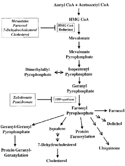
et al. 1991]. The initial antigens from MTB were shown to be small, soluble, non-peptidic, phosphorylated compounds [Pfeffer K et al.,1990; Constant P et al. 1994]. A number of T cell antigens have been identified, mainly anionic molecules that invariably contain a phosphate moiety. Moreover, Aminobisphosphonate (ABs) are synthetic compounds [Tanaka Y et al. 1994], known as potent inhibitors of osteoclast-mediated bone resorption used for the treatment of osteoporosis, bone metastasis and other metabolic bone diseases in which osteoclast number and osteoclastic bone resorption increase and bone mass decreases [Berenson JR., 2005; Kunzmann V et al. 2000; Dieli F et al. 2003; Ferlazzo V et al. 2006]. It has been shown that bisphosphonates exert a stimulatory effect on T cells in vitro and in vivo [Kunzmann V et al. 2000; Dieli F et al, 2003]. Nitrogen-containing Bisphosphonates (N-BPs) such as Pamidronate and Zoledronate, inhibit the mevalonate pathway (Fig.1) [Sato M et al. 1991] acting on farnesyl pyrophosphate syntethase (FPPS) [Bergstrom JD et al. 2000] and cause an accumulation of isopentenyl-pyrophosphate (IPP), dimetyl-allylpyrophosphate (DMAPP) and geranylgeranyl disphosphates (GGPP) that, in turn, stimulate V 9V 2 T lymphocytes [Ferlazzo V et al. 2006], which results in cytotoxicity against tumour cells [Gober HJ et al. 2003].
Figure 1: Mevalonate metabolic pathway. [Gober HJ, et al.” Human T cell receptor gammadelta cells recognize endogenous mevalonate metabolites in tumor cells” J Exp Med. 2003].
Studies on peripheral blood showed that large majority of circulating Vγ9Vδ2 T lymphocytes expresses an effector/memory phenotype. The analysis of differentiation profile can be done by evaluating the expression of CD45RA and CD27 molecules [naïve (CD45RA+CD27+), central memory (CD45RA -CD27+), effector memory (CD45RA-CD27-) and effector (CD45RA+CD27-) T cells]. Using this approach naïve (N), central memory (CM), effector memory (EM) and effector (E) T cells were described to exploit different functional activity such as clonogenic potential, cytokine release and cytotoxicity.[Dieli F et al. 2003; Angelini F et al. 2004].
CB Vγ9Vδ2 T cells are considered to be immature because they have naïve phenotypes and display poor proliferative [Montesano et al., 2001] or cytokine responses [Engelmann et al., 2006]. However, CB Vδ2 T cells proliferate robustly after treatment with non-peptide antigens, as pamidronate, considering them not inherently unresponsive [Cairo et al., 2008]. The activation of Vγ9Vδ2 T cells results in cell proliferation, soluble factor release and cytotoxicity. On encountering antigens in these peripheral sites, Vγ9Vδ2 T cells would be activated to proliferate, secrete cytokines and kill infected cells. Activated Vγ9Vδ2 T cells release inflammatory cytokines including TNF-α, IFN-γ, macrophage-colony stimulating factor, IL-17, and IL-21 and a wide set of chemokines such as MIP1-α, MIP1-β and RANTES [Garcia VE et al, 1997; Poccia F et al 2005]. Vγ9Vδ2 T cells also detect and kill infected cells through perforin and/or Fas–Fas ligand interactions and the intracellular bacteria that they contain through granulysin release [Martino A et al., 2007]. The regulation of Vγ9Vδ2 T cell activation was extensively studied. Human γδ T cells frequently express activatory and/or inhibitory Natural Killer Receptors (aNKRs and iNKRs) that can fine-tune their activation threshold [Halary F et al 1999]. NKG2D, an activating C-type lectin receptor, the ligand of MHC class I-related chain A and B (MICA/MICB) and UL16-binding protein (ULBP) molecules, seems to be a major costimulator of γδ T cells [Das H et al., 2001]. NKG2D is upregulated by inflammatory cytokines [Roberts AI et al., 2001], and NKG2D ligands are induced after a physical or genotoxic stress and/or along infection by intracellular pathogens. Therefore, NKG2D is a key stress sensor that strongly enhances recognition of altered or infected cells by human γδ T cells. It’s already known that Vγ9Vδ2 T cell activation play a wide immunological role in the orchestration of the immune response. They are able to directly inhibit viral replication both through cytolitic and non-cytolitic mechanisms and, on the other hand, Vγ9Vδ2 T cells induce the activation or differentiation of other immune cells. Specifically, they can drive Th1 polarization, dendritic cells (DCs) differentiation and B cell
activation [Poccia et al., 2002; Brandes M et al., 2003]. Extensive studies were performed on DC-Vγ9Vδ2 T cells interaction. PhAgs-activated γδ T cells induce the maturation of DCs by inducing the expression of costimulatory markers, MHC molecules and chemokine receptors for homing in the lymphoid organs, suggesting that Vγ9Vδ2 T cell activation cooperate in the induction of adaptive response. On the other hand, DCs promote γδ T cell activation resulting in the expression of high levels of CD69 and production of pro-inflammatory cytokines such as TNF-α and IFN-γ, suggesting a reciprocal interaction and a positive feedback [Conti L et al., 2005]. Most studies are focused on understanding of immunology of Vγ9Vδ2 T cell population isolated from PBMC, while still little is known about the γδ T cells isolated from human umbilical cord blood. Therefore, the characterization of fetal T cells could be important for the comprehension of their maturation, and for the development of new therapeutic strategies aimed at strengthening the immune defence in neonatal age. The immaturity of the immune system of the newborn is reflected in clinical observations showing that neonates are more highly susceptible to infection than adults and exhibit more severe or prolonged symptoms when infected [Hodge S et al., 2000]. However there is still controversy about the factors underlying this hypo-responsiveness. The dogmatic view that the neonatal immune system has intrinsically less immunological capacity per se than that of adults has recently been challenged by reports suggesting that neonatal cells are naive but nevertheless immunologically competent, that the observed differences are of a quantitative nature and that neonatal cells have the capacity to mount adult-like responses under appropriate conditions of stimulation. Because immune cells mediate many of their functions by secretion of cytokines, differences in cytokine production capacity help to characterize functional differences in neonatal and adult immune responses. [Engelmann et al., 2006]. Further studies are conducted continuously to better understand the mechanisms by which T cells recognize and respond to viruses, parasites and bacteria, and their unique role of innate immunity to infection.
Natural Killer cells
Human natural killer (NK) cells are large, granular and short-lived cells from the lymphocyte lineage [Zhang Y et al. 2007]. NK cells represent 10–15% of lymphocytes and 5% of all mononuclear cells, respectively [Robertson MJ, Ritz J. 1990]. NK cells are also present in non-lymphoid tissues, including the liver, bone marrow and the uterus during pregnancy [Manaster I. et al., 2008]. NK cells, as important sentinels of the immune system, work as
primary responders and alerting the host to the presence of infectious organisms. These cells represent a subset of cytotoxic lymphocytes that is able to recognize and lyse tumor cells and virus-infected cells without previous sensitization [Moretta A. et al., 2008]. They are able to recognize a variety of target cells [Trinchieri G., 1989] and activate cytolytic process in the absence of an initial stimulus and in a non-MHC restricted [Lanier L.L., Kipps T.J., and Phillips J.H., 1985]. They develop primarily in the bone marrow from hematopoietic progenitor cells after activation by Notch ligands [Beck RC et al. 2009] and are characterized by the expression of neural cell adhesion molecule-1 (CD56) and lack of CD3 [Lanier LL et al., 1986]. The level of CD56 expression also delineates two functionally distinct subsets of NK cells: CD56dim NK cells are the more cytotoxic population, whereas the CD56bright subset has limited cytotoxicity but secretes large amounts of cytokines and chemokines [Cooper MA et al., 2001]. Consistent with the known functional differences between the subsets, CD56bright cells express lower levels of activating receptors, including killer-cell immunoglolubin-like receptors, the natural cytotoxicity receptors and CD16, but constitutively express the inhibitory heterodimer CD94/NKG2A [Ferlazzo G. et al, 2004]. In contrast, cytokine receptors tend to be more highly expressed on the CD56bright subset, including those for interleukin (IL)-1, 2, 15 and IL-18 [Poli A. et al., 2009]. Furthermore, the high-affinity IL-2 receptor (CD25) is expressed constitutively on CD56bright NK cells, but is absent from the CD56dim subset [Caligiuri MA et al. 1990].
Unlike T and B lymphocytes, which possess receptors for a single cognate antigen, NK cells express a repertoire of germline-encoded receptors that survey an array of molecular signals of self and the steady-state condition, as well as markers of cell stress, malignancy and infection [Lanier LL, 2008]. This confers NK cells the unique ability to sense and integrate a diverse range of signals of both homeostasis and disease. Owing to their rapid cytotoxic or cytokine-producing responses in the early stage of bacterial, viral and parasitic infection, NK cells are considered predominantly an innate immune cell [Stetson DB et al., 2003; Le-Barillec et al., 2005]. In addition, a modulatory role for NK cells is emerging in which they interact with and modulate cells of both innate and adaptive immune responses [Lapaque et al., 2009; Nedvetzki S et al., 2007; Kloss M et al., 2008].
The vital role of NK cell cytotoxicity in controlling intracellular pathogens is well known, and is shown by severe or fatal viral infections in the few reported cases of selective NK cell deficiency [Eidenschenk C et al., 2006] There is also a large body of evidence showing that NK cells kill malignant cells [Zitvogel L et al., 2006]. Two mechanisms of NK cell cytotoxicity exist:
the deposition of lytic granules into target cells and the expression of death receptor ligands, such as tumour necrosis factor (TNF), TNF-related apoptosis-inducing ligand and Fas ligand [Kashii Y et al., 1999; Arase H et al., 1995]. NK cell granules contain the pore-forming protein perforin, that disrupts the target cell membrane [Motyka B et al., 2000], several serine proteases termed granzymes [Smyth MJ, Trapani JA, 1995] and granulysin [Okada S et al., 2003] by exocytosis to kill target cells, inducing caspase-dependent apoptosis by and incaspase-dependent mechanisms. Natural killer cells also contribute to the innate immune response by cytokine production. They are a major source of interferon- (IFN- ), which has many effects including anti-viral and anti-bacterial actions, promotion of Th1 responses, suppression of proliferation of infected or transformed cells, stimulation of major histocompatibility complex class I expression and DC maturation [Martin-Fontecha A et al. 2004; Schoenborn JR et al., 2007]. Another important pro-inflammatory cytokine released by NK cells is TNF- . In different situation, NK cells can also express the anti-inflammatory cytokines, transforming growth factor- (TGF- ) [Cooper MA et al., 2001] and IL-10 [Grant LR et al., 2008] and the hematopoietic factors, granulocyte-macrophage colony-stimulating factor (GMCSF) and IL-3 [Cooper MA et al., 2001] and promote T helper 2 responses by producing IL-5 and IL-13 [Loza MJ et al., 2002]. In addition to cytokines, NK cells produce several chemokines, including macrophage inflammatory protein-1 and - and RANTES [Roda JM et al., 2006]. Stimulation with different cytokines can induce specific responses by NK cells. For example, the combination of IL-12 and IL-18 induces IFN- expression by NK cells [Strengell M. et al., 2003], whereas IL-12 and IL-15 together induce IL-10. The CD56bright population of NK cells produce cytokines more vigorously than CD56dim cells after stimulation [Cooper MA et al., 2001]. Both the cytotoxic and cytokine-producing functions of NK cells were previously thought to occur without prior sensitization. However, recent work has shown that NK cell responses to bacterial and viral pathogens require priming by DCs, involving the cross-presentation of membrane-bound IL-15 in secondary lymphoid organs [Lucas M et al., 2007], IL-18 may also contribute to in vivo NK cell priming [Chaix J. et al.,2008]. It has been suggested that normal exposure to commensal microbes may be sufficient to induce a population of primed NK cells [Long EO, 2008], which is consistent with the finding that only a small proportion of isolated NK cells respond to activation signals [Bryceson YT et al., 2006]. Their activation is regulated by the balance of signals generated by receptor activator action (CD16) and inhibitory receptors (Killer Inhibitor Receptors KIR), and also by a decreased expression of molecules of Major
Histocompatibility Complex (MHC) class I on surface of target cells [Moretta A et al. 2001]. It is known that NK cells exposed to high concentrations of IL-2 differentiate into "killer cells activated by lymphokines (LAK)”. These cells possess a remarkable cytolytic activity and a broad spectrum specificity: they are indeed capable of killing tumor cells and normal, including epithelial cells. Thus, NK cells play an essential role in innate immune response but at the same time, they are considered a bridge between innate and adaptive immune system.
Role of T lymphocytes and NK cells in immune response to viruses Immunologic "immaturity" is often blamed for the increased susceptibility of newborn humans to infection, but the precise mechanisms and details of immunologic development remain somewhat obscure. Herpes simplex virus (HSV) and cytomegalovirus (CMV) are two of the more common severe infectious agents of the fetal and newborn periods. HSV infection in the newborn most commonly occurs after exposure to the virus during delivery, and can lead to a spectrum of clinical disease ranging from isolated skin-eye-mucous membrane infection to severe disseminated multiorgan disease, often including encephalitis. In contrast to HSV, clinically severe CMV infections early in life are usually acquired during the intrauterine period.
HSV infections can result in a range of clinical disease, including hearing loss and neurodevelopment delay. However, term newborns infected with CMV after delivery are generally asymptomatic, and older children and adults often acquire infection with HSV or CMV with either no or mild clinical symptoms. The reasons for these widely variable clinical presentations are not completely understood, but likely relate to developmental differences in immune responses [Muller WJ et al., 2010]. Herpes simplex virus 1 and 2 (HSV-1 and HSV-2), also known as Human herpes virus 1 and 2 (HHV-1 and -2), are two members of the herpes virus family, Herpesviridae (DNA virus), that infect humans [Ryan KJ, Ray CG, 2004]. Herpes simplex virus (HSV) causes severe infection with a high mortality in neonates. Immaturity of NK cells and a low frequency of specific T lymphocytes responding to HSV at birth are thought to be mechanisms of the severity of primary HSV infection in neonates [Masahiro Ito et al., 1998]. Serological studies have revealed that 50–80% of humans possess neutralizing activity against the virus. This neutralizing activity can be partially protective in inhibiting spread of a replicating virus. In phase I trials, although, there were no apparent differences in toxicities among treated individuals who possessed neutralizing immunity to HSV. In spite of the
phase I findings, it is known that in both immune and naive individuals, wild-type HSV-1 infection will induce initial antiviral innate defenses followed by adaptive, acquired responses. The major differences in such responses between immune and naive individuals reside in the rapidity and magnitude of the acquired response. Innate immunity possesses two primary functions. First, it directly destroys pathogenic microorganisms. Second, it determines the type and initiation of adaptive responses. Innate immune responses against wild-type HSV1 include:
1. activation of the complement cascade
2. activation of macrophages, recruitment, activation, and maturation of NK cells, CD4+ CD11c- type 2 DC precursors, and extrathymic-derived T cells
3. generation and secretion of a variety of cytokines and chemokines that orchestrate the above activities.
The objective of these responses is to limit the initial infection of a tissue, abrogate further propagation of the virus, and begin maturation of dendritic and/or dendritic-like cells that will then activate antigen specific T cells as well the maturation of B cells to begin production of affinity, high-avidity IgG [Wakimoto H et al., 2003].
The role of innate immunity, and in particular and NK cells, in controlling Herpes infections, has also recently received greater attention [Carter C. et al., 2007].
The involvement of T cells, isolated from PB, in natural immunity against infections caused by DNA viruses is well established. In particular, many studies describe a protective role of T lymphocytes in the immunosurveillance against herpes viruses in both rodents and people. HSV-specific T cells can be isolated from infected persons and are able to express HSV-specific cytotoxic activity. To mediate the lytic activity, these virus-specific CTLs require the expression of HLA class I molecules on the surface of target cells. However, the response itself appears to be HLA-unrestricted, suggesting the possible involvement of NK receptors expressed by human T cells [Poccia F et al., 2005]. Specifically, PBMC from HSV-seropositive individuals stimulated with autologous HSV-infected PHA blasts show an expansion of V 9V 2 T cells, and are able to lyse HSV-infected, but not mock-infected targets. Also,V 9V 2 T cells obtained after PHA or mycobacterial stimulation are able to lyse HSV infected as well as unrelated vaccinia-infected targets, but not mock-infected targets. Similarly to the mouse models, human T cell-mediated cytotoxic activity is not restricted by classical HLA class I or class II molecules, and can be blocked by mAbs to CD3 and the TCR. Interestingly, T cells have been shown
to be susceptible to human herpes virus (HHV)-6 infection, and display cytolytic activities against both autologous and heterologous target cells infected with HHV-6. HHV-6 infection induces CD4 expression in T lymphocytes, rendering them susceptible to HIV. Thus, HHV-6 has evolved strategies to interfere with T cell antiviral activities, exploiting the activation of these cells to expand the pool of target cells susceptible to productive infection [Poccia F et al., 2005]. Clearly, T cells play an important role in innate and adaptive immune responses to viral infections. The molecules recognized by T cells during viral infections are probably of cellular rather than viral origin and appear to be metabolites of altered cellular pathways, in particular the products of the mevalonate pathway. Moreover, virus-exposed T cells can be rapidly activated by type I interferons (IFN- , IFN- ), a phenomenon that is likely to contribute to the effective antiviral response [Kunzmann V et al., 2004]. The antiviral role of T cells has been intensively studied in mice and correlated with the production of IFN- by distinct T-cell subsets. In H. sapiens, V 1 T cells are systemically or locally expanded in some chronic viral infections and are probably involved in the accompanying inflammatory processes. V 9V 2 T cells are activated early during the acute phase of most viral infections and can display potent antiviral responses. Moreover, a plethora of soluble factors with antiviral characteristics induced by V 9V 2-stimulatory molecules can influence the outcome of viral infections. In addition to their direct antiviral properties, many of these molecules play crucial immunoregulatory roles and are decisive in controlling the complex antiviral immunosurveillance function as well as in establishing the correct immunological memory environment in vivo [Poccia F et al., 2005]. Another important cell population in the first line of defense against tumors and a wide range of pathogens is represented by the NK cells [Melissa B Lodoen and Lewis L Lanier, 2006].
Natural killer (NK) cells have the ability of lysing targets that have down-regulated the expression of human leukocyte antigen (HLA) class I molecules. Although this recognition mechanism is highly conserved and reproducible, NK cells have been described as a heterogeneous population. Herpes simplex virus (HSV) infection results in a profound reduction of HLA class I molecules on the surface of infected cells. For this reason, NK cell population kills efficiently HSV-infected cells [Pietra G et al., 2000]. It is now recognised that NK cells use a complex array of inhibitory and activating receptors (respectively KIR and KAR) to bind to targets [ Moretta L and Moretta A, EMBO J. 2004]. Decreased HLA class I expression leads to reduced binding to KIR ultimately leading to the disengagement of inhibitory signals and potentially leading to NK cell activation [Moretta L
and Moretta A, 2004]. This indicates that the NK receptors responsible for the induction of cytotoxicity recognize ligands on target cells different from HLA class I molecules. On this basis, the process of NK-cell triggering can be considered as a mainly non-MHC-restricted mechanism [Biassoni R et al., 2001]. In addition, NK cells can also detect the expression cell surface proteins which are only present in times of cellular stress or viral infections. The recent identification of a group of NK-specific triggering surface molecules has allowed a first series of pioneering studies on the functional/molecular characteristics of such receptors. The first four members of a receptor family that has been termed natural cytotoxicity receptors (NCR) are represented by NKp50 [Pietra G et al., 2000], NKp46, NKp44 and NKp30. These receptors are strictly confined to NK cells, and their engagement induces a strong activation of NK-mediated cytolysis [Moretta A et al., 2001; Biassoni R et al., 2001]. Another triggering NK receptor is NKG2D. It appears to play either a complementary or a synergistic role with NCRs. Thus, the triggering of NK cells in the process of infected or tumor cell lysis may often depend on the concerted action of NCR and NKG2D [Moretta A et al., 2001]. Herpes viruses have evolved a number of mechanisms to avoid detection by NK cells [Carter C et al., 2007]. NK cells can respond to infection either directly or indirectly. They respond directly by recognizing virus infected cells, and indirectly by interacting with dendritic cells (DCs), which express Toll-like receptors (TLRs) and secrete cytokines in response to encounter with microbes. NK cells use two main effector mechanisms to control viral infection: the secretion of IFN- and direct lysis of infected cells by exocytosis of granules that contain perforin and granzymes [Melissa B Lodoen and Lewis L Lanier, 2006].
It is clear that NK cells play a central role in the control of infections caused by Herpes viruses in human, but the effector mechanism, at present, seems to be the result of the integration of KIR+ and KAR+ cell activities [Pietra G. et al., 2000]. Thus, the presence of activating receptor on NK cells seems to constitute an important tool acting in close co-operation with other defense mechanisms against viral infections. Future studies will elucidate whether this finding has any clinical relevance. This remain a difficult but important area to study as potential immune defect in patients with susceptibility to a single pathogen are usually subtle but, as in case of HSV-1, can cause a significant morbidity.
Aim of the work
The cord blood is characterized by an immune system, phenotypically and functionally immature, unlike that of peripheral blood. The cord blood mononuclear cells (CBMC) have defective effector functions. The reduced ability to produce cytokines renders CBMC unable to generate significant cytotoxic responses, both specific for alloantigens through the action of CD8+ T lymphocytes, or nonspecific, through the action of NK cells and T lymphocytes. T cells represents less than 1% of cord blood T lymphocytes. Since the V 2 T cells of peripheral blood are able to respond early to polyclonal stimulation and modify their naïve phenotype towards a effector/ cytotoxic subset after ABs stimulation, the purpose of our work is to analyze the phenotypic maturity and function of CB-derived V 2 T lymphocytes after stimulation with phosphoantigens, evaluating:
Cell expansion in response to IPP, ZOL or PAM Phenotypic Differentiation
Expression of activation markers
Production of immunoregulatory cytokine (TNF and IFN ) Cytotoxic activity toward target tumor
The first results show the ability of T cells to respond effectively to PAM or ZOL stimulation, and their subsequent differentiation into effector population able to produce cytokines. Their ability to produce TNF and IFN has focused our attention to a greater understanding of the role of T cells in the immune response to HSV-1, considering that no information has been reported on CB cell immune response against infection with HSV-1. Our aim is to evaluate the CB-derived T lymphocytes activity in co-culture with HSV-1 infected cells, evaluating:
Cell expansion Phenotypic Analysis
Production of immunoregulatory cytokine Assessment of viral titer
Results
Chapter I: Characterization of
T cells isolated from
umbilical cord blood after stimulation with ABs
Expansion of human cord blood V 9V 2 T cells after stimulation
with phosphoantigens.
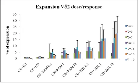
To investigate the effect of different phosphoantigens on V 9V 2 T lymphocytes isolated from human cord blood (CB), CB cells were stimulated with isopentenyl-pyrophosphate (IPP) (3 g/ml) and different concentrations (0.1, 1 and 10 g/ml) of pamidronate (PAM) and zoledronate (ZOL). IL-2 was added in all experimental conditions. The analysis of the expansion of V 9V 2 T lymphocytes in response to those stimuli was performed by flow cytometry, and the percentage of V 2+ T cells measured immediately after isolation (t = 0) and after 3 (t = 1), 7 (t = 2), 10 (t = 3), 13 (t = 4), 16 (t = 5) and 19 (t = 6) days of culture and stimulation (Figure 2). The cytofluorimetric analysis revealed that phosphoantigens, such as PAM and ZOL, increased the percentage of CB T cells by 60-fold over control at 19 days after treatment and a marked expansion of CB T cells was already detectable after 7 days. ZOL triggered significantly greater expansion of T cells than PAM (P <0.05) from 10 days after treatment. In contrast to adult
lymphocytes, CB cells were unresponsive to IPP treatment. These data strongly suggest that phosphoantigens, especially aminobisphosphonates (ABs), induce remarkable expansion of CB-derived V 9V 2 T lymphocytes.
Figure 2. Expansion of CB T cells after non-peptide antigens stimulation. cells derived from CB were stimulated with 3 g/ml IPP or 0.1, 1, 10 g/ml PAM or 0.1, 1, 10 g/ml ZOL in the presence of 30UI/ml recombinant IL-2. Results are shown as mean + SD of 5 experiments from different donors.
Expression of CD25 and CD69 surface antigens on expanded
T
cells
To evaluate the level of activation reached by V 9V 2 T cells expanded with ABs, we investigated the surface expression of CD25 and CD69, two cellular activation markers. The experimental conditions were the same used for cellular expansion. The expression of these receptors was evaluated by flow cytometry analysis. We focused our attention on the treatment with PAM and ZOL at the concentration of 1 g/ml due to the stronger response given by such conditions in our previous experiments. As shown in Fig. 3, the stimulation with IL2 or IL-2+ABs induces a significant increase in the CD25 expression, compared with non-stimulated cells (control). In particular, the receptor expression increase is evident in the sample treated with IL-2+PAM, from 5% to 15% (p <0.05). In cells treated with PAM was observed a lower increase, rising from 5% to 7% (p <0.05). Since the CD25 represents the chain of IL-2 receptor, this difference could be explained by the interaction between IL-2 and CD25 which, as for most of receptor systems, could be
transported into the cell after triggering. In cells stimulated with IL-2 + ZOL and those treated only with ZOL is not found the same dose-dependent response observed with PAM. You can still see a higher percentage of CD25 expression in cells treated with ZOL alone, probably for the same reason, the interaction between IL-2 and CD25 above. The figure 3 also shows that IL-2 treatment induces an increase in the expression of CD69 in CB-derived T cells that is around 32% (p <0.05). Cells stimulated with 2+PAM or IL-2+ZOL show respectively a 40% and 23% increase of CD69 expression. The stimulation with PAM or ZOL without IL-2 shows percentages of 23% and 46% respectively. These data demonstrate a significant expression of CD25 and CD69 48h following ABs stimulation, confirming a strong activation of T cells isolated from CB. In various treatments, the different expression of CD25 and CD69 could be explained by the decrease of cell surface expression of CD25 caused by its internalization after binding with IL-2.
Figure 3. Expression of CD25 and CD69 on ABs expanded T cells. cells derived from CB were stimulated with 1 g/ml PAM or 1 g/ml ZOL in the presence of 30UI/ml recombinant IL-2. Results are shown as mean + SD of 5 experiments from different donors.
Phenotypical characterization of CB-derived
T cells expanded
with ABs.
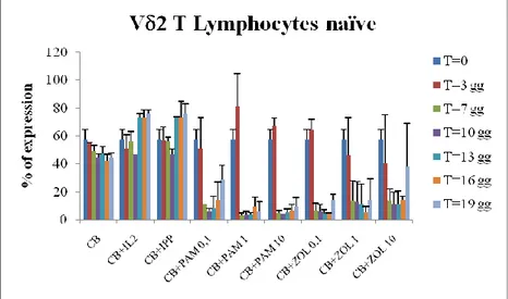
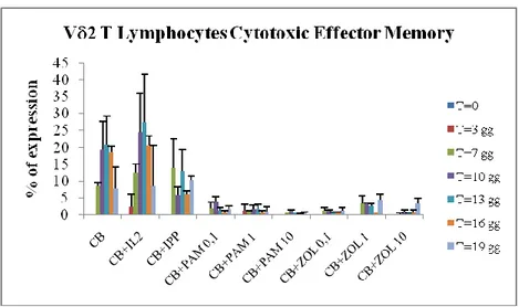
By means of the polychromatic flow cytometry analysis of the expression of different surface markers such as CD27, CD62L, CD45RA and CD16, it has been possible to analyse the differentiation of CB- T lymphocytes expanded with IL-2+ABs. Cell culture conditions for these experiments were the same used for cellular expansion. We found that CB- T cells differentiate from immature cells (naïve, N) that express CD62L+CD27+CD45RA+CD16- phenotype (Fig 4a) to effector cells with regulatory phenotype CD62L-CD27-CD45RA-CD16- (effector memory, EM) (Fig 4b) rather than cytotoxic phenotype CD62L-CD27-CD45RA+CD16+ (T cells Cytotoxic Effector Memory, TEMRA) (Fig 4c). In particular, fig. 4a shows that the majority of V 2+ T cells analyzed immediately after isolation from CB express the phenotype "Naive" (63%). Then, we observed that the lymphocytes stimulated with IL-2+ABs reduce the percentage of cells with N phenotype to 10% in all stimulation conditions (p <0.05). In contrast stimulation with IL-2 alone or with IL-2+IPP does not significantly alter the percentage of effector cells. The decrease in T cells with Naïve phenotype is coupled by a high increase in the percentage of effector cells. In particular, as shown in Figure 4b, the ABs stimulation induces a progressive differentiation of T lymphocytes towards a population of "effector memory", characterized by the functional capacity to produce cytokines (TNF , IFN ). These cells were few in CB after isolation (Fig. 4b) but after 3 days of stimulation with PAM or ZOL they reached about 50%, further increasing at the subsequent time points reaching 80-90% of the V 2
+ population (p <0.05). On the contrary, as shows Fig. 4c, stimulation with PAM or ZOL does not cause the V 2+ T cells to differentiate towards a cytotoxic phenotype, "EMRA". These cells, in fact, are very few in CB at T0, and maintain very low levels (1-10%) during the entire kinetic and after stimulation with different doses of PAM or ZOL (p <0, 05). These results allow us to define T cells isolated from CB as mature and functional immune cells able to undergo activation after ABs stimulation, suggesting an important role in the regulatory immune response after stimulation with phosphoantigens, which could be important in directing the immune response in the newborn.
Figure 4a. Percentage of V 2+ CD62L+/CD27+/CD45RA+/CD16- T lymphocytes
(TN). Results are shown as mean + SD of 5 experiments performed upon CB from
different donors.
Figure 4b. Percentage of V 2+ CD62L-/CD27-/CD45RA-/CD16- T lymphocytes
Figure 4c. Percentage of V 2+ CD62L-/CD27-/CD45RA+/CD16+ T lymphocytes
(Temra). Results are shown as mean + SD of 5 experiments from different donors.
Functional characterization of expanded
T cells from CB
As shown in fig. 4a, b and c, the activation of stimulated T cells is paralleled by their differentiation towards a clearly regulatory rather than cytotoxic phenotype. To confirm the propensity of those cells to exert a regulatory function, we aimed at analysing the production of cytokines, such as IFN and TNF (data contained in the CD, Fig. B). Interestingly, stimulation with IL-2 alone, as with PAM or ZOL in the absence of IL-2, induces a high production of both cytokines. To complete the study of the functionality of these cells, we analyzed the ability of CBMC stimulated and expanded with PAM or ZOL to develop cytotoxic function, evaluating their phenotype differentiation and the production of cytotoxic mediators, such as perforin and granzymes by flow cytometry (data contained in CD, Fig. E and Fig. F). The positivity for the production of granzymes is significantly higher in V 2- T lymphocytes compared to T cells. Nevertheless, it is noteworthy that even among V 2+ T lymphocytes there is positivity to the granzymes, which is about 70% of the total V 2+
T cells, in samples stimulated with IL-2 or IL-2+ABs (data contained in CD, Fig. D). On the other hand, the percentage of T cells able to produce perforin undergoes a significant increase between cells V 2
observe the production of this mediator in T cells (data contained in the CD, Fig. C). The obtained results suggest that CB-derived T cells, after ABs stimulation, develop a cytotoxic immature phenotype, characterized by the production of granzymes but not perforin, confirming their differentiation towards a regulatory phenotype.
Analysis of cytotoxic V
2T lymphocytes induced by treatment
with IL2 + ABs
We previously demonstrated that V 2
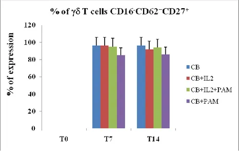
T cells are able to produce mediators of cytotoxicity after stimulation with IL-2 and ABs, therefore we analyzed their phenotypic differentiation. In particular, through the polychromatic flow cytometry, we evaluated the expression of different surface markers such as CD27, CD62L, and CD16 (a marker of cytotoxicity). Also in these experiments we maintained our cell culture conditions, stimulating cells with IL2+ABs for 14 days. As shown in Fig. 5, the stimulation with ABs does not induce the T lymphocytes to differentiate towards a cytotoxic phenotype. In fact, the percentage of expression of the CD16- phenotype is about 90%, indipendently from treatment conditions.
Figure 5. Percentage of V 2+ CD62+/CD27+/CD16- T lymphocytes. Results are
In contrast we observed a drastic reduction of non-effector phenotype in the population V 2- T lymphocytes treated with IL-2 or IL-2+PAM, wherease treatment with only PAM did not cause any effect (Fig. 6)
.
These results suggest their differentiation towards a cytotoxic subset, and in particular an important role of IL-2 in cell-mediated cytotoxicity.Figure 6. Percentage of V 2- CD62+/CD27+/CD16- T lymphocytes. Results are
shown as mean + SD of 5 experiments from different donors.
Phenotypic and functional analysis of expanded cells isolated from
CB after co-culture with K562, a tumor cell line
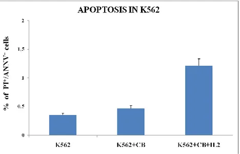
To complete the study of the functionality of these cells, we analyzed their ability to develop an effective cytotoxic function, monitoring the capacity of CBMC expanded and stimulated with IL-2 to lyse target tumor cells. To characterize this cell-mediated cytotoxicity, we performed a cytotoxicity assay in which effector cells (CBMC pre-stimulated with IL-2) were incubated with target cells (K562, human erythromyeloblastoid leukemia cell line). In particular, CBMC were subjected to stimulation with IL-2 for 24 hours. After this period of culture in each well we added K562 and incubated for 6 hours. The amount of tumor cells to be added was determined according
to the average number of CBMC obtained after expansion in order to have an effector:target ratio of 1:1. At 6h after co-culture we evaluated the apoptosis of K562 and the cytotoxic mediators production in effector cells by flow cytometry. As shown in Fig. 7, the percentage of tumor cell death after co-culture with CB cells stimulated with IL-2 increases significantly, by about 70%.
Figure 7. Percentage of apoptotic cells. Results are shown as mean + SD of 5 experiments from different donors.
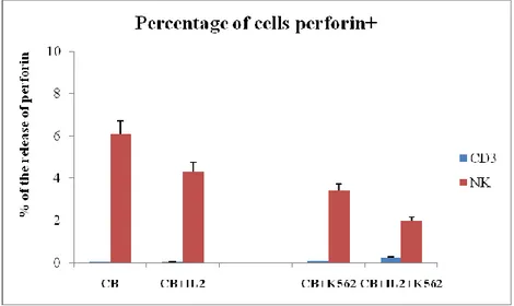
Having observed that T cells isolated from CB, after stimulation with ABs, develop a cytotoxic immature phenotype, characterized by the production of granzymes but not perforin, and that the stimulation with IL2 only induces high cytotoxicity in CBMC, we hypothesized the involvement of other cells capable of performing IL-2 mediated cytotoxic activity. To understand which cell populations could be involved in this mechanism of cytotoxicity, we analyzed by flow cytometry the production of perforin and granzymes in different CBMC subpopulations. In particular, we labeled cells with monoclonal antibodies (mAbs) directed against CD3 and CD56 to study the
functional activity of lymphocytes and NK cells respectively, treated with IL-2, after 24h of co-culture with K562.
Figure 8. Percentage of perforin released by CBMC after IL-2 treatment. Results are shown as mean + SD of 5 experiments from different donors.
To this end, we expanded CBMC through stimulation with IL-2. The percentage of lymphocytes and NK cells producing mediators of cytotoxicity was analyzed by flow cytometry intracellular staining. As shown in Fig. 8, IL-2 treatment induces only NK cells to differentiate towards a cytotoxic phenotype. In fact, co-culture induces in the IL-2 unstimulated and stimulated samples increased perforin release by about 45% and 45%, respectively, demonstrating their functionality. In contrast, there was no activation in lymphocytes, which are unable to produce and release perforin.
Figure 9. Percentage of granzymes released by CBMC after IL-2 treatment. Results are shown as mean + SD of 5 experiments from different donors.
Similarly (Fig. 9), the production and subsequent release of granzymes is attributable only to NK cells, where the co-culture induces in the IL-2 unstimulated and stimulated samples increased granzymes release by about 42% and 50%, respectively. No effect was observed in lymphocytes.
These results suggest that T lymphocytes isolated from cord blood, after stimulation with IL-2, are not able to develop a cytotoxic phenotype, as confirmed by the negativity for the production and release of perforin and granzymes. But they are able to produce immunomodulatory cytokines after stimulation with ABs, confirming their differentiation towards a regulatory phenotype. The data obtained allow us to attribute a cytotoxic effector activity to IL-2-dependent NK cells. We demonstrated that IL-2+ABs stimulation induces phenotypic differentiation in CBMC, in particular, T lymphocytes acquire a regulatory phenotype and NK cells acquire a cytotoxic phenotype. Considering that T lymphocytes and NK cells are two cell subsets mainly involved in innate immune response against several pathogens, such as bacteria and viruses, we evaluate their activity in co-culture with Herpes Simplex Virus- 1 (HSV-1) infected cells.