ORIGINAL RESEARCH
ADULT BRAIN
CT Angiography ASPECTS Predicts Outcome Much Better Than
Noncontrast CT in Patients with Stroke Treated Endovascularly
XF. Sallustio,XC. Motta,XS. Pizzuto,XM. Diomedi,XB. Rizzato,XM. Panella,XF. Alemseged,XM. Stefanini,X S. Fabiano, XR. Gandini,XR. Floris,XP. Stanzione, andX G. KochABSTRACT
BACKGROUND AND PURPOSE: Noncontrast CT ASPECTS has been investigated as a predictor of outcome in patients with acute
ischemic stroke. Our purpose was to investigate whether CTA source images are a better predictor of clinical and radiologic outcomes than NCCT ASPECTS in candidates for endovascular stroke therapy.
MATERIALS AND METHODS: CT scans of patients (n⫽ 124) were independently evaluated by 2 readers for baseline NCCT and CTA source
image ASPECTS and for follow-up ASPECTS. An mRS ofⱕ2 at 3 months was considered a favorable outcome. Receiver operating characteristic curve analysis was used to assess the ability of NCCT and CTA source image ASPECTS to identify patients with favorable outcomes. A stepwise multiple regression analysis was performed to find independent predictors of outcome.
RESULTS: Baseline CTA source image ASPECTS correlated better than NCCT ASPECTS with follow-up ASPECTS (r⫽ 0.76 versus r ⫽ 0.51; P for comparison of the 2 coefficients⬍ .001). Receiver operating characteristic curve analysis showed that baseline CTA source image
ASPECTS compared with NCCT ASPECTS can better identify patients with favorable outcome (CTA source image area under the curve⫽ 0.83; 95% CI, 0.76 – 0.91; NCCT area under the curve⫽ 0.67; 95% CI, 0.58–0.77; P ⬍ .001). Finally, the stepwise regression analysis showed that lower age, good recanalization, lower time to recanalization, and good baseline CTA source image ASPECTS, not NCCT ASPECTS, were independent predictors of favorable outcome.
CONCLUSIONS: CTA source image ASPECTS predicts outcome better than NCCT ASPECTS; this finding suggests CTA rather than NCCT
as a main step in the decision-making process for patients with acute ischemic stroke.
ABBREVIATIONS:CTA-SI⫽ CTA source images; ET ⫽ endovascular stroke therapy
T
he Alberta Stroke Program Early CT Score merges the abilityof quantifying and describing the topography of brain tissue damage produced by acute ischemic stroke in a semiquantitative way.1ASPECTS on noncontrast CT is widely used for the assess-ment of early ischemic changes, and its prognostic value has al-ready been established,2though with poor NCCT sensitivity.3
Re-cent randomized controlled trials on endovascular stroke therapy (ET) have been based on strict inclusion criteria, leading to treat-ment of only those patients with high CT ASPECTS indicating smaller infarct burden.4-6
Many attempts have been made to understand which patients are likely to undergo futile reperfusion.7For instance, it has been recently demonstrated that patients with poor collaterals and lon-ger time to reperfusion do not achieve good outcomes after ET.8 Thus, a careful patient selection for ET should be desirable and should be based on a multimodal neuroimaging approach in ad-dition to onset time and stroke severity. Although not as com-monly available as NCCT in the acute ischemic stroke setting, CT angiography is useful for confirmation of vessel occlusion in can-didates for ET, and hypodensity on CTA source images (CTA-SI) has been shown to reliably correlate with ischemic lesion volume on diffusion-weighted imaging9and final infarct size.10The su-periority of CTA-SI on NCCT in the detection of infarcted areas has been demonstrated for readers of all levels of experience.11 Few data exist on the value of CTA-SI ASPECTS in patients
un-Received October 14, 2016; accepted after revision March 24, 2017.
From the Department of Neuroscience (F.S., C.M., S.P., M.D., B.R., M.P., F.A., P.S., G.K.), Comprehensive Stroke Center, and Interventional Radiology and Neuroradi-ology (M.S., S.F., R.G., R.F.), Department of Diagnostic Imaging, University Hospital of Tor Vergata, Rome, Italy; Santa Lucia Foundation (C.M., G.K.), Rome, Italy; and Department of Medicine and Neurology (F.A.), Royal Melbourne Hospital, Univer-sity of Melbourne, Parkville, Australia.
This work was supported by a grant from the Italian Ministry of Health (RF-2013-02358679).
Ethics approval for this study was obtained from the Tor Vergata Policlinic ethics committee.
Please address correspondence to Fabrizio Sallustio, MD, University Hospital of Tor Vergata, Viale Oxford 81, 00133, Rome, Italy; e-mail: [email protected]
Indicates open access to non-subscribers at www.ajnr.org
dergoing ET for acute ischemic stroke,12,13and this lack of data may explain why only ASPECTS NCCT is currently considered in the guidelines for eligibility for ET. Our purpose was to investigate whether CTA-SI ASPECTS correlate better than NCCT ASPECTS with clinical and radiologic outcome measures in patients with acute ischemic stroke undergoing ET.
MATERIALS AND METHODS
A retrospective analysis of patients identified from a prospective registry at a comprehensive stroke center (University Hospital of Tor Vergata, Rome, Italy) was performed. Patients with anterior circulation acute ischemic stroke secondary to intracranial prox-imal arterial occlusion (M1 MCA, M2 MCA, distal internal ca-rotid artery, and proximal ICA plus intracranial proximal arterial occlusion) admitted within 6 hours of symptom onset were in-cluded. Due to the study period (between 2009 and 2015) before the publication of recent endovascular stroke trials,4-6no exclu-sion criteria other than the time from symptom onset were ad-opted. Patients presenting within 4.5 hours of symptom onset were treated with intravenous thrombolysis, which was continued in the angiographic suite during the endovascular procedure. Pa-tients presenting beyond the time window for intravenous throm-bolysis or with major contraindications to intravenous thrombol-ysis (ie, warfarin therapy with an international normalized ratio of 1.7, recent major surgery, or a history of hemorrhage/hematoma) underwent stand-alone thrombectomy. Demographics, vascular risk factors, and baseline and 24-hour NIHSS scores were re-ported. The modified Rankin Scale was adopted for outcome analysis, and an mRS ofⱕ2 at 3 months was considered a favor-able outcome. The study was approved by the Tor Vergata Poli-clinic Ethical Committee, and informed consent was obtained from all patients or their relatives.
Image Acquisition
The NCCT and CTA were acquired with a standardized protocol. Axial CT was performed on a multisection scanner (Light Speed VCT; GE Healthcare, Milwaukee, Wisconsin) by using 120 kV and 170 mAs with a 5-mm section thickness. Continuous axial sections parallel to the orbitomeatal line were obtained from the skull base to the vertex. CTA was performed with a 64 – detector row scanner. Acquisitions were obtained after single-bolus intra-venous contrast injection of 90 –120 mL of nonionic contrast me-dia into an antecubital vein at 3–5 mL/s. Imaging was autotrig-gered by the appearance of contrast media in the ascending aorta. Standard coverage included the area from the arch to the vertex. Source images were reconstructed at a 1.25-mm thickness in the axial planes at half-thickness intervals. NCCT or DWI was per-formed between 1 and 7 days after stroke onset and used for fol-low-up ASPECTS.
Image Processing
NCCT, CTA-SI, and MR imaging (DWI sequences and apparent diffusion coefficient map) scans were independently screened for ASPECTS by 1 neuroradiologist (M.S.) and 1 stroke neurologist (G.K.) who were blinded to the patients’ symptoms but aware of acute nonlacunar stroke. The readers performed their evaluation at different time periods to make their assessment blinded as
much as possible. Adequate window and optimal level settings were adopted to maximize the contrast produced by attenua-tion differences between normal and ischemic tissue. Our ASPECTS reading includes evaluation of all axial sections (www.aspectsinstroke.com), and as in previous studies, we ex-cluded isolated cortical swelling from the score.14In case of a discrepancy between readers, a third neuroradiologist (R.G.) was involved to achieve a consensus. The interrater reliability was 0.71 for NCCT and 0.75 for CTA-SI, indicating a good interrater agreement for both methods.
Statistical Analysis
The analysis was performed by using STATA/IC, Version 13 (StataCorp, College Station, Texas) and GraphPad Prism soft-ware, Version 6.00 (GraphPad Softsoft-ware, San Diego, California). Continuous variables are summarized as mean⫾ SD or median with interquartile range. Categoric variables are expressed as per-centages. To determine differences between the 2 groups, we used a Student t test or Mann-Whitney U test for continuous variables. Interrater agreement was estimated with the statistic. Compar-ison of frequencies among ASPECTS groups was performed with the Fisher exact test after dichotomization into poor and good ASPECTS. Univariate associations between baseline and fol-low-up ASPECTS were investigated with the Spearman anal-ysis, with confidence limits calculated by means of the Fisher z-transformation. Bubble plots were used to graphically dis-play correlation analyses, and the area of the bubble has to be read as proportional to the number of observations at each point. A nonparametric receiver operating characteristic curve analysis and the area under the curve were used to assess the ability of NCCT and CTA-SI ASPECTS to identify patients with favorable outcomes (mRSⱕ 2).
We then calculated the statistical significance of the differ-ence between the area under the curves using the method of DeLong.15For both NCCT and CTA-SI ASPECTS, a receiver
operating characteristic curve was used to identify the best cutoff point with which to maximize the sensitivity and speci-ficity for discriminating patients with favorable outcomes. A backward and forward stepwise logistic regression analysis was finally performed to determine the independent predictors of good outcome (mRSⱕ 2), including NCCT and CTA-SI ASPECTS as well as all other variables with a significant asso-ciation in univariate analysis, to weigh for potentially con-founding factors. Odds ratios with standard errors and 95% confidence intervals were provided. A P value⬍ .05 was con-sidered statistically significant.
RESULTS
Of 167 patients with anterior circulation stroke, 124 had complete CT, CTA, and clinical data and were included in the analysis. Baseline characteristics are summarized inTable 1. Single-artery occlusion (ICA, anterior cerebral artery, and middle cerebral ar-tery) was diagnosed in 55.6%, whereas tandem lesions occurred in 44.3% of patients. Correlation analysis showed that baseline CTA-SI ASPECTS correlated better with follow-up ASPECTS (r⫽ 0.76; 95% CI, 0.67–0.83; P ⬍ .001) than baseline NCCT ASPECTS (r⫽ 0.51; 95% CI, 0.36–63; P ⬍ .001; P for comparison
of the 2 coefficients⬍ .001) (Fig 1). Furthermore, the ability to identify patients with good outcome (mRSⱕ 2), revealed by re-ceiver operating characteristic curve analysis, was significantly higher for CTA-SI ASPECTS with respect to NCCT ASPECTS (CTA-SI area under the curve, 0.83; 95% CI, 0.76 – 0.91; NCCT area under the curve, 0.67; 95% CI, 0.58 – 0.77; P⬍ .001). Accord-ing to each receiver operatAccord-ing characteristic curve, we determined cutoff values (Table 2), on the basis of which we defined a good CTA-SI ASPECTS asⱖ5 and a good NCCT ASPECTS as ⱖ8. Most interesting, a median baseline NCCT ASPECTS of 9 resulted from a recent meta-analysis of 5 endovascular stroke trials.16No statistical difference was found in onset-to-imaging acquisition time between good and poor ASPECTS groups (good CTA-SI, 135⫾ 56 minutes versus poor CTA-SI, 137 ⫾ 52 minutes; P ⬎ .05; good NCCT, 129⫾ 54 minutes versus poor NCCT, 131 ⫾ 54 minutes; P⬎ .05).
Factors predicting favorable outcome (mRSⱕ 2, n ⫽ 48) in univariate analysis were both good NCCT ASPECTS and good CTA-SI ASPECTS, as well as age, baseline NIHSS, time to recan-alization, and good recanalization (TICIⱖ 2b) (Table 3). To find the best outcome predictors, we finally constructed backward and forward stepwise regression analyses, including all variables sig-nificantly associated with favorable outcome in the univariate
analysis. Both the backward and forward procedures showed that good CTA-SI ASPECTS, age, good recanalization, and time to recanalization remained independent predictors of good clinical outcome, indicating CTA-SI ASPECTS as a better predictor of functional outcome than NCCT ASPECTS (Table 4).
DISCUSSION
ET seems a safe and effective adjuvant treatment strategy for pa-tients with acute ischemic stroke secondary to large intracranial vessel occlusion in the anterior circulation,4,17and many attempts have been made in the recent past to better select those patients who can reliably benefit from ET.7One such effort has been in recent randomized endovascular stroke trials that included only patients with small infarct size as defined by an ASPECTS of ⬎6–7 on NCCT.5,6Among these trials, the Multicenter Random-ized Clinical trial of Endovascular treatment for Acute ischemic stroke in the Netherlands (MR CLEAN) was the only one to in-clude patients on the basis of time from symptom onset and con-firmation of occlusion on neuroimaging.4Nevertheless, a recent meta-analysis of these trials showed a median baseline CT AS-PECTS of 9. Consequently, from all these trials, we have learned that ET works, but we do not know yet whether this kind of rep-erfusion therapy would be safe and effective in patients with lower ASPECTS.
Regarding these findings, a recent subgroup analysis from the MR CLEAN data showed that patients with NCCT ASPECTS of 5–7, not only those with NCCT ASPECTS of 8 –10, may also benefit from ET. This study could not provide further infor-mation on patients with NCCT ASPECTS of 0 – 4 because of a paucity of data.18Therefore, there are not thorough data con-cerning the effects of ET in patients with larger infarct burdens, and we only know that those with lower ASPECTS could be unsuitable for ET19but cannot be excluded from treatment. A recent study of 249 patients showed a rate of good outcome of 5% in the CT ASPECTS group of 0 – 4 and of 38.5% in the CT ASPECTS group of 5– 8,7suggesting a chance for ET in patients
FIG 1. Correlation of ASPECTS on NCCT (A) and CTA-SI (B) with follow-up ASPECTS. The area of the bubble is proportional to the number of
patient observations at that data point.
Table 1: Patient characteristics
Variable Value Age (yr) 68⫾ 13.2 Sex (male) 52.4% Hypertension 79.8% Diabetes 16.1% Atrial fibrillation 32.2% Smoking 19.3% Intravenous thrombolysis 60.4% Baseline NIHSS 19 (IQR, 16–21) NCCT ASPECTS 8 (IQR, 7–10) CTA-SI ASPECTS 4 (IQR, 2–6) Follow-up ASPECTS 3 (IQR, 1–5)
Note:—IQR indicates interquartile range.
Table 2: ROC curve analysis—CTA-SI ASPECTS and NCCT ASPECTS cutoff values indicating patients with good clinical outcomes
AUC (95% CI) Best Cutoff Value Sensitivity Specificity Correct Classification
CTA-SI ASPECTS 0.83 (0.76–0.91) 5 60.42% 88.16% 77.42% NCCT ASPECTS 0.67 (0.58–0.77) 8 64.58% 61.84% 62.90%
with larger infarct burden. Further scientific effort is war-ranted to identify any markers that could better predict out-come and give useful information for the decision-making process.
Thus, CTA can depict the area of ischemia (though not neces-sarily infarcted) much better than NCCT, especially when analyz-ing parenchymal CTA-SI.12 Hypodensity on CTA-SI provides greater demarcation between normal and abnormal tissue, and this finding could be explained by the ability of CTA to detect alterations in cerebral blood volume, as opposed to cytotoxic edema on NCCT, with a threshold insufficient to produce NCCT changes.11In a small previous study, CTA-SI was shown to be more sensitive in the early detection of irreversible ischemia and more accurate in the prediction of final infarct size.13Our results confirm those from Bhatia et al,20suggesting CTA-SI ASPECTS prior to ET as a better predictor of final infarction. In our study, we also compared CTA-SI ASPECTS and NCCT ASPECTS to evaluate whether the former can improve prediction of clinical outcome. We used receiver operating characteristic curve analysis and found that baseline CTA-SI ASPECTS compared with NCCT ASPECTS can better identify patients with functional in-dependence at 3 months.
Moreover, our study, different from the study of Bhatia et al,20 also analyzed reperfusion data. In particular, we found that good reperfusion was a predictor of good outcome in univariate anal-ysis and remains a significant and independent predictor of good outcome in a multivariable regression model together with age, time to reperfusion, and CTA-SI ASPECTS, but not NCCT ASPECTS. In line with this finding, good reperfusion has been recently demonstrated to improve the rate of good outcome irre-spective of ASPECTS.21
Limitations
Our study has several limitations primarily due to the retro-spective methodology of analysis. Moreover, the sample size
was relatively small and may limit the reliability of results. Larger multi-center studies are needed to conclu-sively demonstrate the utility of CTA-SI in clinical decision-making.
CONCLUSIONS
Our study shows that CTA-SI ASPECTS can predict final infarct size and out-come better than NCCT ASPECTS. This finding suggests that CTA-SI, rather than NCCT, should be consid-ered a main step of the decision-mak-ing process in patients with acute isch-emic stroke.
ACKNOWLEDGMENTS
We thank all stroke neurologists of the Comprehensive Stroke Center at the University of Tor Vergata (Angela Giordano, MD, Vittoria Carla D’Agostino, PhD, Barbara Rizzato, PhD, Simone Napolitano, MD, Domenico Sama`, PhD) and interventional ra-diologists of the Department of Diagnostic Imaging at the Uni-versity of Tor Vergata (Daniel Konda, MD, Enrico Pampana, MD) for data collection.
Disclosures: Fabrizio Sallustio—RELATED: Grant: Italian Ministry of Health 02358679)*. Caterina Motta—RELATED: Grant: Italian Ministry of Health (RF-2013-02358679)*. Marina Diomedi—UNRELATED: Payment for Lectures Including Service
on Speakers Bureaus: Italian Society of Neurology (Symposium of Bristol Myers–
Squibb and Pfizer), Comments:€1000 for a lecture. Paolo Stanzione—UNRELATED:
Board Membership: Union Chimique Belge (UCB), Sandoz, Comments: Advisory
Board; Grants/Grants Pending: UCB, Boehringer, Comments: unrestricted grants for sleep disturbances in Parkinson disease, unrestricted grant for a Masters Degree in Neurovascular Disease*; Payment for Lectures Including Service on Speakers
Bu-reaus: UCB, Chiesi, Comments: Continuous Education in Medicine in Italy. *Money
paid to the institution.
REFERENCES
1. Barber PA, Demchuk AM, Zhang J, et al; ASPECTS Study Group.
Validity and reliability of a quantitative computed tomography score in predicting outcome of hyperacute stroke before thrombo-lytic therapy. Lancet 2000;355:1670 –74CrossRef Medline
2. Dzialowski I, Hill MD, Coutts SB, et al. Extent of early ischemic
changes on computed tomography (CT) before thrombolysis: prognostic value of the Alberta Stroke Program Early CT Score in ECASS II. Stroke 2006;37:973–78CrossRef Medline
3. Menon BK, Puetz V, Kochar P, et al. ASPECTS and other
neuroim-aging scores in the triage and prediction of outcome in acute stroke patients. Neuroimaging Clin North Am 2011;21:407–23CrossRef Medline
4. Berkhemer OA, Fransen PS, Beumer D, et al. A randomized trial of
intraarterial treatment for acute ischemic stroke. N Engl J Med 2015;
372:11–20CrossRef Medline
5. Goyal M, Demchuk AM, Menon BK, et al. Randomized assessment
of rapid endovascular treatment of ischemic stroke. N Engl J Med
2015;372:1019 –30CrossRef Medline
6. Jovin TG, Chamorro A, Cobo E, et al. Thrombectomy within 8 hours
after symptom onset in ischemic stroke. N Engl J Med 2015;372:
2296 –306CrossRef Medline
7. Yoo AJ, Zaidat OO, Chaudhry ZA, et al. Impact of pretreatment
noncontrast CT Alberta Stroke Program Early CT score on clinical outcome after intra-arterial stroke therapy. Stroke 2014;45:746 –51 CrossRef Medline
8. Sallustio F, Motta C, Pizzuto S, et al. CT angiography-based
collat-Table 3: Univariate analysis—variables associated with good clinical outcome
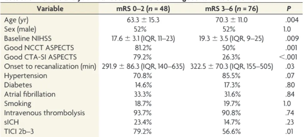
Variable mRS 0–2 (n = 48) mRS 3–6 (n = 76) P
Age (yr) 63.3⫾ 15.3 70.3⫾ 11.0 .004
Sex (male) 52% 52% 1.0
Baseline NIHSS 17.6⫾ 3.1 (IQR, 11–23) 19.3⫾ 3.5 (IQR, 9–25) .009 Good NCCT ASPECTS 81.2% 50% .001 Good CTA-SI ASPECTS 79.2% 26.3% ⬍.001 Onset to recanalization (min) 291.9⫾ 86.3 (IQR, 140–635) 322.5 ⫾ 70.3 (IQR, 155–505) .03 Hypertension 70.8% 85.5% .07 Diabetes 14.6% 17.3% .80 Atrial fibrillation 33.3% 31.6% .84 Smoking 18.7% 19.7% 1.0 Intravenous thrombolysis 93.7% 90.8% .74 sICH 23.4% 14.7% .23 TICI 2b–3 79.2% 56.6% .01
Note:—sICH indicates symptomatic intracranial hemorrhage.
Table 4: Multivariable regression model—best predictors of good clinical outcome
OR SE 95% CI P
Good CTA-SI ASPECTS 15.61 8.24 5.54–43.97 ⬍.001 Age 0.95 0.02 0.91–0.99 .011 TICI 2b–3 3.12 1.70 1.07–9.09 .037 Onset to recanalization (min) 0.99 0.01 0.98–0.99 .009
eral flow and time to reperfusion are strong predictors of outcome in endovascular treatment of patients with stroke. J Neurointerv Surg 2016 Sep 23. [Epub ahead of print]CrossRef Medline
9. Schramm P, Schellinger PD, Fiebach JB, et al. Comparison of CT and
CT angiography source images with diffusion-weighted imaging in patients with acute stroke within 6 hours after onset. Stroke 2002;
33:2426 –32CrossRef Medline
10. Lev MH, Segal AZ, Farkas J, et al. Utility of perfusion-weighted CT
imaging in acute middle cerebral artery stroke treated with intra-arterial thrombolysis: prediction of final infarct volume and clini-cal outcome. Stroke 2001;32:2021–28CrossRef Medline
11. Aviv RI, Shelef I, Malam S, et al. Early stroke detection and extent:
impact of experience and the role of computed tomography angiog-raphy source images. Clin Radiol 2007;62:447–52CrossRef Medline
12. Nabavi DG, Kloska SP, Nam EM, et al. MOSAIC: Multimodal Stroke
Assessment Using Computed Tomography—novel diagnostic ap-proach for the prediction of infarction size and clinical outcome. Stroke 2002;33:2819 –26CrossRef Medline
13. Camargo EC, Furie KL, Singhal AB, et al. Acute brain infarct:
detec-tion and delineadetec-tion with CT angiographic source images versus nonenhanced CT scans. Radiology 2007;244:541– 48 CrossRef Medline
14. Puetz V, Dzialowski I, Hill MD, et al. The Alberta Stroke Program
Early CT Score in clinical practice: what have we learned? Int J Stroke
2009;4:354 – 64CrossRef Medline
15. DeLong ER, DeLong DM, Clarke-Pearson DL. Comparing the areas
under two or more correlated receiver operating characteristic curves: a nonparametric approach. Biometrics 1988;44:837– 45
16. Goyal M, Menon BK, van Zwam WH, et al; HERMES collaborators.
Endovascular thrombectomy after large-vessel ischaemic stroke: a meta-analysis of individual patient data from five randomised tri-als. Lancet 2016;387:1723–31CrossRef Medline
17. Sallustio F, Koch G, Di Legge S, et al. Intra-arterial thrombectomy
versus standard intravenous thrombolysis in patients with anterior circulation stroke caused by intracranial occlusions: a single-center experience. J Stroke Cerebrovasc Dis 2013;22:e323–31 CrossRef Medline
18. Yoo AJ, Berkhemer OA, Fransen PS, et al; MR CLEAN investigators.
Effect of baseline Alberta Stroke Program Early CT Score on safety and efficacy of intra-arterial treatment: a subgroup analysis of a randomised phase 3 trial (MR CLEAN). Lancet Neurol 2016;15:
685–94CrossRef Medline
19. Wahlgren N, Moreira T, Michel P, et al; ESO-KSU, ESO, ESMINT, ESNR and EAN. Mechanical thrombectomy in acute ischemic
stroke: consensus statement by ESO-Karolinska Stroke Update 2014/2015, supported by ESO, ESMINT, ESNR and EAN. Int J Stroke
2016;11:134 – 47CrossRef Medline
20. Bhatia R, Bal SS, Shobha N, et al; Calgary CTA Group. CT
angio-graphic source images predict outcome and final infarct volume better than noncontrast CT in proximal vascular occlusions. Stroke
2011;42:1575– 80CrossRef Medline
21. Noorian AR, Rangaraju S, Sun CH, et al. Endovascular therapy in
strokes with ASPECTS 5–7 may result in smaller infarcts and better outcomes as compared to medical treatment alone. Interv Neurol