Abstract— One post-stroke patient underwent neuro-motor rehabilitation of one upper limb with a novel system combining a passive robotic device, Virtual Reality training applications and high resolution electroencephalography (HR-EEG). The outcome of the clinical tests and the evaluation of the kinematic parameters recorded with the robotic device concurred to highlight an improved motor recovery of the impaired limb despite the age of the patient, his compromised motor function, and the start of rehabilitation at the 3rd week post stroke. The time frequency and functional source analysis of the HR-EEG signals permitted to quantify the functional changes occurring in the brain in association with the rehabilitation motor tasks, and to highlight the recovery of the neuro-motor function.
I. INTRODUCTION
It is estimated that one to three fourths of stroke survivors show chronic motor impairments and require some assistance, or are fully dependent on caregivers for at least one Activity of Daily Living (ADL) [1]. Despite the recent developments in neurorehabilitation techniques [2], there is still a strong need to determine the optimal therapeutic approaches according to the individual level and type of impairment.
Among the non-invasive adjuncts to traditional rehabilitation therapies, noninvasive engineering solutions, such as robot-assisted training and brain-computer interfaces, can offer repetition, task oriented training, appropriate feedback, and a motivating environment, hence maximizing the functional outcome in post-stroke patients [3, 4, 5, 6].
We have recently proposed a system for the neuro-motor rehabilitation of upper limbs in stroke survivors composed of a passive robotic device (Trackhold) working in combination S. Comani and G. Tamburro are with the BIND - Behavioral Imaging and Neural Dynamics Center, University “G. d’Annunzio”, Via dei Vestini 33, 66100 Chieti (Italy). S. Comani is also with the Department of Neuroscience, Imaging and Clinical Sciences of the University “G. d’Annunzio”, and with the Casa di Cura Privata Villa Serena, Viale L. Petruzzi 42, 65013 Città Sant’Angelo (Italy) (e-mail: [email protected]), and G. Tamburro is also with the Department of Medicine and Aging Sciences of the University “G. d’Annunzio” ([email protected]). .
L. Schinaia, L. Velluto and B. Guarnieri are with the Casa di Cura Privata Villa Serena (Italy) (e-mails: [email protected]; [email protected]; [email protected]). L. Schinaia is also with the BIND - Behavioral Imaging and Neural Dynamics Center, University “G. d’Annunzio”, Via dei Vestini 33, 66100 Chieti (Italy).
S. Sorbi is with the Department of Neuroscience, Psychology, Drug Research and Child Health, University of Florence, Viale Pieraccini 6, 50139 Firenze (Italy), and with the Casa di Cura Privata Villa Serena, Città Sant’Angelo (Italy) (e-mail: [email protected]).
S. Conforto is with the Department of Engineering, Roma Tre University, Via della Vasca Navale 79/81, 00146 Rome (Italy), and with the BIND - Behavioral Imaging and Neural Dynamics Center, University “G. d’Annunzio”, Via dei Vestini 33, 66100 Chieti (Italy).
with Virtual Reality (VR) and synchronized with high resolution electroencephalography (HR-EEG) for the simultaneous recording of kinematic and functional data during the execution of rehabilitation tasks [7]. Here, we present the results of the neuro-motor rehabilitation of a post-stroke survivor using this combined system.
II. MATERIALS AND METHODS A. Patient
A right handed, 75 years old male patient with a lesion in the right parietal cortex was recruited for this study at the Private Hospital “Casa di Cura Privata Villa Serena” (Città Sant’Angelo, Italy), after written informed consent. The experimental procedures involving human subjects described in this paper were approved by the Institutional Review Board. The patient, with normal eye sight and no cognitive deficits or psychiatric disturbances, presented a motor impairment in the left arm: he had difficulties in performing simple movements and needed assistance to carry out ADL. He started rehabilitation with the new system 21 days after stroke.
B. Rehabilitation System
The three component parts of the neuro-motor rehabilitation system were synchronized to ensure the simultaneous and synchronous recording of both kinematic and neural data. Therefore, this system permitted to quantify the task-related activation changes occurring in the brain during the recovery of the motor function.
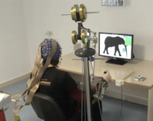
The passive robotic device (Trackhold - PERCRO, Pisa, Italy) had exchangeable counter-weights to adjust the arm weight compensation to the patient's motor abilities recovered during rehabilitation. The patient interacted with the VR environments by moving the Trackhold’s end effector (Fig.1). Being the device passive, only voluntary movements of the patient were registered during training.
The five VR training applications replicated ADL in 2D and 3D environments, and required simple visuo-motor coordination tasks: Sponge (free movements), Bug Hunt,
Grab2D and Grab3D (simple or double reaching in 2 or 3
dimensions), and Twirl (2D coordination movements). The applications were displayed in real time on a 22-inch LCD monitor at eye-level at a distance of 90 cm from the patient. No written or spoken instructions were provided during training. The difficulty level of the VR applications could be adjust to the patient's residual or recovered motor abilities by setting task control parameters and termination criteria [7].
A HR-EEG system including an EBN Galileo MIZAR-PLUS amplifier, an EBN Galileo NT acquisition software
Assessing Neuro-motor Recovery in a Stroke Survivor with
High-resolution EEG, Robotics and Virtual Reality
Silvia Comani, Lorenzo Schinaia, Gabriella Tamburro, Lucia Velluto, Sandro Sorbi, Silvia Conforto,
Biancamaria Guarnieri
(EBNeuro, Italy), and a head-cap with 128 Ag/AgCl electrodes (international 10-5 system, Electro-Cap International, Ohio, USA) [8] was synchronized with the Trackhold and VR components through a pushbutton trigger signal [7]. Therefore, cortical activity was recorded in relation to the execution of each task during rehabilitation training, and the task-related functional changes occurring in the brain during recovery of the motor function could be quantified. Sampling frequency was 1024 Hz.
Figure 1. Example picture of the Trackhold with a volunteer grabbing the end-effector, the HR-EEG system and the VR application Sponge. C. Rehabilitation Protocol
Rehabilitation with the novel system was added to conventional therapy (performed daily) 3 times per week for 4 weeks. The VR training applications were presented with increasing task difficulty, from simple free movements (Sponge) to reaching movements in 2D and 3D (Bug Hunt,
Grab2D, Grab3D), and to 2D coordination movements on a
pre-defined circular trajectory (Twirl). Each application was run for 5 minutes with inter-trial breaks of 7 seconds.
During the first and last rehabilitation sessions, HR-EEG was acquired, and clinical tests assessing the level of motor impairment were administered. The tests were: the Nine
Hole Peg Test, the Motricity Index, the Barthel Index, the Functional Independence Measure, and the Canadian Stroke Scale. The Beck Depression Inventory-II (BDI-II) and the Pittsburgh Rehabilitation Participation Scale (PRPS) were
also administered to investigate depressive symptoms and active participation in the rehabilitation procedure, respectively. The outcome of these tests was used to confirm the results of subsequent kinematic and EEG analyses.
D. Kinematic data analysis
From the reconstructed trajectories of the patient's hand during the execution of the VR rehabilitation tasks, four indices of motor performance were calculated, for each trial [7]: Trial Duration (TD), Path Length (PL), Normalized Jerk (NJ), and Speed (S). A custom-made MATLAB toolbox was used (Trackhold Analysis Toolbox, THA 1.0). The kinematic quantities were averaged across all trials of each VR application within each rehabilitation session. Hence, for each rehabilitation task, we could quantify the changes of the kinematic parameters throughout rehabilitation.
E. Neurophysiological data analysis
Raw EEG data were offline downsampled to 512 Hz,
notch-filtered (50 Hz) and bandpass filtered (0.5–200 Hz). Bad channels and epochs with residual gross artefacts were excluded from further analysis [9]. Independent Component Analysis (ICA, InfoMax algorithm) was used to separate and reject the components related to artifacts of biological and environmental origin [9, 10, 11]. True EEG signals were reconstructed from the retained components (EEGLAB toolbox; http://sccn.ucsd.edu/eeglab).
Time frequency analysis: During motor performance,
oscillatory cortical activity changes in relation to movement [12]. In particular, an amplitude attenuation (event-related desynchronization, ERD) reflecting cortical activation related to movement planning is observed in the alpha and beta bands during voluntary movements. After movement, event-related synchronization (ERS) in the beta band replaces ERD [12]. In sustained movements a small post-movement beta rebound is observed [13, 14].
For each rehabilitation session (first and last) and each trial within the same VR application, true EEG data were epoched around movement onset (time windows from -3 s to + 5 s), and averaged over all epochs. The ERD/ERS patterns were evaluated using the Morlet wavelet decomposition [15] in the [1–50 Hz] frequency range. The first second of the time window was used as pre-movement baseline to visualize the decrease/increase (ERD/ERS) of the oscillatory activity during movement execution.
Source imaging: The coordinates of the EEG sensors and
the anatomical landmarks defined using a realistic head model [8] (http://robertoostenveld.nl) were coregistered with the corresponding standard fiducials from the SPM8 standard T1 MRI template (Fig.2) [16]. Boundary Elements Method (BEM) was used to compute the lead field over a model constructed using 8196 vertex cortical meshes [17] (Fig.2). The inverse solution was calculated for time windows of 6 s locked to movement onset (from -2 s to +4 s) using the true EEG data previously common average referenced and downsampled to 200 Hz [17]. Source estimate was performed using the multiple sparse priors (MSP) algorithm [18], as implemented in SPM8.
Figure 2. A) SPM template meshes (cortex, inner skull, outer skull and scalp). Anatomical fiducials are displayed in light blue. B) Template head model used to compute BEM forward solution. The sensor locations are displayed in light green. C) 3D outcome of the coregistration.
For each rehabilitation session (first and last) and each VR application, oscillatory source power was summarized as one 3D image in terms of contrasts (mixtures of activity estimates) over time and frequency [18]. Time-frequency (TF) contrast images (at intervals [0.5, 30] Hz and [-100, 100] ms) were obtained by convolving the single-trial source activity with a series of Morlet projectors, and then weighting the average power with a Gaussian window centered at movement onset [17]. As a final step, to better highlight the reduction of the over-activation in the impaired 3926
left hemisphere after rehabilitation, for each VR application we subtracted the image obtained for the last session from the one obtained for the first session. With this approach, the brain areas that do not undergo any activation changes during the 4 weeks of therapy will not be shown.
III. RESULTS
Initial and final values of the clinical tests are summarized in Table I. They indicated a significant recovery of the motor function in the impaired limb by the end of rehabilitation. In particular, the Nine Hole Peg Test was reduced to one third, indicating an improvement of motor function; the Motricity Index doubled, , indicating the retrieval of normal movements; the Barthel Index reached the maximum value, showing that the patient was again able to take care of himslef. Also the depression level, always very low, halved at the end of rehabilitation, and the mean rehabilitation participation score was always very good.
All kinematic parameters also showed clear improvements during the performance of the VR training applications (Table II). It is worth noting that the patient reported extensive tiredness during the last session, which might explain the still quite high values of Normalized Jerk and Trial Duration for Sponge, Grab 2D and Twirl.
TABLE I. OUTCOME OF THE CLINICAL TESTS Clinical test First session Last session
Nine Hole Peg Test; impaired limb (s) 180 60 Motricity Index; upper limbs (in %) 39 78
Barthel Index 11 20
Functional Independence Measure 60 109
Canadian Stroke Scale 7 12.5
Beck Depression Inventory-II 8 4
Pittsburgh Rehab. Participation Scale 5 5 TABLE II. MEAN VALUES OF THE KINEMATIC PARAMETERS Kinematic
parameters
VR training applications
Sponge Bug Hunt Grab 2D Grab 3D Twirl sessions sessions sessions sessions sessions first last first last first last first last first last
TD (s) 20 10 8 4 19 8 11 6 38 17
PL (cm) 66 46 18 15 93 36 39 34 152 79
NJ 212 41 45 12 159 34 85 17 871 171
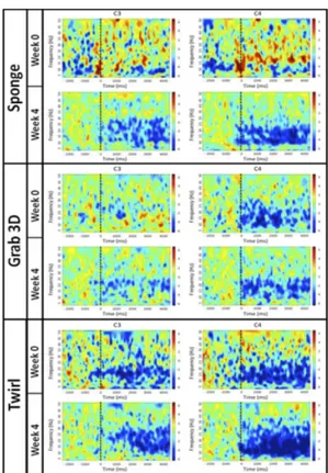
S (cm/s) 3.4 4.7 2.2 3.8 5.1 5.0 3.8 6.1 4.0 4.6 The time-frequency analysis of the oscillatory cortical activity during the performance of all VR applications showed an improvement of the neural response after rehabilitation. Fig.3 shows the time-frequency decomposition of the EEG signals recorded by 3 representative derivations over the central cortex (electrodes C3 and C4, left and right hemisphere respectively) for 3 different VR applications: a clear desynchronization pattern could be recognized in both alpha and beta bands after rehabilitation with Sponge (free movements). A better defined desynchronization pattern occurred also for Grab 3D (reaching in 3D) and Twirl (2D coordination). It is worth noting that major differences and an ERD shift toward trial onset (t=0) could be observed in the impaired left hemisphere (C3) for all applications.
Figure 3. Time frequency decomposition of the EEG signal recorded at the start and end of rehabilitation for 3 representative VR applications and 2 representative electrodes over the central cortex (C3: left hemisphere, C4: left hemisphere). Movement onset is shown by a dashed vertical line.
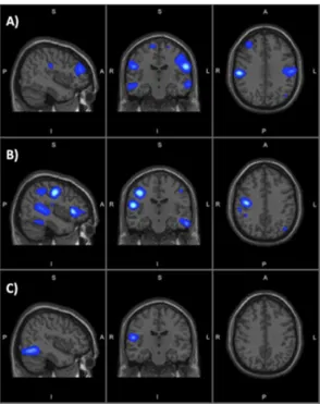
Similarly, the source imaging results showed that, after rehabilitation, major activation reductions occurred in the hemisphere contralateral to the moved arm, i.e. in the ipsilesional hemisphere (the right hemisphere). These reductions mainly regarded the sensorimotor network [19], composed of the primary motor cortex (precentral gyrus), primary somatosensory cortex (postcentral gyrus), superior parietal lobule, supplementary and pre-supplementary motor areas, both lying in the superior frontal gyrus [20]. Fig.4 shows the differential maps obtained for the VR applications selected to represent the 3 main rehabilitation movements.
Differences of activation after rehabilitation with Sponge (Fig.4-A) were observed in both hemispheres in the frontal lobe, temporal and parietal areas. The major activation reductions occurred in the right hemisphere (i.e. the stroke-affected or ipsilesional hemisphere), particularly in the primary motor cortex, inferior temporal gyrus and planum temporale, and primary somatosensory cortex. Sgnificant pre- and post-rehabilitation differences were also found in the right cerebellum, whereas no changes were observed in the occipital cortex. The 3D differential maps recostructed for Grab 3D (Fig.4-B) showed similar results, with bilateral activation decreases in the frontal lobe, temporal and parietal areas. The main differences were localized in the ipsilesional hemisphere, particularly the primary motor cortex. Small changes occurred in the occipital lobe. Pre- and post-rehabilitation differences were found also in the cerebellum (both hemispheres). For Twirl (Fig.4-C), the main activation changes were found in the parietal lobe and, to a minor extent, in the temporal areas. Activations in the occipital areas and right cerebellum were also reduced.
Figure 4. Sagittal, coronal and axial views of the source differential maps for 3 representative VR applications: A) Sponge, B) Grab 3D, C) Twirl. S: superior, I: inferior, P: posterior, A: anterior, R: right, L: left.
IV. DISCUSSION
The clinical tests and the kinematic parameters indicated a significant motor recovery of the impaired limb. These results are particulary valuable because the patient was aged, showed considerable motor impairment, and could not start rehabilitation earlier than 3 weeks after stroke. At a neural level, the initial bilateral over-recruitment of the sensorimotor network (aiming to compensate for the impaired function) was significantly reduced by the end of rehabilitation, especially in the ipsilesional hemisphere. Interestingly, the main differences were found in the areas involved in motor and somatosensory processing. Also the bilateral reduction of the cerebellar activations were associated with the recovery of the motor function, given the well known role of cerebellum in motor coordination. Similarly, the results of the time frequency analysis indicated a tendency towards the recovery of a normal oscillatory processing within the somatosensory network, with better defined desynchronization patterns after rehabilitation, especially in the impaired hemisphere. Overall, these results confirm the potential effectiveness of the proposed neuro-motor rehabilitation system in quantifying the functional changes related to the recovery of the motor abilities in performing specific motor schemes of ADL, such as free movements, reaching or 2D coordination tasks. As assessed by the clinical tests at the end of treatment, functional recovery does not seem to be due to mere habituation to the training applications. The effectiveness of the proposed system in acute and chronic patients is under assessment in an ongoing study on a larger population of stroke survivors including a control group of patients subjected only to traditional rehabilitation.
REFERENCES
[1] J.M. Veerbeek, G. Kwakkel, E.E. van Wegen, J.C. Ket and M.W.
Heymans, "Early prediction of outcome of activities of daily living after stroke: a systematic review," Stroke, vol. 42, pp. 1482–1488, Apr. 2011.
[2] M. Brainin and R.D. Zorowitz, "Advances in stroke: recovery and rehabilitation," Stroke, vol. 44, no. 2, pp. 311-313, Feb. 2013 [3] P. Sale, F. Bovolenta, M. Agosti, P. Clerici and M.Franceschini,
"Short-term and long-term outcomes of serial robotic training for improving upper limb function in chronic stroke," Int J Rehabil Res, 37(1):67-73. Mar. 2014.
[4] S.H. Scott and S.P. Dukelow, "Potential of robots as next-generation technology for clinical assessment of neurological disorders and upper-limb therapy," J Rehabil Res Dev, vol. 48, no. 4, pp. 335-353, 2011.
[5] G. Kwakkel, B.J. Kollen and H.I. Krebs, "Effects of robot-assisted therapy on upper limb recovery after stroke: a systematic review,"
Neurorehabil Neural Repair, vol. 22, no. 2, pp. 111-121, Mar-Apr
2008.
[6] M. Modo, F Ambrosio, RM Friedlander, SF Badylak, L.R.Wechsler. "Bioengineering solutions for neural repair and recovery in stroke"
Curr Opin Neurol, vol. 26, no. 6, pp. 626-31, Dec. 2013.
[7] M. Steinisch, M.G. Tana and S.Comani, "A post-stroke rehabilitation system integrating robotics, VR and high-resolution EEG imaging,"
IEEE Trans Neural Syst Rehabil Eng, vol. 21, no. 5, pp. 849-859, Sep.
2013.
[8] R. Oostenveld and P. Praamstra, " The five percent electrode system for high-resolution EEG and ERP measurements," Clin Neurophysiol vol. 112, no. 4, pp. 713–719, Apr. 2001.
[9] W.B. McMenamin, A.J. Shackman, J.S. Maxwell, D.R.W. Bachhuber, A.M. Koppenhaver, L.L. Greischar and R.J. Davidson, "Validation of ICA-based myogenic artifact correction for scalp and source-localized EEG," Neuroimage, vol. 49, no. 3, pp. 2416–2432, Feb. 2010. [10] A.J. Bell and T.J.Seinowski, "An Information-Maximization approach
to blind separation and blind deconvolution," Neural Comput, vol. 7, no. 6, pp. 1129-1159. Nov. 1995.
[11] T.W. Lee, M. Girolami and T.J. Seinowski, "Independent Component Analysis using an Extended Infomax algorithm for mixed subgaussian and supergaussian sources," Neural Comput, vol. 11, no. 2, pp. 417-441. Feb. 1999.
[12] G. Pfurtscheller and F.H. Lopes da Silva, "Event-related EEG/MEG synchronization and desynchronization: Basic principles," Clin
Neurophysiol vol. 110, no. 11, pp. 1842-1857. Nov. 1999.
[13] F. Cassim, W. Szurhaj, H. Sediri, D. Devos, J.-L. Bourriez, I. Poirot, P. Derambure, L. Defebvre, J.-D. Guieu, "Brief and sustained movements: differences in event-related (de)synchronization (ERD/ERS) patterns," Clin Neurophysiol vol. 111, no. 11, pp. 2032-2039, Nov. 2000.
[14] M. Alegre, A. Labarga, I.G. Gurtubay, J. Iriarte,A. Malanda and J. Artieda, "Movement-related changes in cortical oscillatory activity in ballistic, sustained and negative movements." Exp Brain Res. vol. 148, no. 1, pp. 17–25, Jan. 2003.
[15] A. Delorme, M. Westerfield and S. Makeig, "Medial prefrontal theta bursts precede rapid motor responses during visual selective attention," J Neurosci vol. 27, no. 44, pp. 11949–11959, Oct. 2007. [16] C. Bledowski, D. Prvulovic, K. Hoechstetter, M. Scherg, M. Wibral,
R. Goebel and D.E.J Linden, "Localizing P300 generators in visual target and distracter processing: a combined event-related potential and functional magnetic resonance imaging study," J Neurosci vol. 24, no. 42, pp. 9353–9360, Oct. 2004.
[17] V. Litvak, J. Mattout, S. Kiebel, C. Phillips, R. Henson, J. Kilner, G. Barnes, R. Oostenveld, J. Daunizeau, G. Flandin, W. Penny and K. Friston, "EEG and MEG Data Analysis in SPM8," Comput Intell
Neurosci, Article ID 852961, Mar. 2011.
[18] K. Friston, L. Harrison, J. Daunizeau, S. Kiebel, C. Phillips, N. Trujillo-Barreto, R. Henson, G. Flandin and J. Mattout, "Multiple sparse priors for the M/EEG inverse problem," Neuroimage, vol. 39, no. 3, pp. 1104-1120, Feb. 2008.
[19] M.L. Otten, C.B. Mikell, B.E. Youngerman, C. Liston, Michael B. Sisti, J.N. Bruce, S.A. Small and G.M. McKhann, "Motor deficits correlate with resting state motor network connectivity in patients with brain tumours," Brain vol. 135, no. 4. pp. 1017-1026, Apr. 2012. [20] P. Nachev, C. Kennard and M. Husain, "Functional role of the
supplementary and pre-supplementary motor areas," Nat Rev Neurosci vol. 9, no. 11, pp. 856–869, Nov. 2008.