Università degli Studi di Urbino “Carlo Bo”
Dipartimento di Scienze Biomolecolari
DOTTORATO DI RICERCA IN
SCIENZE DELLA VITA, SALUTE E BIOTECNOLOGIE
Curriculum in Scienze biochimiche, farmacologiche e
biotecnologie
Ciclo XXXI
ERYTHROCYTES LOADED WITH PHENYLALANINE
AMMONIA LYASE (PAL) AS ENZYMATIC REPLACEMENT
THERAPY FOR PHENYLKETONURIA
Settore scientifico disciplinare: BIO/10
RELATORE
DOTTORANDO
Chiar.ma Prof.ssa
Dott.ssa
LUIGIA ROSSI
NOEMI BIGINI
CO-RELATORE
Dott. GIOVANNI MAMBRINI
INTRODUCTION ... 1
THE DISCOVERY OF PHENYLKETONURIA ... 1
CHARACTERISTICS OF THE DISEASE ... 2
MOLECULAR AND GENETICS CHARACTERISTICS OF PHENYLALANINE HYDROXYLASE ENZYME ... 5
PAH working and regulation ... 7
PAH GENE MUTATIONS AND DATABASE ... 9
CLASSIFICATION ... 11
Classification according to blood L-Phe ... 11
Phenylalanine tolerance ... 12
Clinical course of the disease ... 13
SCREENING AND DIAGNOSIS ... 13
Guthrie test ... 14
Fluorimetric assay: ... 14
Reverse-phase liquid chromatography ... 15
Tandem mass-spectrometry (TMS) assay ... 15
Molecular diagnosis... 15
The BH4 loading test ... 15
PATHOGENIC MECHANISMS OF HPA ... 17
Brain development and behavioral outcomes ... 17
L-Phe influence on cholesterol biosynthesis and obesity ... 21
MATERNAL PKU ... 22
THERAPEUTIC STRATEGIES ... 23
Dietary treatment ... 24
Glycomacropeptide ... 26
Large neutral aminoacids (LNAAs) supplementation ... 26
Tetrahydrobiopterin (BH4) treatment ... 27
Gene therapy and liver transplantation ... 29
Enzyme replacement therapy (ERT) ... 30
ENZYME REPLACEMENT THERAPY IN PKU ... 31
DRUG DELIVERY SYSTEMS: RED BLOOD CELLS AS THE BEST CHOICE ... 35
AIM OF THE WORK ... 39
MATERIALS AND METHODS ... 41
ENZYMES ... 41
Recombinant AvPAL ... 41
PCR analysis ... 43
GEL analysis ... 44
DEVELOPMENT OF MURINE rAvPAL-RBCs AND IN VIVO EFFICACY OF REPEATED ADMINISTRATIONS ... 45
PHENYLALANINE AMMONIA LYASE ACTIVITY ASSAY ... 46
L-PHE AND L-TYR EVALUATION IN DRIED BLOOD SPOT (DBS) BY TANDEM MASS SPECTROMETRY (MS/MS) ... 47
BEHAVIORAL ANALYSIS... 48
Behavioral assay in EPM apparatus ... 48
Behavioral assay in OFT apparatus ... 49
Behavioral assay in ORT apparatus ... 49
NEUROCHEMICAL AND MORPHOLOGICAL ANALYSES ... 51
Neurochemistry ... 51
Morphology ... 51
IMMUNOHISTOCHEMICAL AND BIOCHEMICAL ANALYSES ... 52
Western blot analyses ... 52
Immunofluorescence analyses ... 53
EVALUATION OF PLASMA ANTI-rAvPAL IgG TITER ... 53
Antibody titer determination ... 54
STATISTICAL ANALYSES ... 54
Behavioral study ... 54
Neurochemical and Morphological analyses ... 54
Antibody titer determination ... 55
RESULTS ... 56
DEVELOPMENT OF MURINE rAvPAL-RBCs ... 56
BIOCHEMICAL RESULTS ... 57
Blood L-Phe and L-Tyr concentrations ... 57
Brain L-Phe and L-Tyr concentrations ... 58
BEHAVIORAL RESULTS ... 59
Elevated Plus Maze (EPM) results ... 59
Open Field Test (OFT) results ... 60
Object Recognition Test (ORT) results ... 60
NEUROCHEMICAL RESULTS ... 61
MORPHOLOGICAL RESULTS ... 63
IMMUNOHISTOCHEMICAL AND BIOCHEMICAL ANALYSES ... 66
MBP immunoreactivity ... 66
EVALUATION OF ANTI-rAvPAL PLASMA IgG TITER ... 68
DISCUSSION ... 69
CONCLUSION AND FUTURE PERSPECTIVES ... 75
REFERENCES ... 76
1
INTRODUCTION
THE DISCOVERY OF PHENYLKETONURIA
Phenylketonuria (PKU) (OMIM# 261600) was first described in 1934 by the Norwegian endocrinologist dr. Asbjørn Følling, who originally defined this pathology as “phenylpyruvic oligophrenia” because of the typical mental disorders that affected his patients (Mitchell et al., 2011). This definition was modified in 1930s by Penrose (Penrose and Quastel, 1937), who coined the name currently known, PKU, and identified its autosomal recessive nature. He surmised that PKU state had an endogenous chemical cause; in keeping with his hypothesis, he was the first to consider the possible correlation between “nurture” and mutant “nature”. He thought that modifying the nurture might be possible to neutralize the harmful effects of the pathology (Penrose, 1998).
PKU is an inherited metabolic disorder characterized by severe intellectual impairment, motor problems, and skin abnormalities and occupies a unique place in the history of the study of metabolic disease not only for its role as principal inborn error of amino acid metabolism but also because it is the first cause of mental retardation to be discovered. Dr. Følling found that affected individuals could be identified by the abnormal excretion of phenylpyruvic acid in their urine. The credit for the discovery was also due to that caring and stubborn mother, who could not resign herself to the mental retardation of her children without having found a reason (http://pkuworld.org/home/history.asp).
Her 7 years old daughter, could say only few words and had a whimsy and purposeless way of moving about; likewise, her 4 years old son did not walk and was unable to fix his eyes on anything. Their skin was fair and their urine had a peculiar smell. By means of a traditional assay of classical chemistry for the detection of ketones, consisting in the addition of ferric chloride to the urine of diabetic patients, dr. Følling observed the appearance of a deep green color, which he had never seen before. Further chemical analyses and steps of purification on many other urine samples from patients sharing the same neurocognitive and developmental delays, led to the identification of a chemical substance whose empirical formula was C9H8O3,
named phenylpyruvic acid. The analysis of the urine from another 430 mentally impaired subjects, allowed dr. Følling to identify eight patients excreting the same substance and for the first time he understood the correlation between mental impairment and excretion of phenylpyruvic acid. Further studies of family relationships highlighted an autosomal recessive mechanism of transmission (Følling, 1944). Few years later, Jervis (1947, 1953), succeeded in identifying the metabolic block and the enzymatic deficiency of phenylalanine hydroxylase (PAH), the alteration behind this pathological condition; at the same time, Bickel and collaborators (1953) showed the importance of reducing the intake of phenylalanine (Phe) in order to obtain a prognosis improvement. Phenylketonuria was the first known inborn error of metabolism to seriously affect the victims and to give mental disturbance. In addition, its discovery determined an important breakthrough in understanding how metabolic dysfunctions can influence neurological functions and how treatments can heavily influence
2
clinical manifestations: PKU today is considered “the epitome of metabolic disorders” and is often employed as a model to describe and understand many other inborn errors of amino acid metabolism (Scriver and Clow, 1980 Part I and II; Raghuveer et al., 2006). To explain the causes of the phenylpyruvic acid excretion, dr. Følling hypothesized some kind of defect in phenylalanine metabolism, which lead to high concentration of this aminoacid in the blood of PKU affected patients; the effectiveness of his hypothesis was successively confirmed (Følling and Closs, 1938) through a microbiological test developed by dr. Robert Guthrie which exploited the reversal of growth inhibition observed in Bacillus subtilis ATCC 6051 in the presence of a high level of phenylalanine (Guthrie and Susi, 1963). The identification of the first mutations of the PAH gene, codifying for the enzyme PAH, began immediately after its cloning and mapping in 1983 (Woo et al., 1983) opening the way to the in vitro study of the different functionalities of the enzyme. Currently, all the known mutations of the PAH gene (about 859) known, are collected in the "PAHdb" database (http://www.pahdb.mcgill.ca/) created in 1996 (Hoang et al., 1996).
CHARACTERISTICS OF THE DISEASE
Phenylketonuria (PKU) is the most common autosomal recessive disease among Caucasians (overall incidence 1:10.000 on average; 1:2.600 in Turkey; 1:100.000 in Japan). PKU is a result of an inborn error of amino acid metabolism caused by a deficiency of the enzyme phenylalanine hydroxylase (PAH, EC 1.14.16.1) which catalyze the irreversible conversion, via para-hydroxylation, of the amino acid L-phenylalanine (L-Phe) into tyrosine (L-Tyr), a limiting step for the complete oxidation of L-Phe to CO2 and H2O (Scriver and Kaufmann, 2001). The
enzyme PAH needs the pterin cofactor tetrahydrobiopterin (BH4), molecular oxygen (O2) and
non-heme iron (Fe2+) to perform its activity (Figure 1).
Figure 1. Conversion of L-Phe into L-Tyr. The enzyme phenylalanine hydroxylase (PAH) catalyzes the conversion
of L-Phe into L-Tyr using for its activity the cofactor tetrahydrobiopterin (BH4), molecular oxygen (O2) and iron
(Fe2+).
Failure of PAH activity results in L-Phe accumulation in all tissues, brain included. Here, it plays a toxic role and lead to severe neurological and intellectual disability due to the abnormally reduced levels of neurotransmitters for which L-Tyr is a precursor (Scriver, 2007; Donlon et
3
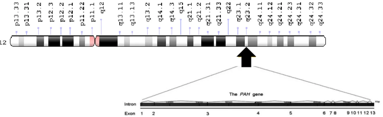
al., 2010). Early diagnosis and a quick treatment are able to reduce toxic levels of this aminoacid, avoiding these serious consequences. Nowadays, many countries include a neonatal screening such as Guthrie test or more modern system based on tandem mass spectrometry for the detection of hyperphenylalaninemia (HPA). Moreover, L-Phe itself is an essential nutrient an it represents a pivotal constituent for protein synthesis. Therefore, PKU treatment requires the balanced reduction of systemic L-Phe levels without its excessive depletion in order to guarantee a satisfactory synthesis of L-Tyr. Mutations of PAH gene, located in chromosome 12 (region 12q22-q24.2, GenBank U49897), is responsible for the insufficient activity of this cytosolic hepatic enzyme and the establishment of the HPA state. HPA can be caused by either mutations at the PAH locus, which results in more or less severe forms of PKU, or mutations in the genes encoding the enzymes involved in the biosynthesis or regeneration of the cofactor BH4, resulting in non-PKU HPA (Scriver and Kaufman, 2001) (Figure 2). This condition was initially referred to as “malignant phenylketonuria” (Matalon et al., 1989).
4
Figure 2. Pathways of BH4 cofactor formation. Here are shown the two possible ways through which the pterinic cofactor is made available. At the top is the de novo synthesis starting from the guanosine triphosphate (GTP), at the bottom the regeneration route starting from 4a-OH-BH4. The biosynthetic enzymes involved are GTP cyclohydrolase I (GTPCH), 6-pyruvoyl-tetrahydropterin synthase (PTPS) and sepiapterin reductase (SR); on the other hand, the recycling enzyme are pterin-4a-carbinolamine dehydratase (PCD) and dihydropteridine
reductase (DHPR) which catalyze the reduction of the oxidized cofactor quinonoid dihydrobiopterin (qBH2) once
L-Phe conversion to L-Tyr has occurred (adapted from Blau et al., 2010).
The final concentration of phenylalanine in the body (Figure 3) is the result of a finely regulated balance between L-Phe input amount, coming from diet and the endogenous recycling of amino acids, and L-Phe output amount, represented by that fraction integrated in newly synthesized proteins and the one oxidized to L-Tyr through the PAH-mediated reaction (Scriver and Kaufman, 2001).
De Novo
synthesis
Recycling
pathway
5
Figure 3. L-Phe metabolism in humans. Representation of the dynamic equilibrium to which L-Phe is subjected in normal conditions. The figure also shows the alternative metabolisms to which L-Phe can meet, which lead to the formation of various metabolites subsequently eliminated through the urine (adapted from Williams et al., 2008).
MOLECULAR AND GENETICS CHARACTERISTICS OF PHENYLALANINE HYDROXYLASE ENZYME
Phenylalanine hydroxylase (also named phenylalanine-4-monooxygenase, symbol PAH or PheOH, EC 1.14.16.1) is part of the enzymatic family of pterin-dependent aromatic amino acid hydroxylases (AAAH). This family also includes two other monooxygenases, i.e. tryptophan hydroxylase (tryptophan-5-monooxygenase, TPH or TrpOH, EC 1.14.16.4) and tyrosine hydroxylase (tyrosine-3-monooxygenase, TH or TyrOH, EC 1.14.16.2); all these proteins share the necessity of BH4, molecular oxygen and reduced iron (Fe2+) to carry out their own activity
(Fitzpatrick, 1999; Bjørgo et al., 2001) and show high sequence identity and similar molecular structure (Fitzpatrick, 2000) even if differ in their substrate specificity.
The human PAH gene, cloned for the first time in 1980s (Woo et al., 1983, 1985; Kwok et al., 1985), is located in the long arm of chromosome 12 (locus PAH 12q22-q24.2) and expressed mainly in liver but also in kidney (Wang et al., 1992; Lichter-Konecki et al., 1999; Tessari et al., 1999) whereas the full-length genomic sequence and cDNA of the gene was obtained about 10 years later (GenBank AF404777) (Konecki et al., 1992) and deposited in the PAHdb knowledgebase (Scriver et al., 2003). Chromosome 12 is particularly rich in disease-associated loci, with 5.2% of known “disease-genes”. PAH gene is composed of 13 exons and 12 big introns, reaching the total length of 90 Kb or about 171 Kb if flanking regions are included (Scriver, 2007; http://www.genecards.org/cgi-bin/carddisp.pl?gc_id=PAH). The gene coding
6
sequence (cds, nt 473-1831) is transcribed into a mature mRNA of approximately 2,6 Kb (2680 bp), which is in turn translated into a 452 amino acid monomer (http://www.ncbi.nlm.nih.gov/nuccore/U49897.1, last update 1997) (Figure 4).
Figure 4. Basic structure and localization of the human PAH gene. Located on the long arm of chromosome 12, the human PAH gene contains 13 exons that encode a polypeptide of 452 amino acids (adapted from Williams et al., 2008).
Eukaryotic sequence of PheOH has several homologies with the other two pterin-dependent hydroxylases: TrpOH and TyrOH. All these three enzymes show high homology close to five common cysteine residues, in the core of the primary sequence, and a minor homology at the N-terminal end (Onishi et al., 1991). This homology is partially maintained not only in eukaryotes (man and rat) but also in prokaryotes such as Chromobacterium violaceum in which the primary structure of PAH has 24% homology with human and rat protein and 11% of homology with TrpOH and TyrOH of the eukaryotes (Onishi et al., 1991).
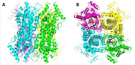
In human, PAH enzyme exists as an assortment of functional homodimer and homotetramer (200 kD as a tetramer), in a pH- and L-Phe-dependent equilibrium, with a marked shift towards the tetrameric form as pH decreases or L-Phe concentration increases (Kappock et al., 1995; Hufton et al., 1995; Martinez et al., 1995). However, both oligomeric forms are functional, as demonstrated by studies on truncated forms of both PAH and TyrOH (where only the tetramerization and catalytic domains are maintained), which still retain the enzymatic activity (Fusetti et al., 1998). Interestingly, the tetramer formed by PAH is asymmetrical because it is a “dimer of dimers” (Erlandesen and Stevens, 1999), where secondary elements switch their mutual position in order to promote a stable oligomerization, together with the formation of an antiparallel coiled-coil structure with the other monomers (Bennett et al., 1995; Fusetti et al., 1998) and present low specific activity (Kaufman, 1987).
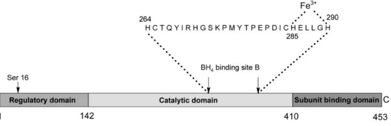
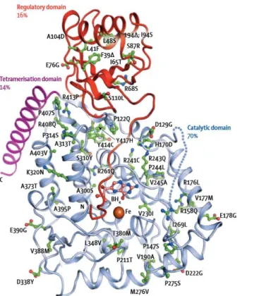
Each monomer is about 50 KDa in size and is comprised of 452 amino acids (Hufton et al., 1995). It consists of three domains: N-terminal regulatory domain (residues 1-142, Glu19-Leu142, also identified as ACT domain), which is thought to be involved in activation by phosphorylation; a catalytic domain (residues 143-410, Asp143-Phe410), responsible for cofactor and ferric ion binding and a short C-terminal tetramerization domain (residues 411-452, Ser411-Lys452) (Erlandsen and Stevens, 1999; Erlandsen et al., 2003; Williams et al., 2008; GenBank AAC51772.1) (Figure 5).
7
Figure 5. PAH structure. (A) Full-length structure of human phenylalanine hydroxylase monomer obtained by superimposing the catalytic domains of the truncated forms. The red sphere represents iron. (B, C) Two perpendicular views of the full-length PAH model structure. The iron is shown as a gray sphere in all four monomers (adapted from Erlandsen and Stevens, 1999).
PAH working and regulation
PAH activity is tightly regulated by a number of possible mechanisms such as reversible phosphorylation and substrate activation. The activity of PAH, as previously mentioned, requires the binding of the cofactor BH4 and molecular oxygen (Figure 1).
The binding of the BH4 cofactor is thought to occur at a sequence of 27 amino acids (from His263 to His289), highly conserved among the three pterin-dependent hydroxylases (Hufton et al., 1995; Jennings et al., 1991). Within this sequence, 10 residues (Phe263, Cys265, Thr266, Thr278, Pro279, Glu280, Pro281, His285, Glu286 and Gly289) belong to the active site. The binding of the cofactor occurs near the Fe (III) and establishes molecular links with two of the three water molecules coordinated to the ion and with the carbonyl oxygen of the main chains of Ala322, Gly247 and Leu249, with the amide chain of Leu249 and with the atom of oxygen γ (Oγ) of Ser251 (Erlandsen et al., 2000; Andersen et al., 2001) (Figure 6).
Figure 6. Structural components of PAH enzyme. The residue of serine 16 involved in the regulation of the enzymatic activity can be seen in correspondence with the regulatory domain, while, in correspondence with the catalytic domain, we can see the sequence of 27 amino acids responsible for the binding of the cofactor BH4 and the Fe (III) (from Williams et al., 2008).
After the binding of the pterinic cofactor at the active site, there is a large conformational change: the residues from 245 to 250 move in the direction of the iron, allowing the formation of numerous hydrogen bonds between the ptery ring and the protein (Erlandsen et al., 2000).
(411-452) (1-142)
8
Studies carried out on PAH have shown that a stoichiometric amount of BH4 can be oxidized in the presence of oxygen by determining the reduction of the enzyme (Marota and Shiman, 1984), i.e. the reduction of Fe3+ to Fe2+, a process necessary for the activation of PAH (Wallick
et al., 1984). This reaction is achieved by transferring an electron from BH4 to Fe3+ and a
second electron to the oxygen molecule (Marota and Shiman, 1984; Wallick et al., 1984; Hill et al., 1988).
The PAH enzyme is particularly sensitive to its substrates concentrations and its activity is strictly regulated through different mechanisms that act together to tightly control the negative effects of an excessive concentration of circulating L-Phe (Heintz et al., 2013). The first thirty residues of the enzyme (residues 19-33) act as a autoregulatory sequence (ARS) and contain a residue of serine 16 (Ser16) which has been demonstrated to be the site of phosphorylation by the cAMP-dependent protein kinase A (cAMP-PKA) (Døskeland et al., 1996). This sequence is named autoregulatory because it sterically limits the access of the substrate to the catalytic site of the enzyme (Heintz et al., 2013). When the first 30 N-terminal residues are removed, PAH shows a higher affinity and a consequent higher rate of L-Phe conversion (Knappskog et al., 1996).
L-Phe acts as an allosteric activator, promoting the activation of the enzyme, thus increasing the formation rate of L-Tyr proportionally to the concentration of L-Phe (Døskeland et al., 1996). This effect is probably due to a conformational modification that the enzyme undergoes following the binding of L-Phe, a change that also leads to changes in the spectroscopic properties of PAH (Kappock et al., 1995). The mechanism of activation involves all the four monomers, inducing modifications in the monomeric structures which promote a stronger interaction at the dimer interface, whereas the interactions between dimers in a tetramer are weakened. As a result, the dimer/tetramer equilibrium is shifted towards the tetrameric form of the enzyme upon binding of L-Phe (Martinez et al., 1995), the volume of the tetramer increases and a competent catalytic site is exposed (Kappock et al., 1995). L-Phe binds in a specific allosteric site located in the regulatory domain (Li et al., 2011), different from the active site of the catalytic domain, and the binding on one site do not automatically excludes the binding on the other one, although the affinity for the allosteric site is seven-fold higher (Shiman, 1980; Shiman et al., 1990). In fact, in each monomer the N-terminal tail stretches over the active site, thus preventing the access of substrate unless L-Phe binds on the regulatory sequence (Fusetti et al., 1998).
On the other hand, BH4 cofactor acts as an allosteric inhibitor, blocking the conformational change induced by the substrate (Kaufman, 1993). Indeed, it binds the N-terminal autoregulatory domain, blocking the access of the substrates to the active site (Teigen and Martinez, 2003). A possible mechanism behind this allosteric inhibition is the decrease of the phosphorylation rate of the Ser16 residue by the c-AMP dependent kinase (Døskeland et al., 1984). The phosphorylation of this residue is facilitated by the conformational change induced by the L-Phe binding to the ARS site, which in turn facilitates the entry of L-Phe at the active site, increasing the activation level of the enzyme. In support of this hypothesis, in some studies it has been observed that the phosphorylated PAH requires less L-Phe to be activated than the unphosphorylated PAH (Døskeland et al., 1984). Nevertheless, BH4 is necessary for
9
the reduction of the Fe3+ ion to Fe2+, an inevitable prerequisite for enzyme activation (Shiman,
1980).
Shiman and his collaborators (1982) demonstrated that both phosphorylated and unphosphorylated forms of the enzyme require L-Phe for their activation; hence, phosphorylation is not equivalent to the allosteric activation, but lowers the energy needed for its occurrence. This happens by means of two mechanisms: by promoting the transition to the active state of the protein, and by reducing BH4 affinity for its inhibitory binding site. In keeping with this evidence it could be explained why in vivo phosphorylated PAH has a higher affinity for the substrate L-Phe, a higher activation rate and a lower sensitivity to BH4-mediated inhibition (Kappock and Caradonna, 1996). This important role of L-Phe in the regulation of PAH activity is reported also in a recent publication (Jaffe, 2017) in which the authors described the enzyme in terms of a sensitive equilibrium between resting-state PAH (RS-PAH) and activated PAH (A-PAH) structures. This position depends on L-Phe availability because when L-Phe levels rise, the PAH structural equilibrium shifts toward A-structures while at low L-Phe level (<50µM) the enzyme is in RS-form.
PAH GENE MUTATIONS AND DATABASE
Many variations of PAH gene have been described during 25 years of research (Scriver et al., 2000) with the most commonly variations occurring in exon 3, 6, 7 and 11 (Blau et al., 2014). The PAH gene is characterized by great allelic heterogeneity, as reported in the open access PAHvdb database (http://www.biopku.org/home/pah.asp) that harbors 957 variants of this gene (Blau et al., 2014). To date, 60% of PAH variants are missense mutations, followed by deletions (13.4%), splice alterations (10.9%), silent or non-sense mutations (7% and 5% respectively) and small insertion (1%). Large deletions, probably account for 3% (Scriver, 2007). The genotypes and clinical phenotypes are tabulated in the BIOPKU database in which it can observed that 55% of PKU patients shows a classical phenotype, 27% has a mild phenotype and the remaining have non-PKU mild HPA. The most common mutations (c.1222C>T and c.1066-11 G>A) are responsible of abolish PAH activity (DiLella et al., 1987; Gjetting et al., 2001). Other type of mutations alters PAH activity in a different manner; for example, alleles c.782G>A and c.1241A>G have respectively about 44% and 57% of the activity when compared with the wild type enzyme (Zurflüh et al., 2008; Wettstein et al., 2015). Other variants of the gene are silent mutations with little or no effect on PAH activity (Wettstein et al., 2015). The alterations which destroy enzyme functionality, named “null” mutations (Zhou et al., 2012; Mitchell et al., 2011) (such as mutations at splice sites, frameshift as well as missense mutations), often occur on exons or between introns and exons, interfering with the correct folding of the protein, the tetramerization process or destroying the catalytic domain, accelerating its degradation and compromising its catalytic activity (Bai and Song, 2003). The mutations called "silent" (Zhou et al., 2012; Mitchell et al., 2011), mainly missense mutations, interfere with the protein folding, with its regulation or with the parameters that regulate and influence PAH activity: however, with these mutations the enzyme maintain a minimal residual activity.
10
These last type of variations are most likely to demonstrate increased activity in presence of BH4. In fact, BH4 seems to be a molecular chaperone for PAH, protecting it against protein misfolding during its synthesis. This activity is therefore likely to be multifactorial in nature (Erlandsen et al., 2004; Gersting et al., 2008).
PKU affected patients are generally not homozygous for a single mutation; they are instead heterozygous (about 75%) for two different allelic alterations. Some patients, who are compound heterozygous, are phenotypically functional hemizygous, due to a combination of a severe mutation (such as a null one) with an allele that still allows the production of enzyme, even if only partially functioning: in those cases, the mutation determines the PKU metabolic phenotype (Guldberg et al., 1998). This is the principal reason underlying the great phenotypic diversity associated with the disease, which makes PKU very widespread in spite of its recessive inheritance pattern (Bercovich et al., 2008; Santos et al., 2010).
Allelic heterogeneity exists also in the gene coding for the enzymes involved in the biosynthesis or regeneration of the BH4 cofactor (Figure 2). Defects of BH4 synthesis result from alterations in GTP cyclohydrolase I (GTPCH) or 6-piruvoyl-tetrahydropterine synthase (PTPS), while alterations of BH4 regeneration result from mutations in the NADH-dependent dihydrobiopterin reductase enzyme (DHPR) or in the carbinolamine pterina dehydratase enzyme (PCD) (Mitchell et al., 2011). About 2% of the HPA cases are due to these impairments whose make very important the careful analysis of the cause responsible for the increase in L-Phe levels (Blau et al., 2001). This cofactor is part of several and important metabolic pathways, making the unavailability of BH4 the basis of various pathological changes such as vascular dysfunction and neurological impairments. Indeed, it influences the synthesis of catecholamines, serotonin and nitric oxide in the central nervous system (CNS), being used as a cofactor by TyrOH e TrpOH as well as by all the three forms of nitric oxide synthase (NOS) and glyceryl ether monooxygenase (Werner et al., 2011).
The population incidence of BH4 deficient forms of PKU is 1 out of 1 million births (Thöny and Blau, 2006) but even if primary disorders of BH4 metabolism are rare, they must be identified during a positive newborn screening test in order to start an appropriate and accurate treatment for the patients. More and comprehensive information about these genes and enzymes could be obtain on a dedicate website (www.bh4.org).
It is therefore clear that the identification of the correct etiologic agent allows the development of a specific treatment aimed at limiting the phenotypic effects of the disease: phenylketonuric patients show a different tolerance with respect to the daily amount of L-Phe intake, and on these basis, a dietary therapy has been proposed in the 1950s, with first positive results published in 1953 (Woolf et al., 1951; Bickel, 1953, 1954). In addition, a correlation between genotype-phenotype does not always exist.
Indeed, PKU has often been defined as a disease born from the discordance between nature and nurture (Scriver, 2007; Donlon et al., 2010), where the nurture component is the essential amino acid L-Phe and the nature is represented by the mutation in the PAH gene. The result of this discordance is HPA, the PKU metabolic phenotype, which leads to the clinical phenotype of impaired cognitive development and function. The possibility to act externally on the metabolic manifestation of the disease makes PKU the first genetic disease to have a
11
pharmacological treatment, thus smoothing the negative effects of the gene alterations (Scriver, 2007).
CLASSIFICATION
Until the late 1980s ”phenylalanine loading test” was applied for the detection of heterozygote in PKU families (Driscoll et al., 1956) when molecular analysis of PAH gene and mutations replaced it. This test was developed by Blaskovics (2006) in the mid-1960s and gained further interest when Guthrie card mass screening allowed to identify not only classic PKU but also some variants. The test consists of three-day loading of natural protein at 6 months age. Through the loading test was possible to distinguish three types of response. Type 1 response corresponds to the classic PKU and was characterized by a 72h L-Phe beyond 1200 µmol/L; type 2 response was characterized by a decline of L-Phe levels - despite continuation of protein loading - from 1200 µmol/L after 2 days to 1200-600 µmol/L after 72h; in the type three response, L-Phe levels was <600 µmol/L after 72h and corresponds to mild HPA. About 10% of the patients belong in type 2 response. Despite the success, the Blaskovics test do not predict the current and future dietary requirements and some patients manifested signs of intoxication during the test: the test has been thus replaced in practice (Blau et al., 2011).
Various forms of clinical phenotypes associated with HPA state have been described, so it is possible to establish different classifications for PKU considering different aspects: the first is based on the severity of HPA due to the type and position of the PAH mutation, which determines the rate of enzymatic activity; the second is based on the tolerance to dietary L-Phe intake and last but not least on the clinical course of the disease and BH4 responsiveness (Blau et al., 2010). Hence, the classification is primarily made on the basis of the severity of HPA, considering that the normal L-Phe concentration in the blood of healthy individuals ranges from 50 to 110 µM (Kure et al., 1999; Blau et al., 2010).
Classification according to blood L-Phe
This classification is primarily made on the basis of the severity of HPA, considering that the normal L-Phe concentration in the blood of healthy individuals ranges from 50 to 110 µM (Blau et al., 2010). Known pretreatment L-Phe levels is important but this values are influenced by some factors such as: the timing of blood L-Phe detection and the diet that the patients have been received before that time but also from the neonatal catabolism. In addition, the current practice of blood L-Phe screening in newborns within the third day of life can result in a negative conclusion (false negative), if L-Phe has no time to reach its maximal concentration (Blau et al., 2010). However, these parameters have been shown to be used for phenotyping patients with PKU in about 80% of the treatment centers (Blau et al., 2011).
The phenotyping of patient according to amino acidic pre-treatment levels was introduced in 1980 by Güttler et al. (1980) and defines the following phenotypes:
Classical PKU: pre-treatment L-Phe >1200 µM. This is the most severe form and the subjects exhibit high risk of suffering from cognitive impairment without treatment
12
Variant PKU: pre-treatment L-Phe between 600 and 1200 µM
Mild HPA or non-PKU HPA: pre-treatment L-Phe between 120 and 600 µM
The class named “variant PKU” was later divided into two subcategories (Guldberg and Guttler, 1994; Guldberg et al., 1998), resulting in:
Classical PKU: L-Phe >1200 µM (>20 mg/dL)
Moderate PKU: L-Phe between 900 and 1200 µM (15-20 mg/dL)
Mild PKU: L-Phe between 600 and 900 µM (10-15 mg/dL)
Mild HPA: L-Phe above 110 µM but <600 µM
A further classification has been made for the values below 600 µM (Camp et al., 2014). In particular, we can distinguish, “Mild HPA-gray zone” to describe blood L-Phe levels between 360 and 600 μmol/L (6-10 mg/dL) and “Mild HPA-NT zone” to describe blood L-Phe levels between 120 and 360 μmol/L (2-6 mg/dL). The difference between the two zones has been made because the NT zone doesn’t require a treatment while for the gray zone it remain unclear if a treatment to avoid negative influence on cognitive and executive functioning is required or not (Hanley, 2011; van Spronsen, 2011).
However, the picture is now more clear thanks to the new Key European guidelines, which suggest that no intervention is required if the blood L-Phe concentration is less than 360 µmol/L but is recommended when this value is between 360 µmol/L and 600 µmol/L up to age of 12 years and lifelong treatment is strongly recommended when the concentration is more than 600 µmol/L (van Spronsen et al., 2017).
Phenylalanine tolerance
Using L-Phe tolerance we can identify three different phenotypes:
Classic PKU: L-Phe tolerance <20 mg/kg body weight/day
Variant PKU: L-Phe tolerance between 20 and 50 mg/kg body weight/day
Mild HPA: L-Phe tolerance >50 mg/kg body weight/day
Subsequently, a subdivision with four phenotypes has been adopted (Guldberg and Guttler, 1994; Guldberg et al., 1998):
Classic PKU: L-Phe tolerance <20 mg/kg/day, corresponding to 250-300 mg L-Phe/day
Moderate PKU: L-Phe tolerance of 20-25 mg/kg/day (350-400 mg/day)
Mild PKU: L-Phe tolerance of 25-50 mg/kg/day (400-600 mg/day)
Mild HPA: patients not requiring dietary restriction
This evaluation is determined with the amount of daily L-Phe intake that a patient can tolerate without L-Phe reaches the maximum level. L-Phe tolerance is usually determined at the age of 5 years (Guldberg et al., 1998), but recently has been shown that also at 2 years old it is possible a reliable determination because the tolerance at 2, 3 and 5 years correlates with that observed at 10 years age (van Spronsen et al., 2009). On the contrary, L-Phe tolerance must be reassessed in adulthood in relation to body weight in order to satisfy as much as
13
possible the criterion of 9.1 mg L-Phe/kg ideal body weight/day (MacLeod et al., 2009). This kind of classification is currently used by 70% of medical centers (Zurfluh et al., 2008).
Clinical course of the disease
Another possibility that allows to discriminate the different phenotypes of PKU, is based on its clinical course (Blau et al., 2011). In particular, it includes parameters such as the intellectual outcome, in terms of patient education and IQ, the maximum L-Phe concentration reached in particular conditions or periods of life (such as non-compliance to the restricted diet or the occurrence of infectious diseases) and, most importantly, the variations in blood L-Phe levels and L-L-Phe/L-Tyr ratio (Luciana et al., 2001; Anastasoaie et al., 2008; Humphrey et al., 2011). The classification is based on the need for treatment as reported:
PKU: patients who need a strict dietary control of L-Phe levels
Non-PKU HPA: patient who do not need any dietary treatment to keep L-Phe under control
BH4-responsive PKU: patients who may take advantage from BH4 supplementation. This type of classification is applied only in 31% of the medical centers (Blau et al., 2011). An additional classification based on BH4-responsiveness has been proposed by Blau and Muntau (2002) and consists in BH4-non-responsive HPA and BH4-responsive HPA, the latter further divided into BH4-responsive PAH deficiency and HPA due to defects in the BH4 pathway. Deficiencies in the BH4 synthesis or recycling enzymes are inherited similarly to the
PAH mutations as autosomic recessive traits, and account for approximately 2% of HPAs detected in babies by newborn screening (Harding, 2010).
The definition of PKU phenotypes is fundamental for establish the best treatments options, in counseling, in the outcome prediction and during pregnancy.
The three different systems of classification above reported may help to discriminate the PKU phenotype but they are not precise parameters (Blau et al., 2011).
SCREENING AND DIAGNOSIS
Hereditary metabolic diseases (HMDs), such as PKU, are rare diseases that if ascertained are treatable, thus preventing intellectual and general disability (Morrissey et al., 2013). In fact, an improved and rapid detection and treatment in pediatric practice of HPA has resulted in increased survival up to adult life.
For this reason, in developed countries all newborns are routinely tested for PKU/HPA soon after birth according to national screening programs (Dhondt, 2006), since 1960s. Blood samples are drawn for analysis between the 2nd and the 5th day of life in most centers
(Zaffanello et al., 2003). Today, it is accepted that the best results in terms of sensitivity of screening tests are obtained in healthy neonate when performed before 24h of life, especially when L-Phe/L-Tyr ratio is monitored for the diagnosis (Chace et al., 1998; Eastman et al., 2000; Zaffanello et al., 2003). However, results of early PKU screening should be carefully analyzed in sick neonate or in neonate under parenteral nutrition or blood transfusion to avoid wrong
14
results (false negatives). Nevertheless, false positives may also origin from an improper sample preparation or an excessive blood spot thickness, or a combination of two or more of these factors (Mitchell at al., 2011). In addition, temporarily higher levels of L-Phe might be due to heterozygosity for PAH deficiency (Hennermann et al., 2004), to maternal PKU or to other non-PKU disorders but also in prematurely infants which display an immaturity of the enzymes involved in amino acid metabolism. In all these cases, generally, a second test on dried blood spot should be performed in order to confirm the first result (Williams et al., 2008).
Diagnosis mainly consists therefore in the biochemical assessment of blood L-Phe and L-Tyr, biopterin and neopterin content in blood or urine, and the measurement of specific enzymatic activities (Blau et al., 2011). The measurement of L-Phe metabolites (phenylpyruvate, phenylacetate, phenylactate) in urine is not accepted as PKU screening method because their levels vary considerably between blood and urine and excretion depends upon transaminase activity which can be low in neonates (Knox, 1970). All the forms of this disease reveal upon neonatal screening a common pattern of blood L-Phe, which is higher than 120 µM, normal or reduced L-Tyr concentrations (with a L-Phe/L-Tyr ratio >2) and normal values for the remaining amino acids (Blau et al., 2010). The analytical methods employed to assess blood L-Phe are briefly described below.
Guthrie test
The first efficient test for the detection of HPA by newborn screening, was developed in 1960s by Robert Guthrie (Guthrie and Susi, 1963). The test was based on Bacillus subtilis activity which requires L-Phe for its grow. This test is very useful for neonatal screening and is performed by a rapid withdrawal, generally carried out in the hospital or in the doctor's office, of a small amount of peripheral blood from the heel prick which is then put onto filter paper cards (Guthrie card). The dried blood spot (DBS) obtained is then submitted to the analysis. This system has become an accepted facet of newborn care throughout the modern world (AAP Newborn Screening Task Force) also considering the elevated conservation time of the cards. Nevertheless, nowadays it is being increasingly replaced by more modern techniques (e.g. tandem mass spectrometry) characterized by an improved precision, sensitivity, practicability, and faster time of analysis. However, in recent years several positive aspects have been emerged on the use of DBS (Demirev, 2013): for this technique a small volumes (µl) of blood are required and the stability of the compounds (amino acids) remain over a long period of time (15 years), thus allowing storage and shipping at room temperature (Strnadovà et al., 2007). For all these reasons, a study carried out by Pecce and colleagues (2013) demonstrated the effectiveness of the determination of L-Phe and L-Tyr from blood spots rather than from blood samples normally used to perform analysis in HPLC, indicating this method as a viable alternative to follow patients with PKU;
Fluorimetric assay:
The fluorimetric assay is a simplified and automated method yielding a lower rate of false positive results compared to the Guthrie’s test (Blau, 1983; Gerasimova et al., 1989);
15
Reverse-phase liquid chromatography
Analysis by reverse-phase liquid chromatography (Vollmer et al., 1990; Pecce et al., 2013);
Tandem mass-spectrometry (TMS) assay
TMS was recently developed as a fast method to obtain quantitative determination of amino acids concentrations in small volumes of blood or plasma (Chace et al., 1998). This method has been also used to simultaneously identify small amounts of amino acids (L-Phe and L-Tyr) in dried blood spots collected on Guthrie’s cards, providing the L-Phe/L-Tyr ratio and yielding a low rate of false-positive results (Schulze et al., 1999; Chace et al., 1993, 1998, 2003). In addition, using TMS it is possible to identify many other inborn errors of metabolism;
Molecular diagnosis
Using molecular diagnosis of PAH locus we can identify mutations and associated polymorphic haplotypes revealing the number and the nature of the alterations and hence allowing an evaluation of the potential residual enzymatic activity. For prenatal diagnosis, this system is really useful since it allows to identify babies with aberrant gene but also to recognize those genotypes resulting in a milder phenotype and thus presenting a higher probability of BH4-responsiveness (Blau et al., 2011);
After the identification of a neonates with HPA it is very important to carry out a differential diagnosis in order to discriminate patients with defect in BH4 synthesis or recycling from those with a defect on PAH enzyme (Blau, 2011). About 2% of all HPA are due to disorder of BH4 metabolism and their frequency is higher in some countries, such as Turkey or Saudi Arabia, where the rate of consanguineous marriages tends to maintain the presence of genetic disorder (Blau et al., 2011).
The discrimination between the two types of disorders should be obtained by the analysis of urinary neopterin and biopterin, as well as the activity of the enzymes of BH4 metabolism in blood, with particular attention to DHPR (Scriver and Kaufman, 2001; Blau and Thöny, 2008; Blau et al., 2011; Mitchell et al., 2011; Blau et al., 2003). In addition, the quantification of neurotransmitter metabolites (5-hydroxyindoleacetic acid and homovanillic acid), pterins and folate in cerebrospinal fluid, in association with the execution of the “BH4 loading test”, provides further information about the disease, thus enabling a correct differentiation among the various severe forms of PAH or BH4 deficiency (Blau et al., 2003; Blau and Thöny, 2008; Longo, 2009).
All the determinations requiring blood samples can be performed on a single dried blood spot by means of tandem mass spectrometry (Blau et al., 2003; Chace et al., 2003).
The BH4 loading test
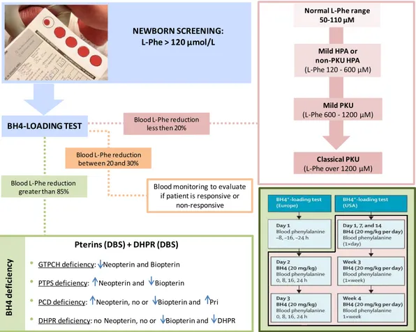
Today, the BH4 loading test is an important tool not only for the discrimination between HPA due to PAH deficiency or to BH4 deficiency, but also for the identification of PKU patients responsive to BH4 administration. An international online survey reported that in the 62% of the metabolic centers this test is an integral part of the diagnosis. In Figure 7 is reported the flow-chart commonly followed to perform the differential diagnosis of PKU or BH4 deficiencies, once HPA has been detected.
16
Figure 7. Differential diagnosis of PKU or BH4 deficiencies. Flow-chart employed to distinguish the different disorders of PAH and BH4 metabolism that can result in HPA. Blood L-Phe reduction less than 20% implies that the patient is a non-responder to BH4 treatment; if blood L-Phe reduced by 20-30 % treatment is continued for a further 1-3 weeks with daily monitoring of L-Phe; if blood L-Phe reduction is greater than 85%, BH4 deficiency is present and the patient can take advantage by BH4 treatment. In the box on the bottom right are reported
BH4 loading test protocols in Europe and USA (adapted from Blau et al., 2010). DBS= Dried blood spot; n= normal;
Neo= neopterin; Bio= biopterin; Pri= primapterin. *BH4 can be either tetrahydrobiopterin or sapropterin
(Kuvan®).
This test was used in Europe for almost 30 years and nowadays is an integral part of the neonatal screening tests. The positive effect of BH4 treatment on PKU patients was described for the first time on Japanese patients and then confirmed in prospective studies with large cohorts of patients (Bernegger and Blau, 2002; Fiege and Blau, 2007; Levy et al., 2007). It is therefore clear that the rapid detection of BH4 responsive PKU patients is absolutely important for a quick treatment with oral administration of sapropterin dihydrochloride-6R-BH4- (Kuvan®, BioMarin Pharmaceutical Inc), in order to obtain decreases of blood L-Phe levels.
The frequency of BH4 responsiveness is higher in patients with non-PKU HPA or mild PKU because of the residual enzyme activity of PAH gene; on the other hand, patients with classical PKU, which typically display absent or very low PAH activity, unlikely respond to BH4 treatment.
There are some differences about the application criteria of the test between different metabolic centers: for instance, about 78% of the centers use the test in all age groups and only 11% of centers on pregnant PKU woman. At the same time a dosage of 20 mg/kg is used in 92% of the centers and the duration of the test can vary from 24 h (33%), 48 h (24%), 72 h
Classical PKU (L-Phe over 1200 µM) Mild PKU (L-Phe 600 - 1200 µM) Mild HPA or non-PKU HPA (L-Phe 120 - 600 µM)
Normal L-Phe range 50-110 µM
NEWBORN SCREENING: L-Phe > 120 µmol/L
BH4-LOADING TEST
Blood monitoring to evaluate if patient is responsive or
non-responsive
Pterins (DBS) + DHPR (DBS) • GTPCH deficiency: Neopterin and Biopterin
• PTPS deficiency: Neopterin and Biopterin
• PCD deficiency: Neopterin, no or Biopterin and Pri
• DHPR deficiency: no Neopterin, no or Biopterin and DHPR
B H4 d e fi ci e n cy
Blood L-Phe reduction greater than 85%
Blood L-Phe reduction between 20 and 30%
17
(16%) and, in some centers - especially from US - from 1 to 4 weeks while in Europe shorter tests were favorite.
The test should be performed early after birth and before the introduction of the low L-Phe diet, so as blood L-Phe variations upon BH4 treatment are more evident. Blood L-Phe must be over 400 µM in order to avoid false negative or false positive results (Belanger-Quintana et al., 2011). Thus, older patient who are already on dietary regimen must increase the protein intake before and during the testing period, or should undergo a concomitant L-Phe load, consisting in a single administration of 100 mg L-Phe/kg BW (Blau, 2008; Blau et al., 2010). In European centers, a reduction of 30% of L-Phe after a twice administration of 20 mg/kg BH4 is considered a positive response while a decrease under the 20% as negative response. For a reduction in the range between 20% and 30% a daily monitoring of L-Phe for about 3 weeks is recommended (Belanger-Quintana et al., 2011). Although the test is effective at all ages, its sensitivity in newborns has been questioned due to liver immaturity and because only 24h protocols can be employed at this age (Belanger-Quintana et al., 2011). Performing the analysis as soon as possible allows the early introduction of the restricted diet in non-responders, favoring breastfeeding or natural protein intake in responder patients but, at the same time, implies the possibility to miss slow responders (who are mistakenly considered negative to the test). Moreover, the association between genotype and responsiveness to BH4 is not really true because if on one side it can identify classic PKU, in all the other situations, above mentioned, it difficulty predict who will respond to the treatment (Blau et al., 2010). Therefore, it is advised to repeat the test according to longer protocols after 3 months of life, that is when the liver has reached complete maturity and longer testing protocols can be applied (Belanger-Quintana et al., 2011). The BIOPKU database (www.biopku.org) reports all mutations that are correlated with BH4 responsiveness.
PATHOGENIC MECHANISMS OF HPA
Brain development and behavioral outcomes
The main clinical manifestations are due to the disruption of PAH metabolism that causes accumulation of high levels of L-Phe in the blood, its excessive and toxic concentration in the brain together with low levels of L-Tyr and its metabolites, which lastly affect different aspects of brain functioning. The effects of liver PAH mutations on the ability to maintain L-Phe homeostasis have been well described and the main clinical effects are related to the normal development of the brain and the physiological development of cognitive functions (Kayaalp et al., 1997). Untreated HPA is the most common biochemical cause of mental retardation (intelligence quotient, IQ <30), seizures, microcephaly, epilepsy, motor deficits, severe intellectual disability and behavioral disturbances, including psychotic, autistic, and aggressive disorders (Mitchell et al., 2011; Bone et al., 2012). Thus, patients with PKU have lower IQ scores than normal subjects and they present other deficits in various neuropsychological functions such as working memory, cognitive and executive functions (Blau et al., 2010; Feillet et al., 2010). All these conditions are due to both aminergic neurotransmitters depletion, i.e. dopamine (DA), serotonin (5-HT), and myelin impairment. As extensively reviewed by several authors (Surtees and Blau, 2000; Bone et al., 2012), many of which use PKU mouse models
18
(Fiori et al., 2017; Pascucci et al., 2002, 2008), the excessive L-Phe exposure is responsible for the altered development of the brain architecture which include abnormal myelination, cortical plate width and altered dendritic arborization together with a reduced number of synaptic spines. In addition, the exposure to high concentration of L-Phe makes the already formed myelin unstable, thus demyelinated axons undergo a reverse maturation, with consequent neuronal dysfunction (Cleary et al., 1995). Myelin is a metabolic active membrane produced by oligodendrocytes and it plays an important role for the fast transmission of action potentials. White matter pathology characterizes the brain of untreated PKU patients where neurological deterioration is evident and the impact of metabolic control on impairment of myelination process is related to specific brain areas. Therefore, in childhood the injury of visuospatial processing is more evident because occipital regions are the first myelinated area during development while the frontal regions are myelinated later, so the damage of complex executive functions is more evident during adolescence and adulthood (Klingberg et al., 1999; Gogtay et al., 2004; Best and Miller, 2010). In addition, the development of the cerebral cortex occurs following a precise sequence of events, well defined in time and space, especially as regards the synapses and dendrites formation in the prefrontal cortex. As reported in a study conducted on mouse model of PKU (Pascucci et al., 2008), during the critical developmental period (PND 14-21), different availability of brain amines, with an initial increase of catecholamines and serotonin which then decrease and return to adult levels, has been observed. This period represents the most susceptible phase to L-Phe-induced damages, as extensively demonstrated by studies on animal models (Goldman-Rakic and Brown, 1982; Thomas et al., 1995; Zhou et al., 1995; Berger-Sweeney and Hohmann, 1997; Chugani et al., 1999; Puglisi-Allegra et al., 2000; Herlenius and Lagercrantz, 2001, 2004; Cabib et al., 2003; Pascucci et al., 2008, 2009, 2012; Andolina et al., 2011).
Indeed, before acting as neurotransmitters, biogenic amines represent fundamental signals for the correct early development of the brain (Lauder, 1993) suggesting that a deficit in the availability of these amines during the critical periods of development, particularly around the third week of life in the murine models of PKU, is associated with cognitive dysfunction (Pascucci et al., 2008). These observations have been confirmed by several studies that highlight how development of the synapses, the growth of the dendritic tree and its remodeling (Van Eden and Uylings, 1985; Huttenlocher, 1991; Vitalis and Parnavelas, 2003) are dramatically affected by the decrease of amines levels in the critical period. In particular, 5-HT was the first neurotransmitter for which the role in brain development, dendritic spines formation and maintenance and amelioration of synaptic connectivity during postnatal life, has been demonstrated (Mazer et al., 1997; Whitaker-Azmitia, 2001; Sodhi and Sanders-Bush, 2004).
On the other hand, the decreased levels of neurotransmitters, including dopamine, are related to cognitive and behavioral disabilities (Diamond, 1996; Puglisi-Allegra et al., 2000; Pascucci et al., 2002; Joseph and Dyer, 2003). L-Phe belongs to the group of the Large Neutral Amino Acids (LNAAs), with valine, leucine, isoleucine, threonine, histidine, tryptophan, methionine and tyrosine. All these amino acids share a common selectively predominant carrier system, the L-amino acid transporter-1 (LAT-1), to cross the blood-brain barrier (BBB) and enter into
19
the brain (Blau et al., 2010). Binding of LNAA to this transporter is a dynamic and competitive process (Pardrige, 1998; Boado et al., 1999; Smith, 2000), in fact for each LNAA taken into the brain another one is excreting (Zielke et al., 2002). Across species LAT-1 appeared to have a higher affinity for L-Phe than the other LNAAs, and this is particularly marked for the human species, making it more susceptible to the negative effects of HPA. In fact, an excessive circulating amount of L-Phe has the ability to saturate the LAT-1 transporter, thanks to its lowest km value for the carrier respect the other LNAAs, resulting in a L-Phe overload and decreased amount of the other LNAAs, particularly L-Tyr and L-Trp, in the brain (Surtees and Blau, 2000; Blau et al., 2010). At the same time, non-L-Phe LNAA export from the brain in exchange for blood L-Phe is increased (de Groot et al., 2010), carrying a reduction in cerebral protein synthesis for reduced availability of these non L-Phe aminoacids (Pardridge, 1998; van Vliet et al., 2015). All this evidence explains why the brain is vulnerable to HPA.
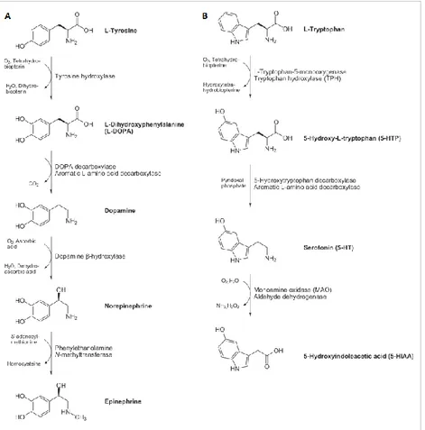
The LNAAs L-Tyr and L-Trp besides their role in protein synthesis, are also precursors for neurotransmitters, namely dopamine (DA), norepinephrine (NE), epinephrine and serotonin (5-hydroxytryptamine, 5-HT) respectively (Figure 8) (Surtees and Blau, 2000). Dopamine plays an important role in motor and cognitive functioning; norepinephrine is involved in learning and memorization processes, in the arousal of attention, fear and anxiety, and in the development of the maternal behaviour in females; serotonin is important for neuronal proliferation, synaptogenesis and morphogenesis (Herlenius and Lagercrantz, 2001, 2004). Thus, there are two possible mechanisms by which L-Phe alters brain functioning: if on the one hand the increased L-Phe concentration in brain results in a decreased level of the other LNAAs including L-Tyr and L-Trp, as described above, on the other hand it acts as a competitive inhibitor of the other two amino acid hydroxylases, TyrOH and TrpOH (McKean, 1972; Curtius et al., 1981; Surtees and Blau, 2000; Ogawa and Ichinose, 2006; Pascucci et al., 2008), generating a lack of their products. In fact, high brain L-Phe was reported to negatively affect the activity of the other hydroxylases (Surtees and Blau, 2000).
The idea that a deficiency of 5-HT neurotransmitter plays an important role in neurological disorders due to HPA (Shimada et al., 1993) is supported by the fact that it is also the most reduced amine in the brain - about 50% - compared with 40% reduction of NE and 30% of DA (Pascucci et al., 2008). In particular, in a study of Pascucci et al. (2008) it has been demonstrated that the reduction of 5-HT around 3 postnatal week overlaps with a critical period for synaptogenesis and dendritic development, thus compromising maturation of prefrontal cortex (PFC) neurons with the subsequent alteration of the cognitive performance. Severe lack of whole brain 5-HT during critical post-natal periods and deficits in the level of its immediate and limiting precursor 5-hydroxytryptophan (5-HTP), is not connected to a decrease in its initial amino acidic precursor L-Trp; this evidence support the hypothesis of TrpOH activity inhibition exerted by L-Phe excess, rather than a hampered access of L-Trp across the BBB (Ogawa and Ichinose, 2006; Pascucci et al., 2009), thus confirming a minor involvement of L-Trp in the L-Phe induced alterations (Pascucci et al., 2002). On the contrary, dopamine and its precursor L-3,4-dihydroxyphenylalanine (L-DOPA) are the less affected by HPA because, when L-Tyr levels are abnormally low and L-Phe is extremely high, the latter can serve as TyrOH substrate for the production of L-DOPA (Joseph and Dyer, 2003; Fernstrom
20
and Fernstrom, 2007). Nonetheless, the reduction of dopamine synthesis in prefrontal cortex often coexists with depressive symptoms which in turn worsen with an increase of L-Phe level, as observed in the study by Sharman and colleagues (2012).
Figure 8. Biosynthetic pathways of neurotransmitters lacking in PKU patients’ brain. (A) Synthesis of catecholamines (dopamine, DA, norepinephrine, NE, and epinephrine); (B) synthesis of serotonin (5-hydroxytryptamine, 5-HT). Adapted from http://www.hdri-usa.com/.
All the neurological alterations encountered in PKU patients ultimately account for deficits commonly belonging to the field of the executive functioning, including also response speed, academic abilities, language-related tasks (including reading and arithmetic), problem solving ability, attention, interhemispheric transfer of information, and visuo-spatial and visual-motor abilities, as observed by Scriver et al. (1995) and then extensively reviewed in the works by Bone et al. (2012) and by Huijbregts et al. (2013). In a meta-analysis study, processes such as planning, working memory, inhibition, processing speed, and cognitive flexibility were found impaired in early diet-treated patients, compared to controls (DeRoche et al., 2008).
The psychological and psychiatric problems documented in adolescent patients concern the area of social life, with negative findings in terms of autonomy, self-esteem, frustration threshold, school achievements, attention, mood disturbances, depression and anxiety (Weglage et al., 1992), even in early treated children (Brumm et al., 2010). An intermittent dietary therapy as well as high levels of L-Phe are associated with a higher incidence of behavioral problems. Untreated individuals show more severe symptoms such as autism, hyperactivity, aggression, social withdrawal, anxiety, depression, psychosis, and profound intellectual disability (Brumm et al., 2010), whereas adult patients early treated in childhood
21
display generalized depressed and anxious mood, lack of autonomy, low self-esteem and a tendency to social isolation; phobias are also typical and the most common one is agoraphobia (Waisbren and Levy, 1991; Pietz et al., 1997; Brumm et al., 2010).
Adequate control of blood L-Phe concentration is therefore very important for the prevention of brain deficit. Children with poor metabolic control (L-Phe >400 µM) have reduced executive functions while children with PKU have behavioral abnormalities, motor dysfunction (Arnold et al., 1998) and memory impairment (White et al., 2002). In addition, even if several publications showed a correlation between blood L-Phe fluctuations and intellectual outcomes, cognition or executive functions, no correlation has been found between this fluctuation and patients IQ (intelligence quotient) even if it has been identified an influence on these parameters (Cleary et al., 2013). However, some studies have shown that high blood L-Phe fluctuation in patients with PKU are associated with lower neurocognitive outcome. Crucial for the improvement of cognitive function is the metabolic control; indeed, a meta-analysis of five studies on PKU patients and control showed a significant inverse correlation between the IQ score and L-Phe levels. This correlation is especially clear during the critical developmental period (age 0-12 years), even in early treated children: each 100 µM rise in L-Phe concentration corresponded to a 1.3-3.1 IQ point decline (Burgard, 2000). This result has been confirmed by a meta-analysis study on children treated since the neonatal age where the IQ decreases of about 1.9-4.1 point for each 100 µM increase in L-Phe (Blau et al., 2010); a similar correlation was also found between lifetime L-Phe levels and IQ scores in early-treated individuals (Waisbren et al., 2007). This condition is due to both dopamine depletion and myelin impairment.
In addition, HPA includes the reduction of pyruvate kinase activity in the brain (Hörster et al., 2006), the alteration of glutamatergic neurotransmission (Martynyuk et al., 2005), the reduction of enzyme 3-hydroxy-3-methyl coenzyme A reductase (HMG-CoA reductase) (Shefer et al., 2000) as well as the impairment of monoamine oxidase B activity (Ghozlan et al., 2004).
L-Phe influence on cholesterol biosynthesis and obesity
L-Phe levels play an important role in the inhibition of the rate-limiting enzyme of the cholesterol biosynthetic pathway in liver and brain, namely 3-hydroxy-3-methylglutaryl-CoA reductase (HMG-CoA reductase; EC 1.1.1.88), reducing the synthesis of mevalonic acid (Castillo et al., 1988). The resulting hypocholesterolemia is hypothesized to have a protective effect against cardiovascular diseases in adults (Williams et al., 2008), but even if PKU children have lower blood total cholesterol and LDL levels with respect to healthy subjects, cardiovascular risk has been reported to be the same (Verduci et al., 2016). Elevated levels of L-Phe have been showed to decrease coenzyme Q10 (ubiquinone-10; CoQ10) concentrations
both in plasma and in lymphocytes. This coenzyme is involved in many functions i.e. acting as a cofactor in the mitochondrial electron transport chain, preventing LDL oxidation and representing an antioxidant molecule in mitochondria and lipid membranes (Colomé et al., 2002).
22
The PKU patients, in particular female, represent those with the highest incidence of obesity but the correlation is not clear yet (Belanger-Quintana and Martínez-Pardo, 2011; Burrage et al., 2012; Rocha et al., 2012; Robertson et al., 2013).
MATERNAL PKU
Non-controlled levels of L-Phe during pregnancy are teratogenic for the fetus and can increase the risk of miscarriage (American Academy of Pediatrics: Committee on Genetics, 2001; Blau et al., 2010). This condition, called maternal phenylketonuria syndrome or maternal PKU (MPKU), was firstly described over 60 years ago (Pinto et al., 2017) and is responsible for intrauterine growth retardation, spontaneous abortion, intrauterine fetal death (IUFD), congenital heart disease, developmental delay and other important fetal alterations (Levy and Ghavami, 1996; Rouse et al., 1997; Williams et al., 2008; Prick et al., 2012). Babies delivered by mothers under MPKU condition show microcephaly, congenital heart disease (CHD), intellectual or developmental disabilities (IDDs), and facial dysmorphism (FD) together with low birth weight (defined as small for gestational age, SGA) (Lenke and Levy, 1980; Levy and Ghavami, 1996; Rouse et al., 1997). Adequate control of L-Phe levels is important not only during pregnancy but also before conception because the toxic effect of this amino acid is dangerous in early stages of pregnancy, especially during the first weeks of embryogenesis. To this purpose, it is essential that affected women follow a strict low L-Phe diet for several months before conception, in order to stabilize the levels of this amino acid between 100 and 360 μmol/L, thus preventing teratogenic effects on the fetus (Lee et al., 2005); moreover, it is essential to maintain an optimal blood L-Phe level throughout the all pregnancy.
Despite during pregnancy the phenylalanine tolerance is slightly increased thanks to the activity of fetal PAH - as has been observed during the second trimester of pregnancy when L-Phe levels decrease and tolerance of proteins intake increased (Prick et al., 2012) - weekly or biweekly controls of L-Phe blood levels remain fundamental to avoid fetal impairments (Australian Society for Inborn Errors of Metabolism). At the same time is important that these women receive an adequate energy intake, in terms of proteins, fats, carbohydrates and multivitamin complexes, vitamin B12 and folic acid, in order to guarantee the best conditions for fetal growth (Koch et al., 2000).
Although some studies have shown that children born by women with untreated concentrations of L-Phe lewer than 400 μmol/L, may be normal, the "Maternal PKU Collaborative Study" (MPKUCS) reports that between children born from women with levels of L-Phe between 120 and 360 μmol/L, 6% showed microcephaly and 4% showed a delay in post-natal growth. If the concentration of L-Phe exceeds 900 μmol/L, the risk of microcephaly rises to 85%, post-natal growth delay rises to 51% and intrauterine growth retardation to 26%. For all these reasons, strictly controlled plasma L-Phe levels should be necessary (Rouse and Azen, 2004). However, a common problem is the difficulty of sick women in adhering to the strict diet for poor intellectual and social skills (Koch et al., 2000). For this reason, a pilot project has been developed to provide a special support and education about the importance of diet control during pregnancy with the aim of guaranteeing a good health for the newborn
23
child (Waisbren et al., 2000). PKU mothers bearing non-PKU babies (i.e. healthy carriers of a single mutated allele) are encouraged to breastfeed their children without restriction, since the single non mutated copy of the PAH gene is sufficient to metabolize the amount of L-Phe introduced with breast milk (NIH consensus panel, 2001), also because breast milk contains only 43 mg L-Phe/dl compared to 59-73 mg/dl of infant formulas and 164 mg/dl of cow’s milk (Berlin et al., 1995).
Following all such recommendations it is possible for PKU mothers to have children with the same expectancy of cognitive development as non-PKU people (Levy and Ghavami, 1996).
THERAPEUTIC STRATEGIES
Prognosis and outcome depend on time of diagnosis, type of mutations and quickness of intervention. In fact, the goal of the various therapeutic strategies is to rapidly restoring the normal levels of L-Phe and L-Tyr in the circulation, in order to eliminate biochemical abnormalities, improve neurological and psychological performance, and prevent the syndrome of maternal PKU (Williams et al., 2008). Clinical goals are not only aimed to reduce L-Phe levels in blood but also to limit its negative effect on other brain aminoacid concentrations. Nevertheless, there is great discrepancy between the different European countries and clinical centers in defining the level of L-Phe beyond which it would be appropriated to intervene (Table 1), especially during the first decade of life, the most important period of development (van Spronsen et al., 2009; Blau et al., 2010).
This discrepancy further increases beyond the first decade of life, where the gap between Europe and U.S.A. becomes more pronounced (van Spronsen et al., 2009; Blau et al., 2010). The most common concentrations identified to this purpose are 360 µM, 400 µM and 600 µM (Blau et al., 2010) and the L-Phe level considered safe is in the range between 120 and 360 µM, at least until 12 years of age (Koch et al., 1996; NIH consensus, 2001), with the upper limits rising up to 900 µM after the 12th year of life (NIH consensus, 2001). However, there is
great inconsistency about the target range to be reached in adolescence and adulthood, resulting in a wide spectrum of disease management and outcomes (Ahring et al., 2009; Blau et al., 2010) and some studies highlighted the greater importance of considering L-Phe/L-Tyr ratio rather than the concentration of L-Phe alone, since is considered more involved in the impairment of executive functions (Sharman et al., 2010).