Pisa
Classe di Scienze
Corso di Perfezionamento in Neurobiologia
2006-2009
PhD Thesis
Food restriction enhances brain
plasticity in adult rats
Candidate
Maria Spolidoro
Supervisor
Prof. Lamberto Maffei
INTRODUCTION 4
The visual system in mammals . . . 7
Phototransduction and information processing in the retina . . . 7
Central visual pathways . . . 11
The Rat Visual System . . . 15
Plasticity in the visual cortex 18 Visual system plasticity and amblyopia . . . 18
Extracellular matrix and plasticity . . . 20
Inhibition and plasticity . . . 21
Environmental Enrichment and fluoxetine treatment: two different meth-ods to enhance plasticity and to reduce inhibition . . . 24
Food restriction and brain health . . . 30
Some notes on metabolism . . . 31
Beneficial effects of food restriction . . . 32
Molecular mechanisms of neuroprotection induced by FR . . . 38
MATERIALS AND METHODS 51 Animals treatment . . . 51
Corticosterone measurement . . . 52
Elevated plus maze test . . . 52
Locomotor activity . . . 53
In vivo electrophysiology . . . 53
Visual cortex LTP . . . 54
In vivo brain microdialysis . . . 55
High Performance Liquid Chromatography (HPLC) . . . 56
Western blotting . . . 56
Immunohistochemistry . . . 58
RESULTS 60 Effects of food restriction on food intake and body weight . . . 60
Corticosterone levels in the blood of food restricted rats . . . 60
Restoration of OD plasticity following FR in adult rats . . . 63
FR is accompanied by a decrease in GABA release and GAD65 expression in the visual cortex . . . 65
FR restores WM-evoked layer III LTP in the visual cortex and enhances CA3-CA1 LTP in the hippocampus of adult rats . . . 65
FR enhances histone acetylation in the visual cortex and hippocampus of adult rats . . . 69 Effects of FR on extracellular matrix remodeling and neurotrophin expression 70
DISCUSSION 71
The term plasticity has been used in brain science for well over a century and it refers to the changes that occur in the organization of the brain as a result of experience (for an historical review on the term plasticity see Berlucchi and Buchtel 2009). The word experience accounts for various form of behavioural modifiability, either short or long-lasting, including maturation, adaptation to a mutable environment, learning and compensatory adjustments in response to functional losses during ageing or brain damage.
Every single behavioural task performed by the mature nervous system, from the perception of the sensory input and the control of motor output to cognitive func-tions such as learning and memory, depends on the definite interconnecfunc-tions of many millions of neurons. Such a defined wiring is largely controlled by molecular programs, but the precise matching of presynaptic neurons to postsynaptic targets widely depends on neural activity, either spontaneous or evoked by sensory-driven experience (Good-man and Shatz 1993; Katz and Shatz 1996). There are periods in early postnatal life, the so called critical periods (CPs), during which neural circuits display a heightened sensitivity to certain environmental stimuli and are shaped by natural sensory experi-ence.
These periods of high plasticity have been found in all species tested so far, from humans to Drosophila (Barth et al., 1997) and affect the development of the primary sensory systems (Hensch, 2004; Berardi et al. 2000).
One crucial question about CP plasticity is why experience is so effective in eliciting great structural and functional changes only during a restricted period of postnatal life. What determines the beginning of the CP? What makes the CP end?
in-dicating that the adult brain is not hard-wired with fixed and immutable neuronal circuits. On the contrary, it keeps a certain degree of plasticity in response to proper stimuli (for a review see Spolidoro et al., 2009). Given these important results, studying the mechanisms that reduce plasticity in adulthood has begun even more challenging considering the great impact that it could have in clinical neurology. Furthermore, the possibility to influence the brain by simply changing some aspects of the individual lifestyle has recently emerged (Sale et al, 2007a). This opened a new fascinating field in the therapeutic neuroscience indicating that non-invasive treatments could exert significant and helpful benefits both at the level of rehabilitation from injuries and amelioration of everyday life conditions.
Nutrition is a fundamental component of our life. The quality and quantity of food consumed by each individual is critical for the development and maintenance of its general physical structure. A great number of studies have identified several spe-cific dietary components that are essential for the correct development of the central nervous system (Mattson et al., 2003). Interestingly, it has been demonstrated that simply reducing the caloric intake can lead to an extension of the mean and maximum lifespan in a wide range of organisms including yeasts, worms, fruit flies, rodents and monkeys (Masoro, 2001). This increase in longevity is often accompanied by an ef-fective counteraction of the aging process both at the level of body healthness with reduced incidence of age-related cardiomyopathy, diabetes, hypertension and neoplas-tic processes (Masoro, 1993; Mattson et al, 2002) and at the level of brain function with a delay in the onset of learning and memory decline (Idrobo et al., 1987; Stewart et al, 1989) and suppression of LTP deficits in the hippocampus (Hori et al., 1992; Eckles-Smith et al, 2000).
The mechanisms by which dietary restriction leads to increased maximal and average lifespan and to decrease of age-related pathologies remain unclear. One fasci-nating hypothesis is that the reduction in caloric intake could be accompanied or could even exert its effects by influencing brain plasticity. The main purpose of my study was to test this possibility and to give, for the first time, evidence that food restriction (FR) increases brain plasticity in adult, not aged, healthy animals.
mammals visual system, the paradigmatic model I used to study plasticity in the CNS. The second chapter will deal with the reinstatement of plasticity to the adult and some of the treatments which have been used in the last years in order to achieve this goal. In the third chapter the general benefits of FR will be analyzed in depth and a discus-sion of the molecular mechanisms involved in the longevity effects of caloric restriction will be provided. Finally I will explain the details of my work, presenting my results an examining their significance and implications.
Phototransduction and information processing in the retina
Visual perception occurs in two stages. Light entering the cornea is projected on the back of the eye where it is converted into an electrical signal by a specialized sen-sory organ, the retina. These signals are then sent through the optic nerve to higher centres in the brain for further processing.
Anatomical organization of the retina. The retina bears careful examination for several reasons. First, it is a useful model system for understanding sensory trans-duction. Indeed, light is converted in electrical signals by specialized retinal neurons called photoreceptors. Second, unlike other sensory structures, such as the cochlea or somatic receptors in the skin, the retina is not a peripheral organ but part of the cen-tral nervous system. Compared to other brain regions, however, the retina is relatively simple. It contains only five major classes of neurons linked in an intricate pattern of connections, but with an orderly, layered anatomical arrangement. This combination of physiological diversity and relatively simple structural organization makes the retina useful for understanding how information is processed by complex neural circuits in the brain.
The eye is foremost an optical device designed to focus the visual image on the retina with minimal optical distortion. Light is focused by the cornea and the lens, then traverses the vitreous humour that fills the eye cavity before being absorbed by the photoreceptor cells. The retina is apposed to the pigment epithelium that lines the back of the eye. Cells in the pigment epithelium are packed with the black pigment melanin, which absorbs any light not captured by the retina, thereby preventing it from being reflected back to the retina (which would degrade the visual image).
Pigment epithelial cells also assist photoreceptors with important aspects of their metabolism. For this reason, photoreceptors directly contact the pigment epithe-lium, while the other retinal cells are closer to the lens.
In one region of the retina, the fovea, the cell bodies of the proximal retinal neurons have been shifted to the side, enabling the photoreceptors to receive the visual image in its least distorted form. Nasal to the fovea is the optic disc where the optic nerve fibers leave the retina. This region has no photoreceptors and therefore creates a blind spot in the visual field.
Photoreceptors. The human retina contains two types of photoreceptors, rods and cones. Cones are responsible for day vision while rods mediate night vision, they function in the dim light (for example at dusk or night) when most stimuli are too weak to excite the cone system.
The brain obtains information about color by comparing the responses of three types of cones, each with a visual pigment that is more senisitive to a different part of the spectrum. In contrast, there is only one rod pigment so that all rods respond in the same way to different wavelenghts. Rod vision is therefore achromatic.
Rods and cones have similar functional regions: 1) a region specialized for pho-totransduction, called the outer segment (because it is located at the outer or distal surface of the retina); 2) a region containing the cell’s nucleus and most of its biosyn-thetic machinery, called the inner segment; 3) a synaptic terminal that makes synaptic contact with the photoreceptor’s target cells.
The outer segments of rods and cones are effective light-catchers because they are densely packed with light-absorbing visual pigments.
The absorption of light by visual pigments in rods and cones triggers a cascade of events that eventually leads to a change in ionic fluxes across the plasma membrane of these cells and a consequent change in membrane potential. A key intermedite in this cascade is the cyclic nucleotide 3’-5’ cyclic guanosine monophosphate, or cGMP.
Phototransduction. Phototransduction occurs in three stages: 1) light activates visual pigments; 2) these activated molecules cause the stimulation of cGMP
phos-phodiesterase, an enzyme that reduces the cytoplasmic concentration of cGMP; 3) the reduction in cGMP concentration closes the cGMP-gated channels, thus hyperpolariz-ing the photoreceptor.
The change in the photoreceptor’s membrane potential during illumination is determined both by the change in the current that flows through the cGMP-gated
channels and by a current flowing across the photoreceptor membrane through K+
-selective, nongated (leakage) channels that are like those of other neurons. In darkness the high resting levels of cGMP in the cell keep the cGMP-gated channels open, al-lowing an inward current of about 50 pA, largely carried by Na+
ions, to flow into the cell. This steady inward current, called the dark current, maintains the photoreceptor membrane potential at around 40 mV.
When light reduces cGMP and the cGMP-gated channels close, the inward Na+
current that flows through these channels is reduced, thus hyperpolarizing the cell.
Retinal interneurons and ganglion cells. The output neurons of the retina are the ganglion cells. Their axons form the optic nerve, which project to the lateral geniculate nucleus (LGN) and to the superior colliculus as well as to brain stem nuclei. Unlike photoreceptors, which respond to light with graded changes in membrane potential, ganglion cells transmit information as trains of action potentials. Sandwiched between the photoreceptors and the ganglion cells are three classes of interneurons: bipolar, horizontal and amacrine cells. These cells transmit signals from the photoreceptors to the ganglion cells. They also combine signals from several photoreceptors. Thus, the electrical responses evoked in ganglion cells depend critically on the precise pattern of the light that stimulates the retina and how this pattern changes with time.
Ganglion cells are never silent, even in the dark, but light modulates their spon-taneous activity. Each ganglion cell responds to light directed to a specific area of the retina. This area is called the receptice field of the cell. The receptive field of a ganglion cell (or any other cell in the visual pathway) is that area of the retina where stimulation of photoreceptors causes either an increase or decrease of the ganglion cell’s firing rate. Ganglion cell receptive fields have three important features:
the primate retina, where visual acuity is greatest, the receptive fields are small, at the periphery of the retina, where acuity is lower, the fields are larger;
• They have a center and an antagonistic surround;
• Ganglion cells process visual information in two parallel pathways. Two classes of ganglion cells can be distinguished by their response to a small spot of light applied to the center of their receptive field. On-center ganglion cells fire few action potentials in darkness, and light directed to the center of their receptive field increases their firing rate. Light applied to the surround inhibits the effect produced by illumination of the center; the most effective inhibitory stimulus is a ring of light on the entire surround. Off-center ganglion cells are inhibited by light applied to the center of their receptive field. Light excites an off-center ganglion cell when it is directed to the surround of the receptive field.
Each type of interneuron in the retina (horizontal, bipolar and amacrine cells) palys a specific role in shaping photoreceptor signals as they are transmitted through the retina. The bipolar cells represent the most direct pathway between the receptors and ganglion cells.
Visual information is transferred from cones to ganglion cells along two types of pathways in the retina. Cones in the center of a ganglion cell’s receptive field make direct synaptic contact with bipolar cells that in turn directly contact the ganglion cells (direct or vertical pathway). Signals from cones in the surround of the ganglion cell’s receptive field are conveyed to the ganglion cell by means of horizontal and amacrine cells (lateral pathways). Horizontal cells tranfer information from distant cones to nearby bipolar cells. Some type of amacrine cells tranfer information from distant bipolar cells to the ganglion cells.
Bipolar cells are the key interneuron in the retina. They have complex receptive field properties like those of ganglion cells.
The cones in the receptive field center of a bipolar cell appear to be directly connected to the bipolar cell. On-center bipolar cells depolarize, while off-center bipolar cells hyperpolarize when light stimulates cones in the center of their receptive field. In
contrast, the inputs of cones in the bipolar cell’s surround are relayed by horizontal cells, which can respond to inputs from distant sources because they have large dendritic trees and because they are electrically connected to other horizontal cells by gap junctions. When light stimulates cones in a bipolar cell’s surround, it produces the opposite response to that evoked by illumination of cones in the center.
The cones release a single neurotransmitter, thought to be the glutamate, which has opposite actions on the two classes of bipolar cells. On-center bipolar cells are inhibited and off-center bipolar cells are excited (recall that the cone is depolarized in the dark).
Glutamate produces different responses in the two classes of bipolar cells by gating different ion channels.
The receptive field properties of a ganglion cell largely reflect those of the bipolar cells connected to it because each type of bipolar cell (on-center or off-center) makes excitatory synaptic connections with the corresponding type of ganglion cell.
Central visual pathways
The regions of the visual field are defined with respect to the two retinas. The regions of the retina are named with reference to the midline: the nasal hemiretina lies medial to the fovea, and the temporal hemiretina is lateral to the fovea. The visual field is the view seen by the two eyes without movement of the head. If the foveas of both eyes are fixed on a single point in space it is possible to define a left and a right half of the visual field. Light originating in the central region of the visual field enters both eyes, this area is called the binocular zone. In either half of the visual field there is also a monocular zone: light from the temporal portion of the hemifield projects only on the nasal hemiretina of the eye on the same side because the nose blocks this light from reaching the eye on the opposite side.
The axons of all the retinal ganglion cells stream toward the optic disc, where they become myelinated and together form the optic nerve. The optic nerves from each eye join at the optic chiasm. There, fibers from each eye destined for one or the other side of the brain are sorted out. Retinal fibers fro both eyes then enter each optic tract, which projects to three subcortical targets. Of the three subcortical regions receiving
direct input from the retina, only one, the LGN, processes visual information that ultimately results in visual perception. The pretectal area of the midbrain uses inputs from the retina to produce pupillary reflexes, whereas the superior colliculus uses its input to generate eye movements.
Lateral geniculate nucleus. The majority of retinal axons terminate in the LGN. Axons from the retina project through the optic chiasm, where the fibers from the nasal half of each retina cross the opposite side of the brain. The axons from ganglion cells in the temporal hemiretina do not cross.
Fibers from the right half of each retina (the nasal hemiretina of the left eye and the temporal hemiretina of the right eye) project in the right optic tract to the right LGN. Similarly, fibers from the left hemiretina of each eye project in the left optic tract to the left LGN.
Ganglion cells in the retina project in an orderly manner to points in the LGN, so that in each LGN there is a visuotopic representation of the contralateral half of the viasual field.
The LGN of primates contains six layers of cell bodies separated by intervening layers of axons and dendrites. The layers are numbered from 1 to 6, ventral to dorsal. An individual layer in the nucleus receives input from one eye only: fibers from the contralateral nasal hemiretina contact layers 1, 4, and 6; fibers from the ipsilateral temporal hemiretina contact layers 2, 3 and 5.
Receptive fields of neurons in the LGN are the same as those found in the retina: small concentric fields either on or off-center. Like the retinal ganglion cells, neurons in the LGN respond best to small spots of light within their receptive field center. This similarity of the receptive properties of cells in the LGN and those of retinal ganglion cells derives in part from the fact that each geniculate neuron receives its main retinal input from only a very few ganglion cell axons with very little transformation of the incoming information.
As in the retina, the on and off-center pathways in the LGN are independent.
receptive field properties change significantly is the primary visual cortex (V1). Like the LGN and superior colliculus the V1 in each cerebral hemisphere receives informa-tion exclusively from the contralateral half of the visual field.
The human visual cortex is about 2 mm thick and consists of six layers of cells (layers 1-6) between the pial surface and the underlying white matter. The cortex contains two basic classes of cells. Pyramidal cells are large and have long spiny den-drites; they are projection neurons whose axons project to other brain regions. Non pyramidal cells are small, stellate in shape, and have dendrites that are either spiny or smooth. They are local interneurons whose axons are confined to the primary visual cortex. The pyramidal and spiny stellate cells are excitatory and many use glutamate or aspartate as their transmitters; the smooth stellate cells are inhibitory and many contain γ-aminobutyric acid (GABA).
Once afferents from the LGN enter the V1, information flows systematically from one cortical layer to another, starting with the spiny stellate cells, which predom-inate in layer 4. These cells receive direct input from the LGN and project up to layers 2 and 3. Cells in layers 2 and 3 project down to pyramidal cells in layer 5, which then feed via axon collaterals to pyramidal cells in layer 6. The pyramidal cells of layer 6 complete the local excitatory circuit by sending axon collaterals to layer 4 to excite the inhibitory smooth stellate cells. These in turn contact and modulate the firing of the excitatory spiny stellate cells, completing an inhibitory feedback loop. Thus the spiny stellate cells distribute the input from the LGN to the cortex and the pyramidal cells feed axon collaterals upward and downward to integrate activity within the layers of V1.
Hubel, Wiesel and their collegues found that most cells above and below layer 4 respond only to stimuli that are substantially more complex than those that excite cells in the retina and LGN (Hubel and Wiesel 1959). The most astonishing finding was that small spots of light are completely ineffective in all layers of the visual cortex (except some regions called blobs). Cells in all regions except blobs respond only to stimuli that have linear properties, such as a line or bar. Hubel and Wiesel categorized the cells into two major groups, simple and complex, based on their reponses to linear stimuli. Simple cells resemble cells of the LGN, except that the on and off zones of
their receptive fields are rectangular with a specific axis of orientation. A stimulus with an orientation perpendicular or even oblique to the orientation of the cell’s receptive field will be ineffective. Every axis of rotation is represented for every retinal position. Hubel and Wiesel suggested that a rectilinear receptive field could be biult up from many circular fields in appropriate connections with stellate cells in the layer 4 of V1 (Hubel and Wiesel 1962). This idea has received direct support from studies by Stryker and collegues (1990) who found that the distribution of geniculate input on simple cortical cells predicts their axes of orientation.
The receptive fields of complex cells are usually larger than those of simple cells. These fields also have a critical axis of orientation, but the precise position of the stim-ulus within the receptive field is less crucial because there are no clearly defined on or off zones. Movement across the receptive field is a particularly effective stimulus for certain complex cells. Although some complex cells have direct connections with cells of layer 4, Hubel and Wiesel proposed that a significant input to complex cells comes from a family of simple cortical cells that have the same axis of orientation but slightly offset receptive field positions.
The convergent actions of cells in V1 are the initial steps in percetion. In its simplest form, this scheme suggests that each complex cell surveys the activity of a group of simple cells. The simple cells survey the activity of a group of geniculate cells, which themselves survey the activity of a group of retinal ganglion cells. The ganglion cells survey the activity of bipolar cells that survey a group of receptors. At each level, each cell has a greater capacity for abstraction than do the cells at the lower level.
At the lowest level of the system, the level of retinal ganglion and geniculate cells, neurons respond primarily to contrast. This elementary information is repatterned by the simple and complex cells of the cortex into rectangular fields with reltively precise line segments and boundaries. Thus, the stimulus requirements necessary to activate a cell become more precise at each level of the afferent system. In the retina and LGN stimulus position is important. In simple cells, in addition to position, the axis of orientation is important. In complex cells, whose receptive fields are larger, the axis of orentation is also important, but these cells have the ability to detect orientation over a wide range of positions. The V1 is organized in narrow colums, running from the pial
surface to the white matter. In each column there are simple cells with almost identical retinal positions and identical axes of orientation. For this reason these groupings are called orientation columns. Each orientation column also contains complex cells.
Detailed mapping of adjacent columns by Hubel and Wiesel, using tangential penetrations with microelectrodes, revealed a precise organization with an orderly shift in axis of orientation from one column to another. The systematic shifts in axis of ori-entation from one column to another is occasionally interrupted by blobs, peg-shaped regions of cells in layer 2 and 3 of V1. Cells in the blobs receive direct connections fom the LGN and are concerned with color.
In addition to columns devoted to axis orientation and blobs related to color, a third alternating system of columns is devoted to the left or the right eye. These ocular dominace columns are important for binocular interactions.
Hubel and Wiesel introduced the term hypercolumn to refer to a set of columns responsive to lines of all orientations from a particular region in space. A complete sequence of ocular dominance columns and orientation columns is repeated regularly and precisely over the surface of the V1. These columnar systems communicate with one another by means of horizontal connections that link cells within a layer. These connections integrate information over many millimeters of cortex. As a result, a cell can be influenced by stimuli outside its normal receptive field. Indeed, a cell’s axis of orientation is not completely invariant but is dependent on the context on which the feature is embedded. The psychophysical principle, called the contextual effect, whereby we evaluate objects in the context in which we see them, is thought to be mediated through horizontal connections.
The Rat Visual System
The general concept of the visual system operating as a set of pathways working largely in parallel can be applied to all Mammals. However, results and interpretations of visual studies in the widely used cat and primate should not be uncritically extrap-olated to the rat. There are some peculiarities of this species which are worth a note. The rat, a nocturnal murid rodent, has sophisticated and effectively functioning visual system. Its laterally placed eyes provide it with a panoramic view but there is
a binocular overlap, estimated to be 40-60◦ in front of the animal.
Although rods are the predominant photoreceptors, cones are also present (about 0,85% of photoreceptors) and the retina is therefore capable of functioning in both sco-topic and phosco-topic conditions (Cicerone, 1976; Sz´el and R¨ohlich, 1992). Of the two types of cones identified, the majority (93%) contains a photopigment with a peak sensitivity at about 500-520 nm (Deegan and Jacobs, 1993). Unlike other dichromatic animals, but like some other nocturnal rodents, the rat appears to have photoreceptors sensitive to ultraviolet light.
As already said, visual information is processed in the retina and sent to differ-ent structures of the cdiffer-entral nervous system through retinal ganglion cell axons, which represent the output of the retina. The largest retinal projection terminates in the dorsal LGN (dLGN). There is extensive literature on the small ipsilateral pathway in the rat that contributes to the binocular representation of the visual field in target nu-clei. Only about 3% of all ganglion cells project ipsilaterally (Jeffrey, 1984). In albinos the ipsilateral projection is even smaller (about 1,5%) (Ahmed et al., 1996; Dreher et al., 1985; Lund, 1965). Despite the lack of lamination in dLGN of the rat, several au-thors have discerned regional variations in the distribution of cells of different sizes, in the composition of afferent axons, and in the patterns of retrograde degeneration after cortical lesions. Reese (1988) emphasized that the nucleus consists of an outer shell located caudodorsally and an inner core constituting the ventromedial part. Ipsilateral axons with fast and medium conduction velocities terminate medially, in the inner core (Fukuda et al., 1981; Hale, 1980; Hayhow et al., 1962; Reese, 1988; Reese and Cowey, 1983).
In primates and cats the talamic input representing the two eyes parcel cortical layer 4 into alternating, equal-size stripes, allowing to distinguish a clear anatomi-cal organisation in columns of ocular dominance (Hubel and Wiesel, 1963; Shatz and Stryker, 1978). The major difference between the rat visual cortex and that of cat and other mammals is the lack of anatomical ocular dominance columns. However, Thurlow and Cooper (1988) found hints of a patchy organisation of ipsilateral and contralateral input in the visual cortex of the rat, using a functional mapping by means of deoxyglu-cose. This issue has been confirmed through electrophysiological techniques by Caleo
and collegues (1999).
The physiological properties of the visual cortical neurons of the rat are imma-ture at the opening of the eyes (postnatal day 15, P15) and develop gradually during the first month of postnatal life.
Briefly, in the rat, visual cortical responses are sluggish and variable at P17, in particular they present habituation, that is the tendency of cell response to diminish after several stimulations. These condition has been found also in kittens. Cell respon-siveness, evaluated in term of amplitude of cell discharge in response to an optimal stimulation, increases progressively from P19-23 to P30, when values are superimpos-able to adult values. Spontaneous discharge is almost absent at P17 and it increases to reach adult values at around P30.
Ocular dominance (OD) distribution does not change significantly through de-velopment. Indeed, the vast majority of visual cortical neurons are binocular and preferentially driven by the contralateral eye. The major component of age-dependent changes in OD distribution is the increase of monocular, contralaterally driven cells. Furthermore, there is a clear decrease in the number of unresponsive cells, which is pretty high in very young animals but rapidly declines, reaching adult values at P30.
Properties like selectivity for orientation and for the direction of movement are absent at P17 and they increse progressively to reach adult values at around P30.
Receptive fields (RF) in adult rats are small and well defined, but this is not the case of younger animals. At P17, RF are very large, they extend through almost all the binocular hemifield and they are difficult to define since cell responses are weak and tend to habituate. At P19-21 RF size is around 34 degrees (deg), while it reaches the values of 10 deg or less in the adult.
The visual acuity is about 0,5 cycles/degrees (c/deg) at P17 whereas it reaches the value of 1,0-1,2 c/deg in the adult. It has to be noticed that development of visual acuity is correlated with the decrease of the mean RF size (Fagiolini et al., 1994).
The visual cortex has been a model for plasticity in the CNS since the pioneering work of Wiesel and Hubel (1963). Still now, it is considered one of the best example of the nature-nurture interaction during brain development and, due to the relative simplicity of triggering sensory alteration in the visual system, it can be used as a paradigm of experience-dependent modifications of neuronal connections.
Visual system plasticity and amblyopia
One of the best described pathologies of the visual system development is am-blyopia. Also known as lazy eye, it has traditionally been defined as a decrease of visual acuity caused by pattern vision deprivation or abnormal binocular interaction for which no causes can be detected by the physical examination of the eye.
Amblyopia is estimated to afflict 1-4% of children, with recent large population studies falling in the range of 1.6-3.6%, and with evidence that the rate is even higher in medically underserved populations (for a review see Simons, 2005).
The problem is caused by either no transmission or poor transmission of the visual image to the brain for a sustained period of dysfunction or during early child-hood. Amblyopia normally only affects one eye, but it is possible to be amblyopic in both eyes if both are similarly deprived of a good, clear visual image.
This pathology can be distinguished in three main types depending on its eti-ology. It can be caused by deprivation of vision (vision-obstructing disorders such as congenital cataracts), by strabismus (misaligned eyes), or by anisometropia (different degrees of myopia or hyperopia in each eye).
As already said, the functional abnormalities have long been attributed to ab-normal visual experience in early life, and for this reason Hubel and Wiesel started
investiganting the effects of ocular occlusion and strabismus on the LGN and visual cortex of cats very soon after they had established the normal properties of single neu-rons recorded in these places. Their first experiment was to occlude, by suturing the eyelids, one or both eyes of kittens as soon as they opened (monocular deprivation, MD). Several weeks or months later they reopened the occluded eye and recorded from single neurons in the LGN or visual cortex.
Due to the decussation of retinal inputs, cells recorded from a normal visual cortex can be classified according to their relative response to the stimulation of the two eyes. Thus, Hubel and Wiesel indicated with the class 1 the cells that can be excited only by the contralateral eye, with class 7 the cells guided exclusively by the ipsilateral eye and with class 4 the cells equally driven by either eye. The other classes correspond to cells judged to have intermediate degrees of dominance of each eye.
With this paradigm Hubel and Wiesel demonstrated that closing one eye at the time of eye opening dramatically decreases the number of neurons of the visual cortex that respond to that eye and cortical cells become strongly dominated by the non-occluded eye.
Thus, apparently the disuse of an eye weakens the connections it makes with the LGN and visual cortex. Since usage of the pathway appears to be necessary to preserve normal functions, it may be thought that suture of both eyelids, or total visual deprivation by rearing in the dark, would produce even greater changes than those described. Nevertheless, these two treatments do not cause any change in term of ocular dominance distribution. This evidence has been classically interpreted calling into question a form of competition of the two eyes. In other words, the pathway from one eye to a cortical neuron is not disconnetted or impaired, unless there is another pathway from the other eye competing for control of the same cortical neuron.
This competition leads not only to physiological but also to anatomical rear-rangements. For example, in the lateral geniculate nucleus of rodents it has been observed a reduction in the number of projections to the visual cortex contralateral to the deprived eye accompanied by an hypotrophy of the neuronal cellular bodies (Domenici et al. 1993; Antonini et al. 1999).
neurons and in producing detectable morphological changes declines with age (Hubel and Wiesel 1970; Fagiolini et al. 1994; Prusky et al. 2000; Mataga et al. 2002; Prusky and Douglas 2003; Liao et al. 2004; Mataga et al. 2004). Even in the mouse, in which a greater potential for experience-dependent plasticity in the adult visual cortex has been recently demonstrated (Sawtell et al. 2003), plasticity in response to short term (3 days) MD is restricted to the critical period (for a review see Morishita and Hen-sch, 2008). Moreover, It has been demonstrated that if the visual defect is removed early in development (for example through reverse suture, i.e. opening of the formerly closed eye and deprivation of the open eye), the ensuing recovery is very good; on the contrary, recovery is very limited if defect removal is delayed to adulthood.
Despite decades of research, the cellular and molecular factors contributing to the decline in plasticity after the critical period are still poorly understood (see Berardi et al. 2003). In the last few years, several candidates have emerged as critical in reg-ulating developmental plasticity: the maturation of myelin (Mc Gee et al. 2005), the condensation of extracellular matrix molecules into perineuronal nets (Hockfield et al 1990; Pizzorusso et al. 2002) and the maturation of intracortical inhibition (Hensch et al. 1998; Huang et al. 1999; Hensch 2005). In particular, several papers converged in indicating the last two factors as key components not only for the regulation of critical period but also for the restoration of plasticity in the adult visual system.
Extracellular matrix and plasticity
A substantial amount of brain volume consists of extracellular space interposed between brain cells. This space is filled with a matrix of molecules interconnected and linked with membrane bound molecules. These interactions are essential for the me-chanical properties of brain tissue and are also able to activate intracellular signaling pathways. A number of studies, reviewed by Dityatev and Schachner (2003), have involved elements of this network, such as integrins, cadherins, NCAM, tenascins, and heparinsulfate proteoglycans, in synaptic plasticity (LTP and LTD) and in learning and memory processes. Furthermore, several studies have shown an important role for key components of the brain extracellular matrix (ECM), the chondroitin-sulfate proteoglycans (CSPGs), in OD plasticity of the visual cortex. CSPGs are major
con-stituents of the ECM of the CNS and comprise a core protein and chondroitin-sulfate glycosaminoglycan chains. CSPGs are inhibitory for axonal sprouting, and after injury they are upregulated in the CNS, with the effect of blocking axon regeneration (Brad-bury et al., 2002; Silver and Miller, 2004). During development, CSPGs condense at high concentration in lattice-like structures, denominated perineuronal nets (PNNs), which completely envelop visual cortical neurons.
PNNs are fenestrated at sites of synaptic contact, where they assume a perisy-naptic localization (Dityatev and Schachner, 2003). The process of condensation of CSPGs into PNNs begins during late development and is completed after the end of the critical period. Dark rearing, which prolongs critical period closure, also prolongs CSPGs condensation (Hockfield et al., 1990). Degradation of CSPGs from the adult visual cortex with the enzyme chondroitinase ABC reactivates ocular dominance plas-ticity in monocularly deprived adult rats, suggesting that the CSPG-enriched adult ECM exerts a powerful repressive control on OD plasticity (Pizzorusso et al., 2002). This could be partially due to a non-permissive action on the remodeling of visual cor-tex pyrmidal neurons, as suggested by the well documented effect of the ECM on spine motility (Majewska and Sur 2003; Mataga et al. 2004; Oray et al. 2004; Pizzorusso et al. 2006). However, as already said, toward the end of the critical period CSPGs con-dense at an extremely high concentration in PNNs surrounding parvalbumin-positive inhibitory interneurons (Hartig et al. 1992, 1999). Therefore, at least some of the effects of chondroitinase ABC could be mediated by modifications of intracortical inhibitory circuits occurring after PNNs degradation. This is a very interesting point, since it establishes a link with a second crucial regulating factor of plasticity, the inhibitory system.
Inhibition and plasticity
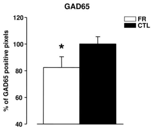
GABA (γ-aminobutirric acid) is the most abundant inhibitory neurotransmitter in the brain. It is synthesized by two distinct isoform of glutamic acid decarboxylase, GAD65 and GAD67 (Soghomonian and Martin 1998). The latter is the primary syn-thetic enzyme, it is localized to cell bodies and its deletion is lethal causing death at birth. The other isoform, GAD65, is concentrated in axon terminals and bound to
synaptic vescicles. GAD65 knockout mice are viable even if both overall GABA levels, and the amounts released by stimulation, are markedly reduced. Although the cortex is not epileptic (as occurs when inhibition is totally eliminated) neurons in these mice show prolonged responses to visual stimuli, suggesting that the inhibitory circuits are less effective. Interestingly, Hensch et al. (1998) found that these GAD65 knockout mice showed no shift in electrophysiologically measured ocular dominance preference when one eye was closed during the critical period. Remarkably, local infusion of di-azepam, a use-dependent GABA agonist, restored the ability of neurons to undergo an ocular dominance shift in response to eye closure, starting a normal critical period; this rescue of plasticity was possible at any age, which indicates that the beginning of the critical period is dependent on the proper level of inhibitory transmission. Con-versely, using the same approach, Fagiolini and Hensch (2000) provided strong evidence that increased intracortical inhibition is a major factor responsible for triggering the initial onset of the critical period. Indeed, premature enhancement of inhibition with diazepam just after the eye opening triggers the onset of a precocious critical period in normal animals. Importantly, once diazepam induced plasticity has occurred in a given animal, no further ocular dominance plasticity can occur later in life, suggesting again that diazepam initiate a normal critical period that, once overtaken, prevents subsequent plasticity.
Another evidence comes from the BDNF transgenic mice generated by Huang and colleagues. In these mice, BDNF overexpression accelerates the development of interneurons, thus increasing inhibition levels earlier than in wild-type animals (Huang et al., 1999). Interestingly , it has been shown by single cell extracellular recordings that, in these mice, the critical period for ocular dominance plasticity starts earlier than in wild-type (Hanover et al., 1999).
A close relationship between neural activity, BDNF release and GABA func-tion also explains the classic effect of dark-rearing. Raising animals in darkness from birth reduces BDNF levels (Castren et al., 1992) and development of cortical inhibition (Benevento et al., 1995) and delays the peak of plasticity into adulthood. Diazepam infusion (Iwai et al., 2003), BDNF overexpression (Gianfranceschi et al., 2003) or se-cretion by environmental enrichment (Bartoletti et al., 2004) abolishes the delay of
critical period triggered by darkness. Together, all these striking results seem to sug-gest that during development, the inhibitory tone surpasses two functional thresholds in the visual cortex: the first one that enables plasticity probably being necessary to detect activity differences between the two eyes, the other that subsequently closes the critical period by further reducing activity levels (Feldman, 2000).
Interestingly, a certain amount of inhibition is not only needed to close critical period but it is still effective during adulthood in limiting plasticity. Indeed, in a recent study by our group it has been demonstrated that the pharmacological reduction of intracortical inhibition reactivates ocular dominance plasticity in response to monocu-lar deprivation in adult rats (Harauzov et al., in press). This could have implications also for brain repair, making intracortical inhibition a target for behavioural or phar-macological interventions in presence of brain lesions. A change in inhibitory tone has indeed been found in perilesional regions in the visual cortex and also in patients with stroke in the motor cortex (Liepert, 2006). Moreover the hippocampal learning and memory deficits of an animal model of Down’s syndrome (the Ts65Dn mice) appear to be due to enhanced GABAergic inhibition (Kleschevnikov et al. 2004). A recent ground-breaking study (Fernandez et al. 2007) showed that although acute treatment (1 day) with GABAA receptor antagonist (picrotoxin) had no effect, a 2 week treatment with several GABAA receptor antagonist (picrotoxin pentylenetetrazole or bilobabide) rescued cognitive deficits in adult Ts65Dn mice. Surprisingly, it was shown that the behavioural and phisiological recovery was persistent, lasting for at least two months after the end of treatment.
The shift toward higher inhibition in this model of neurodevelopmental disorder raises the possibility for premature closure of critical period in these mutant mice, a mechanism that could contribute to their deficits. Accordingly, strategies that decrease inhibition may reactivate developmental levels of plasticity in the adult brain (Spoli-doro et al. 2009).
The work by Harauzov and collegues pointed out another important result. The reactivation of ocular dominance plasticity in the adult visual cortex caused by the reduction of intracortical inhibition is accompanied by rearrangements in the ex-tracellular matrix (ECM) with a reduction in perineuronal nets. Together with the
evidence that in the CA1 region of the hippocampus the disruption of PNNs is associ-ated with a reduction in perisomatic inhibition (Saghatelyan et al., 2001), this result strongly suggests that a crosstalk between the two systems probably exist and that both must be taken into account looking for possible strategies to recover plasticity in the adulthood.
Although the above mentioned work is the first direct evidence that the in-hibitory system has a fundamental role in reducing plasticity in the adult, several recent papers have shown that the restoration of plasticity to adulthood is often ac-companied by a change in the inhibitory/excitatory balance in the brain. Exposure of adult rats to complete darkness enhances experience-dependent visual cortical plastic-ity, leading to a reduced expression of GABAA receptors relative to AMPA receptors, thus altering the balance between inhibition and excitation in the visual cortex (He et al., 2006).
In the following section we will discuss very recent results in which two differ-ent strategies, namely environmdiffer-ental enrichmdiffer-ent and chronic treatmdiffer-ent with fluoxetine, have been shown to restore plasticity in adulthood by a reduction of the inhibitory tone. If this reduction was counteracted, restoration of plasticity was prevented, demonstrat-ing that a reduced inhibition is necessary for the plastic outcome at the functional level.
Environmental Enrichment and fluoxetine treatment: two different methods to enhance plasticity and to reduce inhibition
Environmental enrichment (EE) is a widely used paradigm to investigate the influence of sensory experience on brain and behaviour (for a review see Rosenzweig and Bennett 1996; van Praag et al 2000; Diamond 2001). Enriched animals are reared in large groups in wide cages where a variety of toys, tunnels, nesting materials and stairs are present and changed frequently. In addition, animals are typically given the opportunity for voluntary physical activity on running wheels.
This definition of EE is based on the comparison of the enriched condition with alternative conditions frequently used in the laboratory, the standard condition, in which animals are reared in little groups of individuals in small cages where no partic-ular objects other than nesting material, food and water are present, and the so-called
impoverished condition, in which even normal social interactions are prevented because animals are reared in isolation. The naturalistic environment recreated in the enriched condition gives animals the opportunity for enhanced social interactions, a continuous multi-sensorial stimulation provided by the novelty of new objects attracting the ex-plorative curiosity of most laboratory animals and allows the organism to benefit from a balanced schedule of feeding and physical activity.
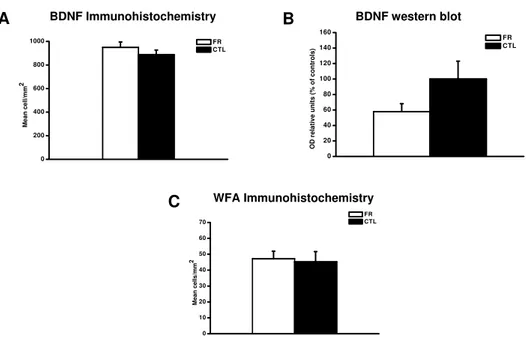
Rearing animals in an enriched environment has profound effects on the devel-opment of the nervous system. Indeed, it has been demonstrated that EE from birth leads to a profound acceleration of important properties of visual system development at behavioural, electrophysiological and molecular level (Cancedda et al., 2004; Sale et al., 2004; Ciucci et al. 2007) and accelerates the postnatal development of the retina (Landi et al., 2007; Sale et al. 2007b). Furthermore, it affects BDNF expression in the visual cortex at very precocious ages (Cancedda et al., 2004).
Environmental enrichment has strong effects also on the adult organism, leading to anatomical changes (observed, for instance, in the cortex, hippocampus and cere-bellum) in dendritic arborization, spine density and number of synapses per neuron ( Rosenzweig, 1966; Greenough and Volkmar, 1973; Renner and Rosenzweig, 1987 and Rampon et al., 2000). These morphological changes are associated with improved learn-ing and memory and enhanced neural plasticity (reviewed in van Praag et al., 2000) and reorganization of cortical somatosensory maps ( Polley et al., 2004). Moreover, EE has been demonstrated to be beneficial in reducing cognitive deficits and the progres-sion of the disease in several models of neurodegenerative pathologies related to human diseases, such as Huntington’s and Alzheimer’s, and in preventing neurodegeneration caused by ischemic or traumatic insults (Lazarov et al., 2005; Nithianantharajah and Hannan, 2006; Berardi et al., 2007).
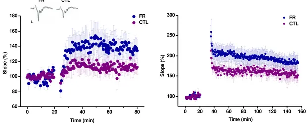
Only recently, environmental enrichment was explored as a strategy to enhance plasticity in the adult. It has been shown that EE promotes a complete recovery of visual acuity and ocular dominance in adult amblyopic animals (Sale et al., 2007a). Recovery of plasticity was associated with a marked reduction of GABAergic inhibition in the visual cortex, as assessed by in vivo brain microdyalisis. Moreover, a decreased cortical inhibition has been demonstrated also at the synaptic level, using the in vitro
paradigm of long-term potentiation of layer II-III field potentials induced by theta-burst stimulation of the white matter (WM-LTP). This form of LTP is normally not present in the adult as a result of the maturation of inhibitory circuits (Kirkwood and Bear 1994; Huang et al. 1999), but it can be restored if GABA.mediated inhibition is reduced (Artola and Singer 1987; Kirkwood and Bear 1994). Notably WM-LTP was fully restored in the visual cortex of EE adult rats (Sale et al. 2007a). The authors also demonstrated that the reduction of inhibition is a crucial molecular mechanism underlying the enhancement of plasticity induced by EE, since restoration of plasticity was completely prevented by benzodiazepine cortical infusion during EE period. The reduction of cortical inhibition in EE rats was also paralleled by an increased expression of the neurotrophin BDNF and a lower density of PNNs in the visual cortex contralat-eral to the recovering (previously amblyopic) eye.
It is well known that EE has a profound effect on diffuse projections’ system in the brain, increasing acetylcholinesterase activity, noradrenalin and serotonin (Rosen-zweig et al. 1962, 1967; Escorihuela et al. 1995; Rasmuson et al. 1998; Naka et al. 2002). Since these neuromodulators have been reported to influence plasticity in the adult brain, it would be interesting to test whether it is possible to reproduce the EE effects on adult visual cortex plasticity by an artificial modulation of their levels.
This possibility was addressed recently by Maya Vetencourt and collegues (2008) The authors found very similar results to those reported for EE using chronic treat-ment with fluoxetine, a selective serotonin reuptake inhibitor (SSRI) widely prescribed in the treatment of depression.
Clinically used antidepressant drugs (ADs) increase extracellular serotonin and/or norepinephrine levels, but the relationship between acute increases in these neuro-trasmitters and the clinical antidepressant effect , which develops with a time delay of several weeks, lies unclear (Nestler, 1998; Castren, 2005). Chronic antidepressant administration promotes neurogenesis and synaptogenesis in the adult hippocampus (Malberg et al., 2000; Hajszan et al., 2005) as well as increased expression of the neu-rotrophin BDNF and its primary receptor TrkB (Nibuya et al., 1995; Castren, 2004). These cellular and molecular events seem to be necessary to mediate the therapeutic effects of antidepressants. The behavioural response to ADs is blocked if the induced
neurogenesis is disrupted (Santarelli et al., 2003), whereas direct infusion of BDNF into the hippocampus or the overexpression of its receptor in transgenic mice induces an antidepressant effect (Shirayama et al., 2002; Koponen et al., 2005).
Recent experimental evidences also suggest that a disturbance of brain plasticity is involved in animal models of depression and that chronic antidepressant treatment may counteract these alterations. Indeed chronic mild stress facilitates LTD in the hip-pocampus of adult rats and has no effect on LTP, while treatment with ADs prevents the induction of LTD and increases the extent of LTP (Holderbach et al., 2007).
Taken together, these data suggest an influence of antidepressant in neuronal plasticity. However, as mentioned before, in a very recent work this influence was in-vestigated directly. In fact, in the work by Maya-Vetencourt it has been demonstrated that chronic treatment with fluoxetine, restores platicity to the adult visual system of the rat. This was assessed using two classical models of plasticity: 1) the ocular dominance shift of visual cortical neurons after MD and 2) the recovery of the visual function in the amblyopic adults (Maya Vetencourt et al., 2008).
Fluoxetine-treated rats displayed a coplete shift in OD in response to MD as-sessed by VEPs. Inportantly this shift was not due to an enhanced response to the open eye (as in the case of OD palsticity in adult mice; Sawtell et al. 2003; Frenkel and Bear 2004), but to a reduction of VEPs amplitude elicited by stimulation of the deprived eye, as tipically observed in juvenile cortex. Fluoxetine exerted also a pow-erful effect as a therapeutic agent for amblyopia, leading to the complete recovery of normal visual functions in long-term monocularly deprived animals (Maya-Vetencourt et al. 2008). As found for EE, the effects induced by fluoxetine in adult visual cortex plasticity were associated with a marked reduction of GABAergic inhibition. Once more, cortical diazepam administration totally prevented the OD shift induced by MD in adult rats chronically treated with fluoxetine.
The fact that both EE and fluoxetine administration lead to reduced GABAer-gic neurotransmission highlights the importance of the intracortical inhibitory tone as a key element regulating visual cortex plasticity in the adult brain. Both treat-ments, in addition, have been shown to increase BDNF expression, a critical factor for experience-dependent plasticity. Thus, these results may be explained through a
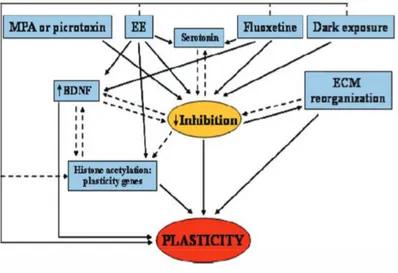
Figure 1: Schematic diagram showing key molecular events underlying restoration of plasticity in the adult visual system. Maturation of the inhibitory circuitry is thought to be one of the most important limiting factors that restrain visual cortex plasticity to the critical period. The restoration of visual cortex plasticity in adult animals is accompanied by a reduction in intracortical inhibition in different paradigms as MPA treatment, EE, chronic administration of SSRIs and dark exposure. We propose a model in which a decreased level of inhibition is the central hub triggering plasticity in the adult visual cortex. The reduction in the inhibitory tone may be accompanied by a reorganization of the ECM and an enhancement of BDNF expression. ECM remodeling could be the starting point for structural modifications at the level of synaptic connectivity, while BDNF could upregulate other genes that promote plasticity. Furthermore, an influence on the epigenetic control of gene transcription has been suggested at least for EE. Histone acetylation could be the final gate to reopen plasticity in the adult visual cortex. Continuous lines represent well-documented interactions between boxes; dashed lines indicate likely interactions in the context of visual cortical plasticity deserving further experimental characterization
model in which enhanced sensory-motor activity under environmental enrichment or enhanced serotoninergic transmission elicited by SSRI treatment, decrease cortical lev-els of inhibition and, in parallel or in series, increase BDNF expression, which could in turn upregulate other genes that promote plasticity (fig.1).
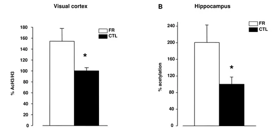
Interestingly, it has been recently demonstrated that EE has an influence on chromatine remodeling. Histone posttranslational modifications regulate chromatin susceptibility to transcription with high level of histone acetylation on a specific DNA segment being generally correlated with increased transcription rates (Mellor 2006; Workman 2006). EE enhances histone acetylation in the hippocampus and, to a lesser extent, in the cortex of P25 mice (Fischer et al. 2007) rescuing the ability to form new
memories and re-establishing access to remote memories even in the presence of brain degeneration. A similar relationship between histone acetylation and EE effects couls also occur in the adult visual system. Visual experience activates histone acetylation in the visual cortex during the critical period, but this capacity is downregulated in adult animals (Putignano et al. 2007). Trichostatin treatment, which promotes histone acetylation, also enhances plasticity in the adult visual cortex (Putignano et al. 2007). Thus, it is possible that the cellular and molecular mechanism proposed by Sale et al. (2007a) to mediate the effects of EE on the adult visual system could ultimately regulate the pattern of histone acetylation, modulating the expression of genes crucial for plasticity.
The influence that dietary factors have on the function of the nervous system, and on its susceptibility to disease, is an active and important area of biomedical re-search. Studies have identified several specific dietary components that are critical for the proper development of the nervous system. Folate deficiency during pregnancy can result in neural tube defects in babies (Refsum 2001), choline deficiency during brain development may result in learning and memory problems (Zeisel 1997), and certain fatty acids are critical for optimum brain function in the adult (Chalon et al. 2001). Recent findings suggest that folate deficiency (and a consequent increase in the levels of homocysteine) may increase the risk of Alzheimers disease (AD), Parkinsons disease (PD), stroke and psychiatric disorders (Duan et al. 2002; Kruman et al. 2002; Seshadri et al. 2002). Folate plays a critical role in one-carbon metabolism by facilitating the remethylation of methionine from homocysteine (Fenech 2001). By increasing homo-cysteine levels and impairing DNA synthesis, methylation and repair, folate deficiency can damage cells including neurons (Kruman et al. 2000, 2002).
Other examples of dietary supplements that may improve brain function and/or protect against age-related disease include antioxidants such as vitamin E (Halliwell 2001), Ginko biloba extract (Youdim and Joseph 2001) and creatine (Sullivan et al. 2000).
While vitamins, minerals and antioxidants may improve the healthspan of the brain, a more fundamental aspect of diet is emerging as a major factor in brain health. This factor, which is the focus of the present chapter, is caloric intake.
Some notes on metabolism
Metabolism is the set of chemical reactions that occur in living organisms in order to maintain life. These processes allow each individual to grow and reproduce, maintain its structure, and respond to its environment. Metabolism is usually divided in two categories. Catabolism breaks down organic matter, while anabolism uses en-ergy to construct components of cells such as proteins and nucleic acids.
The chemical reactions of metabolism are organized in metabolic pathways, in which one chemical is transformed into another by a sequence of enzymes. Enzymes are crucial to metabolism because they allow organisms to drive desirable but ther-modynamically unfavorable reactions by coupling them to favorable ones, and because they act as catalysts to make these reactions proceed quickly and efficiently. Enzymes also allow the regulation of metabolic pathways in response to changes in the cell’s environment or signals from other cells.
Most of the structures that make up animals, plants and microbes are made from three basic classes of molecule: amino acids, carbohydrates and lipids (often called fats). As these molecules are vital for life, metabolism focuses on making these molecules, in the construction of cells and tissues, or breaking them down and using them as a source of energy. Many important biochemicals can be joined together to make polymers such as DNA and proteins. These macromolecules are essential parts of all living organisms.
The carbohydrates, fats and proteins in food are metabolized to glucose which is then utilized as the major source for ATP production in cells. Unlike other cells which can use different fuels for a period of time (for example fatty acids), the only source of energy for brain cells is glucose. Moreover, because neurons cannot store this sugar, they depend on the blood stream to deliver a constant supply of this precious fuel. For this reason, blood glucose levels normally needs to remain within a proper range.
The homeostatic mechanism which keeps the blood value of glucose in a re-markably narrow range is composed of several interacting systems, of which hormone regulation is the most important. The two main hormones implicated in this phe-nomenon are insulin and glucagon.
These two hormones are secreted by islet cells within the pancreas and work antagonistically. Insulin is normally secreted by the β cells and the stimulus for its secretion is a high level of blood glucose. Similarly, as blood glucose falls, the amount of insulin secreted by the pancreatic islets goes down. This hormone has an effect on a number of cells, including muscle, red blood cells, and fat cells. In response to insulin, these cells absorb glucose from bloodstream, having the net effect of lowering the high blood glucose levels into the normal range.
Conversely, glucagon is secreted by the α cells of the pancreatic islets when blood glucose diminishes (for example, between meals and during exercise). It has an effect on many cells type but it influences mostly the liver function. Indeed, its principal role is to promote the release of glucose from liver storage, with the net effect of increasing blood glucose.
In addition to these mechanisms, during long periods of fasting the organism ac-tivate the so called stress response which involves the stimulation of the hypothalamic-pituitary-adrenal axis (HPA axis). This is a complex set of direct influences and feed-back interactions among the hypothalamus, the pituitary gland, and the adrenal glands. Briefly, in response to almost any type of stress physical or psychological, cells in hy-pothalamus produce the corticotrophin-releasing factor (CRF) which binds to specific receptors on pituitary cells stimulating the production of adrenocorticotropic hormone (ACTH). ACTH is then transported to the adrenal gland and promotes the production of cortisol (corticosterone in rodents). The loop is completed by the negative feedback of cortisol on the hypothalamus and pituitary.
The simultaneous release of cortisol has a number of effects, including elevation of blood glucose for increased metabolic demand. Indeed, one of the actions of cortisol is to counteract insulin producing gluconeogenesis and promoting breakdown of lipids (lipolysis), proteins, and mobilization of extrahepatic amino acids and ketone bodies.
Beneficial effects of food restriction
Both the number of calories consumed over time and the time interval between feedings affect the physiology of brain cells in quite profound ways. The dietary restric-tion (DR) protocols used in the animal studies, indeed, involve a reducrestric-tion in overall
calorie intake, and/or an increase in the intermeal interval, with maintenance of the composition of the diet in terms of vitamins, minerals, protein, etc. In other words, in a first kind of procedure the animals receive daily a quantity of food that is 30-60% of the amount consumed by the controls fed ad libitum, this protocol is better known with the term of caloric restriction (CR); the second paradigm, called intermittent fasting (IF), involves a fasting regimen on alternate days, i.e. the animals have not access to food for a full day, every other day, while they are allowed to eat ad libitum on the intervening days. The expression of food restriction (FR) refers to both methods indistinctly.
Lifespan and susceptibility to desease. The first widely recognized scientific study of restricted diets was published by McCay et al. (1935). They showed that feeding rats with a diet containing indigestible cellulose dramatically prolonged both mean and maximum life-span in these animals. Since then, many studies have con-firmed this result and extended it to mice (Weindruch and Walford, 1988; Sprott, 1997) and other species including fruitflies (Chapman and Partridge, 1996) nematodes (Houthoofd et al., 2002), water fleas, spiders and fish (Weindruch and Walford, 1988). FR reduces the incidence of age-related cancers, cardiovascular desease an deficits in immune function in rodents (Weindruch and Sohal, 1997). Conversely, overeating is a risk factor for cardiovascular desease, many type of cancers, type-2 diabetes and stroke (Levi, 1999; Brochu et al., 2000). Less documented is the evidence suggesting that FR reduces desease risk and extends lifespan in indivuduals that are not over-weight (Roth et al., 2002; Walford et al., 2002).
The FR regimens have also been shown to have beneficial effects on the brain. For example, FR retards age-related increases in the levels of glial fibrilary acidic pro-tein and oxidative damage to propro-teins and DNA (Dubey et al., 1996; Major et al., 1997). Analysis of the levels of mRNAs encoding thousands of proteins in the brains of young and old rats, which had been fed either ad libitum or FR diets, revealed numerous age-related changes in gene expression that were attenuated by FR (Lee et al., 2000a). The genes whose expression was affected by ageing and counteracted by FR included those involved in oxidative stress responses, innate immunity and energy
metabolism.
Synaptic plasticity and neuorgenesis. Because deficits in learning and memory, motor control and other behaviours occur during ageing, some of the earliest studies of the impact of FR on the nervous system involved testing the function of the nervous systems of old rodents that had been mantained on ad libitum or calorie-restricted diets during their adult lives.
Mice mantained on a diet with a 40% reduction in calories beginning at the time of weaning did not exhibit the deficits in motor coordination and spatial learning seen in control mice fed ad libitum (Ingram et al., 1987). FR beginning at 3 months of age prevented age-related deficits in a radial maze learning task in mice (Idrobo et al., 1987). Similarly, life-long CR prevented age-related deficits in the performance of rats in radial arm maze and Morris water maze learning and memory tasks (Stewart et al., 1989). FR retarded age associated deficit in sensorimotor coordination and avoidance learning in mice (Dubey et al., 1996).
Long-term potentiation (LTP) of synaptic transmission is believed to be a cel-lular correlate of learning and memory. Aged rats exhibit a deficit in LTP in the hippocampus, and this deficit is largely abolished in age matched rats that are fed a reduced calorie diet during their adult life (Hori et a., 1992; Eckles-Smith et al., 2000). Moreover, beneficial effects of FR were evident in aged (22-month-old) mice in which caloric restriction was initiated in mid-life (14 months of age); strenght and coordina-tion were preserved such as age related changes in spontaneous alternacoordina-tion behaviour (Means et al., 1993).
A few studies have examined synapses from animals that had been maintained of FR. In one study, neocortical synaptosomes were isolated from rats under IF and from controls. The synaptosomes from FR rats exhibited improved glucose tranport and mitochondrial function following exposure to oxidative and metabolic insults (Guo et al., 2000).
Effects of FR on neurotransmitters have also been documented. For example, FR prevented age-related alterations in the levels of serotonin and dopamine in the cerebral cortex of rats (Yeung and Friedman, 1991), and enhanced evoked dopamine
accumulation in the striatum of aged rats (Diao et al., 1997). Preservation of neu-rotransmitter signaling is likely to be crucial for the ability of FR to maintain the function of the nervous system during ageing.
The adult brain contains populations of cells that are capable of dividing and then differentiating into neurons (neurogenesis) or glial cells (gliogenesis) (Gage, 2000). In mammals, including humans, neural stem cells are most abundant in the subventric-ular zone and the dentate gyrus of the hipocampus. Stem cells in the adult brain may provide a cellular reserve to replace neurons and glia that die as the result of various injuries and deseases; evidence suggesting that neurogenesis can be stimulated by is-chemic and excitotoxic brain injuries is consistent with the cellular reserve hypothesis (Parent et a., 1997; Liu et al., 1998).
Interestingly, more subtle physiological signals can regulate neurogenesis, sug-gesting that neural stem cells may be continuously responding to functional demands. For example, raising rats or mice in an enriched environment or increasing their level of physical exercise can enhance neurogenesis (Kemperman et al., 1997; Nilsson et al., 1999; van Praag et al., 1999). In addition, neurogenesis and synaptic connections are affected by changes in the levels of the sex steroids testosterone and estrogen (Alvarez-Buylla and Kirn, 1997; McEwen, 2001).
It has been reported that FR can increase neurogenesis in the brains of adult rats and mice (Lee et al., 2000b; 2002 a,b) promoting the survival of newly generated neural cells.
Neuroprotection. The number of people with age related neurodegenertive con-ditions such as Alzheimer’s desease (AD), Parkinsons desease (PD), stroke and hearing and vision loss is increasing rapidly as life expectancy continues to raise.
Different but overlapping populations of neurons degenerate in AD, PD and stroke. Neurons in brain regions involved in learning and memory processes, such as the hipocampus and the cerebral cortex, are affected in AD (Ray et al., 1998). In PD dopaminergic neurons in the substantia nigra degenerate resulting in motor dysfunc-tion (Jenner and Olanow, 1998). A stroke occurs when a cerebral blood vessel becomes occluded or rupted resulting in the degeneration of neurons in the brain tissue supplied
by that vessel (Schulz and Dichgans, 1999).
Overeating is a well established risk factor for stroke, and some epidemiological data suggest that individuals with high caloric intake may also be at increased risk for AD and PD (Bronner et al., 1995; Logroscino et al., 1996; Mayeux et al., 1999).
A series of studies have employed animal models of neurodegenerative disorders to directly determine the effects of FR on neuronal vulnerability and functional out-come; the models are based on genetic and environmental factors that may initiate or promote the neurodegenerative process in the corresponding human disorder. AD models include transgenic mice expressing mutant forms of human amyloid precursor protein and/or presenilin-1 that cause early onset inherited AD (Games et al., 1995; Duff et al., 1996; Hsiao et al., 1996; Guo et al., 1999) and infusion of amyloid ß-peptide and excitotoxins into the brains of rats and mice (Geula et al., 1998; Bruce-Keller et al., 1999). PD models include administration of the toxin 1-methyl-4-phenyl-1,2,3,6-tetrahydropyridine (MPTP), 6-hydroxy-dopamine or rotenone to rodents or monkeys resulting in the selective degeneration of substantia nigra dopaminergic neurons and as-sociated motor dysfunction (Duan et al., 1999), and transgenic mice expressing mutant human a-synuclein, which exhibit degeneration of dopaminergic neurons and a behav-ioral phenotype that mimicks several features of PD (Masliah et al., 2000). A stroke can be induced in rodents by transient or permanent occlusion of the middle cerebral artery (Dirnagl et al., 1999; Yu and Mattson 1999). Many of the neuronal deaths that occur in these animal models are believed to involve a form of programmed cell death called apoptosis (Mattson, 2000).
Rats maintained on FR for 2-4 months exhibit increased resistance of hippocam-pal neurons to excitotoxic degeneration in a model relevant to the pathogenesis of epilepsy and AD; this neuroprotection resulted in a preservation of learning and mem-ory ability that is normally compromised in this model (Bruce-Keller et al., 1999). In addition to its neuroprotective actions, FR may be beneficial for epilepsy patients by reducing seizure incidence and severity (Mahoney et al., 1983; Greene et l., 2001; Yudkoff et al., 2001).
In other studies, presenilin-1 mutant knockin mice and amyloid precursor pro-tein (APP) mutant transgenic mice were maintained on FR or ad libitum control diets.