(19) United States
(12) Patent Application Publication (10) Pub. No.: US 2017/0115277 A1
RATHBONE et al.US 201701 15277A1
(43) Pub. Date:
Apr. 27, 2017
(54) (71) (72) (73) (21) (22) (63)
G-PROTEIN COUPLED RECEPTOR 22 TRANSFORMED CELL LINES AND USES THEREFOR Applicant: Inventors: Assignee: Appl. No.: Filed:
LIBRAMEN NATURALS INC.,
Ontario (CA)
Michel P. RATHBONE, Ontario (CA); Shucui JIANG, Ontario (CA); Francesco CACIAGLI, Chieti (IT): Renata CICCARELLI, Chieti (IT): Patrizia BALLERINI, Chieti (IT): Patrizia DI IORIO, Chieti (IT): Patricia GIULIANI, Chieti (IT): Iolanda D’ALIMONTE, Chieti (IT)
LIBRAMEN NATURALS INC.,
Ontario (CA)
15/331,372 Oct. 21, 2016
Related U.S. Application Data
Continuation of application No. PCT/CA2015/ 050326, filed on Apr. 20, 2015.
(60) Provisional application No. 61/984,618, filed on Apr. 25, 2014, provisional application No. 61/985,373, filed on Apr. 28, 2014. Publication Classification (51) Int. Cl. GOIN 33/50 (2006.01) CI2N 5/00 (2006.01) C07K I4/705 (2006.01) (52) U.S. Cl. CPC ... G0IN 33/5041 (2013.01); C07K 14/705 (2013.01); C12N5/00 (2013.01); G0IN 2333/705 (2013.01); C12N 2510/00 (2013.01) (57) ABSTRACT
Disclosed herein are cell lines transformed to express G-Pro tein Coupled Receptor GPCR22 and uses thereof for iden tifying guanosine analogues and/or other ligands to the receptor. In particular, techniques for transforming Droso phila Schneider 2 cells and human astrocytoma 1321N1 cell to express GPCR22 are disclosed as well as transformed
cells lines. The transformed cell lines of the instant disclo
Sure may be useful in identifying guanosine analogues and functional equivalents thereof.
Patent Application Publication
Apr. 27, 2017. Sheet 1 of 25
US 2017/01 15277 A1
B
8
6
t
Patent Application Publication
Apr. 27, 2017. Sheet 2 of 25
US 2017/01 15277 A1
Patent Application Publication
Apr. 27, 2017. Sheet 3 of 25
US 2017/01 15277 A1
:3 365: 8e
Patent Application Publication
Apr. 27, 2017. Sheet 4 of 25
US 2017/01 15277 A1
i
US 2017/01 15277 A1
Apr. 27, 2017. Sheet 5 of 25
Patent Application Publication
Patent Application Publication
Apr. 27, 2017. Sheet 6 of 25
US 2017/01 15277 A1
G
8 &8
B5
i
Patent Application Publication Apr. 27, 2017. Sheet 7 of 25
US 2017/01 15277 A1
O. - O vm V S.Patent Application Publication
Apr. 27, 2017. Sheet 8 of 25
US 2017/01 15277 A1
w- A(5
SasF
3
S5
t
5.
e 2 s n (Y) vaUS 2017/01 15277 A1
Apr. 27, 2017. Sheet 9 of 25
e/
?un3|-
Patent Application Publication
US 2017/01 15277 A1
Patent Application Publication
O D O O O (D
Patent Application Publication Apr. 27, 2017. Sheet 11 of 25
US 2017/0115277 A1
c
e
P
(Y)
8ss S
s
E St
E. O.
P.
9
fans
(Y)
D
(f axial O O s5
R
O
on
O
LL-
LO
N2 ><
O
2
s o 55 A.
O
d
L
D
c
O O O O O O D GN N O S(uu OLulehoud au/loud)
dWWol
US 2017/01 15277 A1
Apr. 27, 2017. Sheet 12 of 25
Patent Application Publication
NNNNNONVINNNNNNN LNNNN
JLNVN NVVVNINNV NNNVVN)LNNOVVN NVVVNV NNNNNNNNNNNNNONNONNINNILNINNOVNI
NNONNULNIN ONNIN, LNNNNNOLNO-ONNVNVN NVV LONNINNV NNNNNO ONON LLOJEVILLALNION
NON LVVV LJLLONOJLLNVONNINN OLNJLONNALOJLLLNINNNNN, LLN NIVOJLNVULLNOJLON VVVOL
OLNVOVVV NVO OVONNV NOV OOVVO!){DOOVOV OOVVVOLNOONVNOÐOOLOOOÐNINNVIL
{D, LOLLLOVOLILOVNNVNJLON?) LLLVILOOLOJIVVOOV OOÐ LOÐVLOEDVOÐVVVOÐOLO?VOVOVO
VOOV?VOJVOVOVO OVVLOLOLO IVVOV OVVVVVõV90COOÐVVVOVVõVOVOOOOVOVVO
JLO LIVOVALOVOÐAÐ VIVOÐOOLVIV VILLO LOÐ{DVOOLOVIV!)VVOOVOVIVOVOJVOLO?D?W?D?5
OLVILLOVOVOLLOJLLOJLLOJVOOOOLVOVOLLOVALOOLOOVO IVLOVLÁDIVADO?O LOÐVÕIOVO
OOOV OLLLLLLVVOLOVVOLI VOLLOOO LIVOLOOLIVOLOJLLOJLL LOJALLALLIVÕÕRLIIVOOLO
OVOJ V VLOOJVOLO LOÐVOVOÐ9?IVVOVOLO LIVVOVOVVVÕÕRLOOVVVVIÐ LOLO?VOVÒ
OVVLOLOOLI VLADIVOÐ{D_LOLOLOJVV LVV LOLVOJLLOVLÁDJEVOOLOJIVVOIVVOVÕLVL ?VOVÝ7
JLOVOLO LOLOOVOJOVVILLOVVOOLVVVOJVOÐJLOVILL LOVLOED LOVLÁDOOVILLOOVVOÐVIÐIÐ
OLOLOVO OVVOOVILOJ VIVVOOVIVOOJVOVOÐVOLIVOVOJVOVOOÐIÐAÐOVOLDOVVVO??
????VÕVVÕÕ?WOWWO?WWWTWOOÕ??WWWuuuu.opvp LOOhuuuuo^^^^oepeão LOLL?eNNNNNN
US 2017/01 15277 A1
Apr. 27, 2017. Sheet 13 of 25
Patent Application Publication
NNNeVVõe?v?)VVIVVV>|>{3VO
TIETOWDWWWW757555(757575575577755555(757557775 T??557555555T????I
TWOWOWZ57557575TW75,7575577WIJIJIJIWW7575777777775777555555(77,75V775,7757
?R?NOT?J?S LONJLOÐVVOONNONLLO LOÐ LOLNOLNJLOÐJLOVNNVOÐVOVOÐ LOVO LOLOOLO
JLNN LVONNO VIVO OVVLN, LOOJLLVN ONVOOÐJLNNNNO LVNNINNOIVOJLLOVNOJLVONNIVVN
NVNÍONNNNNLIVOV LOVOLNANNALNOVOJ VLN VNONINVVN)LVOÐNINN LLOVNONOVNO ON LNN
NONNINOOLNO OOONNNNNNV NNN LLNINNON LLALVONNNNNNNNNNNNNNOLLO{DVILNINNN
NJLNNNNNNNONVIVONNNNNNNNNNNNNNNNNNNNNN LNNNNNNNV NNJ VNN LNNNNNNN
US 2017/01 15277 A1
Apr. 27, 2017. Sheet 14 of 25
Patent Application Publication
:æsedseO
?o
%,
US 2017/01 15277 A1£
?sedSeO
:\/-O
LI
Apr. 27, 2017. Sheet 15 of 25
e
TT
?un
31.-)
Patent Application Publication
:9Sedse.O jo
9%,
US 2017/01 15277 A1€
?sedseO :\/-O LI
Apr. 27, 2017. Sheet 16 of 25
Patent Application Publication
US 2017/01 15277 A1
:esedseO
?o
%
Apr. 27, 2017. Sheet 17 of 25
Patent Application Publication
20
| - OZ. 00 V,:esedseO
?o
%
US 2017/01 15277 A1
Apr. 27, 2017. Sheet 18 of 25
Patent Application Publication
Patent Application Publication Apr. 27, 2017. Sheet 19 of 25
US 2017/0115277 A1
Figure 12b
Patent Application Publication Apr. 27, 2017. Sheet 20 of 25
US 2017/0115277 A1
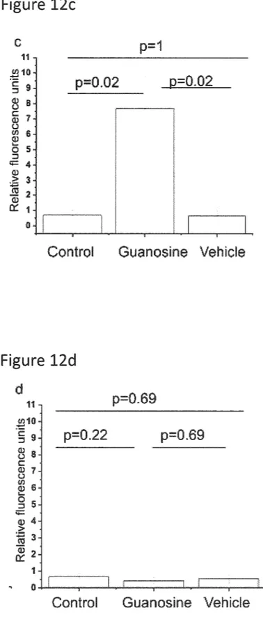
Figure 12C
Control
Guanosine Wehicle
Figure 12d
d
p=0.69
E10
S 9.
p=0.69
8 s proporoporossi
g
a. c) 4 e3"
--Control
Guanosine Wehicle
Patent Application Publication Apr. 27, 2017. Sheet 21 of 25
US 2017/0115277 A1
Patent Application Publication Apr. 27, 2017. Sheet 22 of 25
US 2017/0115277 A1
Fi
8.
U r e1.
3
e :1.2
|
.Contral
GL; tire
Vehicle
Figure 13f
. . . . . . . . . . . . . . . . . . . . . . . p=).U84
ga
-0.647 p
(25, ill 35U. M. (51.JLJ Mai 2.13 drift.
Patent Application Publication Apr. 27, 2017. Sheet 23 of 25
US 2017/0115277 A1
s
i S i. i. : s : e tO-- Sur, a La Sajor L, an ea
Patent Application Publication Apr. 27, 2017. Sheet 24 of 25
US 2017/0115277 A1
s
s
--of d Lifs e O Luc flip L. S.
eds
(u?u)
bull
US 2017/01 15277 A1
Patent Application Publication
US 2017/01 15277 A1
G-PROTEIN COUPLED RECEPTOR 22 TRANSFORMED CELL LINES AND USES
THEREFOR
RELATED APPLICATIONS
0001. The present application is a U.S. Continuation application which claims benefit of priority to International Patent Application serial number PCT/CA2015/050326 entitled “G-Protein Couple Receptor 22 Transformed Cell Lines and Uses Therefor', filed Apr. 20, 2015 which in turn claims benefit of priority to U.S. Provisional Patent Appli cation Ser. No. 61/984,618 entitled “G-Protein Coupled Receptor 22 Transformed Cell Lines And Uses Therefor', filed Apr. 25, 2014 and, U.S. Provisional Patent Application Ser. No. 61/985,373 entitled “G-Protein Coupled Receptor 22 Transformed Cell Lines And Uses Therefor', filed Apr. 28, 2014, the subject matter of which are herein incorporated by reference.
TECHNICAL FIELD
0002 The present disclosure relates to transfected cell and cell lines transformed to express G-Protein Coupled Receptor 22 and uses therefor.
BACKGROUND
0003 Extracellular guanosine, like adenosine, has been shown to have a plurality of physiological effects both in vitro and in vivo. It affects the growth, differentiation and survival of various cells (Di lorio P. Benfenati et al. 2004,
2006; Ballerini et al., 2006; Tavares et al., 2005; Molz et al.,
2005, Rathbone et al., 2008). Guanine-based purinergic signalling has been particularly investigated in the nervous system. For example, exogenously added guanosine stimu lates the division of certain cells in culture including astro cytes, fibroblasts and certain tumour cells, including brain tumour cells (Rathbone et al., 1990; Kim et al., 1991; Ciccarelli et al., 2000: Su et al., 2009; 2010; 2013: Jiang et al., 2006: Rathbone et al. NNNA 2008). It promotes differ entiation of fetal neurons (Rathbone et al., 1999; 2008) and PC12 cells, stimulates outgrowth of nerve processes (neu rites) (Gysbers and Rathbone, 1992: 1996: Bau et al., 2005; Dilorio et al., 2002). Guanosine also prevents apoptosis in astrocytes induced by several stimuli (Pettifer et al., 2004: 2007; Jiang et al., 2007: Su et al., 2009). Furthermore, Guanosine has been shown to protect the CNS from insults such as hypoxia-ischemia (Moretto et al., 2009: Thauerer et al., 2012: Thomazi et al., 2008; Zur Nedden et al., 2008), stroke (Chang et al., 2008; Connell et al., 2013: Rathbone et al., 2011), spinal cord injury (Jiang et al., 2008a; Jiang et al., 2007; Jiang et al., 2003a; Sam, 2004), and seizure (Schmidt et al., 2005; Schmidt et al., 2000; Soares et al., 2004), and Parkinson's Disease (Giuliani et al., 2012: Su et al., 2009).
0004 Extracellular guanosine is known to stimulate the synthesis and release of several growth factors from cells, which promotes, for example, astrocyte proliferation (Rath bone et al., 1990; Kim et al., 1991: Ciccarelli et al., 2000), partly by stimulating Small numbers of microglia in the astrocyte cultures to produce soluble factors, such as IL-1 (Ciccarelli et al., 2000). Additionally, guanosine promotes the synthesis and release of several potentially neuroprotec tive trophic factors from a variety of cells, including nerve growth factor (NGF) from astrocytes (Middlemiss et al., 1995) as well as basic fibroblast growth factor (FGF/FGF-2)
Apr. 27, 2017
(Su et al., 2009) and transforming growth factor-D (TGF-D) (Di lorio et a, 2001). It has been recently shown that exogenous Guanosine can increase intracellular cyclic GMP concentrations through activation of the enzymes hemeoxy genase-1 and hemeoxygenase-2 (HO-1 and HO-2) (Bau et al., 2005).
0005. In some cases guanosine produces its effects by entering cells and interacting with an NGF-dependent pro tein kinase (Jiang et al., 2006) and under certain circum stances, guanosine acts synergistically with certain growth factors, such as NGF to produce its effects. Guanosine has also been shown to promote the release of adenosine from cells (Ciccarelli et al., 2000); however, most of the effects of guanosine are different from those of adenosine and cannot be explained by adenosine release (Di lorio et al., 2002).
0006 To date the mechanism through which guanosine exerts its biological effect remains unclear. Many effects of extracellular purine nucleosides and nucleotides are medi ated through G-Protein Coupled purine receptors—puri noceptors (Burnstock G., 2007) that have common struc tural features. Preliminary evidence for the existence of a high-affinity binding site, specific for guanosine in rat brain membranes (Traversa U., et al., 2002; Traversa et al., 2003: Volpini et al., 2011), analogous to the adenine receptor (Wengert Met al., 2007) has been obtained. Accordingly, it is believed that guanosine is responsible for the activation of a number of intracellular signalling pathways. These intra cellular signalling pathways, for example, result in the elevation of cAMP in rat brain membranes (Traversa et al., 2003) and in primary stem cells (Su et al., 2013), PI3 kinase/ Akt/PKB and mitogen-activated protein kinase and ERK1/2 phosphorylation which are characteristic responses of acti vated G-Protein Coupled Receptors (Dilorio Pet al., 2001;
Pettifer et al., 2007: Traversa U., et al., 2002; Ballerini et al.,
2006; Di lorio et al., 2004: Giuliani et al., 2012). The addition of exogenous guanosine to cultured mouse primary astrocytes also leads to the elevation of intracellular calcium concentrations, although it is unclear whether this is medi ated via the putative Guanosine receptor (Chen et al., 2001). Interestingly, the effects of guanosine on the production of trophic factors by astrocytes and the anti-apoptotic effects of guanosine are sensitive to pertussis toxin, an inhibitor of Gi and Go coupled G-protein Receptors (Fields T A and Casey PJ, 1997), and are not inhibited by inhibitors of nucleoside or nucleobase transport, which lends Support for the hypoth esis that guanosine acts extracellularly. Furthermore, rat brain membranes have been shown to have high affinity cell Surface binding sites specific for guanosinesinosine that do not bind adenosine (Traversa U., et al., 2002).
0007. On the basis of pharmacological and molecular cloning studies, the International Union of Pharmacology Committee on Receptor Nomenclature and Drug Classifi cation (NC-IUPHAR) has subdivided purinoceptors into two major classes: adenosine (P1) receptors and nucleotide (P2) receptors (Fredholm et al., 2001; Abbracchio and Burnstock, 1994). Four subtypes of adenosine receptors have been identified: A, AA, AB, and A. Each has a unique tissue distribution, signal transduction mechanism, and ligand affinity. All adenosine receptor Subtypes are coupled to heterotrimeric G proteins. Activation of A or As Subtypes inhibits adenylate cyclase activity, whereas activa tion of AA and AB Subtypes stimulates adenylate cyclase activity. Additionally, A and A Subtypes are coupled to
US 2017/01 15277 A1
other signal transduction pathways, including phosphoino sitol hydrolysis and potassium channels (Ramkuran et al., 1993: Linden, 1991).
0008. In addition to their different effects, these receptor Subtypes can also be distinguished by the potency order of a series of agonists and antagonists (Palmer and Stiles, 1995). Adenosine is the preferred endogenous agonist at all adenosine receptors. But the naturally-occurring purine inosine, for which no unique receptor has been identified, binds to, and activates A receptors (Jin et al., 1997: Linden et al., 1985), producing immunomodulatory and neuropro tective effects (Gomez and Sitkovsky, 2003; Hasko et al., 2004). A receptor subtypes exhibit the lowest degree of amino acid sequence identity either with different species homologues (Palmer and Stiles, 1995; Fredholm et al., 2001) or with other adenosine receptor subtypes, resulting in unique pharmacological properties (Linden et al., 1993).
0009 P2 receptors are further divided into P2X ligand gated ion channels, which are activated solely by ATP, and G-Protein Coupled P2Y receptors which are activated both by extracellular adenine and uracil nucleotides. Eight P2Y receptors (P2Y 1, 2, 4, 6, 11, 12, 13, 14) have been cloned from mammalian tissues (Abbracchio 2003, Zhang 2002, North 2002, Fredholm, 1997). P2Y receptor activation stimulates phosphoinositol hydrolysis. Additionally, activa tion of P2Y12, 13, 14 receptors inhibits adenylyl cyclase whereas P2Y11 activation stimulates this enzyme (von Kugelgen and Wetter, 2000).
0010 P1 and P2Y purinoceptors are integral membrane proteins that belong to the class A. rhodopsin-like, G-Protein Coupled Receptor (GPCR) superfamily. These purinocep tors are predicted to share a conserved molecular architec ture consisting of seven hydrophobic transmembrane domains (TMDS), which span the plasma membrane, con nected by three intracellular and three extracellular loops (Watson et al., 1994).
0011. It is difficult to extract and crystallise GPCRs. Indeed, only the rhodopsin receptor has been crystallised and studied by X-ray diffraction. However, it has been possible to predict the tertiary structure of other GPCRs based on the atomic co-ordinates of the rhodopsin receptor. Since P1 and P2Y receptors have not been crystallised, an understanding of how purinoceptors bind their cognate ligands is based on modelling studies combined with site directed mutagenesis. GPCRs have low amino acid sequence homology. However, there are several highly con served key amino acid residues which may be essential for either the structure or function of the receptors. As in other receptor families, similarity between sequences of the P1 or P2Y receptor family is greatest in the hydrophobic TMDs. 0012 Certain well-conserved amino-acid residues of the P2Y receptor subfamily have been used to identify and clone new P2Y receptors (Chambers et al., 2000; Communi et al., 2001; Joost and Methner et al., 1997: Jiang et al., 1997). In contrast, no orphan receptor related to the P1 receptor subfamily has been identified. However, mutational data have identified some amino acid residues important for ligand binding to adenosine receptors (Fredholm et al., 2001).
0013 This background information is provided to reveal information believed by the applicants to be of possible relevance. No admission is necessarily intended, nor should it be construed, that any of the preceding information constitutes prior art.
Apr. 27, 2017
SUMMARY
0014. The following presents a simplified summary of the general inventive concepts described herein to provide a basic understanding of Some aspects of the invention. This Summary is not an extensive overview of the invention. It is not intended to restrict key or critical elements of the invention or to delineate the scope of the invention beyond that which is explicitly or implicitly described by the fol lowing description and claims.
(0015 Based on the evidence of G-Protein Coupled Receptors specific for adenosine and the Suspected existence of a receptor for guanosine as well as the intracellular responses of exogenously added guanosine to tissue cul tures, inter alia, an investigation was undertaken to locate motifs in the primary sequence of the TMDs of P1 adenosine receptors, which may be important for the binding of guanosine. Accordingly, adenosine A receptor-like sequences among orphan receptors were searched given that A receptors bind inosine as discussed in more detail below. The sequences of orphan receptors were analyzed to identify similar motifs. Through such studies, G-Protein Coupled Receptor 22 (GPCR22 SEQ. ID NO: 3) was identified as a possible G-Protein Coupled Receptor able to bind guanos ine. Surprisingly, further research verified GPCR22 as a novel guanosine binding receptor. Guanosine was shown in further studies, discussed below, to make functional binding to GPCR22 when expressed in cells which do not normally express the GPCR22 receptor. Additionally, Guanosine, binding to GPCR22 was also shown, using biochemical, pharmacological and physiological techniques, to elicit cel
lular effects in such cells.
0016. It would be desirable to identify a method useful for identifying guanosine analogues which may act as ago nists, partial agonists and inverse agonists on a G-Protein Coupled Receptor able to bind guanosine. Furthermore, in some embodiments it would be desirable to provide a means for identifying various cell types expressing a receptor able to bind guanosine.
0017. In one aspect there is provided a transformed cell line expressing a non-endogenous G-Protein Coupled Receptor. The transformed cell line is proliferated from a host cell having been transformed to express a cell-surface G-Protein Coupled Receptor able to bind guanosine and guanosine analogs. In some embodiments, the host cell is transfected with a recombinant cDNA sequence coding for an amino acid sequence of the cell-surface G-Protein Coupled Receptor able to bind guanosine and guanosine analogs. In preferred embodiments, the amino acid sequence corresponds to GPCR22. Furthermore, in some embodi ments, the recombinant cDNA sequence comprises SEQ. ID NO: 1 or SEQ. ID NO: 2. In some embodiments, the amino acid sequence comprises SEQ. ID NO: 3. In preferred embodiments, the host cell is a Drosophila Schneider 2 cell or a human astrocytoma 1321N1 cell.
0018. In another aspect, there is provided a method for producing the transformed cell line as defined above. The method comprises transfecting the host cell with an expres sion vector, where the expression vector comprises a poly nucleotide and the polynucleotide comprises a nucleotide sequence encoding for a polypeptide comprising the amino acid sequence of SEQ. ID NO. 3.
0019. In yet another aspect, there is provided a method for identifying a compound bindable to a selective guanos ine responsive G-Protein Coupled Receptor. The method
US 2017/01 15277 A1
comprises contacting one or more candidate compounds with a host cell transformed to express a receptor comprising an amino acid sequence comprising SEQ. ID NO: 3; and measuring the ability of the one or more candidate com pounds to stimulate or inhibit a cellular function associated with said receptor. In some embodiments, the amino acid sequence is encoded by DNA comprising SEQ. ID NO: 1 or SEQ. ID NO: 2. Furthermore, in some embodiments, the cellular function evaluation comprises measuring apoptosis regulation, intracellular calcium mobilization, intracellular protein phosphorylation or a second messenger intracellular pathway activation. In some embodiments, the host cell is a Drosophila Schneider 2 cell or a human astrocytoma
1321N1 cell. Furthermore, in some embodiments, the one or
more candidate compounds may be an agonist, a partial agonist or an inverse agonist of the receptor.
0020. In another aspect, there is provided a method for transforming a host cell to express a guanosine bindable G-Protein Coupled Receptor. The method comprises trans fecting a host cell with an expression vector, where the expression vector comprises a polynucleotide sequence comprising SEQ. ID NO: 1 or SEQ. ID NO: 2, encoding for a polypeptide comprising an amino acid sequence compris ing SEQ. ID NO: 3; and wherein the host cell under the appropriate culture conditions, produces a polypeptide com prising the amino acid sequence of SEQ. ID NO: 3. In preferred embodiments, the polypeptide comprising SEQ. ID NO: 3 corresponds to GPCR22 and is expressed at the cell surface. In preferred embodiments, the host cell is a Drosophila Schneider 2 cell or a human astrocytoma
1321N1 cell.
0021. In yet another aspect, there is provided a G-Protein Coupled Receptor capable of binding guanosine or analogs thereof. In some embodiments the G-Protein Coupled Receptor has an amino acid sequence having at least 70% homology to SEQ. ID NO: 3. In some embodiments the G-Protein Coupled Receptor has an amino acid sequence having at least 80% homology to SEQ. ID NO: 3. In some embodiments the G-Protein Coupled Receptor has an amino acid sequence having at least 90% homology to SEQ. ID NO:3. In some embodiments the G-Protein Coupled Recep tor has an amino acid sequence having at least 95% homol ogy to SEQ. ID NO:3. In some embodiments, the G-Protein Coupled Receptor capable of binding guanosine or analogs thereof, has an amino acid sequence corresponding to SEQ.
ID NO: 3.
0022. In another aspect, there is provided a method for identifying a cell having a G-Protein Coupled Receptor able to bind guanosine or analogues thereof. The method com prises extracting DNA from a population of Subject cells, executing a polymerase chain reaction (PCR) technique on said DNA extracted from said population of subject cells using a forward primer corresponding to SEQID NO: 5. and a reverse primer corresponding to SEQ. ID NO: 6 and analyzing the PCR product resultant therefrom.
0023. In still yet another aspect, there is provided a transformed cell line expressing a non-endogenous G-Pro tein Coupled Receptor capable of binding guanosine or analogues thereof wherein the non-endogenous G-Protein Coupled Receptor has a guanosine or analogue thereof binding domain located in transmembrane domain 6, where the binding domain comprises SEQ. ID NO: 7 or SEQ ID NO: 8. In preferred embodiments, the transformed cell line
Apr. 27, 2017
is from a Drosophila Schneider 2 cell or a human astrocy toma 1321N1 cell transfected to express GPCR22.
0024. Other aims, objects, advantages and features of the invention will become more apparent upon reading of the following non-restrictive description of specific embodi ments thereof, given by way of example only with reference to the accompanying drawings.
BRIEF DESCRIPTION OF THE FIGURES
0025. In order that the invention may be better under stood, exemplary embodiments will now be described by way of example only, with references to the accompanying drawings, wherein:
0026 FIG. 1 is a photograph of a gel electrophoresis showing the expression of GPCR22 (G1) determined by real-time PCR in cultured rat astrocytes;
0027 FIG. 2a is a photograph of a gel electrophoresis showing the silencing of GPCR22 (G1) by siRNA deter mined by real-time PCR in cultured rat astrocytes compared to cultured rat astrocytes not treated with siRNA for
GPCR22;
0028 FIG.2b is a photograph of a Western Blot showing the silencing of GPCR22 protein in cultured rat astrocytes by siRNA;
0029 FIG. 3 is a photograph of a gel electrophoresis of Plasma DNA indicating positive cloning for GPCR22;
0030 FIG. 4 is a photograph of a gel electrophoresis showing the expression of GPCR22 determined by real-time PCR from rat striatum, non-transfected normal Drosophila Schneider 2 cells and GPCR22 transfected Drosophila
Schneider 2 cells;
0031 FIG. 5 is a photograph of a gel electrophoresis showing that 1321N1 human astrocytoma cells do not express GPCR22 (G1) as determined by real-time PCR;
0032 FIG. 6a is a photograph of a gel electrophoresis showing the transfection of GPCR22 (G1) DNA into 1321N1 human astrocytoma cells determined by real-time
PCR;
0033 FIG. 6b is a photograph of a Western Blot showing the expression of GPCR22 protein following GPCR22 (G1) DNA transfection into 1321N1 human astrocytoma cells;
0034 FIG. 7a is a photograph of a Western Blot showing Erk 1/2 phosphorylation by guanosine in cultured rat astro cytes and cultured rat astrocytes treated with siRNA for
GPCR22 and ns-siRNA;
0035 FIG.7b is a photograph of a Western Blot showing Akt phosphorylation by guanosine in cultured rat astrocytes and cultured rat astrocytes treated with siRNA for GPCR22
and ns-siRNA;
0036 FIG. 7c is a photograph of a Western Blot showing Erk 1/2 phosphorylation by guanosine in 1321N1 human astrocytoma cells and 1321N1 human astrocytoma cells
transfected with GPCR22 DNA;
0037 FIG. 7d is a photograph of a Western Blot showing Akt phosphorylation by guanosine in 1321N1 human astro cytoma cells and 1321N1 human astrocytoma cells trans
fected with GPCR22 DNA;
0038 FIG. 7e is a graph showing the effect of guanosine on increasing intracellular cAMP concentrations in 1321N1 cells transfected with GPCR22 (G1) receptor DNA in the absence and in the presence of 3 uM forskolin and 3 uM forskolin plus pertussis toxin (200 ng/ml, 16 h);
US 2017/01 15277 A1
0039 FIG. 8 shows the sequence confirmation results of GPCR22-pMT-GPCR22-Forward GPCR22 transfection in Drosophila Schneider 2 transformed cells;
0040 FIG. 9 shows the sequence confirmation results of GPCR22-pMT-GPCR22-Reverse GPCR22 transfection in Drosophila Schneider 2 transformed cells;
0041 FIG. 10 is a photograph of a Western Blot showing GPCR22 (G1) protein expression in transfected Drosophila
Schneider 2 cells;
0042 FIGS. 11 a to d are graphs of flow cytometry data showing the anti-apoptotic effects of guanosine on GPCR22 transformed Drosophila Schneider 2 cells in relation to Caspase-3 activity;
0043 FIG.12a is a fluorescence image showing the effect
of guanosine on the level of intracellular Ca" in non
GPCR22-transfected Drosophila Schneider 2 cells; 0044 FIG.12b is a fluorescence image showing the effect
of guanosine on the level of intracellular Ca" in GPCR22
transfected Drosophila Schneider 2 cells;
0045 FIG. 12c is a graph showing the effect of guanosine
on intracellular Ca" concentration in GPCR22-transfected
Drosophila Schneider 2 cells;
0046 FIG. 12d is a graph showing the effect of guanosine
on intracellular Ca" concentration in non-GPCR22-trans
fected Drosophila Schneider 2 cells;
0047 FIGS. 13 a to d are fluorescence images showing
the effect of guanosine on the level of intracellular Ca" in
GPCR22-transfected Drosophila Schneider 2 cells over a time course;
I0048 FIG. 13e is a graph showing an intracellular Ca"
response to guanosine in GPCR22-transfected Drosophila
Schneider 2 cells;
0049 FIG. 13f is a graph showing high dose (25 M-400
uM) guanosine-induced intracellular Ca" response in
GPCR22-transfected Drosophila Schneider 2 cells; 0050 FIG. 13g is a graph showing a time course of low
dose (0.1 uM-10 uM) guanosine-induced intracellular Ca"
response in GPCR22-transfected Drosophila Schneider 2
cells;
0051 FIG. 14 is a Saturation Binding curve graph of guanosine to GPCR22 receptors; and
0052 FIG. 15 is a graph showing GPCR22/guanosine
mediated intracellular Ca2+ increase in GPCR22-trans
fected Drosophila Schneider 2 cells.
BRIEF DESCRIPTION OF THE SEQUENCES 0053 SEQ. ID NO: 1 is an exemplary cDNA polynucle otide sequence having 100% homology to Rat GPCR22 and 97% homology to mouse GPCR22 using the exemplary forward primer of SEQ. ID NO: 5 of the instant disclosure; 0054 SEQ. ID NO: 2 is a cDNA polynucleotide sequence having 100% homology to Rat GPCR22 and 97% homology to mouse GPCR22 using the exemplary reverse primer of SEQ. ID NO: 6 of the instant disclosure;
0055 SEQ. ID NO: 3 is an exemplary amino acid poly peptide sequence corresponding to GPCR22 of the instant
disclosure;
0056 SEQ. ID NO. 4 is an exemplary mRNA polynucle otide sequence corresponding to a mouse GPCR22 of the
instant disclosure;
0057 SEQ. ID NO: 5 is oligonucleotide sequence corre sponding to an exemplary forward primer for GPCR22 of
the instant disclosure;
Apr. 27, 2017
0.058 SEQ. ID NO: 6 is oligonucleotide sequence corre sponding to an exemplary reverse primer for GPCR22 of the
instant disclosure; and
0059 SEQ. ID NO: 7 is a polypeptide sequence of an exemplary GPCR22 transmembrane 6 binding domain of the
instant disclosure; and
0060 SEQ. ID NO: 8 is a polypeptide sequence of an exemplary GPCR22 transmembrane 6 binding domain of the
instant disclosure.
DETAILED DESCRIPTION
0061. With reference to the disclosure herein and the appended figures, cell lines transformed to express GPCR22 and a method for identifying guanosine analogues which may be agonists, partial agonists and inverse agonists of
GPCR22 are described in accordance with various embodi ments of the invention.
Example 1: Identification of the G1 Receptor Using Bioinformatic Approaches
0062 Based on the pharmacological and transductional evidence Supporting the existence of the putative G1 recep tor, preliminary structural data using bioinformatic approaches was used to identify possible orphan G-Protein Coupled Receptors capable of binding guanosine. Using DNA and Protein databanks, the sequence motifs of the 7 transmembrane segments (TMs) for the adenosine A, A, A and A, receptors, which are highly conserved among these receptor subtypes were used to identify possible can didates for guanosine bindable orphan G-Protein Coupled Receptors. Inosine, a purine similar structurally to guanos ine, is known to interact with the adenosine A receptors, as well as having been shown to bind to the putative G1 binding site in rat brain. Therefore, the sequence for the TMs 3, 5, 6 and 7 of the human adenosine A receptors was selected, which constitutes the hydrophobic binding pocket for both, agonists and antagonists (Moro et al., 2005), and screened against the sequences of some of the orphan G-Protein Coupled Receptors (GPCRs) in the databanks. Based on this sequence homology to the human adenosine A receptor several orphan GPCRs with potential G1 (guanosine) receptor activity were identified.
0063. Many of the amino acid residues important for ligand binding to ATP or adenosine receptors are located in the sixth TMD (Fredholm et al., 2001). Accordingly, a multiple sequence alignment (Clustal-W program, blosum matrix) and Sequence motif search (http://expasy.org) was used to develop novel patterns within the sixth TMD char acteristic of P2Y and P1 receptors.
0064. It was noted that the local sequence: hhxCSIFY hPhHhxRK is shared among all P2Y sequences reported at SwissProt and GeneBank databases. This consensus pattern, when Scanned against sequence databases, matched only with proteins belonging to P2Y class. However, this pattern
also contains residues which are maintained in all metabo
tropic purinoceptors. In particular at 254, 255, 259 and 261 positions (referring to the numbering of the human P2Y2 receptor) a hydrophobic residue (h) is strictly conserved in all P1 and P2 receptors. The role of these hydrophobic residues is unknown. A Pro residue (at position 261) is also strictly maintained in all metabotropic purinoceptors. This represents one of the most conserved amino acid residues in the GPCR superfamily. Analysis of the crystal structure of
US 2017/01 15277 A1
the rodhopsin receptor and mutagenesis studies indicates that the corresponding Pro267 plays a critical role in the overall protein conformation (Okada et al., TiBS 2001; Ridge et al., 1999). The function of the ICS residue, strictly maintained at position 257 in all P2Y and P1 receptors, has never been investigated.
0065 Mutagenesis studies indicate that single substitu tion of the conserved His-262 and Arg-265 with leucine decreased the potency but not the efficacy of ATP and UTP on the P2Y2 receptor, Suggesting that these amino acid residues are mainly involved in the ligand binding (Erb et al., 1995). Molecular modelling studies also indicate that His-262 and Arg-265 interact with negatively charged phos phate residues of ATP (Erb et al., 1995). Arg-265 is always present in P2Y nucleotide receptors except for P2Y 1 and P2Y6 in which a Lys residue occurs in an analogous position. Thus a positive charged residue at position 265 seems to be needed for nucleotide binding (Erb et al., 1995; von Kugelgen and Wetter, 2000).
0066. In the TM6 domain, the consensus pattern of P1 receptors is clearly distinct from that of the P2Y. In particu lar the AAA and AB Subtypes are generally characterised by the conserved local motif hhx|CSIWhPhHxxNxhTxF. whereas. A receptors contain a different consensus pattern hhx|CSIWhPhICShxNxhxxF. The sequence alignment showed that His-262 residue is strictly maintained in P2Y as well as in all A. Aa and Ab receptor Subtypes, but not in A receptor sequences. Several mutagenesis studies have demonstrated that in A, Aa and Ab Subtypes this highly conserved His is involved in ligand binding (Olah et al., 1992: Kim et al., 1994). In the TM6 domain of all As adenosine receptors, the His-262 is always replaced by a Ser residue. Mutagenesis studies indicate that Ser-247 of human A receptors is not involved in agonist binding but, rather, in that of antagonists (Gao et al., 2002). Although adenosine is the main endogenous agonist at A receptors, inosine can also activate them (Jin et al., 1997; Linden et al., 1995; Fredholm et al., 2002), as noted above. The presence of a Ser, instead of His, residue is peculiar to A receptors. Whether this His plays a role in inosine binding is unknown. 0067. The alignment also revealed that the Lys/Arg resi due (265 in P2Y2), involved in the interaction with charged phosphate residues of ATP and present in all P2Y receptors (Erb et al., 1997), is replaced by an Asn residue in an analogous position (250A and 253 A-A) in all P1 subtypes.
Mutational studies indicate that this conserved Asn is
involved either in receptor-ligand recognition or in main taining receptor structure (Kim et al., 1995; Gao et al., 2002). In contrast to P2Y receptors, a Trp residue (Trp-243 in the human A receptor) is rigorously maintained in all P1 sequences. This residue is appears to be needed for activa tion of the A receptor but not for agonist binding (Gao et al., 2002), since substitution of the Trp-243 by a Phe residue impairs the ability of agonists to activate PLC (Gao et al., 2002). A Thr residue (Thr-257 in human A) is highly conserved in A, Aa and Ab Subtypes, distinguishing A and A. from A receptor sequences. The consensus residue Phe-262 (in human A) is unique to the P1 class. No characteristic residue is found at a homologous position in P2Y receptors.
0068. The identification of a specific pattern for the As has been shown to be a good approach to predict ligands for additional A-related orphan receptor. This pattern was built so as to contain residues that are of the P1 receptor subfam
Apr. 27, 2017
ily, especially those that can form plausible intermolecular interaction with the ligand class (inosine). Among orphan GPCRs, only one met the above-note characteristics as
discussed below.
0069. Therefore, a consensus pattern of hlhxCSIWXPh CShxNxhxxF, was defined that contained residues specific for the A subtype of P1 receptors, for which inosine as well as adenosine is an agonist. When scanned against sequence databases the consensus pattern matched only with A receptor Subtypes. However when the consensus sequence was shortened to hhxCSWXPhICShxN, the pattern also matched the orphan G-Protein Coupled Receptor 22 (GPCR22) from human, rat and mouse.
0070. By comparing the amino acid sequences of the As receptors against that of GPCR22, it was surprisingly dis covered that the overall percentage of identity between adenosine A receptors and GPCR22 is low, being about 13%. However, if only TM6 is considered, the percentage of identity rises to 30%. Therefore, as result of the above analysis, it was determined that there may be a binding domain in TM6 of GPRC22 for guanosine represented by hhxCSWXPhICShxN. For example, using the A receptor amino acid alignment as a reference, the binding domain may be located beginning at TM6 amino acid residue 239, preceded by two hydrophobic residues at positions 239 and 240, and additional hydrophobic residues at locations 246 and 248. Accordingly, the binding domain may be as fol lows, where h denotes hydrophobic residues and X denotes
unknown amino acid residues: h-239, h-240, X-241, Ser-242,
Trp-243, X-244, Pro-245, h-246, Ser-247, h-248, X-249. Asn-250 or h-239, h-240, X-241, Cys-242, Trp-243, X-244. Pro-245, h-246, Cys-247, h-248, X-249, Asn-250, noted below as SEQ. ID NO: 7 and SEQ. ID NO: 8, respectively. 0071 Notably, in addition to strictly conserved residues typical of the P1 receptors, GPCR22 shares a Ser residue analogous to Ser-247 of the human A receptor. GPCR22 shares a higher sequence identity with the cholecystokinin-B receptor (34%) (O'Dowd et al., 1997) than with the As receptor. Nevertheless, GPCR22 (SEQ. ID NO: 3) shares with A receptors residues in TM6 that are specific for P1 receptor subfamily, and which are believed to be likely important in purine nucleoside ligand binding.
(0072. These data indicate that GPCR22 may have sub strate binding characteristics related to an A receptor. Although adenine nucleotides and adenosine are the proto typical purinergic signalling molecules, non-adenine-based purines, including guanosine and inosine, have recently been shown to have important neuromodulatory roles (Soares et al., 2004: Gomez and Sitkovsky, 2003). Moreover, as noted above, a receptor-like cell Surface binding site for guanosine has been identified (Traversa et al., 2002; Traversa et al., 2003). Therefore, it was investigated as to whether the orphan receptor GPCR22 is a novel nucleoside receptor. (0073 Based on the above-noted analysis, GPCR22 was selected as a potential candidate for a G-Protein Coupled Receptor capable of binding guanosine, also referred to herein as the G1 receptor, and producing cellular effects.
Example 2: Inhibition of GPCR22 by siRNA in Primary Astrocytes
(0074 GPCR22 Expressed in Cultured Rat Astrocytes: (0075. With reference to FIG. 1, RT-PCR experiments showed that GPCR22, the G1 receptor, as also referred to herein, is expressed in cultured rat astrocytes. The primers
US 2017/01 15277 A1
used for the RT-PCR experiments were GPCR22 forward: CTC ATC TGC TGTTTC CAC GA (SEQ. ID NO. 5) and
GPCR22 reverse CGG ATG TTA AGA GCC TGG AG
(SEQ. ID NO: 6).
0076 siRNA for GPCR22 Experiments on Cultured Rat Astrocytes:
0077. Transient transfection of siRNAs for the rGPCR22 gene on rat cultured astrocytes and cell extracts were assayed for gene silencing 24 h after transfection was carried out. siRNA duplex specific for the rGPCR22 receptor were designed and chemically synthesized (http://www.Dharma con.com), as well as their non specific control (NSC). Transfection of siRNAS for targeting endogenous genes was carried out using Oligofectamine (Invitrogen, Life Tech nologies) according to the manufacturers instruction. Briefly, astrocytes at 60-70% confluence were cultured for 1 day in culture medium without antibiotics. 10 Dl siRNA (20
M) (Dharmacon research, Inc.) were diluted in 175 D1 optimem for 5 min at room temperature. In the meantime 3 1 oligofectamine was mixed in 15 Dl optimem for 5 min. RNA interference was performed by mixing diluted oligo fectamine and siRNA for 30 min at room temperature and was then added to the cells. The same scheme was per formed on other samples using a siRNA-scrambled (NSP-non specific control). Cells were incubated at 37°C. for 24 hours. Twenty-four hours after transfection, the cells were lysed and assayed for gene silencing. The effect of GPCR22 silencing on MAPK and AKT phosphorylation, and cAMP assay was analysed.
0078. Using siRNA for GPCR22 in cultured rat astro cytes, the expression of GPCR22 (G1) was shown to be abolished by RT-PCR, as shown in FIG. 2a. FIG. 2a shows the results of RT-PCR for the GPCR22 DNA (G1) in cultured rat astrocytes and cultured rat astrocytes treated with siRNA for GPCR22 (G1-siRNA). Additionally, through Western Blot analysis (FIG. 2b) using a rabbit polyclonal anti-GPCR22 antibody (NovateinBio, SH-A12465, MA, USA) it was shown that the GPCR22 protein expression in cultured rat astrocytes was inhibited by siRNA. Furthermore, in astrocytes, inhibition of GPCR22 by siRNA was shown to abolish both guanosine binding and activation of intracellular signaling by guanosine (data not shown).
Example 3: Transfection of hCPCR22 in 1321N1 Human Astrocytoma Cells
0079 GPCR22 is not Expressed in 1321N1 Human Astrocytoma Cells:
0080. With reference to FIG. 5, RT-PCR experiments showed that GPCR22 (G1) is not expressed in 1321N1 human astrocytoma cells. The primers used for the RT-PCR experiments were GPCR22 forward: CTC ATC TGC TGT TTC CAC GA (SEQ. ID NO: 5) and GPCR22 reverse CGG ATG TTA AGAGCC TGG AG (SEQ. ID NO: 6).
0081 Transient Transfection of hCGPCR22 in 1321N1: I0082. A full length cDNA clone of human GPCR22 was provided by Origene Technologies. Transient expression in the 1321N1 cell line was performed using a pCMV expres sion vector. cDNA fragments present in an expression vector (pCMV) is located downstream of a transcriptional promoter capable of driving heterologous gene expression in a variety of mammalian cells. 1321N1 cell lines were transiently transfected with hGPCR22 cloned into pCMV expression vectors (Invitrogen). DNA transfection was performed by
Apr. 27, 2017
lipofection using Lipofectamine (Life Technologies). Twenty-four hours after transfection, the cells were lysed and assayed. RT-PCR analysis using GPCR22 forward: CTC ATC TGC TGT TTC CAC GA (SEQ. ID NO: 5) and
GPCR22 reverse CGG ATG TTA AGA GCC TGG AG
(SEQ. ID NO: 6), was used to confirm the transfection of GPCR22 (G1) (labelled “1321N1 G1-pCMV Trans’) into 1321N1 cells as shown in FIG. 6a and evidenced by the band at 411 bp. Furthermore, the expression of GPCR22 (labelled “1321N1 G1-pCMV Trans’) in 1321N1 cells as compared to non-G1 transfected cells (labelled “1321N1) was con firmed by Western Blot analysis using a rabbit polyclonal anti-GPCR22 antibody (NovateinBio, SH-A12465, MA, USA), as shown in FIG. 6b.
0083 Stable Transfection of hCGPCR22 in 1321N1: I0084. It was determined from the above-noted assays that the pCMV vector could not be used for stable transfection. Therefore hCPCR22 was cloned into a pCDNA3.1 vector. The cloning procedures were performed by mean of total RNA extraction from the cells expressing high levels of the receptor. Subsequently a retro-transcription reaction was carried out according to the manufacturers instructions. Specific oligonucleotide PCR primers external to the puta tive open reading frame (ORF) of the human receptor sequence were designed for PCR confirmation of stable
transfection. PCR reactions to ascertain stable transfection
were performed in MgCL (1.5 mM), dNTP (1 mM), oli gonucleotides (100 pmol), Taq Gold (0.5 U, PerkinElmer), with an annealing temperature of 55° C. The PCR product was ligated to an expression vector by utilizing the pCDNA3.1N5-HisCTOPORTA Expression kit (Invitrogen, Italy) according to the manufacturers instructions. Cloned sequences were verified by sequencing.
I0085 1321N1 cell lines were stably transfected with GPCR22 cloned in pCDNA3.1 expression vectors (Invitro gen). 1321N1 cells were seeded at 150,000 cells/dish. Trans fections were performed 24 hours after seeding with 2 ug DNA and 4 ul lipofectamine (Life Technologies). DNA Selection was performed using 600 ug/ml geneticin. After selection individual colonies were maintained in 300 ug/ml geneticin.
Example 4: GPCR22 Mediated Intracellular Effects I0086) Phosphorylation of Erk 1/2 and Akt:
I0087 FIGS. 7a and 7b, respectively, show that the phos phorylation of Erk 1/2 and Akt by guanosine (300 uM) is inhibited in cultured rat brain astrocytes treated with si-RNA for GPCR22; thus lending support that GPCR22 is indeed a G-Protein Coupled Receptor for guanosine, and analogues thereof. Similarly, as shown FIGS. 7c and 7d, respectively, in 1321N1 cells transfected with GPCR22 DNA (labelled “G1-pCMV Trans) so as to express the GPCR22 receptor, guanosine (300 uM) treatment of the cells resulted in phosphorylation of both Erk 1/2 and Akt compared to non-GPCR22-transfected 1321N1 cells (labelled “Non Trans’) which do not endogenously express GPCR22. I0088 Intracellular cAMP Increase by Guanosine in
1321N1 Cells Transfected with GPCR22:
I0089 FIG. 7e shows the effects of increasing concentra tions of exogenously applied guanosine on 1321N1 cell line transfected with GPCR22 (G1) receptor in the absence and in the presence of PTX (200 ng/ml, 16 h). Accordingly, it is shown that guanosine acts to produce cellular effects through GPCR22 and increase intracellular cAMP levels.
US 2017/01 15277 A1
Example 5: Transfection of GPCR22 in Drosophila Schneider 2 (S2) Cells
0090. A Full Length cDNA Clone of Rat GPCR22: 0091 RNA from Rat striatum area was extracted using the Trizol method and then purified with Ambion RNA mini kit according to the manufacture instruction (Ambion RNA mini Kit, Cat: 12183020). cDNA of rCPCR22 was synthe sized using Quanta Biosciences 95049-100 (cScript Flex cDNA synthesis kit), PCR polymerase (Invitrogen Plati num(a) Taq DAN polymerase High Fidelity) and PCR water (GIBCO 10977 Ultrapure distilled water) following the
manufacture’s instructions. GPCR22 forward: CTC ATC
TGC TGTTTC CAC GA (SEQ. ID NO. 5) and GPCR22 reverse CGG ATG TTA AGAGCCTGGAG (SEQ. ID NO: 6) were used. GPCR22 PCR product was tested by running 1% agarose gel following the instructions (Invitrogen). Gene Ruler 1 KB ladder was purchased from Thermo Scientific (SM3011). 5x loading buffer was used to load PCR product, and the gel was run at 100V until the front of the ladder reached the bottom of the gel. The results were detected under UV light. After confirmation of GPCR22 size with agarose gel, the correct band was cut and PCR product (cDNA of GPCR22 whole sequence) was extracted from the gel using QiaeX II Gel extraction kit (Qiagen 20021) as instructed by the manufacture. The extracted GPCR22 whole sequence was sent for sequencing to confirm the identity (Mobix service at McMaster University).
0092 Preparation of GPCR22/pMTN5/His A Plasmid
DNA for Transfection:
0093. The restriction enzyme sequences were added to the primers described as follows. Primers for GPCR22: Kpn I was added to the forward sequence and Apa I to the reverse sequence for whole cloned sequence of GPCR22. Extra base was also added for easier digestion. Restriction enzyme recognition sequence for the restriction enzymes used is underlined as follows GPCR22 forward: Kpn I 5'-TAG
GGTACC ATG TCA GAA TTG TCA AT-3'; GPCR22
reverse: Apa I5'-CTAGGGCCCCTA GTCTGTGAC AAC CT-3'. pMTV5-His-A, pMTV5-His-Lacz and pCoHygro vectors were used. The inducible expression vector pMT V5-His-A has a metallothionein (MT) promoter, and was induced by the addition of copper sulfate to the culture
medium. It was stored in DH5O. E. Coli. in a -80° C. freezer.
Vector extraction was done using Qiagen Qia prep-SPIN minikit (Qiagen 27104) or Qiagen Hispeed Plasmid Max kit (Cat: 12662) according to the instructions from the manu facture. Using the following primer to clone PMT and COPIA sequences from the extracted vector DNA, and gel extracted PCR product were sent for sequencing to confirm the pMT vector (Mobix service at McMaster University). Primer for pMT and COPIA: PMT forward: CAT CTC AGT GCA ACT AAA; Copia forward: TGTTGG AAT ATACTA
TTC AAC CTA CAA
0094. The GPCR22 PCR products or the pMT vector plasmid DNA extraction was prepared through the sequen tial digestion, first using the restriction enzyme Apa I (Fermentas, Catil ER 1415). The digested products were then separated with electrophoresis and extracted with Qiaex II Gel extraction kit (Qiagen 20021). The extracted product was then Subjected to a second digestion with the restriction enzyme Kpn I (Fermentas, Cati ER0521), and then sepa rated using gel electrophoresis and extracted with QiaeX II Gel extraction kit for further ligation. GPCR22 PCR product was digested at 37° C. for 2.5 hr, and the vectors were
Apr. 27, 2017
digested at 37° C. for 1.5 hr. Both of the reactions were deactivated at 60° C. for 20 min after the digestion. (0095 To calculate the amount of DNA required, 1:1 ratio (pMT/V5/His A plasmid DNA: GPCR22 DNA), the amount was calculated according to the following formula: The amount of GPCR22 DNA-bp GPCR22 PCR productxthe amount of Vector/bp of vector. Ligation was performed at 14°C. for overnight. Transformation of the ligated mix was done using one-short Top 10 competitive E. Coli (Invitrogen, Cath()4040-03) following the manufactures instruction. Clones were picked from LB plates and cultured separately at 250 rpm, 37° C., LB medium with 100 ug/ml Amp. Plasma DNA was extracted from clones and double diges tion was performed with Apa I and Kpn I. After digestion, an electrophoretic gel was run to separate the DNA. The band size and number of bands was judged to foretell whether positive clones had been obtained, as shown in FIG. 3. The potential positive clones were sent for sequencing for confirmation. Once the positive E. Coli clones were con
firmed, the culture was continued until a sufficient amount of
GPCR22/pMTN5/His Aplasmid DNA for future transfection
was obtained.
0096. Transfection of Clone-Recombinant GPCR22 DNA to Drosophila Schneider 2 Cells (S2 Cells):
0097. A calcium method was used for GPCR22 transfec tion into Drosophila Schneider 2 cells (S2) according to the manufacture's instruction (CalPhos Mammalian Transfec tion kit, Clontech, Cat No: 631312). Briefly, the Drosophila Schneider 2 cell line (also referred to herein as S2 cells) was purchased from Invitrogen (Cat: R690-07). Cells were cul tured, passaged and stored according to the manufacture
instruction. S2 cells were maintained at 25-28°C. in Schnei
der's Drosophila medium (GIBCO) supplemented with 10% heat-inactivated fetal calf serum, penicillin and streptomy
cin. 1.5x10 S2 cells per well were seeded with Drosophila
Schneider 2 cell medium (S2 medium) and cultured for a day before transfection. Then, transfection was performed fol lowing the manufacture's recommended procedure. Follow ing 24-hour culture, the S2 cells were collected into 1.5 ml eppendorf tube and labeled properly. The cells were then centrifuged at 3000 rpm for 2 min. in an eppendorf tube. The supernatant was re-suspended in 1 ml of S2 medium with 5 ml of 100 mM CuSO solution (final concentration is 500 mM), then transferred one by one to new 12 well plate and incubated for 2 days.
Example 6: RT-PCR to Determine the Expression
of GPCR22 from the Transfected S2 Cells
(0098. For real-time PCR analysis total RNA was purified from S2 cells using Trizol (Invitrogen) according to the
manufacturers instructions and incubated with DNase.
cDNA was prepared from 5 ug purified RNA using oligo nucleotide dT primers. Real-time PCR was performed in an Eppendorf realplex2 PCR machine using SyBr green as a detection reagent. The following primers were used to detect the respective transcripts: Forward Primer (SEQ. ID NO. 5) for GPCR22, 2 ul: Reverse primer (SEQ. ID NO: 6) for GPCR22: 2 ul. RatclNA was used as positive control. FIG. 4 shows the PCR testing results that DNA for GPCR22 was successfully transfected into Drosophila Schneider 2 cells.
US 2017/01 15277 A1
Example 7: Clone of Recombinated GPCR22 for
Transfection
0099 Sequences of Cloned GPCR22:
0100. According to the agarose gel image of FIG. 4, the major band cloned (labeled “Trans-S2) from rat tissue is
close to 1.5 KB. This band was cut and DNA was extracted
for further sequencing confirmation. The sequencing results, using the forward primer of SEQ. ID NO: 5 and the reverse primer of SEQ. ID NO: 6, to obtain SEQ. ID NO: 1 and SEQ. ID NO: 2, showed that the GPCR22 (Trans-S2 band) cloned from rat stratum is 100% homology to rat GPCRC22 and 97% and 96% to mouse GPCR22 (SEQ. ID NO: 4).
0101
0102 Sequence analysis (using Pubmed blast and align function) of vector showed that it is identical to the theo retical sequence for GPCR22.
(0103)
0104 DNA was extracted from 8 E. Coli clones and double digested with Apa I and Kpn I. Both the undigested DNA and cut DNA samples were loaded for electrophoresis. Judging from the image electrophoresis image (FIG. 3), 6 out of 8 clones were positive for GPCR22. Samples from those positive DNA was sent for further sequencing confir
mation.
01.05
0106 The sequencing result showed that there were some “failed sequence reaction” identified as “N', as shown FIG. 8 at both ends of GPCR22-pMT-GPCR22-Forward. There fore, the data in the middle (underlined sequence, 894 letters, in FIG. 8) was used to do further analysis (to blast in NCBI website) and the result are reported as following: features in this part of Subject sequence: component of oligomeric golgi complex 5: G protein-coupled receptor 22; Score=1596 bits (864), Expect=0.0; Identities=869/871 (99%), Gaps=2/871 (0%); Strand-Plus/Minus. Similarly, the sequencing result showed that there were some “failed sequence reaction” identified as “N', as shown FIG. 9 at both ends of GPCR22-pMT-GPCR22-Reverse. Therefore, the data in the middle (underlined sequence, 871 letters, in FIG.9) was used to do further analysis. Features in this part of Subject sequence: component of oligomeric golgi com plex 5: G protein-coupled receptor 22; Score=1578 bits (854), Expect=0.0; Identities=865/871 (99%), Gaps=1/871 (0%); Strand-Plus/Minus.
Confirmation of Vector:
Electrophoresis Identification of Positive Clone:
Sequencing Confirmation of the Positive Clone:
Example 8: Confirmation of Successful
Transfection of Cloned GPCR22 into S2 Cells
0107 Expression of GPCR22 from Transfected S2 Cells Determined by RT-PCR:
0108 FIG. 4 (noted above), shows the expression of GPCR22 determined by real-time PCR from rat striatum (band labeled “Rat), non-transfected normal S2 cells (band labeled “Non-transS2) and GPCR22 transfected S2 (band labeled “Trans-S2), from left to right, respectively, using Forward Primer/SEC). ID NO: 5) for GPCR22, and Reverse primer/SEQ. ID NO: 6) for GPCR22. This data indicates recombinated GPCR22 cl DNA was successfully transfected
into the S2 cells, while the non-transfected S2 cells did not
express any detectable amount of GPCR22.
Apr. 27, 2017
Example 9: Expression of GPCR22 Protein from Transfected S2 Cells Determined by Western
Blotting
0109 Western Blotting and Protein Quantification: 0110 S2 cells were harvested by centrifugation at 1000 g for 3 minutes and lysed on ice for 10 minutes in lysis buffer (Thermofisher with protease inhibiter added, M-PER(R) Mammalian Protein Extraction Reagent, Thermo Scientific cati78503; Halt TM Protease and Phosphatase Inhibitor Cocktail, thermo cati78440). Lysate was spun for 10 min utes at maximum speed, and the Supernatant was added to sample loading buffer. Samples were separated by SDS PAGE and analyzed by western blotting (Rathbone et al., 2011; Connell et al., 2013). Briefly, a rabbit polyclonal anti-GPCR22 antibody (NovateinBio, SH-A12465, MA, USA) was incubated over night at 1:1200 in 1.5% BSA in
1XTBST and a secondary anti-rabbit antibody 1:1000 in
1.5% BSA in 1XTBST was incubated for 1 hr 30 min. Bands
were visualized using enhanced chemiluminescence (Amer sham Biosciences, Piscataway, N.J.), and quantified on a Kodak Image Station 440CF (New Haven, Conn., USA). Antigens of interest were normalized to anti-GAPDH mea Sured in the same sample.
0111 Western blot technique and analysis using antibody against GPCR22 receptor showed GPCR22 protein expres sion in S2 cells following transfection indicating that the GPCR22 receptor DNA was successfully transfected into S2 cells and the encoded protein expressed. FIG. 10 shows the results of the Western blotting.
Example 10: Flow Cytometry Experiments 0112. In order to determine whether guanosine produced any anti-apoptotic effects through a GPCR22-mediated mechanism, the effect of guanosine on apoptosis in GPCR22-transfected S2 cells was measured using flow cytometry techniques. Briefly, S2 cells were maintained in Schneider's Drosophila medium (GIBCO) supplemented with 10% heat-inactivated fetal calf serum, penicillin and streptomycin. For induction of apoptosis both receptor
transfected and non-transfected S2 cells were incubated in 1
uM actinomycin D (Sigma) for 6 hours at 25°C. Prior to the induction of apoptosis, to the S2 culture medium were either added 100 uM guanosine or the same amount of vehicle. After a 48 hr incubation with guanosine or vehicle, the cells were fixed with 4% PFA for fluorescent-immunostaining. The primary antibody Caspase-3 (10 ug/ml, R&D systems AF835) was incubated at room temperature for 1.5 hr and the secondary antibody (donkey anti rabbit Alexa488 (1:1800) was incubated at room temperature for 1 hr. After rinsing with PBS cells were re-suspend with “PBS+0.5% BSA solution for flow cytometry reading using a 8 colour, 2 laser (488 nm, 633 nm, 405 nm) FACS (Canto) machine. 0113. Effect of Guanosine Protecting Cells from Actino mycine-Induced Apoptosis is Regulated by GPCR22:
0114. As shown in FIGS. 11 a to d, guanosine protected transfected S2 cells from apoptotic cell death when it was given to the cells before adding actinomycine into the
medium, but had no effect on the normal non-transfected S2
cells, thus indicating that guanosine-induced anti-apoptosis is mediated by GPCR22 as indicated by Caspase-3 activa
US 2017/01 15277 A1
Example 11: Ca" Determination and Quantification
Experiments 0115 Loading of Cells with Fluo-4:
0116 Fluo-4 AM calcium imaging was used to visualize
intracellular calcium concentration in the GPRC22-trans fected and non-transfected S2 cells. A 2.5 mM stock solution
of Fluo-4 Am dye (molecular Probes, Burlington, ON) was prepared in DMSO and 20% Pluronic F-127 and used to within a week. The S2 cell preparations were incubated with a final concentration of 5 uM fluo-4AM and 0.02% Pluronic
F-127 in 1 ml S2 culture medium for 20 min at room
temperature, then washed to allow for full de-esterification of AM esters. Imaging was carried out on an upright Nikon Eclipse FN-S2N upright fluorescence microscope using 10x/40x objectives with a filter to excite Fluo-4, a dye at 488 nm wavelength (peak excitation 494 nm and peak emis sion=516 nm) using a Lambda DG-4 illuminator (Sutter Instrument Co., Novato, Calif., USA). Images were captured using a Quantem 512SC camera with a frame rate of 5-10 frames per second and Nikon-NIS Elements, advanced research, imaging Software.
0117 Intracellular calcium concentration was created in the Nikon-NIS elements program using regions of interest (ROIs) per each experiment. ROIs were selected free hand and encompassed the cell body to be analyzed. The ROIs were tracked and the average pixel intensity was measured in that region for the duration of the recording. These measurements were then exported to Excel and Clampfit for further analysis of frequency and amplitude.
0118 Intracellular calcium concentration of S2 cells was monitored at room temperature. 20-30 S2 cells were calcu lated to determine the average overall fluorescence signal
intracellular calcium concentration. The fluorescence inten
sity was presented as the ratio F1/F0 in which the raw fluorescence intensity signals of a region of interest (ROI) was compared to the first fluorescence signal in that ROI throughout the recording. The Wilcoxon matched pairst-test was used on two samples with equal variances to determine statistically significant differences at P=0.05. In each set of experiments, the exposure protocols for control and test
conditions were identical. The results are shown in FIG. 15
where exogenously added guanosine is shown to elevate
intracellular Ca" levels.
Example 12: Guanosine-Induced Elevation of
Intracellular Ca" Levels is Mediated by GPCR22
0119) Effects of Guanosine Binding GPCR22 on the
Intracellular Concentration of Ca" (Ca"i):
0120 FIG. 12a shows the effects of guanosine on intra
cellular Ca" levels in normal, non-GPCR22 transfected
cells, whereas FIG. 12b shows the effect of guanosine on
intracellular Ca" levels in S2 cells expressing GPCR22.
Both FIG.12a and FIG.12b are fluorescence images show
ing intracellular Ca" levels. It is clear from FIG. 12a and
FIG. 12b that guanosine acts on GPCR22 to elevate intra
cellular Ca" levels. FIG. 12c graphically shows guanosine
induced Ca2+i increase in transfected S2 cells expressing the GPCR22 receptor and FIG. 12d shows that guanosine does not induce a Ca2-Fi increase in normal S2 cells. No significant difference of the intracellular calcium concentra
tion on non-transfected S2 cells between vehicle treated and
guanosine treated S2 cells were noted. Data are mean+/-
Apr. 27, 2017
SEM. *P<0.05, **P<0.001. Data are representative of at least 20 cells in each group. Scale bar=200 um.
I0121 Effects of Guanosine Binding GPCR22 on the
Intracellular Concentration of Ca" (Ca+li) when Mea
Sured Immediately after Adding Guanosine:
0.122 FIGS. 13a to d show the sequence calcium imaging at time points 0 min, 0.5 min, 1 min and 2 min recording, respectively. Accordingly, a fast and transient fluorescence
increase in intracellular Ca" after guanosine (100 uM)
addition to GPCR22-transfected S2 cells was observed. FIG.
13e shows a significant increase in intracellular Ca" in
GPCR22-transfected S2 cells following guanosine (25 M) addition (at time point-1 min recording). FIG. 13f shows no significant difference after high dose guanosine (25 M-400 uM) addition to GPCR22-transfected S2 cells, at time point-1 min recording. FIG. 13g shows a time course of low dose (0.1 uM to 10 uM) guanosine-induced Ca2+i increase
in GPCR22-transfected 82 cells.
I0123. The data shows that guanosine significantly
increases intracellular calcium level in GPCR22-transfected
S2 cells in time (within 2 mins. after adding guanosine) and in a dose-depend manner (peak with 5 uM), while it had no
effect on the calcium level of non-transfected S2 cells. This
indicates that guanosine-dependent intracellular level of
Ca" elevation is dependent on and meditated by the
GPCR22 receptor.
Example 13: GPCR22 Binding Using Radioactive
Labeled Guanosine
0.124. The ability of GPCR22 receptor to bind to guanos ine was assessed by performing competitive binding studies in the presence of cold guanosine. GPCR22 protein extracted from transfected 82 cells were used in performing
receptor-binding studies. The binding of Hi-guanosine
(“hot” guanosine) to GPCR22 receptor was performed in triplicate in a final volume of 125ul assay buffer (pH 7.4, 50 mM Tris-HCl, 1 mM EDTA, 5 mM MgCl2, 0.1 mM DTT, 0.1 mM PMSF, 100 mg/mlbacitracin and 5 mg/ml soybean
trypsin inhibitor) containing 10 nM H-guanosine and the
following concentrations of unlabeled guanosine ("cold' guanosine) (0.01 uM, 0.1 uM, 1.0 LM, 10 uM, 100 uM and 1 mM) and 50 ug of protein. The incubation of the GPCR22 receptor protein with ligands was carried out at 25°C. for 75 min. At the end of the incubation period, the unbound ligands were separated by vacuum filtration through What man GF/B filters. The bound ligand-protein was further washed 3 times with 5 ml of Tris-EDTA buffer, pH 7.4 (50 mM Tris-HCl, 1 mM EDTA). The radioactive filters were placed in plastic counting vials containing 4 ml of Scintil lation fluid, equilibrated overnight in a dark environment and counted the next day in a Beckman LS5000 liquid scintillation counter Model 1780. Non-specific binding was also calculated by the addition of excess guanosine (10 mM).
0.125
I0126) The competition studies of the H-guanosine with
unlabeled guanosine (cold) show that guanosine binds to GPCR22 in a single high affinity binding site with a KD of
170.9-39.79 nM and Bmax of 1.05+0.12 pmol mg protein
as shown in FIG. 14. The FIG. 14 saturation curve analysis
results are as follows: