Cite this article as: Di Mauro M, Iacò AL, Bencivenga S, Clemente D, Marcon S, Asif Met al. Left ventricular surgical remodelling: is it a matter of shape or volume? Eur J Cardiothorac Surg 2015;47:473–9.
Left ventricular surgical remodelling: is it a matter of shape or volume?
†
Michele Di Mauro
a,b, Angela L. Iacò
a, Sabrina Bencivenga
b, Daniela Clemente
b, Serena Marcon
b,
Mahmood Asif
a, Maria Cristina Di Saverio
b, Silvio Romano
b, Sabina Gallina
c, Maria Penco
band Antonio M. Calafiore
a,*
a Department of Adult Cardiac Surgery, Prince Sultan Cardiac Center, Riyadh, Kingdom of Saudi Arabia b Department of Cardiology, University of L’Aquila, L’Aquila, Italy
c Department of Clinical Science and Bioimaging, University of Chieti, Chieti, Italy
* Corresponding author. Tel: +966-566-299948; fax: +966-1-4760543; e-mail: am.calafi[email protected] (A.M. Calafiore). Received 15 October 2013; received in revised form 20 March 2014; accepted 28 March 2014
Abstract
OBJECTIVE: Left ventricular surgical remodelling (LVSR) can be targeted to volume reduction (VR), (independently of thefinal shape) or to
conical shape (CS). The aim of this study was to evaluate the long-term clinical and echocardiographic results of these two surgical strategies.
METHODS: From January 1988 to December 2012, 401 patients underwent LVSR: 107 in Group VR (1988–2001) and 294 in Group CS
(1998–2012). The latter group of patients had lower ejection fraction (EF) and higher mitral and tricuspid regurgitation grade, with higher incidence of pulmonary hypertension. A propensity score model was built to adjust long-term results for preoperative and operative profiles.
RESULTS: Thirty-day mortality was 6.0%. Median follow-up interval time was 100 (3–300) months. Overall 20-year and event-free survival
were 36.1 ± 7.8 and 19.4 ± 7.2, respectively. No differences were found regarding 10-year survival (Group VR: 55.1 ± 4.8 vs Group CS:
64.2 ± 4.2,P = 0.16) and event-free survival (Group VR: 41.1 ± 4.8 vs Group CS: 50.5 ± 4.8, P = 0.12). However, Group CS provided better
10-year freedom from cardiac deaths (74.5 ± 3.7 vs 60.4 ± 4.8,P = 0.03) and from cardiac events (55.6 ± 5.0 vs 45.0 ± 4.9, P = 0.04). After
propensity score adjustment, all the main outcomes were significantly better in Group CS. Multivariate Cox analysis confirmed this result;
furthermore, to avoid any bias related to improved experience, 30-day mortality being higher in Group VR, we excluded thefirst month
from Cox analysis: left ventricle VR (independently of thefinal shape) was still confirmed as the wrong approach. At the follow-up, Group CS
showed significant improvement in EF (+18 vs +8%), end-systolic volume index (−35 vs −20%) and sphericity index (−6 vs +9%). CONCLUSIONS: LVSR should aim to provide a more physiological shape (conical) rather than simple VR.
Keywords:Left ventricular reshaping• Surgical ventricular restoration • Akinesia • Left ventricular remodelling
INTRODUCTION
The change of healthy myocardium into scar tissue after acute myocardial infarction (AMI) causes deep changes in morphology and function of the left ventricle (LV), especially after anterior AMI. The chamber dilates, the wall stress increases and the pump
efficiency reduces, all these changes being at the basis of heart
failure symptoms.
Surgical treatment of ventricular dilatation, which follows AMI, started in the 1950s, but till now is not a widely accepted form of treatment. In particular, there is no agreement as to whether the purpose of surgery has to be only volume reduction (VR) or to be the recovery of a more conical shape (CS), in addition to VR.
Recently, the STICH trial, based on a technique aimed for VR [1],
questioned the benefit of adding left ventricular surgical
remodel-ling (LVSR) to coronary artery bypass grafting in patients who
had a previous anterior AMI [2]. Even if widely criticized (the
eligi-bility criteria were changed during the study, and in 2003 the heart failure symptoms were abolished, the LV volume was not
anymore an eligibility criterion, only ejection fraction (EF) of≤35%
was kept and so on), the conclusions of the trial cannot be ignored. The debate, in our opinion, has not to defend precon-ceived hypothesis, but has to identify a better patient selection, proposing a new algorithm for the surgical treatment of this com-plication of AMI.
In this study, we evaluate our experience, started in 1988, in order to report the long-term results of two different strategies that had, as their targets, restoration of ventricular volume, as in the STICH trial, or of ventricular shape.
†Presented at the 27th Annual Meeting of the European Association for
Cardio-Thoracic Surgery, Vienna, Austria, 5–9 October 2013.
© The Author 2014. Published by Oxford University Press on behalf of the European Association for Cardio-Thoracic Surgery. All rights reserved.
AD UL T C ARDIA C
doi:10.1093/ejcts/ezu186 Advance Access publication 6 May 2014
MATERIALS AND METHODS
Patient population
From January 1988 to December 2012, 401 patients underwent
LVSR; the entire cohort was split into two groups: thefirst one
included patients in whom the target was LV VR, independently of
thefinal LV shape (Group VR, n = 107, from 1988 to 2001); the
second group included patients in whom the target was, together with
VR, afinal CS (Group CS, n = 294, from 1998 to 2012). The latter group
of patients had lower EF, higher grade of mitral and tricuspid regurgita-tion and higher incidence of pulmonary hypertension. Retrospective analysis of our database was approved by the Institutional Review Board, which waived patient consent. Echocardiographic trans-thoracic assessment was performed preoperatively, at discharge from the hospital and during follow-up. Echocardiographic
methods have already been reported [3]. Table 1 summarizes
some clinical and echocardiographic characteristics.
Surgical techniques
The Dor procedure (Group VR) was performed as previously
reported [3]. The modified Guilmet procedure (Group CS) has
been already described [4]. The septal reshaping (Group CS) was
used when the septum was more involved than the anterior wall
[5]. The septo-apical Dor procedure (Group CS) was used only in
the case of scars that involved the distal portion of the septum and the apex. Mitral valve prosthesis was inserted inside the native mitral valve when coaptation depth was >10 mm, whereas in the remaining cases the mitral valve was repaired.
Definition of terms
Early mortality was defined as any death that occurred in the first 30 days from surgery and late mortality as any mortality that occurred during the follow-up. Cardiac death was defined as
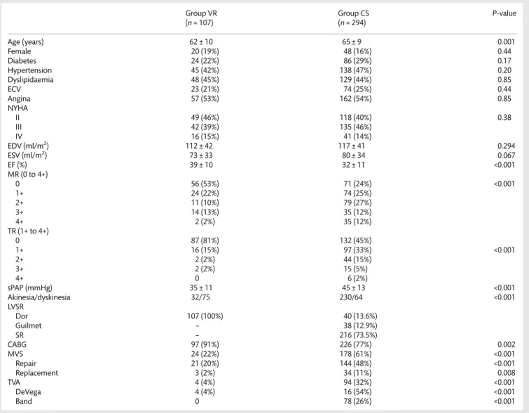
Table 1: Clinical, echocardiographic and surgical data
Group VR Group CS P-value
(n = 107) (n = 294) Age (years) 62 ± 10 65 ± 9 0.001 Female 20 (19%) 48 (16%) 0.44 Diabetes 24 (22%) 86 (29%) 0.17 Hypertension 45 (42%) 138 (47%) 0.20 Dyslipidaemia 48 (45%) 129 (44%) 0.85 ECV 23 (21%) 74 (25%) 0.44 Angina 57 (53%) 162 (54%) 0.85 NYHA II 49 (46%) 118 (40%) 0.38 III 42 (39%) 135 (46%) IV 16 (15%) 41 (14%) EDV (ml/m2) 112 ± 42 117 ± 41 0.294 ESV (ml/m2) 73 ± 33 80 ± 34 0.067 EF (%) 39 ± 10 32 ± 11 <0.001 MR (0 to 4+) 0 56 (53%) 71 (24%) <0.001 1+ 24 (22%) 74 (25%) 2+ 11 (10%) 79 (27%) 3+ 14 (13%) 35 (12%) 4+ 2 (2%) 35 (12%) TR (1+ to 4+) 0 87 (81%) 132 (45%) 1+ 16 (15%) 97 (33%) <0.001 2+ 2 (2%) 44 (15%) 3+ 2 (2%) 15 (5%) 4+ 0 6 (2%) sPAP (mmHg) 35 ± 11 45 ± 13 <0.001 Akinesia/dyskinesia 32/75 230/64 <0.001 LVSR Dor 107 (100%) 40 (13.6%) Guilmet – 38 (12.9%) SR – 216 (73.5%) CABG 97 (91%) 226 (77%) 0.002 MVS 24 (22%) 178 (61%) <0.001 Repair 21 (20%) 144 (48%) <0.001 Replacement 3 (2%) 34 (11%) 0.008 TVA 4 (4%) 94 (32%) <0.001 DeVega 4 (4%) 16 (54%) <0.001 Band 0 78 (26%) <0.001
ECV: extracardiac vasculopathy; NYHA: New York Heart Association; ED: end-diastolic volume; ESV: end-systolic volume; EF: ejection fraction; MR: mitral regurgitation; TR: tricuspid regurgitation; sPAP: systolic pulmonary artery pressure; LVSR: left ventricular surgical remodelling; SR: septal reshaping; CABG: coronary artery bypass grafting; MVS: mitral valve surgery; TVA: tricuspid valve annuloplasty.
any death due to cardiac causes; patients who experienced sudden death or unexplained death were considered as having
cardiac death. Cardiac events were defined as cardiac death,
cardiac reoperation, hospitalization for heart failure, heart trans-plant and New York Heart Association (NYHA) Class III/IV. The
term‘any event’ was defined as all of the above events, including
all deaths and any cause.
Follow-up
All patients were clinically followed up: the most recent informa-tion was obtained by calling the patient or the referring cardiolo-gists. Follow-up was 100% complete and ended in March 2013. In 197 patients, an echocardiographic control was collected.
Statistical analysis
Results are expressed as mean (±standard deviation) and median value. Categorical variables were reported as counts and percen-tages. Differences between the two groups were evaluated by
means of independent t-test (continuous variables) andχ2 test
(categorical variables). A saturate logistic regression model was used to obtain the propensity score using Group A as reference
(goodness-of-fit c-statistic 0.83). Different parametric models
were used to assess changing of hazard function across time; in all cases, hazard risk peaked at 1 month (early phase). Hence, risk factors for early mortality were investigated by means of stepwise binary logistic regression, entering into the initial model all
vari-ables already reported [3]. The results were reported as odds ratio,
95% confidence limits (CLs) and P-value. Ten-year survival curves
were obtained with the Kaplan–Meier method and adjusted using
the propensity score; significant difference was evaluated with the
log-rank test. Time-to-event analysis was performed by a multi-variable Cox proportional-hazard regression (see stepwise logistic regression). The results of Cox analysis were reported as hazard
ratio (HR), 95% CI and P-value. Changes in LV volumes and EF
from preoperative to follow-up period have been evaluated by means of longitudinal linear mixed-model regression for repeated measurements. Changes in NYHA class and mitral regurgitation (MR) grade across time have been evaluated by means of longitu-dinal orlongitu-dinal logistic regression for repeated measurements. The
propensity score was forced in all the regression analyses to adjust all the models for preoperative and operative differences. For all
tests, aP-value of <0.05 was significant. The SPSS software (SPSS,
Inc., Chicago, IL, USA) was used.
RESULTS
Table1reports the clinical, echocardiographic and surgical data of
the two groups. All patients had a Q-wave anteroseptal myocardial infarction of different extents. Operative mortality was 6.0% (24
cases), significantly higher in Group VR (12, 11.2 vs 12, 4.0%,
P = 0.014). Causes of death were cardiac in 19 cases (low cardiac output in 17 and intractable arrhythmias in 2) and non-cardiac in the remaining 5 cases ( pneumonia in 3 and stroke in 2).
Overall 20-year and event-free survival were 36.1 ± 7.8 and 19.4 ± 7.2, respectively. No differences were found regarding 10-year unadjusted survival (Group VR: 55.1 ± 4.8 vs Group CS:
64.2 ± 4.2,P = 0.16) and event-free survival (Group VR: 41.1 ± 4.8
vs Group CS: 50.5 ± 4.8,P = 0.12). However, Group CS provided
better unadjusted 10-year freedom from cardiac deaths (Group
VR: 60.4 ± 4.8 vs Group CS: 74.5 ± 3.7,P = 0.03) and from cardiac
events (Group VR: 45.0 ± 4.9 vs Group CS: 55.6 ± 5.0, P = 0.04);
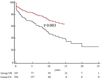
after propensity score adjustment, all the main outcomes were
significantly better in Group CS than in Group VR (Table2, and
Figs 1 and 2). Stepwise logistic regression and Cox analyses
adjusted for propensity score showed that simple LV VR
(independ-ent of thefinal shape) was a poor choice for both early and
long-term cardiac outcomes (Table 3). To avoid any bias related to
improved experience, 30-day mortality being higher in Group VR,
we excluded thefirst month from Cox analysis: LV VR (independent
of thefinal shape) was still confirmed as the less effective approach.
After a median follow-up of 63 (IQR = 19–103) months, 96
patients died, 74 of cardiac and 22 of non-cardiac causes. Further cardiac procedures were performed in 9 cases (4 heart transplants, 1 for MR recurrence, 2 for worsening untreated MR and 2 for LV
assist device implant); 84 of 294 patients survivingfirst
periopera-tive months were readmitted into the hospital due to new onset of heart failure, 41 in Group VR and 43 in Group CS.
At the end of follow-up, 281 survived, with a median follow-up of 100 months (minimum 3 months and maximum 300 months), 48 in Group VR and 233 in Group CS. Among them, 241 (85.8%) were in NYHA Class I or II (38, 79.2% in Group VR and 203, 87.1%
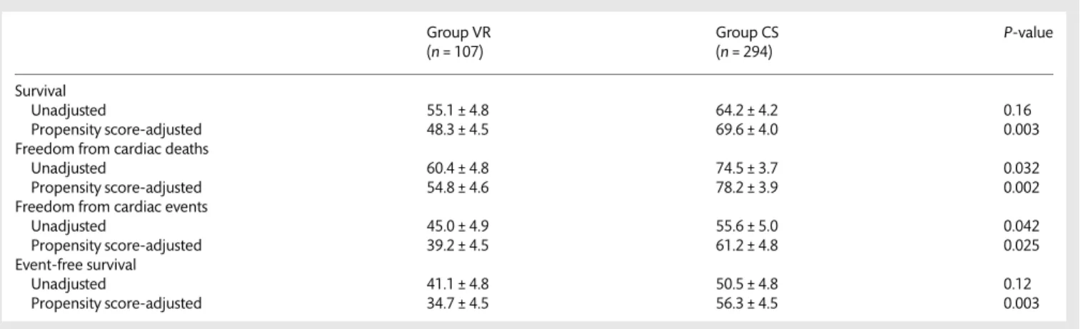
Table 2: Ten-year unadjusted and adjusted results
Group VR Group CS P-value
(n = 107) (n = 294)
Survival
Unadjusted 55.1 ± 4.8 64.2 ± 4.2 0.16
Propensity score-adjusted 48.3 ± 4.5 69.6 ± 4.0 0.003
Freedom from cardiac deaths
Unadjusted 60.4 ± 4.8 74.5 ± 3.7 0.032
Propensity score-adjusted 54.8 ± 4.6 78.2 ± 3.9 0.002
Freedom from cardiac events
Unadjusted 45.0 ± 4.9 55.6 ± 5.0 0.042 Propensity score-adjusted 39.2 ± 4.5 61.2 ± 4.8 0.025 Event-free survival Unadjusted 41.1 ± 4.8 50.5 ± 4.8 0.12 Propensity score-adjusted 34.7 ± 4.5 56.3 ± 4.5 0.003 AD UL T C ARDIA C
in Group CS). LV reshaping rather than VR was associated with
NYHA class improving across time (coefficient −0.80 ± 0.21,
P = 0.002).
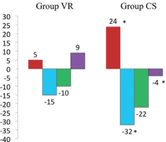
A postoperative echocardiogram, within 15 years from surgery, was obtained in 201 (70%) cases, 48 in Group VR and 153 in
Group CS. Median follow-up was 67 months. Figure3 shows a
higher improvement in LV EF (VR: +5% vs CS: +24%), end-systolic
volume (−15 vs −32%) and sphericity index (+9 vs −4%) in Group
CS than in Group VR. Final sphericity index was 0.75 ± 0.13 (more spherical) in Group VR compared with Group CS (0.67 ± 0.10),
P < 0.001 (Table4).
DISCUSSION
Ventricular remodelling after AMI includes changes in shape and, more importantly, in volume, together with a reduction of the sys-tolic function, measured as EF or other parameters. The LV dilata-tion and thinning, which follows AMI, increase wall stress and, due to the inability of the remaining myocardium to compensate, cause further LV remodelling. Moreover, extensive ventricular
dilatation limits the benefits of isolated myocardial
revasculariza-tion [6,7], as patients with larger ventricular sizes show no
im-provement in the systolic function and higher incidence of cardiac death, myocardial infarction and hospitalization for heart failure.
LVSR was conceived to reverse symptoms of heart failure by reducing LV size, improving, as a consequence, the systolic func-tion in patients with remodelled heart after a myocardial infarc-tion in the left anterior descending (LAD) territory. On these theoretical bases, LVSR was applied since decades with different technical approaches. In the late 1980s, Dor reported a technique
that remained, with some modifications (addition of the Fontan
stitch and introduction of a shaper [8]), the most diffuse procedure
for LVSR [9]. The aim of the Dor technique is reduction in the LV
volume, irrespective of the shape obtained.
In the same decade, Guilmetet al. [10] published another
tech-nique, which combined the reduction in volume with a more CS,
the ‘overcoat technique’. This procedure remained obsolete for
many years until our group, in search of more physiological tions for surgical LV remodelling, rediscovered this elegant
solu-tion in 1998 and, with some modifications, started its clinical
application [4]. The linear suture (anterior wall to septum) was
later changed into an oval patch of different sizes (to obtain a rea-sonable end-diastolic volume), tailored in such a way that the new apex was as distal as possible, to maintain a sphericity index as low
as possible. This evolution was called septal reshaping [5] and was
one of the surgical strategies our group used to maintain a post-operative CS. In fact, the normal LV shape is a prolate ellipsoid
with its long axis directed from apex to base, and its inflow and
outflow being in continuity. LV ejection and filling is a function of
systolic twisting and diastolic untwisting, which depends on the
angular orientation of the oblique muscularfibres that are unique
to the LV [11]. A minor change of 5–10° in the fibre orientation, as
happens in more spherical heart, affects ventricular torsion and
myocardial performance [12]. A change in sphericity seriously
Figure 2:Long-term event-free survival adjusted for propensity score. Group CS (solid line); Group VR (dashed line).
Figure 1:Long-term survival adjusted for propensity score. Group CS (solid line); Group VR (dashed line). CS: conical shape; VR: volume reduction.
Table 3: Stepwise logistic regression and Cox analyses,
adjusted for the propensity score
OR (95% CL) P-value 30-day mortality LV volume reduction 3.3 (1.3–8.5) 0.013 MVS 3.2 (1.2–8.4) 0.018 Age 1.06 (1.01–1.12) 0.031 NYHA class 3.1 (1.6–6.5) 0.001 HR (95% CL) P-value 10-year any deaths
LV volume reduction 1.3 (1.1–2.1) 0.033
MVS 2.7 (1.4–3.3) 0.000
10-year cardiac deaths
LV volume reduction 1.8 (1.2–2.9) 0.002
MVS 2.6 (1.7–4.0) 0.000
10-year cardiac events
LV volume reduction 2.0 (1.4–2.3) 0.001
MVS 2.3 (1.6–3.4) 0.000
10-year any events
LV volume reduction 1.7 (1.1–2.6) 0.003
MVS 2.6 (1.8–3.5) 0.000
LV: left ventricle; MVS: mitral valve surgery; NYHA: New York Heart Association; OR: odds ratio; HR: hazard ratio.
affects the LV function. A myofibril contraction of 15% in a ven-tricle with a normal sphericity index (0.5, ellipsoid shape)
gener-ates an EF of 62%. At the same, with 15%fibre contraction, the EF
falls below 40% if the sphericity index approaches 1 (spherical
shape) and goes up to≥80% if the sphericity index approaches 0
(extreme ellipsoid) [13]. Thus, maintaining a sphericity index as
low as possible must be one of the aims of LVSR.
The benefit of ventricular reduction in patients with
postinfarc-tual akinesia or diskinesia of the anteroseptal wall was challenged
by the STICH trial [2], which demonstrated that there was no
sur-vival or clinical advantage in patients who had LVSR and coronary artery bypass grafting (CABG) when compared with equivalent patients who had CABG without LVSR. Results of this trial caused a strong debate, as inclusion criteria were changed during the re-cruitment of patients and, more importantly, because of the lack
of demonstration of left ventricular end-systolic volume≥ 60 ml/
m² and akinesia≥35% in the LAD territory. Nevertheless, the
con-clusions of the STICH trial, based exclusively on the Dor procedure
[1], are focusing the core problem of ventricular reduction surgery:
the benefits anticipated with surgical reduction in LV volume are
counterbalanced by a reduction in diastolic distensibility [2].
The pathophysiological effects of the Dor procedure have been extensively studied. Even if, after surgery, the wall stress was reduced in the remote zone, in the border zone and in the infarct zone, this reduction was not able to restore the wall stress to
normal values [14]. Wall stress reduction, furthermore, does not
improve contractility in the border zone, as myocytes in that region are in most cases irreversibly damaged. Globally, it seems that reduction in wall stress cannot improve symptoms or increase survival, as suggested by the STICH trial.
The Dor procedure causes an increase in the EF, which is not a valid surrogate of the improvement in the systolic function, as
LVSR, reducing by definition the end-diastolic volume, causes an
in-crease in EF to different extents, independently of the effective
stroke volume. Reduction in mechanical dyssynchrony [15, 16]
improves mechanical efficiency and global LV performance through
an improved synergic distribution of regional stress during the iso-volumic contraction and relaxation phases. Globally, we can agree that the Dor procedure improves systolic function and mechanical
efficiency by reducing LV wall stress and mechanical
dyssyn-chrony, and increasing EF. Revascularization of dysfunctional myo-cardium contributes to improved systolic performance; the impact of the single procedure on global improvement is then impossible to quantify.
The most common negative effect of the Dor procedure is failure to restore a conical LV shape, as demonstrated by a
spher-icity index increase [14,17], which can be at the basis of
post-operative diastolic dysfunction. This effect is independent of the
use of a ventricular shaper [14,17]. The regional shape shows no
significant changes, as indexes of curvature increase, but not to
normal values, failing to optimize the ventricular shape. The
remaining distortion may cause non-optimalfilling and diastolic
dysfunction [14]. Choiet al. [18] showed that a low curvature wall
was characterized by a maximum in the transmural fibre stress
and strain in the mid-wall region, while a steep subendocardial transmural gradient was present in a high curvature wall. In a clinical setting, end-diastolic elastance increases after surgery,
showing a worsening diastolic function [19], insensitive to the
end-diastolic LV pressure [17]. The diastolic dysfunction causes the
Starling relationship to be depressed in most of the patients. Of 12
patients studied by Leeet al. [17], the predicted Starling
relation-ship remained unchanged in 3, worsened in 8 and improved only
in 1. Other studies reported similarfindings [16]. This is reflective
of reduced stroke volume at rest [14,20] and failure to increase
adequately the cardiac output under effort. In general, the improvement in systolic function is counterbalanced, in most cases, by worsened diastolic function.
Other important information was provided by afluid dynamic
model of the normal LV and of a LV after the Dor procedure,
described by Doenstet al. [21]. They found that, in the normal
heart, thefluid dynamic was such to remove blood from the apex
Figure 3: Echocardiographic evolution expressed as percentage variation between preoperative and follow-up values. Ejection fraction (red column), end-systolic volume index (blue column), end-diastolic volume index (green column) and sphericity index (violet column). *P < 0.05 (evaluated with longitu-dinal linear mixed-model regression for repeated measurements).
Table 4: Ecocardiographic results
Group VR Group CS P¹
Pre (48) Post (48) P Pre (153) Post (153) P
EDV (ml/m2) 105 ± 34 95 ± 29 0.040 110 ± 34 86 ± 27 0.000 0.049
ESV (ml/m2) 67 ± 28 57 ± 19 0.043 73 ± 34 50 ± 23 0.000 0.058
EF (%) 39 ± 11 41 ± 10 0.216 33 ± 10 41 ± 11 0.000 1.00
Sphericity index 0.69 ± 0.10 0.75 ± 0.13 0.010 0.70 ± 0.10 0.67 ± 0.10 0.009 0.000
MR 1.0 ± 0.9 0.8 ± 0.6 0.156 1.8 ± 1.1 0.7 ± 0.6 0.000 0.31
EDV: end-diastolic volume; ESV: end-systolic volume; EF: ejection fraction; SI: sphericity index; MR: mitral regurgitation;P: pre versus post; P¹: post group VR versus post group CS.
AD UL T C ARDIA C
of the LV and to generate a vortex, which redirects the blood towards the outlet. Only 2% of the blood that entered the LV during diastole remains in the cavity after four cycles. After
surgery, LV geometry became apple-shaped and thefluid
dynam-ics were completely changed. Blood stagnated into the apex and
was no longer redirected towards the outflow tract. As a
conse-quence, 39% of the blood entering LV in diastole is still present in the cavity after four cardiac cycles. Blood washout was in any case similar before and after surgery.
Even with these potential disadvantages, the outcome of patients who underwent the Dor operation is satisfying. Survival at 5 years
ranges from 61.5 to 72.9% [3,22]. In this study, our 10- and 20-year
freedom from death of any cause have been 60 and 36.1%, respectively.
Both survival and clinical results seem to be better according to the end-systolic volume index (ESVI) obtained after surgery. The best results are obtained when the postoperative values are <60 ml/
m², with values≥60 ml/m² representing a risk factor for lower
sur-vival [23]. Patients who reach an ESVI of <60 ml/m² have a better
preoperative profile, with higher EF and lower volumes. As a
con-sequence, VR has to be less extensive (−26 ml/m², from 85 to 59
compared with−50 ml/m², from 109 to 50 [23]). Preoperative ESVI
is important as well, as values higher than 70 ml/m² (73 [23] or 80
[7]) are necessary to obtain a favourable reverse remodelling of
the LV cavity. However, whereas the EDV can be reduced as much
as the surgeon considers necessary, thefinal ESV will depend not
only on the EDV obtained. The systolic contraction will depend on the residual wall stress, on the curvature of the myocardial wall and on the amount of contractile recovery of the remote area,
which will depend as well on the amount offibrosis present
pre-operatively. Most of these variables are not foreseeable and the final outcome can be unpredictable.
Only a few papers compared volume-related techniques with
shape-related techniques. Isomura et al. [24] reported a 7-year
survival of 61.5 vs 72.1%, respectively (P = 0.041), and our group showed improved freedom from cardiac death in patients who
underwent shape-related LVSR (86.6 vs 76.3%,P = 0.032) [3]. In this
study, we confirmed that, after 10 years from surgery, clinical
results still favour techniques aimed to obtain a more CS together with a reduction in volume. We recently started to apply again the
Guilmet principle, as modified by us, in large ventricles, since
elimination, when possible, of the akinetic area represented by a
patch can be related to a improved stroke volume [25].
Limitations of the study
This study has several limitations. It is a retrospective analysis of many patients undergoing operations over a long period of time,
when techniques, strategies and experience progressively
increased and improved, and the retrospective nature of this study
causes, by definition, a selection bias. Moreover, this article
reports the evolution of a concept across time, starting from the VR to septal reshaping, and therefore although its retrospective nature represents a limitation, we believe that a retrospective study is the only chance we had to report our experience. However, even if propensity score could reduce the selection bias, it cannot protect against the time bias due to the fact that enrol-ment was made in different decades. Finally, being retrospective, some preoperative and follow-up data are missing or incomplete, and therefore, a complete vision of the preoperative status of the patients was not possible in some cases: Echocardiographic data
were obtained for 70% of the entire surviving population. Other preoperative data such as renal function and chronic obstruc-tive pulmonary disease were incomplete (only 55 and 63%, respectively) and so not reported. Aortic atherosclerosis, neo-plasms, smoking and provocative test data to assess myocardial viability were completely lacking.
Most descriptions of the effect of shape on function are only experimental or rely on assumptions never validated in human diseased hearts.
Conclusion
Our long-lasting experience with LVSR shows that long-term
outcome is satisfying and confirms the validity of this procedure.
Results at 10 years seem to be better when VR combined with CS, rather than a mere VR, is pursued, and there are many physio-pathological considerations in favour of this concept. We believe that, as surgery must address different anatomical features, it is not possible to apply to every patient the same procedure. Surgeons must be ready to adapt to each patient the technique
necessary to reach the best correction for the specific case, rather
than attempting to adapt the patient to the technique they are
able to master. Definitively, surgical flexibility is the key point to
achieve an optimal treatment of these complex patients.
Conflict of interest: none declared.
REFERENCES
[1] Jones RH, Velazquez EJ, Michler RE, Sopko G, Oh JK, O’Connor CM et al. Coronary bypass surgery with or without surgical ventricular reconstruc-tion. N Engl J Med 2009;360:1705–17.
[2] Eisen HJ. Surgical ventricular reconstruction for heart failure. N Engl J Med 2009;360:1781–4.
[3] Calafiore AM, Iacò AL, Amata D, Castello C, Varone E, Falconieri F et al. Left ventricular surgical restoration for anteroseptal scars: volume versus shape. J Thorac Cardiovasc Surg 2010;139:1123–30.
[4] Calafiore AM, Gallina S, Di Mauro M, Pano M, Teodori G, Di Giammarco G et al. Left ventricular aneurysmectomy: endoventricular circular patch plasty or septoexclusion. J Card Surg 2003;18:93–100.
[5] Calafiore AM, Mauro MD, Di Giammarco G, Gallina S, Iacò AL, Contini M et al. Septal reshaping for exclusion of anteroseptal dyskinetic or akinetic areas. Ann Thorac Surg 2004;77:2115–21.
[6] Bax JJ, Schinkel AF, Boersma E, Elhendy A, Rizzello V, Maat Aet al. Extensive left ventricular remodeling does not allow viable myocardium to improve in left ventricular ejection fraction after revascularization and is associated with worse long-term prognosis. Circulation 2004;110(Suppl 1):II18–22.
[7] Liu J, Liu Z, Zhao Q, Chen A, Wang Z, Zhu D. Role of surgical ventricular restoration in the treatment of ischemic cardiomyopathy. Ann Thorac Surg 2013;95:1315–22.
[8] Menicanti L, Di Donato M. The Dor procedure: what has changed after fifteen years of clinical practice? J Thorac Cardiovasc Surg 2002;124: 886–90.
[9] Dor V, Saab M, Coste P, Kornaszewska M, Montiglio F. Left ventricular aneurysm: new surgical approach. Thorac Cardiovasc Surg 1989;37:11–9. [10] Guilmet D, Popoff G, Dubois C, Tawil N, Bachet J, Goudot Bet al. A new
surgical technique for the treatment of left ventricular aneurysm: the over-coat aneurysmoplasty. Preliminary results. 11 cases. Arch Mal Coeur Vaiss 1984;77:953–8.
[11] Shapiro EP, Rademakers FE. Importance of obliquefiber orientation for left ventricular wall deformation. Tech Health Care 1997;5:21–8. [12] Helm PA, Younes L, Beg MF, Ennis DB, Leclercq C, Faris OPet al. Evidence
of structural remodeling in the dyssynchronous failing heart. Circ Res 2006;98:125–32.
[13] Sallin EA. Fiber orientation and ejection fraction in the human left ven-tricle. Biophys J 1969;9:954–64.
[14] Zhong L, Su Y, Gobeawan L, Sola S, Tan RS, Navia JLet al. Impact of surgi-cal ventricular restoration on ventricular shape, wall stress, and function in heart failure patients. Am J Physiol Heart Circ Physiol 2011;300: H1653–60.
[15] Di Donato M, Toso A, Dor V, Sabatier M, Barletta G, Menicanti Let al. Surgical ventricular restoration improves mechanical intraventricular dys-synchrony in ischemic cardiomyopathy. Circulation 2004;109:2536–43. [16] Tulner SA, Steendijk P, Klautz RJ, Bax JJ, Schalij MJ, van der Wall EEet al.
Surgical ventricular restoration in patients with ischemic dilated cardiomy-opathy: evaluation of systolic and diastolic ventricular function, wall stress, dyssynchrony, and mechanical efficiency by pressure-volume loops. J Thorac Cardiovasc Surg 2006;132:610–20.
[17] Lee LC, Wenk JF, Zhong L, Klepach D, Zhang Z, Ge Let al. Analysis of patient-specific surgical ventricular restoration: importance of an ellips-oidal left ventricular geometry for diastolic and systolic function. J Appl Physiol 2013;115:136–44.
[18] Choi HF, D’Hooge J, Rademakers FE, Claus P. Influence of left ventricular shape on passivefilling properties and end-diastolic fiber stress and strain. J Biomech 2010;43:1745–53.
[19] ten Brinke EA, Witkowski TG, Delgado V, Klein P, Klok M, Marsan NAet al. Myocardial collagen turnover after surgical ventricular restoration in heart failure patients. Eur J Heart Fail 2011;13:1202–10.
[20] Di Donato M, Fantini F, Toso A, Castelvecchio S, Menicanti L, Annest L et al. Impact of surgical ventricular reconstruction on stroke volume in patients with ischemic cardiomyopathy. J Thorac Cardiovasc Surg 2010; 140:1325–31; e1-2.
[21] Doenst T, Spiegel K, Reik M, Markl M, Hennig J, Nitzsche S et al. Fluid-dynamic modeling of the human left ventricle: methodology and application to surgical ventricular reconstruction. Ann Thorac Surg 2009; 87:1187–95.
[22] Suma H, Anyanwu AC. Current status of surgical ventricular restoration for ischemic cardiomyopathy. Semin Thoracic Surg 2012;24:294–301. [23] Di Donato M, Castelvecchio S, Burkhoff D, Frigiola A, Raweh A, Menicanti
L. Baseline left ventricular volume and shape as determinants of reverse remodeling induced by surgical ventricular reconstruction. Ann Thorac Surg 2011;92:1565–71.
[24] Isomura T, Hoshino J, Fukada Y, Kitamura A, Katahira S, Kondo Tet al. RESTORE Group. Volume reduction rate by surgical ventricular restoration determines late outcome in ischaemic cardiomyopathy. Eur J Heart Fail 2011;13:423–31.
[25] Dang AB, Guccione JM, Zhang P, Wallace AW, Gorman RC, Gorman JH IIIet al. Effect of ventricular size and patch stiffness in surgical anterior ventricular res-toration: afinite element model study. Ann Thorac Surg 2005;79:185–93.
APPENDIX. CONFERENCE DISCUSSION
Dr R. Klautz (Leiden, Netherlands): Your elegant presentation points to the fact that reshaping the ventricle is probably more important than reducing the volume. Many people have already said that, but you just gave us the proof that it has not only direct but also long-term consequences. But as you’ve found that the shape of the ventricle is so important, not every reshaping is the same. Have you tried to make it more quantifiable – for example, that patients with a higher conical index or sphericity index have a better outcome? Was there a
difference - not just that those with reshaping had better outcome - but was there a difference between patients with severe and patients with more limited sphericity index change?
Dr Di Mauro: We had patients with poorer sphericity index who in the long term probably had worse outcome. But when we started to change our minds and to reshape the ventricle, we no longer found sphericity index to be a problem because we addressed the issue of LV sphericity with a very long patch (median 60 cm) to create a very long longitudinal axis of the left ventricle and to reduce the transverse diameter of the ventricle. Using this technique, we are able to improve the sphericity index. So the sphericity index probably now is no longer an issue.
Dr Klautz: Did you use any measurement to decide how to reshape the ven-tricle? All of us have used the Mannequin which directs the volume, but it also directs the shape where the new apex needs to come.
Dr Di Mauro: No. We don’t use the Mannequin because the Mannequin is set on the physiological LV volume, but the“new” ventricle has a wall which is completely akinetic which could result in the repaired ventricle being restrict-ive. So we prefer to use a long patch from deep septum to the apex with the aim of obtaining a more conical chamber. We don’t use any particular meas-urement. We just set the 6 cm patch for 2 cm or for 3 cm depending on the volume of the ventricle. This is enough to obtain a conical chamber.
Dr Klautz: I have a final technical question. How do you deal with the re-mainder of the scar you leave in the ventricle? We usually plicate that.
Dr Di Mauro: The remaining scar is sutured on the new apex. Thus we have a third chamber that clots after surgery. We found in the new echocardiographic control that all the third excluded chambers were clotted inside.
Dr E. Mostafa (Cairo, Egypt): Considering myself as a student in the school of Foch working with Daniel Guilmet, I myself am inclined to use the technique of Guilmet that you used in about 15% of cases, as shown in your slide.
Dr Di Mauro: Yes, we used a modified Guilmet technique in 40 cases before the use of a patch. I think the overcoat technique is a good technique, but we preferred to move to the septal reshaping technique believing that it is more comfortable for the surgeon to place the patch and to obtain a very long longi-tudinal diameter.
Dr Mostafa: Yes, I completely agree because it’s not considered to be the best approach in large aneurysms. I have two questions. Firstly, have you found a difference between the Guilmet technique and the Dor technique in your physiological shaping rather than anatomical shaping? My second question is about the incidence of ventricular dysrhythmias in these types of patients and how you dealt with them.
Dr Di Mauro: We have already published a paper comparing the Dor and Guilmet techniques before the advent of this new technique, and we found that probably the overcoat technique, the Guilmet technique, should be applied when we have a large ventricle with huge involvement of the septum so that we can obtain a very conical shape. When we use the Dor, we had some difficulties in excluding all the septum. Sometimes we had some restrictive chambers and these patients died because of a restrictive syndrome. So prob-ably it’s not a matter of comparison between two techniques but two different approaches. We should tailor the technique to the patient.
Dr R. Przybylski (Zabrze, Poland): Did you check the diastolic dysfunction? In our series we found that diastolic dysfunction correlates with volume rather than shape.
Dr Di Mauro: We are now moving to the second part of this paper to investigate diastolic dysfunction. But diastolic dysfunction is not easy to investigate because we do not have a lot of tools to define it. Since we started with this new technique, we have had no significant diastolic dysfunction with this new approach.
AD UL T C ARDIA C