High Relaxivity Gadolinium-Polydopamine Nanoparticles
Zhao Wang, Fabio Carniato, Yijun Xie, Yuran Huang, Yiwen Li, Sha He, Nanzhi Zang,
Jeffrey D. Rinehart,* Mauro Botta,* and Nathan C. Gianneschi*
Z. Wang, Y. Xie, Y. Huang, Dr. Y. Li, N. Zang, Prof. J. D. Rinehart, Prof. N. C. Gianneschi Department of Chemistry and Biochemistry University of California San Diego La Jolla, CA 92093, USA E-mail: [email protected]; [email protected] Z. Wang, Prof. N. C. Gianneschi Department of Chemistry Northwestern University Evanston, IL 60208, USA Dr. F. Carniato, Prof. M. Botta
Dipartimento di Scienze e Innovazione Tecnologica Università del Piemonte Orientale
“A. Avogadro” Viale Teresa Michel 11 15120 Alessandria, AL, Italy
E-mail: [email protected]
The ORCID identification number(s) for the author(s) of this article can be found under https://doi.org/10.1002/smll.201701830.
DOI: 10.1002/smll.201701830
of detection than what should be attain-able.[3] This is mainly because of rapid
molecular tumbling (τR < 100 ps) and low
hydration states (q = 1). Low r1 values limit
the use of small molecule Gd(III) che-lates in targeted imaging because a large local concentration of the CAs is needed to produce a detectable change in R1. To
overcome this limit of sensitivity, different strategies have been explored. The most common strategy involves the preparation of nanoscale or macromolecular formula-tions in which multiple Gd(III) chelates are associated.[4] Since r
1 is linearly
pro-portional to the concentration of the CA, a high payload (>> 100 ions) is critical to attain a high local concentration at the biological target site; that is, for each tar-geting event of the macromolecule or particle species, multiple Gd(III) centers are localized. Ideally then, these systems would possess high molecular r1 resulting from the combined effect of the large
number of Gd(III) ions and the slow global rotational motion that increases r1 of each individual Gd-complex.[5] Noncovalently
bound MRI CAs often suffer from weak associations limiting applications in vivo. In one example, protein engineering has enabled the design of Gd-specific binding proteins that show excellent metal selectivity and stability.[6] However, the
sophisti-cated preparation processes prevent large-scale production with immunogenicity also being a concern. Covalently linking small molecule chelates with synthetic polymers or nanoparticles
This study reports the preparation of a series of gadolinium-polydopamine nanoparticles (GdPD-NPs) with tunable metal loadings. GdPD-NPs are ana-lyzed by nuclear magnetic relaxation dispersion and with a 7-tesla (T)
mag-netic resonance imaging (MRI) scanner. A relaxivity of 75 and 10.3 mM−1 s−1
at 1.4 and 7 T is observed, respectively. Furthermore, superconducting quantum interference device magnetometry is used to study intraparticle magnetic interactions and determine the GdPD-NPs consist of isolated metal ions even at maximum metal loadings. From these data, it is concluded that the observed high relaxivities arise from a high hydration state of the Gd(III) at the particle surface, fast rate of water exchange, and negligible antifer-romagnetic coupling between Gd(III) centers throughout the particles. This study highlights design parameters and a robust synthetic approach that
aid in the development of this scaffold for T1-weighted, high relaxivity MRI
contrast agents.
Y. Xie, Y. Huang, N. Zang, Prof. J. D. Rinehart Materials Science and Engineering
University of California
San Diego, La Jolla, CA 92093, USA Dr. Y. Li
College of Polymer Science and Engineering
State Key Laboratory of Polymer Materials Engineering Sichuan University
Chengdu 610065, China Dr. S. He
Department of NanoEngineering University of California
San Diego, La Jolla, CA 92093, USA Prof. N. C. Gianneschi
Department of Materials Science and Engineering Northwestern University
Evanston, IL 60208, USA Prof. N. C. Gianneschi
Department of Biomedical Engineering Northwestern University
Evanston, IL 60208, USA
Contrast media are used in a third of the ≈80 million mag-netic resonance imaging (MRI) exams performed worldwide each year.[1] Those approved for clinical use are low molecular
weight, paramagnetic, Gd(III) chelates. They increase the signal intensity on T1-weighted images, shorten the
examina-tion time, improve the diagnostic confidence, and enhance the image contrast.[2] Relaxivity, r
1, is an important parameter
that measures the ability of a Gd-complex to change R1 (1/T1)
of nearby water protons. Currently used Gd-based contrast agents (CAs) exhibit only a fraction of the efficacy (r1)
often results in r1 values that are lower than theory would
sug-gest based on the tumbling rates of the macromolecule or par-ticle. The major limiting factors in many of these formulations have been the relatively slow rate of exchange of the bound water (kex = 1/τM) and the occurrence of fast, local rotational
motion of the Gd(III)-complex about the linker that connects it to the nanoparticle.[5]
The strong metal binding ability of catechol-based functional groups has led to an interest in polydopamine (PD)-based nan-oparticles (PD-NPs) in a growing number of research areas, including bioimaging,[7] battery materials,[8] catalysis,[9] and
environmental remediation.[10] In addition, PD-NPs and
natu-rally occurring melanin-based nanoparticles doped with Fe(III) and Mn(II) have been reported as MRI contrast agents.[11]
Recently, a prepolymerization doping strategy was reported to prepare Fe(III)-loaded PD-NPs in a single pot.[12] However,
adapting this procedure to synthesize high and tunable Gd(III)-content PD-NPs has remained a challenge.[13] In addition,
pre-vious strategies do not result in ultrahigh relaxivity, and we expect to generate high relaxivity close to the theoretic limit.[13c]
Unlike transition metals such as Zn(II), Fe(III), and Mn(III), the methodology of introducing metal salts during the polymer-ization of dopamine is not compatible for the incorporation of the Gd(III) ion. Therefore, we developed a metal displacement method for the synthesis of a series of Gd(III)-polydopamine nanoparticles (GdPD-NPs) with variable Gd loadings, allowing an investigation of the basic properties of these materials in the context of their MRI contrast agents’ performance. Given
the high coordination number of Gd(III)-complexes, fast H2O
exchange, weak antiferromagnetic superexchange interactions, and strong anchoring chelation provided by the PDA catechol moiety, we expect to generate high relaxivity close to the theo-retic limit.
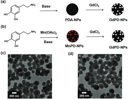
We first prepared GdPD-NPs using a postparticle forma-tion doping strategy (Figure 1). PD-NPs were synthesized via auto-oxidation and polymerization of dopamine under basic conditions.[14] The resulting nanoparticles were characterized
by transmission electron microscopy (TEM) and dynamic light scattering (DLS) to quantify size and uniformity (Figures S1 and S2, Supporting Information). TEM shows spherical parti-cles with diameters of ≈160 nm. After complexation with Gd, the morphology remains unchanged (Figure S1, Supporting Information). This is consistent with previous studies on iron-loaded polydopamine nanoparticles (FePD-NPs). Inductively coupled plasma atomic emission spectroscopy (ICP-OES) analysis was used to determine the Gd loadings to be 2 wt% (massGd/massGdPD-NP). Though this postparticle formation
doping strategy was successfully applied to prepare GdPD-NPs, the approach is limited to low Gd loading. Regardless Gd(III) could not be incorporated via the prepolymerization doping route as described in previous sections. This route does not produce uniform nanoparticles (Figure S3, Supporting Infor-mation). Additionally, the resulting nanoaggregates are col-loidally unstable after polymerization and thus unsuitable for the analysis of their MRI performance. Therefore, we devised a strategy to first synthesize MnPD-NPs with controllable
Figure 1. Preparation of GdPD-NPs (Gd-i (i = 1–5)): a) Postparticle formation doping strategy used to generate Gd-1. b) The prepolymerization doping strategy used to generate particles Gd-2, Gd-3, Gd-4, and Gd-5. White and red dots represent Gd(III) and Mn(III) ions, respectively. c) TEM micrograph of Mn(III)-doped PD-NPs (Mn-4) and d) corresponding GdPD-NPs (Gd-4). Gd loadings for Gd-1, Gd-2, Gd-3, Gd-4, and Gd-5 are 2%, 3%, 5%, 11%, and 17%, respectively.
Mn(III) loadings. These structures served as templates with Mn(III) binding sites primed for displacement when treated with GdCl3. MnPD-NPs were prepared through the oxidative
polymerization of dopamine in the presence of Mn(III) salts under basic conditions as previously reported.[15a] We initially
observe a red solution suggesting the formation of an Mn(III)-catecholate species,[15b] which is polymerized to form black
MnPD-NPs. The incorporation of Mn can be confirmed by X-ray photoelectron spectroscopy (XPS). As shown in Figure S4 (Supporting Information), the XPS spectrum of MnPD-NPs showed a typical binding energy (641.7 eV) at the charac-teristic peak of Mn(2p3/2), which can be assigned to Mn(III).
Recent magnetometry studies on materials prepared via this method for MnPD-NPs have corroborated the trivalent oxida-tion state, likely due to the highly oxidative condioxida-tions of the polymerization.[15a]
After work-up and isolation of the MnPD-NPs, a simple ion exchange procedure was attempted. MnPD-NPs were sus-pended in an aqueous solution of excess GdCl3 and incubated
for ≈12 h. The extent of the displacement reaction was deter-mined by the analysis of XPS and ICP-OES data before and after the Gd(III) incubation. As shown in Figure S4 (Supporting Information), the initial survey scan spectrum of MnPD-NPs exhibits binding energies at the characteristic peaks of C(1s), O(1s), N(1s), and Mn(2p), thus confirming their presence in the original MnPD-NPs. After the displacement reaction, the survey scan spectrum exhibits the characteristic peaks of C(1s), O(1s), N(1s), and Gd(4d), and the characteristic peaks of Mn2p have been eliminated. The resulting nanoparticles show a Gd loading of 11 wt% and a negligible amount of Mn (0.006 wt%) by ICP-OES measurement, consistent with the XPS data. Therefore, we hypothesize that the Mn(III) was replaced by Gd(III) due to weaker binding of Mn(III) to polydopamine compared with Gd(III).[15c] These GdPD-NPs were
character-ized by TEM and DLS with no change in morphology compared to the initial MnPD-NP species (Figure 1d and Figures S1 and S2 and Table S1, Supporting Information). In addition, by choosing templates with increased amounts of Mn(III), we can achieve higher Gd loadings of up to 17 wt% (Table S1, Sup-porting Information). We also measure zeta potential of all five samples. All of these samples exhibited highly negative zeta potential value (Gd-1: −17.8 V; Gd-2: −31.3 V; Gd-3: −50.6 V;
Gd-4: −20.7 V; Gd-5: −24.7 V), suggesting the high stability of these systems in aqueous solution.
In our previous study describing tunable Fe(III) loadings in PD-NPs via the prepolymerization chelation strategy, we found that antiferromagnetic coupling between Fe(III) centers low-ered their r1 as we increased Fe(III) loadings in the particles.[12]
We hypothesized that Gd(III) would exhibit much less antifer-romagnetic coupling because of the poor radial extension of the f-orbitals. This implies that increasing the Gd(III) loading would not lead to the r1 losses observed for Fe(III), but rather a
dramatic increase in per-particle relaxivity, with the only poten-tial for diminishing returns being core-localized metal ions having less access to water. Therefore, we aimed to determine optimal loading conditions by measuring r1 as a function of
Gd(III) loading.
The measurement of r1 and transverse water proton
relaxa-tion rates (r2) as a function of applied magnetic field for the
five GdPD-NPs (Table 1) was undertaken to assess their T1
con-trast enhancing ability. Nuclear magnetic relaxation dispersion (NMRD) allows accurate determination of the field dependence of r1 that arises from magnetic interactions between the metal
centers and the solvent (Figure 2).[3b,16] The NMRD profiles are
characteristic of slowly rotating systems with a shallow decrease of r1 at about 1–5 MHz and a broad and pronounced peak
cen-tered at 30–50 MHz. All five curves share similar characteristics across the entire frequency range, but appear offset from one
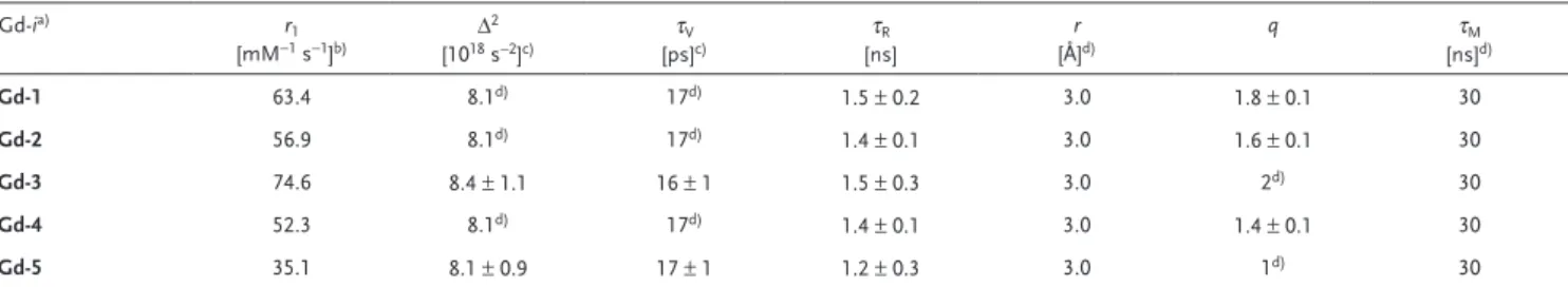
Table 1. Selected relaxation parameters obtained from the analysis of NMRD profiles (298 K).
Gd-ia) r 1 [mM−1 s−1]b) Δ 2 [1018 s−2]c) τV [ps]c) τR [ns] r [Å]d) q τM [ns]d) Gd-1 63.4 8.1d) 17d) 1.5 ± 0.2 3.0 1.8 ± 0.1 30 Gd-2 56.9 8.1d) 17d) 1.4 ± 0.1 3.0 1.6 ± 0.1 30 Gd-3 74.6 8.4 ± 1.1 16 ± 1 1.5 ± 0.3 3.0 2d) 30 Gd-4 52.3 8.1d) 17d) 1.4 ± 0.1 3.0 1.4 ± 0.1 30 Gd-5 35.1 8.1 ± 0.9 17 ± 1 1.2 ± 0.3 3.0 1d) 30
a)The outer-sphere component of the relaxivity was estimated using standard values for the distance of closest approach a (4 Å) and the relative diffusion coefficient of
solute and solvent D (2.2 × 10⁻⁵ cm2 s−1); b)60 MHz; c)The parameters for electronic relaxation are used as empirical fitting parameters and do not have a real physical
meaning for slowly tumbling nanosized systems. Low-field data, those most affected by electronic relaxation, were not included in data analysis; d)Fixed during the fit.
Figure 2. 1H NMRD profiles for Gd-i (i = 1–5). The x-axis is proton Larmor
another by a scaling factor. The fact that the NMRD profiles exhibit a common shape but offset in this way suggests that the origin of the relaxivity differences must be a parameter that directly influences r1 without being field-dependent. Relaxivity
scales either with the hydration number q or with the distance,
rGdH, between the coordinated water proton and the Gd(III)
S = 7/2 electron spin. According to data for small Gd(III) che-lates, the rGdH values are typically found in the narrow range of
≈3.0–3.2 Å, with no evidence of a dependence on the coordina-tion geometry of the metal complex.[17] Therefore, it is plausible
that the different amplitude of the five NMRD profiles reflects a change in the hydration state of the Gd(III) ions. Therefore, we first analyzed Gd-5 assuming the presence of one water molecule in the inner coordination sphere of Gd(III) centers throughout the structure, as the relaxivity values are in line with those of other macromolecular Gd-based systems with
q = 1.[2a,5,18] We note that only frequencies above 3 MHz were
analyzed, as Solomon–Bloembergen–Morgan theory is not ade-quate to account for the data at low magnetic fields in the case of slowly rotating systems. The temperature dependency of r1
at high field clearly indicates the occurrence of fast exchange conditions. That is, r1 is not limited by water exchange. We then
fixed this parameter at 30 ns and performed the fit treating three parameters as variables, namely, the overall rotational cor-relation time (τR), the correlation time for the transient
zero-field splitting (ZFS; τV), and the amplitude of the transient ZFS
(Δ2). The best-fit parameters obtained for the GdPD-NPs from
the analysis of 1H NMRD data are shown in Table 1. We
con-clude that the tight packing of Gd-centers at catecholate groups throughout crosslinked polymers within the NPs results in a rotationally restricted system at the metal centers and their che-lates. The absence of internal rotation of the Gd-chelates leads to a very good fit, even at high frequencies, with a single global rotational correlation time of 1.2 ns. Likewise, the NMRD pro-file of Gd-3 can be fitted by simply assuming q = 2. The other parameters vary only marginally reinforcing the hypothesis that relaxivity changes are associated with changes in the state of hydration of the paramagnetic centers. A q value of 2 could be associated with a larger number of Gd ions exposed to the surface and more accessible to water molecules, whereas q = 1 might arise as an average between the Gd centers on the surface (q = 2) and those in the inner core of the NPs, not accessible to water (q = 0). The NMRD profiles of NPs Gd-4, Gd-2, and Gd-1 were analyzed by fitting only q and τR. The best results were
obtained with the parameters shown in Table 1, where the rota-tional correlation time is essentially unchanged and the effec-tive q value scales with the relaxivity, from 1.4 to 1.8.
The r1 values (1.4 T; 60 MHz) demonstrate the presence
of facile relaxation in all five samples (Table S2, Supporting Information). Gd-3 has an r1 value of 75 mM−1 s−1, 20-fold
higher than commercially used Gd-based contrast agents (i.e., Gd-DOTA) and superior to many previously reported macromolecules and nanoscale formulations (such as liposomes and dendritic species).[19] It is also the highest one
among the five tested samples. Therefore 5% is the optimal loading conditions for achieving the highest per Gd ion relaxivity, which is approaching the theoretical limit. When increasing the Gd loading to 17% (Gd-5), the r1 value remains
relatively high (35 mM−1 s−1). The decrease of r
1 may be due
to the larger size of Gd-5 preventing water coordination of paramagnetic centers within the core of the particle. The r2/r1
ratios at various field strengths (20–70 MHz) were determined (Figure S5, Supporting Information) and not surprisingly, these ratios increase with increasing proton Larmor frequency, with the highest value being under 3, indicating that Gd-1–5 act as
T1-weighted contrast agents with relatively little interference
from T2 darkening.
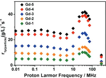
Given that each particle contains many chelated Gd(III) ions, it is interesting to consider the “per-particle relaxivity” (r1p(particle)) to describe the local concentration necessary to
achieve the desirable T1 MRI contrast under different magnetic
fields (Figure 3).[20] Interestingly, plots of r
1p(particle) and r1p(Gd(III))
show different trends. Whereas r1p(Gd(III)) reveals a maximum
relaxivity for Gd-3, the plot of r1p(particle) shows that relaxivity
increases monotonically with doping amount on a per-particle basis. Thus, Gd-5 exhibits the highest per-particle relaxivity, about five times larger than the Gd-1 at 60 MHz (Figure 3). These data lead to the intuitive result that highly paramagneti-cally doped particles are superior to those doped with lower levels of Gd(III) ions.
To evaluate the potential utility of GdPD-NPs as T1-weighted
contrast agents at high magnetic fields, we calculated their r1
values and captured T1-weighted MR images using a volume
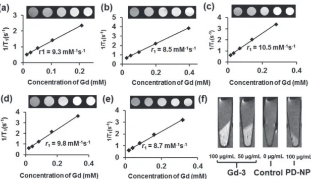
coil with a 7-tesla (T) MRI scanner. The T1 relaxation times
became shorter and the phantom image became brighter as the Gd(III) concentrations increased (Figure 4a–d), suggesting that the GdPD-NPs accelerated the recovery of net magnetiza-tion. At 7 T, Gd-3 has the highest r1 value (r1 = 10.5 mM−1 s−1),
while the other four samples exhibit only slightly lower values. This value is roughly four times higher than common com-mercial contrast agents such as Gd-DOTA (r1= 3.0 mM−1 s−1)
and Gd-DTPA (r1= 3.1 mM−1 s−1).[21] Additionally, the rough
equivalence of Gd-1–5r1 values means that a significantly lower
Gd(III) ion dosage can be used without sacrificing the contrast enhancement ability. The r2/r1 values (Table S3, Supporting
Information) are larger compared with the ones at 1.4 T due to the enhanced T2 effect at high field. To demonstrate the
poten-tial for Gd-i to be used as contrast agents, they were incubated with HeLa cells and shown to provide enhanced positive
con-Figure 3. 1H NMRD profiles for Gd-i (i = 1–5). The x-axis is proton Larmor
trast in T1-weighted MR images as compared to control cells
(Figure 4). Overall, the high T1 relaxivity and low r2/r1 ratio of
GdPD-NPs could make them suitable as low dosage, Gd-based contrast agents for T1-weighted MRI measurements.
A key prerequisite for in vivo imaging applications is low toxicity. For Gd-i to be useful, Gd(III) ions must not be sus-ceptible to transmetallation as this would result in a release of potentially harmful free Gd(III) ions.[22] Thus it is important to
examine the selectivity of these nanoparticles for Gd(III) over physiological metal ions, such as Ca(II) and Zn(II). We use
Gd-3 to examine stability of Gd(III) coordination over Ca(II)
and Zn(II) at their maximum human blood concentration (2.5 × 10⁻³m for Ca(II) and 0.15 × 10⁻³m for Zn(II)). Gd-3 was
incubated with aqueous CaCl2 and ZnCl2 solutions for 7 d,
fol-lowed by multiple washing and centrifugation steps to remove free metal ions. The sample was then analyzed via ICP-OES to show no significant release of Gd(III) ion on this timescale (Figure S6, Supporting Information). The stability of Gd-3 in various media was also examined. We observe that the amount of chelated Gd(III) ion does not change significantly over time in water, 4-(2-hydroxyethyl)-1-piperazineethanesulfonic acid (HEPES) and Phosphate Buffered Saline (PBS) buffer (Figure S7, Supporting Information). Although the cell viability of PD-NPs had already been confirmed,[23] the cytotoxicity
of GdPD-NPs has not yet been evaluated. Nanoparticles with negatively charged surfaces often exhibit a lower cytotoxicity compared to those with positively charged ones.[24] We tested
the toxicity of samples with various loadings of Gd(III) (Gd-1,
Gd-3, Gd-4, Gd-5, and PD-NP). The results showed no obvious
cytotoxicity after incubation with Gd-1, Gd-3, or Gd-4 at levels up to 125 µg mL−1 within 48, while the Gd-5 sample showed
some cytotoxicity at high concentration (125 µg mL−1) that may
come from the high content of Gd(III) inside the nanoparticle
(Figure S8, Supporting Information). We recently found that polydopamine nanoparticles undergo a similar degradation pathway as natural melanosomes.[25] In addition, Gd(III) is also
known to have high affinity to catecholate units.[26] Therefore
we anticipate that the GdPD-NPs, upon possible degradation in vivo over the long term, would stay in chelated form.
In our previous work,[12] it was shown that increasing Fe(III)
ion concentration within polydopamine nanoparticles led to antiferromagnetic coupling that correlated with decreases in MR contrast. To investigate whether Gd(III)–Gd(III) coupling or the formation of Gd oxide phases at high concentrations could be playing a role in the decrease in r1, the magnetic
prop-erties of GdPD-NPs were investigated by variable-temperature magnetic susceptibility measurements from 2 to 300 K under a 5000 Oe magnetic field (Figure S9, Supporting Information). Unlike in FePD-NPs, there is no appreciable deviation from single-ion, spin-only behavior (χMT = 7.88 emu K cm−3 mol−1)
for the three GdPD-NP samples that were tested. This indicates a nearly complete lack of antiferromagnetic coupling between Gd(III) centers in the polydopamine systems. These data indi-cate that the drop in per-Gd(III) relaxivity at high concentrations must be dependent on the lack of solvent access to core ions in the larger particles. In addition, the magnetic susceptibility of the S = 2 Mn(III) ion is much smaller than that of the S = 7/2 Gd(III) ion (χMTMn(III), 300 K = 3 emu K mol−1 < χMTGd(III), 300 K =
7.88 emu K mol−1). Even if some small amount of Mn(III) ions remains chelated, their contribution to the magnetic suscepti-bility can be considered negligible.
In conclusion, we report a simple and scalable synthetic method of generating GdPD-NPs. We describe Gd PD-NPs as contrast agents with several potential advantages: (1) the nanoscale formulation and crosslinked nature of the Gd-chelates leads to slow molecular tumbling increasing contrast;
Figure 4. MRI characterization of Gd-i (i = 1–5) on a Bruker 7-T magnet. Plots of 1/T1 versus Gd(III) concentration and its corresponding image of
a) Gd-1, b) Gd-2, c) Gd-3, d) Gd-4, and e) Gd-5. f) In vitro T1-weighted MR images of HeLa cells incubated with Gd-3 and PD-NP at different
(2) restriction of the free rotation of the chelates extends to the surface bound Gd, due to the crosslinked polydopamine 3D network; (3) the catechols in the 3D crosslinked network are strained, leading to the formation of unsaturated Gd-catecholate, allowing high hydration states (q > 1). Finally, these factors result in our observation that the as-prepared nanopar-ticles show significant MRI signal enhancement and the value of longitudinal relaxivity is as high as 75 mM−1 s −1 at 1.4 T and 10.5 mM−1 s−1 at 7 T for Gd-3. While the relatively large
particle size (>100 nm) limits the use in targeting to the interstitial space of tissues,[27] these nanoparticles may have
potential applications in tumor imaging due to the enhanced permeability and retention effect.[11b,13b,28]
Supporting Information
Supporting Information is available from the Wiley Online Library or from the author.
Acknowledgements
The authors thank the AFOSR for generous funding through a PECASE to N.C.G. (FA9550-11-1-0105). M.B. acknowledges support of the “Compagnia di San Paolo” (CSP-2012 NANOPROGLY Project). Y.L. acknowledges financial support from the State Key Laboratory of Polymer Materials Engineering, Sichuan University (Grant No. sklpme2016-3-03). This work made use of the Keck-II facility of Northwestern University’s NUANCE Center, which has received support from the Soft and Hybrid Nanotechnology Experimental (SHyNE) Resource (NSF ECCS-1542205); the MRSEC program (NSF DMR-1121262) at the Materials Research Center; the International Institute for Nanotechnology (IIN); the Keck Foundation; and the State of Illinois, through the IIN. Dr. S.H. acknowledges the support from FISP award (C6007) and Air Force Offfice of Scientific Research (AFOSR) FA9550-15-1-0273.
Conflict of Interest
The authors declare no conflict of interest.
Keywords
antiferromagnetic coupling, contrast agent, magnetometry, MRI, polydopamine
Received: May 31, 2017 Revised: August 4, 2017 Published online: October 10, 2017
[1] J. Enders, E. Zimmermann, M. Rief, P. Martus, R. Klingebiel, P. Asbach, C. Klessen, G. Diederichs, T. Bengner, U. Teichgräber, B. Hamm, M. Dewey, BMC Med. Imaging 2011, 11, 4.
[2] a) P. Caravan, J. J. Ellison, T. J. McMurry, R. B. Lauffer, Chem. Rev.
1999, 99, 2293; b) L. M. Randolph, C. L. M. LeGuyader, M. E. Hahn,
C. M. Andolina, J. P. Patterson, R. F. Mattrey, J. E. Millstone, M. Botta, M. Scadeng, N. C. Gianneschi, Chem. Sci. 2016, 7, 4230; c) Z. Zhou, Z.-R. Lu, Wiley Interdiscip. Rev.: Nanomed. Nano
biotechnol. 2013, 5, 1.
[3] a) P. Caravan, Chem. Soc. Rev. 2006, 35, 512; b) S. Aime, M. Botta, E. Terreno, Adv. Inorg. Chem. 2005, 57, 173; c) Z. Wang, Y. Li, Y. Huang, M. P. Thompson, C. L. M. LeGuyader, S. Sahu, N. C. Gianneschi, Chem. Commun. 2015, 51, 17108; d) K. R. Maravilla, J. A. Maldjian, I. M. Schmalfuss, M. J. Kuhn, B. C. Bowen, F. J. Wippold, V. M. Runge, M. V. Knopp, S. Kremer, L. J. Wolansky, N. Anzalone, M. Essig, L. Gustafsson, Radiology
2006, 240, 389; e) S. He, N. J. J. Johnson, V. A. Nguyen Huu,
E. Cory, Y. Huang, R. L. Sah, J. V. Jokerst, A. Almutairi, Nano Lett.
2017, 17, 4873.
[4] a) A. J. L. Villaraza, A. Bumb, M. W. Brechbiel, Chem. Rev. 2010, 110, 2921; b) N. J. J. Johnson, S. He, V. A. Nguyen Huu, A. Almutairi,
ACS Nano 2016, 10, 8299.
[5] M. Botta, L. Tei, Eur. J. Inorg. Chem. 2012, 2012, 1945.
[6] J. J. Yang, J. Yang, L. Wei, O. Zurkiya, W. Yang, S. Li, J. Zou, Y. Zhou, A. L. W. Maniccia, H. Mao, F. Zhao, R. Malchow, S. Zhao, J. Johnson, X. Hu, E. Krogstad, Z.-R. Liu, J. Am. Chem. Soc. 2008,
130, 9260.
[7] a) X. Zhang, S. Wang, L. Xu, L. Feng, Y. Ji, L. Tao, S. Li, Y. Wei,
Nanoscale 2012, 4, 5581; b) Y. Li, Y. Huang, Z. Wang, F. Carniato,
Y. Xie, J. P. Patterson, M. P. Thompson, C. M. Andolina, T. B. Ditri, J. E. Millstone, J. S. Figueroa, J. D. Rinehart, M. Scadeng, M. Botta, N. C. Gianneschi, Small 2016, 12, 668; c) Q. Fan, K. Cheng, X. Hu, X. Ma, R. Zhang, M. Yang, X. Lu, L. Xing, W. Huang, S. S. Gambhir, Z. Cheng, J. Am. Chem. Soc. 2014, 136, 15185.
[8] a) H. Jiang, L. Yang, C. Li, C. Yan, P. S. Lee, J. Ma, Energy Environ.
Sci. 2011, 4, 1813; b) H. Jiang, T. Sun, C. Li, J. Ma, RSC Adv. 2011, 1, 954.
[9] K. Ai, Y. Liu, C. Ruan, L. Lu, G. M. Lu, Adv. Mater. 2013, 25, 998.
[10] J. Fu, Z. Chen, M. Wang, S. Liu, J. Zhang, J. Zhang, R. Han, Q. Xu,
Chem. Eng. J. 2015, 259, 53.
[11] a) K.-Y. Ju, J. W. Lee, G. H. Im, S. Lee, J. Pyo, S. B. Park, J. H. Lee, J.-K. Lee, Biomacromolecules 2013, 14, 3491; b) Z.-H. Miao, H. Wang, H. Yang, Z.-L. Li, L. Zhen, C.-Y. Xu, ACS Appl. Mater.
Interfaces 2015, 7, 16946.
[12] Y. Li, Y. Xie, Z. Wang, N. Zang, F. Carniato, Y. Huang, C. M. Andolina, L. R. Parent, T. B. Ditri, E. D. Walter, M. Botta, J. D. Rinehart, N. C. Gianneschi, ACS Nano 2016, 10, 10186.
[13] a) W.-W. Cai, L.-J. Wang, S.-J. Li, X.-P. Zhang, T.-T. Li, Y.-H. Wang, X. Yang, J. Xie, J.-D. Li, S.-J. Liu, W. Xu, S. He, Z. Cheng, Q.-L. Fan, R.-P. Zhang, J. Biomed. Mater. Res. A 2017, 105, 131; b) S. Cho, W. Park, D.-H. Kim, ACS Appl. Mater. Interfaces 2017, 9, 101; c) P. Caravan, C. T. Farrar, L. Frullano, R. Uppal, Contrast Media Mol.
Imaging 2009, 4, 89.
[14] Y. Liu, K. Ai, J. Liu, M. Deng, Y. He, L. Lu, Adv. Mater. 2013, 25, 1353.
[15] a) Z. Wang, Y. Xie, Y. Li, Y. Huang, L. R. Parent, T. B. Ditri, N. Zang, J. D. Rinehart, N. C. Gianneschi, Chem. Mater. 2017, https://doi. org/10.1021/acs.chemmater.7b02262; b) M. J. Sever, J. J. Wilker,
Dalton Trans. 2004, 0, 1061; c) B. Larsson, H. Tjälve, Acta Physiol.
1978, 104, 479.
[16] S. H. Koenig, R. D. Brown, Prog. Nucl. Magn. Reson. Spectrosc. 1990,
22, 487.
[17] P. Caravan, A. V. Astashkin, A. M. Raitsimring, Inorg. Chem. 2003,
42, 3972.
[18] A. Accardo, D. Tesauro, L. Aloj, C. Pedone, G. Morelli, Coord. Chem.
Rev. 2009, 253, 2193.
[19] J. Tang, Y. Sheng, H. Hu, Y. Shen, Prog. Polym. Sci. 2013, 38, 462. [20] G. Sitbon, S. Bouccara, M. Tasso, A. Francois, L. Bezdetnaya,
F. Marchal, M. Beaumont, T. Pons, Nanoscale 2014, 6, 9264. [21] I. M. Noebauer-Huhmann, P. Szomolanyi, V. Juras, O. Kraff,
[22] a) P. H. Kuo, E. Kanal, A. K. Abu-Alfa, S. E. Cowper, Radiology
2007, 242, 647; b) J.-M. Idée, M. Port, I. Raynal, M. Schaefer,
S. Le Greneur, C. Corot, Fundam. Clin. Pharmacol. 2006, 20, 563.
[23] K.-Y. Ju, Y. Lee, S. Lee, S. B. Park, J.-K. Lee, Biomacromolecules 2011,
12, 625.
[24] E. Fröhlich, Int. J. Nanomed. 2012, 7, 5577.
[25] Y. Huang, Y. Li, Z. Hu, X. Yue, M. T. Proetto, Y. Jones, N. C. Gianneschi, ACS Cent. Sci. 2017, 3, 564.
[26] G. E. Freeman, K. N. Raymond, Inorg. Chem. 1985, 24, 1410. [27] S. Barua, S. Mitragotri, Nano Today 2014, 9, 223.
[28] R. Ge, M. Lin, X. Li, S. Liu, W. Wang, S. Li, X. Zhang, Y. Liu, L. Liu, F. Shi, H. Sun, H. Zhang, B. Yang, ACS Appl. Mater. Interfaces 2017,