2
INDEX
Introduction pag. 3
Synthesis of testosterone pag. 9
Mechanism of action pag. 12
Organ damage pag. 14
AAS and hypogonadism pag. 16
Aim of the thesis pag. 29
Materials and methods pag. 31
Results pag. 38
In vitro
Testosterone production pag. 38
Levels of enzymes involved
In testosterone synthesis pag. 38
Expression of StAR and CYP17A1 pag. 40
In vivo
Body weight pag 40
Effect of ND administration
on testosterone biosynthesis pag. 41
histological analisys pag. 43
effect of ND stimulation
on gene expression levels of BTB components pag. 43
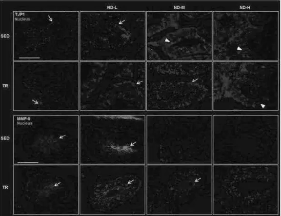
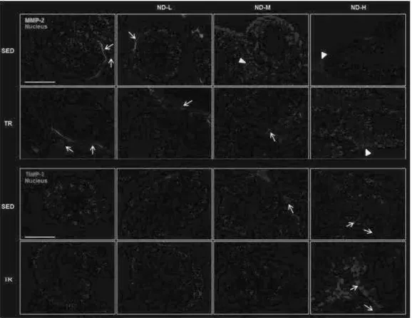
Immunofluorescence analysis pag. 45
Immunohistochemical analysis pag. 47
Discussion pag. 49
Conclusion pag. 56
3
INTRODUCTION
Anabolic androgenic steroids (AASs), or anabolic steroids as they are commonly known, are one of the leading and most widely used classes of doping drugs. AASs are synthetic derivates of the male sex hormone, testosterone, and they have androgenic (development and sustainment of secondary sex characteristics), anabolic (tissue building) and hedonic effect (Turillazzi et al. 2011).
First identified in 1935, testosterone is the principal androgen controlling the development of the male body with androgenic masculinizing effects along with anabolic properties that increase lean muscle mass (Coward et al. 2013). For this AASs are, however, used by some healthy men and more rarely by women, to gane muscle and lose body fat. Structural modifications to the testosterone molecule were introduced in the 1950s to increase its anabolic effects while minimizing androgenic effects, resulting in the AASs family (Neri et al. 2011). Initially, AASs were restricted to professional athletes and bodybuilders, becoming gradually more popular among recreational and non-professional power athletes. In fact the prototype of an AASs users is shifting from the competitive body builders/athlete to men seeking to optimize their physical appearance (Rahnema et al. 2014). It is currently estimated that there are as many as three million AASs users in the USA and, interestingly, two third of US users are non-competitive bodybuilders, or even non-athletes, who use these substances for aesthetic purpose only. In fact the data show that four out of five AASs abusers are not competitive athletes. The lifetime prevalence of AASs use for men is estimated to be from 3.0% to 4.2% and is increasing. Use among the male gym is estimated to be as high as 15-30%. However, it has been provided by a study entitled "Monitoring the Future” that the use of AASs will decline in the coming years thanks to the success of education and the numerous prevention campaigns having as targets high school students both athletes and non-athletes (Rahnema et al. 2014).
For regards the availability and the procurement of AASs it has been suggested that the “Internet” is the most common source for men to obtain AASs as well as ancillary drugs. Access to these suppliers can vary from open access to special invitation offered by internet forum members or blogs or via word of mouth at local gymnasium. Then by these mediatic means AASs users around the world can anonymously offer or request advice, share drug sources, chronicle
4
results and collaborate on dosing schedules. Therefore the AASs user’s rationale for choosing various drugs and protocol is typically based on anecdotal evidence and interpretations of “quasi-scientific literature” propagated via internet forums. Another source of information involves “specific nutritionists” who, for a fee, usually yearly, give advice to these men about AASs, dietary supplements and nutritional planes. Beyond non physicians sources, in nutritional supplements sold legally online or in retails stores have been found to contain AASs or other ancillary drugs that may or may not be listed as ingredients on the product label. Indeed, >20% of legally sold nutritional supplements have been found to be contaminated with AASs. With global sales of nutritional supplements exceeding $32 billion in 2012 this ubiquitous impurity poses significant public health problems (Rahnema et al. 2014).
The basic chemical structure of all steroids is a perhydrocyclopentano phenantrene ring system which comprises of three fused six-membered rings (A, B and C) and one five-membered ring (D)(figure 1) (Turillazzi et al. 2011):
These rings may be modified in order to obtain designed chemical modifications sought to obtain different anabolic and/or androgenic effects (Turillazzi et al. 2011):
5
Testosterone derivates have been categorized in three classes:
· Class A: alkylation at the 17-b-hydroxy esters requiring intramuscular injection; in fact they usually have a longer half-life and a slower absorption rate, bringing much less hepatic stress than the orally taken steroids. Pain at injection sites is common, because of the oily base. In this group are involved four groups of AASs: testosterone esterified (i.e. testosterone cypionate, propionate and others), nandrolone (trade mark: Decadurabolin), boldenone and finally trenbolone.
· Class B: alkylation at the 17-a-hydroxy position with a methyl or ethyl group. These compounds can be given orally; in fact the alkylation prevents deactivation of steroids by hepatic first-pass metabolism promoting oral administration. They usually have short half-life, making several daily doses necessary
Fig.2: Structural features influencing the expression of the androgenic and anabolic activities. By Turillazzi et al. 2011.
6
to maintain appropriate blood levels. This class include: stanazolol (trade mark: Winstrol), oxandrolone and others.
· Class C: alkylation in the A,B,C ring of the steroid backbone results in orally available AASs resisting hepatic metabolism (de Souza et al. 2010, Neri et al. 2011).
Nowadays, the most commonly used AASs include: nandrolone, oxandrolone, stanazololo and oxymetholone (Neri et al. 2011).
Synthetic androgens are interesting not only for their injury and organ damage but also for their medical use. In fact since 1930s, androgens have been approved for the treatment of a variety of clinical conditions, including: delayed puberty, hypogonadism, impotence, infertility, sarcopenia, osteoporosis, decline of cognitive functions, anemia, hepatic regeneration (following an injury or a transplantation), endometriosis, breast cancer and, finally, cachexia (because among the other effects AASs have also the “anti-glucocorticoid effect” mediated by testosterone occupation of cortisol receptors which have a remarkable affinity with testosterone and for this they create an anti-catabolic effect) (Neri et al. 2011). On the contrary, non-medical AASs use was primarily confined to elite athletes and bodybuilders in the 1960s who used it as a means to enhance performance. In the last three decades, however, AASs use has spread into the general population. Actually, AASs are been used worldwide by millions of men, many of whom having no athletic ambition wishing to increase and improve their physical strength and appearance for the aesthetic purposes (MPahil et al. 2104). Use of AASs among athletes and specific subsets of the general population (high school and college students) is a major public health tissue (MPahil et al. 2014). For regards the side effects as well as the organ damage that the abuse of AASs can cause, it can established that in the short–term AASs abuse seems to have few serious medical consequences and, besides, in this case the damage seems to be reversible (Neri et al. 2011, Riezzo et al. 2011); but in the long – term, taken in supraphysiological doses, AASs show various adverse medical effects, especially cardiovascular toxicity that usually can lead to sudden death, influence of total cholesterol, activation of hemostatic system, hepatotoxicity, renal failure, musculoskeletal damages, psychological symptoms (in fact behavioral effects by AASs abuse include hypomanic or manic symptoms, accompanied by aggression or violence, which usually occur while taking AASs, and depressive symptoms occurring during AASs withdrawal) and increased general mortality (Kostic et al. 2011, Kanayama et al. 2011, Neri et al. 2011, Turillazzi et al. 2011). Additionally, for men who have previously used AASs, a
7
unique condition known as anabolic steroid–induced hypogonadism (ASIH) becomes a real concern. Clearly described in 1990 by Jarow and Lipshultz, ASIH has recently been identified as a potentially underrecognized cause of hypogonadism in young men (Rahnema et al. 2014). Again, AASs abuse may be accompanied by the consumption of other drugs such as alcohol, tobacco or marijuana and among the other well known side effects of AASs there are: acne, alopecia, LUTS (Lower Urinary Tract Symptoms due to prostate enlargement related to 5DHT-effect), erectile dysfunction, libido loss (these two latter expecially after discontinuation when endogenous testosterone levels are usually low) and finally gynaecomastia (due to higher aromatization of testosterone which results very high during the “cycles”) (de Souza et al. 2010). Finally, AASs may cause a dependence syndrome, which may pose a growing public health problem in future years (Kanayama et al. 2010).
Another real problem of AASs abusers is that the athletes and non-athletes, to minimize the risk of developing tolerance to any particular agent, take them as a cocktail of different agents taken at one time. This process is commonly called “stacking”. In fact different oral and injectable compounds are generally combined (“stacked”), creating large-dose regimens usually self-administered during periods (“cycles”) lasting 4-12 weeks. Stacking is based also on the “idea” that smaller dosages of multiple drugs might reduce the chance of complications than larger dosages of a single drug. This may also facilitate the administration of multiple AASs necessary to achieve supraphysiological doses for long periods. The aim of “stacking” is to rationally combine different characteristics, avoiding overlap of benefits or side effects. For example combination of testosterone and nandrolone are the basis of the “mass-building stacks”, used to maximize muscular and strength gains (Fineschi et al. 2005, de Souza et al. 2010).
Moreover, frequently, AASs users may also combine AASs with other “performance drugs”, such as painkillers (including oppioids), diuretics, insulin, Gh, stimulants, aromatase inhibitors and tyroxine to improve physical performance (de Souza et al. 2010).
For the reasons described above mortality risk among chronic users is estimated to be 4.6 times higher than among non-users (de Souza et al. 2010).
The culture of using ergogenic (performance enhancing) compounds among athletes dates back to the initial Olympiad in ancient Greece where herbal remedies and animal extracts were apparently used by the athletes before the competition. Hence, the history of “doping” began long before the discovery of
8
androgens. Not surprisingly, soon after their discovery, androgens made their way on the list of ergogenic compounds. Although androgens were used in various sporting events, they only received major attention during the Olympic Games. In fact the most notorious era in the doping history of the Olympic Games regarded the athletes (mainly women) of the German Democratic Republic since 1960s to 1980s. Nevertheless, in 1974, AASs abuse was prohibited by the International Olympic Committee (IOC) and, currently, the scrutiny of doping in sports is conducted by a subsidiary of IOC called World Anti-Doping Agency (WADA), which was formed a decade ago (Basaria et al. 2010).
Among the anabolic androgenic steroids, nandrolone decanoate (ND) is a synthetic testosterone analog considered one of the most commonly abused anabolic androgenic steroids by adolescents and athletes. ND is alleged to promote an increase in muscle mass and improves both physical appearance and sporting performance (Nilsson et al. 2004). Nowadays, ND abuse is often associated with serious adverse effects, interfering with the musculoskeletal system, the endocrine system, and the reproductive system (Socas et al. 2005). Moreover, ND may suppress the hypothalamus-pituitary-gonadal axis resulting in a decreased production of endogenous testosterone (Kostic et l. 2011). Testosterone is usually produced by testicular Leydig cells and its production is regulated by a neuroendocrine feedback mechanism which regulates the pulsatile release of luteinizing hormone (LH). The result is therefore activation or repression of the steroidogenic signaling cascade as well as gene transcription of key enzymes (Janjic et al. 2012; Wu et al. 2007). The steroidogenic enzymes involved in testosterone biosynthesis include: steroidogenic acute regulatory protein (StAR), cholesterol side chain cleavage enzyme (CYP11A1), 3ß-hydroxysteroid dehydrogenase (HSD3B1), 17α-hydroxylase/17,20-lyase (CYP17A1), and 17ß-hydroxysteroid dehydrogenase (Bjelic et al. 2015). The role of these steroidogenic enzymes is further described in Table 1. Although ND is frequently abused in sports, there are limited animal studies which compare the relationship between physical activity/exercise with and without ND use. Shokri et al. demonstrated that exercise associated with supraphysiological doses of ND in rats increased apoptosis in spermatogenic cells (Shokri et al. 2010). In testes, germ cell development is supported by Sertoli cells that reside within the basal epithelial lining within the seminiferous epithelium (Kotaja et al. 2010). These cells create a specialized microenvironment through the formation of the blood-testis barrier (BTB), preventing free passage of solutes, ions, and water that might affect the development of germ cells (Mruk et al. 2010). The BTB is formed by tight junctions (TJs), basal ectoplasmic specializations (ES) and desmosome-gap
9
junctions (D-GJs) that compartmentalize the seminiferous tubule into the basal and adluminal compartments (Mruk et al. 2010). The aim of this study is to investigate the effects of ND administration on testosterone biosynthesis in a mouse exercise model. Moreover, testis morphological alterations associated with the dysregulation of factors that confer BTB integrity will also be determined.
Synthesis of testosterone
Testosterone, the principal human androgen, controlles the development of the male body with “androgenic effects” (or also called “masculinizing effects”) that are most evident during puberty (a period in which testosterone elicites dramatic psyco-physiological changes in the male body), including the onset of secondary male characteristics, hair growth pattern, sebaceous gland activity and maturation of sperm and libido. All these effects are associated to “anabolic properties” of testosterone that increase lean muscle mass. In fact “anabolism” is defined as any physiologic state in which nitrogen is differentially retained in lean body mass through the stimulation of protein synthesis and/or reduction in protein breakdown. Besides it includes growth promotion, protein and collagen synthesis and an increase of size and bone metabolism (de Souza et al. 2010).
Regarding AASs abusers, the anabolic effects are dose-dependent, and they usually occur when supraphysiological testosterone levels (>1000ng/dl) are found, which generally requires weekly doses of 300 mg or more (de Souza et al. 2010).
Daily testosterone synthesis ranges from 2.1 to 11 mg in individual males with normal plasmatic level of 300-1000 ng/dl, which progressively decline with age (de Souza et al. 2010):
Endocrine tissues such as the gonads and the adrenals are tissues that are able to produce active steroid hormones (from cholesterol) and to deliver them into the circulation to exert their action away from the site where they are produced. It is well recognized that gonads produce sex steroids while the adrenals produce primarily glucocorticoids and mineralcorticoids and sex steroids in small part (Kostic et al. 2011).
The biosynthesis of testosterone is dependent on both acute and chronic stimulation of Leydig cells by the pituitary luteinizing hormone (LH).
10
Testosterone regulates release of the hypothalamic gonadotropin-releasing hormone (GnRH), the signal that initially stimulates the pituitary to synthesize and release LH characterizing the hypothalamic-pituitary-gonadal axis (HPG) (figure 3) (de Souza et al. 2010, Kostic et al. 2011).
Fig.3: Negative feedback of HPG axis in men and women respectively. By http://dsdgenetics.org/index.php?id=48.
11
Steroidogenic process is mainly produced by the Leydig cells of testes in males and ovaries in females, although smaller amounts are synthesized by adrenal glands in both sexes. Steroidogenesis is initiated with cholesterol that is the common substrate for all steroids hormones. In fact cholesterol and steroids hormones have the same basis chemical structure characterized by the tetracyclic structure of the cyclopentaneperhydrophenanthrene also called Gonano (figure 4) that is formed by four rings of which three are hexanes and one pentane. Cholesterol can be divided into endogenous and exogenous, the latter taken with the diet. Cholesterol is then converted into esters that are stored in vesicles (Kostic et al. 2011).
Steroidogenesis process begins in the cytosol and is completed with the passage through the mitochondria and the smooth endoplasmic reticulum. The transfer into the mitochondria, and particularly to the inner membrane of the mitochondria, is mediated by the steroidogenic acute regulatory (StAR) protein. Through the mobilization and delivery from the outer to the inner mitochondria membrane, cholesterol is converted to pregnenolone by the cytochrome P450
Fig.4: Chemical structure of Ciclopentanoperidrofenantrene (also called Gonano). By Turillazzi et al. 2011.
12
cholesterol side chain cleavage enzyme (CYP11A1 also known as P450scc). Pregnenolone is then transported to endoplasmic reticulum and converted to progesterone by 3b-hydroxysteroid dehydrogenases (HSD3b). After this, the maturation of progesterone to androstenedione is catalyzed by the 17a-hydroxylase/C17-20lyase (CYP17A1 also known as P45017A1) and finally to testosterone by 17b-hydroxysteroid dehydrogenase (17bHSD), steroid dehydrogenase specific for androgen production (Kostic et al. 2011).
Once synthesized, testosterone has several possible metabolic fates. First, it binds to the androgen receptor (AR) in target tissue to exert its effects. Second, it is reduced to 5a-dihydrotestosterone (5DHT), which also acts on the AR. Finally, testosterone may be aromatized to estradiol to exert estrogenic effects, tipically characterized by water retention, breast tissue growth and other effects (de Souza et al. 2010, Kostic et al. 2011).
Mechanism of action
AASs are believed to exert their action by several different mechanisms by increasing synthesis of a wide variety of structural, enzymatic and receptorial proteins. Nevertheless the various clinical effects are determined by the type and concentrations of androgen receptors and enzymes controlling steroid metabolism in a given organ (Turillazzi et al. 2011).
AASs are synthetic derivatives of testosterone and therefore, their mechanism of action is similar to all other steroid hormones (Turillazzi et al. 2011).
The classical pathway of androgen action involves steroid binding to the androgen receptors (ARs), an intracellular ligand-activated transcription factor, and a member of the nuclear receptor superfamily, acting on the genome. After the formation of the complex AR-steoids, it migrates to the nucleus and it binds to palindromic DNA sequences, and specifically, to hormone response elements (HRE) and initiates gene transcription. This type of patway is also called “genomic mechanism” (Turillazzi et al. 2011).
The genomic action of ARs is modulated by a large variety of coregulators, which are proteins that target gene expression by enhancing (coactivator) or restraining (corepressor) transcription (Turillazzi et al. 2011).
However, AASs may also have a direct rewarding or hedonic properties, mediated not so much by their genomic effects (although these may well
13
contribute) but more directly by the effects of AASs and their metabolites on plasma membranes. This alternative and/or contemporary pathway of steroid is called “non-genomic” and most of its effects involves a membrane receptor (Turillazzi et al. 2011).
Non-genomic effects of steroids androgens are distinguished from genomic ones by: 1) rapid onset (seconds to minutes) that is faster than genomic mechanism, 2) insensivity to inhibition of RNA and protein syntesis, 3) effects produced by steroids unable to access the nucleus either covalently linked to membrane impermeable macromolecules or in cell lacking a nucleus), and 4) not usually blocked by classical antagonists due to different steroidal specificity from classical cognate nuclear receptors (Turillazzi et al. 2011).
Nevertheless, non-genomic androgen effects characteristically involve the rapid induction of conventional second messanger signal transduction cascades, including increase in cytosolic calcium and activation of protein kinase A, protein kinase C and MAPK (mitogen-activated protein kinase), leading to diverse cellular effects including smooth muscle relaxation, neuromuscular and junctional signal transmission and neuronal plasticity. Non-genomic effects were hypothesized be also at the basis of cardiotoxicity as we shall see later (Turillazzi et al. 2011).
14 Organ Damage
The analysis of AAS is an issue of main interest in many field of clinical and forensic toxicology. AASs toxicity, in fact, may cause both morbidity and mortality; the death in fact may occur suddenly or be related to the chronic damage of vital organs, as it’ll expressed later (Strano et al. 2011).
The incidence of sudden death in athletes is estimated 2.5 times higher than in non-athletes; in only about 1% of the examined cases could drug abuse be confirmed. So even if a possible or probable cause of death is found at the autopsy, there remains doubt about substance-triggered or facilitated sudden death (this latter because of the stacking process). In literature, the main pathologies found out in case of sudden death were myocardial injuries but in other sudden death cases pulmonary venous thrombosis and right cerebellar hemorrhage were the cause of death. At this point it is important to emphasize that for regards the sudden death, in many cases only microscopic examination and/or blood and/or urine samples or even the only inspection revealed the
Fig.5: Genomic and Non-genomic mechanism of action of AASs. By Turillazzi et al. 2011.
15
modifications organ and/or the presence of AASs and their main metabolites in victims from steroid abuse. In fact, it often happened that to the autoptic relief, macroscopically organs appeared normal; for example, it happened that the heart was normal in size and weight, the valvular apparatus was normal, that the coronary arteries were normal hiding the real cause of death. Toxicological investigations are therefore fundamental in those cases of sudden death in subjects suspected of consuming AASs (Strano et al. 2011).
Biological specimens used for toxicological investigation are usually: urine, blood, vitreous humour and hair; while for antidoping analyses the only specimen used is urine because the majority of AASs metabolites undergo to the phase 2 metabolism, and therefore excreted mainly as glucuroconiugate, and in minor extent, as sulfo-coniugates. For this purpose Fineschi et al have designed a new method to extract main AASs ematic and urinary metabolites, that occurs through C18 cartridges after enzymatic hydrolysis to obtain trimethylsylil derivates that are more simple to detect (Strano et al. 2011).
Recommendation on post-mortem sampling regarding in particular the collection of blood from peripheral sites and heart blood, urine, vitreous humour, bile, cerebrospinal fluid, gastric contents, hair, nails and skin samples necessary to extend the chronological window assumption (Strano et al. 2011).
However many studies report that the determination of AASs in hair is a good application for forensic toxicology. In fact main advantages linked to hair analysis are: the easy, non invasive sample collection (that is not easily adultered), and preservation without specific precautions. Besides, hair analysis could be useful for epidemiological or prevalence studies for the evaluation of AASs abuse. Furthermore, hair samples permit a retrospective evaluation of drug use history corresponding to many months before the actual sampling moment, depending, obviously, on hair length. Besides has been demonstrated that hair has an isoelectric pH close to 6 and thus favours incorporation of the undissociated drugs and of their metabolites (Strano et al. 2011).
In the field of doping, the hair analysis is always considered second to the sampling of urine (Strano et al. 2011).
For the “classic” drugs of abuse, the isolation of the drugs always occurs after alkaline digestion with NaOH followed by an extraction phase characterized by liquid/liquid extraction or liquid/solid extraction or both. This latter phase is then followed by analysis in GC/MS or in LC/MS (Strano et al. 2011).
16
Therefore, it can say that AASs abuse analysis has become indispensable procedure routinely carried out in antidoping laboratories; recently the determination of AASs constitutes a new analytical challenge also for forensic laboratories, owing to the increased interest of the forensic sciences in the investigation about “anabolic steroids misuse”, particularly in case of sudden death related to AASs toxicity (Strano et al. 2011).
AAS and hypogonadism
The male genital apparatus consists of: gonads (testes or didimi), sperm ducts (epididymis, vas deferens and ejaculatory ducts), glands (seminal vesicles, prostate and bulbo-urethral glands) and external genital organs (scrotal sac and penis) (Gray et al. 2000).
Fig.6: Anatomy of the male reproductive apparatus. By http://galleryhip.com/male-and-female-reproductive-system.html.
17
Anatomically, the testes are paired organs, ellipsoidal in shape and slightly flattened. In adult humans, the testes have an average weight of about 30 grams and are located slightly in height difference relative to one another, with the left most place in the bottom of the right. They are elastic and soft in consistency, bluish-white in color and have two faces (one medial and one lateral), two margins (one anterior and one posterior), and two poles (one upper and one lower) and finally they have a vertical axis of about 3cm and a diameter of about 2cm (Gray et al. 2000).
The testes are placed in the scrotal sac and are separated between them by the scrotal septum and because sensitive to temperature, they need to stay at a lower temperature than that of the body because, otherwise, they would not be able to produce sperm (Gray et al. 2000).
The testes have two main functions:
· Reproductive: because of the spermatogenesis process (from puberty until andropause);
· Endocrine: because they secrete hormones called androgens, including testosterone that is the most important androgen hormone but its production is not constant over the life: it is evident from birth, but increases dramatically at puberty and lasts for the whole adulthood.
The testicle is wrapped of connective tissue, from which originate the septa which extend inward, separating it into lobules, in which lie the seminiferous tubules. Within the seminiferous tubules occurs the production of sperm. In fact, in each seminiferous tubule are arranged in layers, germ cells, which since by the spermatogonii through successive cell divisions are transformed into spermatozoa. The seminiferous tubules continue in the epididymis through a network of convergence; their wall also consists of flask-shaped cells called Sertoli cells, which are responsible of nutrition and guide the maturation of sperm, activities regulated primarily by the hormone pituitary FSH (Gray et al. 2000).
The production and secretion of testosterone is carried out by groups of cells called Leydig cells, interposed between the seminiferous tubules, and whose activity is regulated by the pituitary hormone LH (Grey et al. 2000).
Regarding steroidogenesis, this latter is a biological process regulated in a pulsatile manner by modulating circulating LH levels. In the testes, it is established that the binding of LH to its G protein-coupled receptor (LHR) on Leydig cells acutely increases the levels of cAMP and cytoplasmic Calcium, which are both required for steroidogenesis. The transient synthesis of steroids
18
in response to LH occurs through modulation of a key factor, steroidogenic acute regulatory protein (STAR), whose expression highly correlates with testosterone level (Clark et al. 1994). STAR is the rate limiting factor in hormone-dependent steroidogenesis because it is essential for the transport of cholesterol, the precursor of all steroids hormones, from the outer to the inner mitochondrial membrane (Clark et al. 1994). The cAMP pathway induces Star expression and steroidogenesis through activation of several transcription factors (Manna et al. 2002). Besides Star activation also requires de novo synthesis of the nuclear receptor 4A1 (NR4A1) also known as NUR77 (Martin et al. 2008). In addition induction of NUR77 and STAR involves the Calcium pathway (Martin et al. 2008, Martin et al. 2009). An increase in intracellular Calcium is nonetheless required. Indeed, cytosolic calcium concentration increases in parallel with testosterone production (Sullivan et al. 1986) probably coming from both extracellular environment and from intracellular calcium store (i.e. endoplasmic reticulum) (Costa et al. 2007). This hypothesis is validated by experimental studies on Leydig cells using calcium channel inhibitors drugs demonstrating that blocking the release of calcium into the cytoplasm has got the hang of the steroidogenic process. In fact the block of calcium channels inhibits expression both of STAR and of its regulator NUR77 (Costa et al. 2010). Therefore, induction of steroidogenesis requires the induction of STAR expression trough NUR77 activated itself by calcium signaling in these cells (Costa et al. 2010). Moreover, inhibiting the calcium channels could decrease steroidogenesis by preventing the translocation of cholesterol from the outer to the inner mitochondrial membrane and/or by affecting the expression/activity of any of the downstream steroidogenic enzymes. Thus, calcium signaling is critical for male reproductive health (Costa et al. 2010).
The steroidogenic-enzyme genes are generally regulated at the transcriptional level (Chin-Hee Song et al. 2012). Nur77 and SF-1 are major transcription factors which are known to regulate steroidogenic-enzyme genes (Parker et al. 1997, Mellon et al. 1998, Chin-Hee Song et al. 2012). Nur77 family members regulate the expression of steroidogenic-enzyme genes such as P450-17A1 (Wilson et al. 1993), 3b-HSD (Martin et al. 2005), STAR (Houk et al. 2004) and others.
In the testes, androgen action is mediated by androgen receptor (AR), which is a ligand-inducible transcription factor (Kokontis et al. 1999). Has been shown that androgen-bound AR physically interacts with Nur77, and inhibits Nur77 transactivation through competitive binding with Nur77 coactivators, which results in the downregulation of steroidogenic enzyme gene expressions in
19
mouse Leydig cells (Chin-Hee Song et al. 2012). By the results of some studies, in fact, has been strongly supported the idea of androgen’s local action on testicular steroidogenesis that suppresses the level of steroidogenic enzyme (such as STAR, P450-17A1, 3b-HSd) (Chin-Hee Song et al. 2012) . Moreover, AR suppresses the transactivation of Nur77 in a dose-dependent manner. Interestingly, there is also a ligand-independent effect of AR on Nur77 transactivation, although it is much less compared to its ligand-dependent effect. This is probably due to minor translocation of AR to the nucleus because of the androgens. There results imply that transcription of steroidogenic enzyme genes enhanced by Nur77 is suppressed by androgen/AR in Leydig cells. Nur77 was predictably localized in the nucleus and the presence of DHT did not affect its localization. On the other hand, AR is present in the cytoplasm without androgen and translocated into the nucleus upon addition of androgen. The overlay revealed a nice co-localization of Nur77 and AR in the nucleus in an androgen-dependent fashion. These results support the possibility that AR protein, translocated into the nucleus in the presence of androgen, modulates Nur77 transactivation. In fact many studies suggest that AR and NUR77 coactivators compete for the modulation of NUR77 transactivation. Therefore AR inhibits Nur77 transactivation by interfering with the interaction between Nur77 and its coactivators (Chin-Hee Song et al. 2013).
Therefore, these studies have suggested that androgens regulate the testicular steroidogenesis locally in the testis, in addition to long-loop negative feedback regulation through the hypothalamus–pituitary–testis axis (Hong et al. 2004, Lee et al. 2009). In addition, has been speculated that the local androgen action on testicular steroidogenesis might be accomplished by cross-talk between androgen signaling and major transcription factors responsible for the expression of steroidogenic enzyme genes. The endocrine system consists of dynamic biological processes involved in the regulation of a complex array of physiologic activities. The synthesis of sex steroids in male is elaboratively regulated by various local factors including testicular paracrines/autocrines in addition to the negative feedback through the hypothalamic–pituitary–gonadal (HPG) axis (Chin-Hee Song et al. 2012). Since the change of androgen level is associated with various physiological activities, the acute response may be necessary to keep hormone level rapidly within normal ranges. All these findings just sayd strongly support the local action of androgen/AR on testicular steroidogenesis, and may provide an insight into its regulatory mechanism with regard to the steroidogenesis in Leydig cells (Chin-Hee Song et al. 2012).
20
In this thesis, in particular, it was discussed of the gonadic organ damage and in particular of its pathological state consequent to AASs abuse as hypogonadotropic hypogonadism with its correlated problem of fertility. This pathological state is seen as a social evil continually increasing in the general population. In fact the impact of AASs abuse on male fertility is one of the least reported but certainly not the least important. In fact, partially due to a lack of research in the field, AASs use is a rarely considered as an etiology of hypogonadism (de Souza et al. 2010).
Epidemiologically, in some studies it has underlined that contrary to the common public perception that AASs is used primarily by professional athletes, it has found that AASs are much more common and their use is significantly correlated negatively with the age, the educational level and number of children. In addition, AASs seem to be appealing to abusers because when taken in conjunction with exercise, they increase muscle mass and strenght, and increase libido (de Souza et al. 2010).
Clinically, hypogonadism is defined as a pathological condition of inadequate gonadal function, which manifests itself as an insufficient gametogenesis and/or secretion of androgens. In fact total testosterone is at 50ng/dl or less (de Souza et al. 2010).
Infertility, instead, is defined as the failure to achieve a successful pregnancy after 12 months or more of regular unprotected intercourse (de Souza et al. 2010).
The data reveal that in the world, approximately, 80 million people suffer from infertility and that male infertility is present in up to 50% of all infertile couples (de Souza et al. 2010).
Usually hypogonadism is classified into three forms:
· primary hypogonadism (also known as
hypergonadotropic) in which the damage of Leydig cells impairs the production of androgens (testosterone) and/or alters the seminiferous tubules, resulting in oligospermia or azoospermia and increased of gonadotropins. The more frequent example of this type of hypogonadism is Klinefelter Syndrome but there is also hypogonadism caused by liver disease, bacterial infections, autoimmune diseases, orchitis, kidney disease, radio/chemotherapy and others.
21
· secondary hypogonadism (also called hypogonadotropic) in which abnormalities of the pituitary gland or hypothalamus affect the secretion of gonadotropins and this can lead to impotence and/or sterility. Some example can be the panhypopituitarism syndrome, hypothalamic and pituitary tumors, steroids/opiates abuse, vitamin and/or nutritional deficiency, hemochromatosis, cerebral bleeding, infection, HIV, obesity, trauma and others.
· tertiary hypogonadism in the case of resistance to the action of androgens, or if the response to androgens available is inadequate.
Etiologically, as just seen, several clinical conditions both congenital and acquired (such as: body weight, the smoking impact, drugs and the strenuous sport) may be related to male hypogonadotropic hypogonadism and its consequent state of infertility (Schilling et al. 2012). Nevertheless, the etiology of pathologic state of hypogonadotropic hypogonadism by AASs abuse is under-estimated because many studies are based on the honesty of patient responses on a sensitive topic; data in fact are often subject to bias as well as secondary to partecipant embarrassment or distrust (de Souza et al. 2010) .
In the hypogonadotropic hypogonadism by AASs abuse, the organ damage, as we will see, may be either primary (due to the direct damage caused by ROS resulting in apoptosis) (Shokri et al. 2009) and secondary (since it involves the HPG axis). Hypogonadotropic hypogonadism is a typical example of a (ir)reversible side effect by AASs abuse. It is clearly known that FSH and LH from pituitary gland have growth-promoting effects on testis development and that AASs abuse suppresses the serum LH, FSH and testosterone levels in humans (Rahnema et al. 2014).
Pathophysiologically, in fact, hypogonadotropic hypogonadism is a pathological condition induced, characterized by decreased in circulating levels of total and free testosterone concentrations as well as of LH with consequently testicular atrophy and impaired spermatogenesis (Rahnema et al. 2014). These effects result from the negative “systemic” feedback of exogenous androgens on the hypothalamic-pituitary-gonadal axis (also known as anabolic steroid-induced hypogonadism ASIH) which in turn results in suppression of endogenous testosterone, and from “local” suppressive effects of exogenous androgens on the testis (Rahnema et al.2014, Chin-Hee Song et al. 2012, Shokri et al. 2009). In fact FSH and LH concentrations are typically low. What sets hypogonadotropic
22
hypogonadism from hypergonadotropic hypogonadism is the presence in the first case of an inhibition of the pulsatile release of GnRH whose levels are very low in this case. All this is followed, therefore, by a sequent decrease in LH and FSH levels (de Souza et al. 2010).
In addition, during AASs use, serum androgen concentrations may be supraphysiologically high, but the hypogonadotropic state lowers the intratesticular testosterone (ITT) concentrations required to maintain normal spermatogenesis (for this it is not surprising that AASs users have presented to fertility clinics with azoospermia and oligospermia as well as dysmorphia and dysmotility) (Shokri et al. 2012, de Souza et al. 2010). ASIH arises from the combination of hyperandrogenism (resulting from the supraphysiologic supplementation of AASs) and subsequent hypogonadism. This testosterone deficiency occurs because typical AASs users alternate between “on cycle” supraphysiologic plasma androgen levels and periods of androgen deficiency where ancillary drugs such as SERMs, hCG are used in attempt to recover the HPG axis (Rahnema et al. 2014). The duration of suppression and the resultant symptomatic ASIH is highly variable and due to multiple factors, including differences in the choices of drugs, amounts used and duration use (Rahnema et al. 2014).
Clearly described in 1990 by Jarow and Lipshultz, ASIH has recently been identified as a potentially under-recognized cause of hypogonasism in young men (Coward et al. 2013).
23
On the other hand, many studies have been demonstrated that also AASs use coupled with endurance exercise induce alterations in the hypothalamic-pituitary-gonadal (HPG) axis. Therefore not only hormonal aspect influences the hypogonadotropic hypogonadism state (Shokri et al. 2009).
Nevertheless, less attention has been directed towards another fundamental aspect of change in male reproductive organ, that is the germ cell apoptosis (Shokri et al. 2009). This latter phenomenon has been reported to play an important role in normal testicular physiology, and apoptotic control is important for regulating the germ cell population in the adult testes (Shokri et al. 2009). Successful germ cell maturation requires a precise balance between the numbers of germ cell population and the neighbouring Sertoli cells, which support their survival, proliferation and differentiation (Shokri et al. 2009). Also in this latter case both exercise and AASs abuse are known to exert apoptotic alterations in male reproductive tissues(Shokri et al. 2009). Many studies have reported a significant increase in caspase-3 activity and a decrease in the number of the germ cell layer (Shokri et al. 2009). This phenomenon occurs because the exercise increases the oxygen consumption rate by 10-20 times, resulting in an enhanced production of reactive oxygen species (ROS) in the cells/tissues and
Fig.7: Illustration of pathophysiology of anabolic-androgenic steroids-induced hypogonadism (ASIH) and the mechanism of action of selected treatment strategies.
24
exerts oxidative stress, which increase the rates of cellular damage (Shokri et al. 2012). Besides, exercise impairs the induction of antioxidant enzyme activities (Shokri et al. 2012). Therefore, it seems logical that the physical/chemical-induced oxidative stress may affect the testicular antioxidant system and lipid peroxidation in humans (Shokri et al. 2009). In fact oxidative stress has been shown to be a major cause of male infertility; a large proportion of infertile men have elevated levels of seminal ROS (Shokri et al. 2012). Several forms of sperm DNA damage are caused by ROS (e.g. chromatin cross-linking, chromosome deletion) (Shokri et al. 2012). In addition, it has been underlined that testes contain a low amount of antioxidant enzymes compared to other tissues such as the liver and the kidney (Shokri et al. 2012). Therefore high level of apoptosis will be detected in those body-builders submitted to AASs abuse and strenuous exercise because of increased vulnerability of the testes (Shokri et al. 2009).
Histologically, many experiments report AASs-induced Leydig cell alterations in hypogonadotropic hypogonadism. In fact the decrease in this population of cells is accompanied by low testosterone and LH levels, accompanied by decreased steroidogenesis in testicular tissue and spermatogenesis, with the lack of advanced forms of spermatids. Nevertheless, after AASs discontinuation, Leydig cells tend to proliferate but remain below the regular counts.
In some studies has been underlined that the reproductive organ weight, the weight of testes and other accessory sex organs (such as epididymis, prostate and others) are significantly reduced in patients submitted to strenuous exercise and AASs abuse (Shokri et al. 2012). This pathological state is consequent to the decreased level of intratesticular testosterone that can evolve also in testicular atrophy (Shokri et al. 2012). Studies show that also the physical exercise exasperates this state of atrophy because endurance exercise reduces the blood flow to the testicles and causes low level of testosterone secretion thus affecting some degree of spermatogenesis (Sjodin et al. 1990, Shokri et al. 2012). Therefore, also the weight of epididymis (both left and right) is decreased (Shokri et al. 2012). Besides in these patients is usually observed also a decrease in “total epididymal sperm count” (Shokri et al. 2012). In fact many body-builders report azoospermia as well as oligospermia (de Souza et al. 2010, Shokri et al. 2012, Rahnema et al. 2014).
25
For regards the “semen quality”, among the AASs users is common a low-quality semen associated to morphologically abnormal (dysmorphia), hypokinetic (dysmotility) and dead spermatozoa (de Souza et al. 2010, Shokri et al. 2012, Rahnema et al. 2014).
For the “sperm motility”, usually expressed as a percentage of motile sperm of the total sperm counted, it has been showed by many studies that also this spermatic parameter is decreased in patient submitted to exercise and AASs abuse (de Souza et al. 2010, Shokri et al. 2012, Rahnema et al. 2014).
Regarding symptoms and signs, sexual dysfunction and decreased libido are reported by 25% of users and symptoms of androgen deficiency including fatique and depression, lethargy and insomnia are common complaints, especially during a post-cycle period (Shokri et al. 2012, Rahnema et al. 2014).
The diagnosis requires a trusting patient-physician relationship, which may be difficult to achieve during an initial visit (Rahnema et al. 2014). Obtaining an AAS use history can be uncomfortable for the patients and
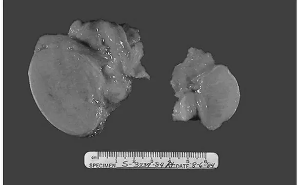
Fig. 8: Comparison between a normal testis (on the left) and an atrophic testis (on the right) after exposition to AASs chronic abuse. By http://library.med.utah.edu/WebPath/MALEHTML/MALE082.html.
26
physicians (Rahnema et al. 2014). Besides an initial testing tipically consists of an hormonal panel (LH, FSH, E2, T, free T, SHBG and PRL), complete blood cell count (for the diagnosis of polycythemia), lipid profile, prostate specific antigen and metabolic profile (Rahnema et al. 2014). Physical examination should include height, weight, blood pressure and body mass index and common signs consistent with AASs abuse such as acne, gynecomastia, testicular atrophy, skin striations and alopecia (de Souza et al. 2010, Rahnema et al. 2014).
For regards the management of AASs-induced male hypogonadotropic hypogonadism and its consequent state of infertility, there are different solutions (de Souza et al. 2010). First, following establishment of a nonjudgmental, healthy, and trusting physician-patient relationship, the patient should be counseled to discontinue the AASs as well as any self-administered ancillary drugs and supplements (de Souza et al. 2010, Rahnema et al. 2014). In fact the already simple discontinuation of AASs use may lead to fertility recovery within 4-6 months in certain proportion of male users, but there is little literature and considerable disagreement regarding the management of such cases (Rahnema et al. 2014).
Nevertheless, the classic treatment of hypogonadotropic hypogonadism is based on testosterone and this type of therapeutic approach has shown a significant growth in recent years because testosterone prescriptions is increased more than of 170% since 2007 and than 500% since 1993 (Coward et al. 2013). The paradigm in the treatment of hypogonadotropic hypogonadism with testosterone is that this latter alone has a negative impact on spermatogenesis and fertility (Coward et al. 2013, Rahnema et al. 2014). In fact, for this is preferable to administre in conjunction with testosterone low doses of hCG to maintain spermatogenesis in men who wish to preserve fertility (de Souza 2010, Coward et al. 2013, Rahnema et al. 2014). For its mechanism of action, testosterone acts both centrally (on hypothalamus and on amygdale to modulate libido) and peripherally (acting on endothelial cells of the arteries, increasing nitric oxide synthase activity leading to increased nitric oxide levels, smooth muscle relaxation and increased penile blood) (Ramasamy et al. 2014).
Aside the testosterone, moreover, patients may also be actively treated, with other options, in a manner similar to those used for other forms of hypogonadotropic hypogonadism infertilities, requiring the induction of spermatogenesis with gonadotropins or gonadotropin analogues, including i.m. injections of hCG, human menopausal gonadotropin (hMG), recombinant FSH,
27
selective estrogen receptor modulators and aromatase inhibitor (Coward et al. 2013, Rahnema et al. 2014).
In fact the use of hCG alone, or in combination with hMG, has been reported to be a successful treatment for this group of patients. Therefore, considering the favourable results after treatment, it is resonable to consider it during the infertility consultation (de Souza et al. 2010).
On the other hand, there are many cases in which above seen therapy had no effect. This problem is due to the fact that AASs up to 90% of abusers often “stack” multiple androgens (a practice that users believe provides the greatest results while minimizing unwanted side effects) and at much higher doses of 500-1500 mg/week for 4-12 weeks and sometimes for years (de Souza et al. 2010, Nangia et al. 2014). Because of this multiple different AASs regimens used, of variable durations and of cycles of treatment, the exact recovery from AASs is not known (Nangia et al. 2014). Besides, a prospective study of different real world AASs regimens and recovery would be difficult to conduct in a structured and ethical manner since these are controlled drugs that are obtained illicitly from a number of questionable sources of variable quality and given in doses that are much higher than are considered safe (Nangia et al. 2014).
Therefore, the duration of ASIH is unclear. Some studies suggest that ASIH lasts only 4 to 12 months after AASs discontinuation but others describe a subset of men with long-term or permanent gonadotropin suppression (de Souza et al. 2010, Rahnema et al. 2014). Depending on the cumulative dose and type of AASs used and duration of AASs exposure, ASIH may last indefinitely (Rahnema et al. 2014).
After these just seen therapy, if total serum testosterone remains low and the patient continues to be symptomatic, primary testicular failure is likely. These patients will require a longer duration of testosterone treatment to avoid permanent ASIH. In fact the recovery of hormonal function may be limited in men with testicular failure, and a close monitoring is recommended ( de Souza et al. 2010, Coward et al. 2014, Rahnema et al. 2014).
However, many studies based on the experience suggest that younger men may have more “elastic axis” capable of recovering GnRH pulsation and gonadotropin secretion faster and more completely than older AASs users (Rahnema et al. 2014). It is possible that shorter durations, lower doses, younger
28
ages, and higher levels of testosterone at baseline are associated with a quicker recovery of HPG axis function after AASs use (Rahnema et al. 2014).
Therefore, when counseling and developing a treatment plan for the hypogonadal patient with ASIH, it is critical to have an understanding of what supplements the patient is on and where they were obtained. Besides, AASs users are often hesitant to stop their regimens and often present to physicians with request for diagnostics or unwarranted therapies without the intent of stopping illicit AASs use (de Souza et al. 2010).
Therefore, as such, the treatment of AASs users poses a unique challenge for the physicians. So central is the need for doctors to deal not only with the physiopathologically AASs-abusers but also by using psicologically, maybe, even a surgery team (de Souza et al. 2010, Nangia et al. 2014, Rahnema et al. 2014).
29
AIM OF THESIS
Anabolic androgenic steroids are one of the most commonly used drugs among athletes to improve physical performance. The use of AASs was prohibited by the International Olympic Committee in 1976 and more recently the World Anti-Doping Agency included these compounds in the list of prohibited substances (Hartgens et al. 2004). Despite those bans, AASs abuse is continuing to increase particularly in the general population at fitness centers, for aesthetic purposes (Hartgens et al. 2004) and more prevalent among teenagers than those older than 19 year (McPhail et al. 2014). The non-medical use of AASs among athletes and specific subsets of the general population (high school and college students) is nowadays considered a major and widespread public health issue (Basaria et al. 2010).
There are clear evidences that the abuse of AASs is associated with serious adverse effects that include the reproductive, hepatic, musculoskeletal, endocrine, renal, immunologic, cardiovascular, cerebrovascular, and hematological systems (Turillazzi et al. 2011, Van Amsterdam et al. 2010, Shahidi et al. 2001). Significant side effects of AASs abuse have been described involving the negative sequelae on male fertility (Kanayama et al. 2009, Fronczak et al. 2012). AASs are known to suppress gonadotropin releasing hormone, luteinizing hormone and follicle-stimulating hormone (Rahnema et al. 2014).
Human studies mimicking the real entity of self–underground administration of AASs activity are infeasible since it would be unethical to administer the high dose regimens in controlled studies over prolonged periods of time to evaluate the risks to health (Hartgens et al. 2004). Hence experimental studies conducted on animal models are useful to investigate the mechanisms underlying to AASs toxicity on male fertility and the organ alterations due to these substances. The evidence coming from studies performed on animal models suggest a direct testicular toxicity due to synthetic AASs use (Rahnema et al. 2014). However, the significance of these findings is not well established (Rahnema et al. 2014, Janic et al. 2012). Consequently, the use of in vitro assays could help to assess the effects of AASs on Leydig cells and to understand the complex pathophysiology as well as the molecular mechanism of AASs-induced reproductive disorders.
The aim of the present study was to investigate the effects of nandrolone, one of most commonly abused AASs (Basaria et al. 2010, Coward et al. 2013),
30
on the main enzymes involved in testosterone biosynthesis in a cellular model of Leydig cells and the effects in mouse model on the biosynthesis of testosterone and integrity of blood-testis barrier.
31
MATERIALS AND METHODS Cell cultures
R2C cells (cat. No. 89031606, ECACC, Health Protection Agency Culture Collections, Salisbury, United Kingdom), a rat Leydig cell line used for testing the steroidogenic function, were cultured in M-199 medium (Invitrogen Corp., Carlsbad, CA, USA) supplemented with 15% horse serum (Invitrogen Corp.), 2.5% fetal bovine serum (FBS; Invitrogen Corp.) and an antibiotic and antimycotic solution (Invitrogen Corp.) containing 100 U/ml penicillin, 100 µg/ml streptomycin, 0.25 µg/ml amphotericin B. Cells were incubated at 37°C in a humidified atmosphere with 5% CO2 and maintained in culture as previously described (Macaluso et al. 2012). When cells reached approximately 80% confluence, they were treated for 48 h with different concentrations (0 to 15.6 µM) of nandrolone working solution (NWS) prepared by first dissolving Nandrolone Vetranal analytical standard (Sigma–Aldrich, St. Louis, MO, USA) in absolute ethanol and then in FBS containing 1% bovine serum albumin (BSA; Sigma - Aldrich, St. Louis, MO).
Viability test
3-[4,5-Dimethylthiaoly]-2,5-diphenyltetrazolium bromide (MTT) assay was conducted to detect cell proliferation. A total of 5x10³cells per well were seeded in 96-well plates, and after the incubation period of 24 and 48 hours with NWS (0-500 µM), 100 µl of fresh MTT solution dissolved in PBS (0.5 mg/ml final concentration) was added to the cells that were cultured for 3h at 37°C. The culture media were discarded and replaced with 100 μl of Dimethyl Sulfoxide (DMSO). Absorbance was measured at 570 nm in a spectrophotometer. The colorimetric assay was performed in triplicate. At concentrations of 250 µM and 500 µM nandrolone induced cell death after 24 hours (data not shown); at concentrations higher than 31.25 µM cell death was induced after 48 hours (30-90% of mortality). Absorbance of untreated cells (UTs) was considered 100%, values after 48 hours are shown in Figure 9. Then, for the purposes of the present study we used concentrations of 1.95, 3.9, 7.8 and 15.6 µM for 48 hours.
32
Animals and animal care This experiment was carried out on forty-eight healthy
male, 3-month-old CD1 mice (46,1 ± 3,2 g body weight). The mice were housed in cages and maintained in an animal room with controlled lighting (12-hours light-dark cycle) and temperature (21 ± 1 °C). Animals were allowed free access to standard food and water. After one week of acclimatization, the animals were assigned to one of the eight experimental groups described in Table 2. Experiments on animals were performed at the Department of Human Anatomy, Faculty of Medicine and Surgery, University of Malta, Msida, Malta. The care and treatment of all animals were carried out in accordance with the EU Council Directive 86/609/EEC, the Animals Scientific Procedures Act 1986. All experimental protocols were approved by the Faculty of Medicine and Surgery Animal Care and Use Committee, University of Malta.
Training Protocol and Nandrolone administration A motorized
treadmill (Exer 3/6, Columbus, U.S.A.) was used to train the mice. The TR mice ran 5 days/week at a progressively increasing duration and intensity of training. During the first week the mice ran for 15 min at a speed of 10 m/min; the second
Fig.9: Viability Test conducted for evaluating cellular replication at various concentration of Nandrolone Decanoate using an absorbance at 570 nm. The best Nandrolone Decanoate concentration utilized in the present study were: 1.95, 3.9, 7.8, 15.6 µM for an exposition of 48h.
33
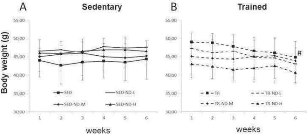
and third week the mice ran for 30 and 60 min, respectively, at the same speed; the fourth and fifth week for 60 min at a speed of 12 m/min. Finally during the last week the mice ran for 90 min at a speed of 14m/min. All the mice were weighed weekly. Mice of all experimental groups were treated with intramuscular injections (IM) of ND (Sigma-Aldrich, St. Louis, MO, USA) or peanut oil twice a week in the hindlimb for six weeks (Table 1). ND was dissolved in peanut oil with 10% of benzoic alcohol and the dose of ND was selected according to the literature. Mice of the SED and TR groups (controls) were administered IM peanut oil and 10% benzoic alcohol. Forty-eight hours after the last training session, mice were sacrificed via cervical dislocation. The blood was collected in tubes, centrifuged and serum was stored at -80 °C. Testes were dissected and preserved in liquid nitrogen or embedded in paraffin for morphological and molecular evaluation.
Measurement of Testosterone level with Liquid chromatography-mass spectrometry Testosterone levels were assessed by “Locorotondo Labs srl,
Palermo”. Testosterone inserum was quantified using a validated method for the analysis in serum/plasma of testosterone by liquid chromatography-mass spectrometry (LC-MS/MS). The method was performed as described previously. Total testosterone analysis in serum was performed in all experimental groups (n=6 per group).
Real-Time quantitative PCR (qPCR) Total cellular RNA was isolated
from cell cultures using TRIzol® REAGENT (Sigma-Aldrich, USA), according to the manufacturer’s instructions. RNA (12 ng) was retro-transcribed using the ImProm-II Reverse Transcriptase Kit (Promega Corporation) to obtain cDNA, which was amplified using the GoTaqqPCR Master Mix (Promega Corporation, USA) as previously described (Di Felice et al. 2013).The mRNA levels were normalized to the levels obtained for hypoxanthine phosphoribosyltransferase 1 (HPRT1), for beta-glucuronidase (GUSB) and for Glyceraldehyde 3-phosphate
34
dehydrogenase (GAPDH). Changes in the transcript level were calculated using the 2-ΔΔCtmethod (Di Felice et al. 2013, Livak et al. 2001).
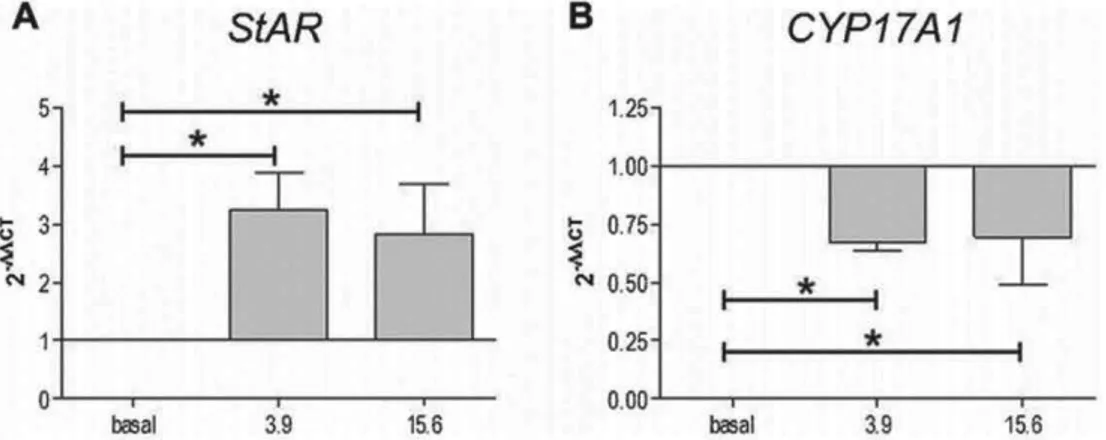
For the in vivo experiments Reverse transcription was performed using the ImProm-II Reverse Transcriptase Kit (Promega, Madison, WI, USA) according to the manufacturer’s instructions. qRT–PCR analysis was performed using GoTaq qPCR Master Mix (A6001, Promega). mRNA levels were normalized to that of GAPDH and GUSB. Changes in the transcript level were calculated using the 2–∆∆CT method (Schmiigen et al. 2008). Complementary deoxyribonucleic acid (cDNA) was amplified using primers indicated in Table 4. cDNA was amplified using the Rotor-gene™ 6000 Real-Time PCR Machine (Qiagen GmbH, Hilden, Germany).
The cDNA was amplified using different primers as in table 2:
Western blotting Western blotting was performed as previously described
(Barone et al. 2013). Equal amounts of proteins (40µg) were separated on SDS-PAGE and transferred onto a nitrocellulose membrane (BioRad, Segrate, Italy). After blocking with 5% albumin bovine serum (Sigma Aldrich), membranes were probed with primary antibodies; rabbit anti-CYP17A1 M-80 clone polyclonal antibody, mouse anti-β actin C-4 clone monoclonal antibody, Santa Cruz Biotechnology) diluted at 1:1.000 overnight at 4 °C. Protein bands were visualized using the enhanced chemiluminescence (ECL) detection system (GE Healthcare Life Sciences, Milan, Italy), and the data were evaluated and quantified using ImageJ Free software (NIH, Bethesda, MD). For the in vivo
35
experiments testes homogenization were performed as described previously. The membrane was incubated in a blocking solution containing 5% milk in Tris-buffered saline (20 mM Tris, 137 mM NaCl, pH 7.6) containing 0.05% Tween-20 (T-TBS) for 1 h. Next, the membrane was further incubated in a primary antibody overnight at 4 °C (see Table 3). All the primary antibodies were diluted in T-TBS containing 5% BSA and incubated overnight at 4 °C. The following day, the membrane was washed with T-TBS and incubated with an HRP-conjugated secondary antibody (anti-rabbit NA934V, or anti-mouse NA931, Amersham Biosciences, NY, USA) diluted in T-TBS containing 5% milk for 1 h. The detection of the immunopositive bands was performed using ECL Western Blotting Detection Reagent (Amersham Biosciences) according to the manufacturer’s instructions. Each experiment was performed at least three times.
Immunofluorescence Staining Immunofluorescence was performed as
described previously (Gorska et al., 2013). Cells were fixed in ice-cold methanol, incubated with unmasking solution (10 mM trisodium citrate, 0.05% Tween-20) for 10 min, and treated with blocking solution (3% BSA in PBS) for 30 min at 24°C. Then, a primary antibody against CYP17A1 and StAR (Santa Cruz
Tab 3 List of antibodies used in wester blot, immunofluorescence and immunohistochemestri analisys
36
Biotechnology) was added and incubated in a humidified chamber overnight at 4°C, diluted 1:100. Cells were incubated for 1 h at RT with a conjugated secondary antibody (anti-rabbit IgG–FITC antibody produced in goat, Sigma– Aldrich, 1:200 dilution); followed by 15 min incubation with HOECHT33342 (Sigma–Aldrich) diluted 1:1000. Finally, slides were mounted with cover slips, and images were taken immediately with a Leica Confocal Microscope TCS SP8 (Leica Microsystems, Heidelberg, Germany). The confocal microscopy analysis was carried out using the Leica application suite advanced fluorescences software. The staining intensity was measured as the mean pixel intensity (PI) normalized to the CSA (cross-sectional area) of all cells in five fields per slide. Each experiment was quadrupled.
For immunofluorescence in testes samples, deparaffinized sections of 4-5 µm were incubated in the antigen unmasking solution (10 mM tri-sodium citrate, 0.05% Tween-20) for 8 min at 75 °C and treated with a blocking solution (3% BSA in PBS) for 30 min. Next, the primary antibody (Table 3) was applied, and the slides were incubated in a humidified chamber overnight at 4 °C. Then, the sections were incubated for 1 h at 23 °C with a conjugated secondary antibody (rabbit IgG–FITC antibody produced in goat, F0382, Sigma-Aldrich; anti-mouse IgG-TRITC antibody produced in goat, T5393, Sigma-Aldrich). Nuclei were stained with Hoescht Stain Solution (1:1,000, Hoechst 33258, Sigma-Aldrich). The slides were treated with PermaFluor Mountant (Thermo Fisher Scientific, Inc. Waltham, MA, USA) and cover slipped. The images were captured using a Leica Confocal Microscope TCS SP8 (Leica Microsystems, Heidelberg, Germany).
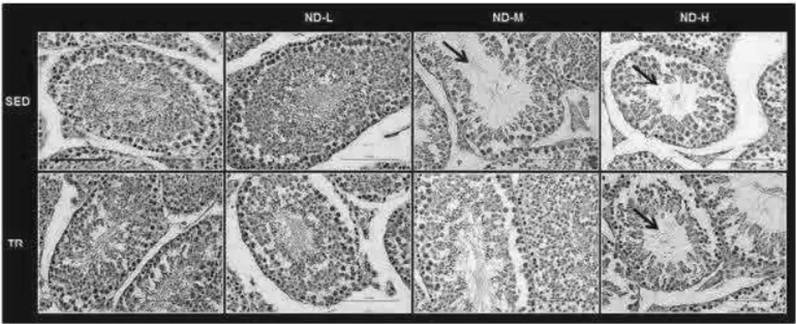
Histological examination After the sacrifice of animals, samples of
testes were taken from each mouse for histological analysis as described previously [18, 19]. Sections were stained with hematoxylin and eosin, mounted with coverslips and finally observed with a Leica DM5000 upright microscope (Leica Microsystems). Two independent observers (F.C and F.R) examined the specimens on two separate occasions, in a blind manner, using coded slides without knowing their source. For the histological evaluation, 10 sections which had a 20 µm distance from each other were observed with the light microscopy and the images were taken at × 40 magnification.
Immunohistochemistry Analysis For immunohistochemical analysis,
serial sections (4-5 µm) were incubated in an antigen unmasking solution for 8 min at 75 °C. Then, the MACH1 kit (M1u539g, Biocare, Concord, CA, USA)
37
was used according to the manufacturer’s instructions. The sections were incubated with the primary antibody in a humidified chamber overnight at 4 °C. The following day, the sections were incubated for 1 h with the secondary antibody. Finally, the slides were coverslipped, and images were captured with a Leica DM5000 upright microscope.
Statistical analyses
A one-way ANOVA followed by a Bonferroni post-hoc test for multiple comparisons was used as an appropriate analysis for the data. All statistical analyses were performed using the GraphPadPrismTM 4.0 program (GraphPad Software Inc., San Diego, California, USA). All data are presented as the means ± SD, and the level of statistical significance was set at p<0.05.
38
RESULTS In vitro
Testosterone production To determine testosterone secretion, the
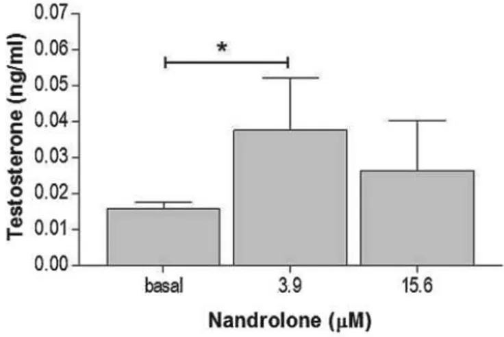
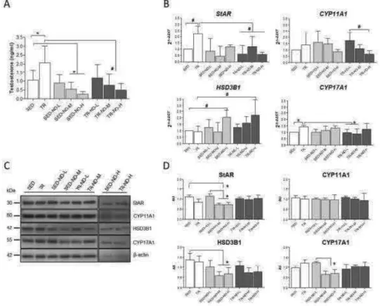
medium was collected at the end of the incubation period and subsequently analyzed. The nandrolone working solution induced, after 48 h, an increase of the secreted testosterone in R2C cells treated with 3.9 mM nandrolone, compared to the basal sample (P < 0.05) (Fig. 10). Interestingly, a higher concentration (15.6 mM) did not induce a significant testosterone increment compared to basal values.
Levels of enzymes involved in testosterone synthesis Lysates of R2C
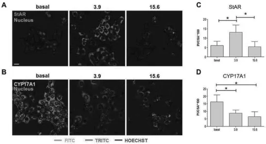
cells treated with different nandrolone concentrations were analyzed by western blotting analyses to verify the effects of nandrolone stimulation on the levels of proteins involved in testosterone synthesis. Our results showed that levels of StAR (Fig. 11) increased significantly in R2C cells treated with 3.9 mM nandrolone compared to both basal and 15.6 mM (P < 0.05). These results were confirmed by confocal analyses (Fig. 12A and C). Interestingly, the levels of CYP17A1 decreased upon treatment with 15.6 mM of nandrolone compared to both basal and 3.9 mM (P < 0.05) (Fig. 11). In addition, these results were in agreement with confocal analyses (Fig. 12B and D). Finally, western blotting analyses for CYP11A1 and HSD3B1 protein levels did not show any significant
Fig 10 testosterone production in R2C cells treated with 3,9 and 15,6 mM of nadrolone for 48 h. Basal are untreated cells.
39
differences between untreated and treated cells (Fig. 11), and this datum was also in accordance with confocal analyses (not shown).
Fig 11 effect of nandrolone stimulation on steroidogenic proteins
Fig 12 effects of nandrolone supplementation on StAR and CYP17A1 levels. In A and B representative picture at confocal microscopy of R2C cells treated with 3,9 and 15,6 mM of nadrolone for 48 h. basal are untreated cells