Università degli Studi ROMA TRE
SCUOLA DOTTORALE IN BIOLOGIA (XXII CICLO) Sezione: SCIENZE BIOMOLECOLARI E CELLULARI
A possible molecular mechanism
for parasitic inhibition by Lactoferrin
Dottorando: Docente Guida:
Dr. Loris Leboffe Prof. Giovanni Antonini
A
POSSIBILE
MOLECULAR
MECHANISM
FOR
PARASITIC
INHIBITION BY LACTOFERRIN
Summary
Transferrins (Tfs) belong to a family of iron-binding glycoproteins possessing similar aminoacid sequence though they have different biological functions and locations. Lactoferrin (Lf) is expressed and secreted from glandular epithelial cells and from mature neutrophiles of mammalian and it is an important component of the aspecific host defence or natural immunity, including resistance to parasitic infections. Serum transferrin (sTf) is synthesized by the liver of mammals and secreted into the blood stream; its primary function is iron transport. Ovotransferrin (Otrf), synthesized by avians, displays both iron transport and protective functions.
Parasites synthesize papain-like cysteine proteases that are relevant for the virulence and pathogenicity of parasites, being involved in several aspects of the parasite life cycle, it is therefore possible that the antiparasitic activity of Lf could be due to the inhibition of parasitic papain-like cysteine protease that we have recently observed.
In this study we have investigated the thermodynamic parameters of hLf, bLf and Otrf inhibition of the parasitic papain-like type I cysteine proteases from Leishmania infantum, Trypanosoma cruzi and Trypanosoma
brucei. bLf, hLf and Otrf, both in the apo- and olo-forms, showed time- and
concentration-dependent inhibition of the catalytic activity of papain and of type I proteases from L.infantum, T. cruzi and T. brucei. The KI values
observed for bLf and hLf inhibition of L. infantum, T. cruzi and T. brucei proteases were in the nanomolar range (KI = 3.1 nM), lower than KI values
observed for papain inhibition (KI = 24 nM). Otrf showed lower inhibition
of cysteine proteases (KI = 0.6 µM). On the contrary, sTf did not display any
inhibition towards parasitic proteases, according to its different role in mammals. The inhibition of parasitic cysteine proteases by hLf, bLf and Otrf appeared to conform to a competitive mechanism. The observed pH
optimum for bLf inhibition of parasitic proteases was around neutrality, while it was acidic for hLf and alkaline for Otrf. The further quantitative analysis of pH dependence of the intrinsic ligand-independent inhibition constant KI allowed the evaluation of pKa values that define the acid-base
equilibrium of amino acidic residue(s) modulating the enzyme(s)-inhibitor recognition events. SDS-PAGE showed that hLf, bLf and Otrf were easily degraded by either papain or parasitic type I protease during the assay incubation time (few minutes) and it is likely that one or more protease inhibitory peptides were generated from protein hydrolysis.
As a matter of fact, a sequence present near the C-terminus of human (hLf) and bovine (bLf) lactoferrin shows homology with the sequence of the active site of cystatins, which are competitive inhibitors of papain-like cysteine proteases. The same sequence is present, though with lower homology, in Otrf and, with even lower homology, in sTf.
Therefore, we have charactherized by MALDI-TOF the profile of Lf cleavage by papain and preliminary data suggest the presence of a cystatin-like peptide in two proteolytic fragments of hLf and in one proteolytic fragment of bLf.
Le transferrine (Tfs) sono una famiglia di glicoproteine in grado di legare reversibilmente il ferro, e presentano un’elevata omologia di sequenza tra tutti i membri di questa famiglia. In particolare la lattoferrina (Lf) è prodotta e secreta nelle cellule ghiandolari epiteliali ed è presente nei granuli dei granulociti neutrofili dei mammiferi. Essa rappresenta un’importante componente della difesa immunitaria aspecifica dell’ospite. La siero transferrina (sTf) invece è sintetizzata nel fegato, ed è coinvolta unicamente nel trasporto del ferro. L’ovotransferrina (Otrf), l’omologa aviaria della Lf , svolge sia una funzione di difesa che di trasporto del ferro.
Le proteasi a cisteina papaina-simili da protozoi e metazoi parassiti, secretorie e di membrana, partecipano ai processi di morfogenesi degli organismi patogeni, sono implicate nell’invasione delle cellule e dei tessuti da parte dei parassiti, riducono la risposta immunitaria dell’ospite e costituiscono uno dei fattori più rilevanti nelle patologie associate alle infestazioni da parassiti. Pertanto, a fronte della capacità della Lf di svolgere un’azione antiparassitaria, noi abbiamo ipotizzato che la Lf possa svolgere tale azione grazie all’inattivazione delle suddette proteasi a cisteina.
Nel presente lavoro abbiamo studiato le proprietà termodinamiche dell’inibizione delle proteasi a cisteina di alcuni parassiti, quali Leishmania
infantum, Tripanosoma cruzi e Trypanosoma brucei. Tale studio è stato
esteso anche verso la papaina, capostipite di questa famiglia. Innanzitutto è emerso una maggiore affinità delle transferrine studiate (bLf, hLf e Otrf, in forma apo e olo) verso le proteasi parassitarie (KI = 3.1 nM) rispetto a
quella verso la papaina (KI = 24 nM). Otrf risulta essere l’inibitore con
minore affinità (KI = 0.6 µM). Inoltre è emerso che la sTf non è in grado di
svolgere una’zione inibitoria, coerentemente con la sua funzione nei mammiferi. L’inibizione di tutte le proteasi risulta essere conforme ad un meccanismo di inibizione competitivo.
Da un’analisi della dipendenza del pH dei suddetti fenomeni di inibizione si è osservato una maggiore attività della bLf a pH fisiologico mentre la hLf inibisce più efficacemente a valori di pH acidi (pH = 5.0) e la Otrf a valori di pH alcalini (pH = 9.0). La dipendenza dal pH del parametro
KI relativo all’interazione degli inibitori studiati con gli enzimi, ha permeso
di evidenziare la variazione di diversi valori di pKa, ascrivibili ad eventi di
protonazione e deprotonazione di differenti gruppi ionizzabili.
Inoltre si è osservato, a seguito di SDS-page, una parziale idrolisi della Lf e della Otrf durante diverse incubazioni con la papaina e con le proteasi parassitarie, e l’insieme di tali dati suggerisce la liberazione di un peptide, che può quindi inibire competitivamente le suddette proteasi a cisteina.
A supporto di tale ipotesi, si è osservato in presenza della porzione C-terminale della Lf, una sequenza aminoacidica che presenta un’elevata omologia con il sito attivo delle cistatine, una (super)famiglia di proteina, riconosciute essere gli inibitori reversibili per eccelenza delle proteasi a cisteina papaina-simili. Abbiamo quindi caratterizzato, mediante MALDI TOF, il profilo di idrolisi della Lf ad opera della papaina, ed è stata riscontrata la presenza di frammenti contenenti tale sequenza cistatino-simile, sia nella bLf che nell’hLf.
Abbreviations
ACN Acetonitrile
apo-Lf, lactoferrin in its iron free state (Fe3+)
bLf, bovine lactoferrin
CEW Cys chicken egg-white cystatin
CHCA α-Cyano-4-hydroxycinnamic acid
CMV, Cytomegalovirus DTT, (-)-1,4-Dithiothreitol DMSO, dimethylsulfoxide HBV, Human hepatitis B HCV, Human hepatitis C hLf, human Lactoferrin
holo-Lf, lactoferrin iron bound (Fe3+)
HSV, Herpes simplex virus
Lf, lactoferrin
Otrf, ovotransferrin
sTf, human serum transferrin
Tf, transferrin
TFA Trifluoroacetic acid
Z-Phe-Arg-AMC, N-α -benzyloxycarbonyl-L-phenylalanyl-L-arginine-(7-amino-4-methyl coumarin)
Introduction
Among parasitic Protozoa, trypanosomatids belonging to the
Leishmania and Trypanosoma genres comprise etiological agents of
endemic diseases mainly localised in developing countries. All the human pathogenic trypanosomatids are dixenic parasites: namely with a complex life-cycle characterized by a vector host (an ematophagous insect) which became infected after a blood meal on a infected mammalian and which is responsible to bear and to transmit the flagellate parasite to the next mammalian host. The last one becoming in turn a source of infection for further insect vectors (Cox, 1993; Gilles, 1999; WHO, 2002).
In particular, Trypanosoma cruzi is the agent of the American trypanosomiasis (Chagas’ disease), affecting at least 20 million people, being responsible of the chagasic cardiopathy, the most relevant clinical manifestation of Chagas’ chronic disease, affecting about a third of the infected people (Cox, 1993; Gilles, 1999; WHO, 2002).
Tripanosoma brucei is the causative agent of African sleeping
sickness (human African trypanosomosis), a vector-borne disease that is fatal if untreated (Barrett et al., 2003) and ranks second among parasitic diseases in sub-Saharan Africa only to malaria in terms of mortality (Cox, 1993; Gilles, 1999).
Furthermore, several species of the genre Leishmania (i.e. those belonging to the L. mexicana, L. brasiliensis, L. dononvani and L. tropica complexes) cause a broad spectrum of diseases, affecting 12 million of people in both the Old and New Worlds. Leishmaniasis can be fatal (visceral leishmaniasis), grossly disfiguring (mucocutaneous leishmaniasis), or relatively mild, localized, and in some cases self-healing (some forms of cutaneous leishmaniasis) (Cox, 1993; Gilles, 1999; WHO, 2002).
These obligate parasites have two hosts - an insect vector and mammalian host. Because of the large difference between these hosts the trypanosome undergoes complex changes during its life cycle to facilitate its survival in the insect gut and the mammalian bloodstream (Barret et al., 2003; Matthews, 2005).
In the last years growing evidences arose pointing to the importance between the parasitic cisteine proteases and the virulence and pathogenicity of parasites. In fact, cysteine proteases belonging to the
papain family are relevant to several aspects of the parasite life cycle and of the parasite-host relationship and are thus seen as promising therapeutic target of parasitic diseases. As an example, the major papain-like cysteine proteases from T. Cruzi, T. brucei and L. infantum, are mainly expressed in all stages of the parasites life cycle. Among others, cruzipain participates in the penetration of T. cruzi trypomastigotes into host cells, in the nutrition of the parasite at the expense of the host, and in the escape mechanisms of the parasite from the immune system of the host. Similar biological roles, even though with distinct molecular mechanisms, are been proposed for cysteine proteases from Leishmania (Coombs & Mottram, 1997; Del Nery et al., 1997; McKerrow, 1999; McKerrow et al., 1993, 1995, 1999; Frame et al., 2000; Caffrey et al., 2000; Lecaille et al., 2002; Sajid & McKerrow, 2002; Mottram et al., 2004). In fact the cysteine proteases of L. mexicana are critical in suppressing protective immune responses, inhibiting host TH1 responses (Buxbaum et al., 2003).
1.1. Cysteine proteases: the papain family
Cysteine proteases are divided into four main groups referred to as clans, CA, CB, CC and CD. Conventionally, proteases are assigned to clans and families depending on a number of characteristics including sequence similarity, common peptide loops, and substrate specificity to small peptide substrates. The clans CB and CC include viral proteases, while in the clan CD (Legumain-like family) there are asparagynil peptidase and transamidase. Asparaginyl endopeptidases exclusively hydrolyse peptides and proteins on the carboxyl side of asparagines residues. These enzymes are often referred to as ‘legumain-like’ as the template protease was first identified and characterised from the plant legume, Canavalia ensiformis, the jack bean. Legumain-like proteases have been identified in many plants, mammals including, human, mouse, rat and pig, and in many parasite organisms like Fasciola hepatica, Schistosoma mansoni and Caenorhabditis
elegans (Lecaille et al., 2002; Sajid & McKerrow, 2002; Mottram et al.,
2004).
In 1879 the first cysteine protease was purified and characterised from Carica papaya, the papaya fruit, and was thus named papain. Papain was also the first cysteine protease structure to be solved. Since its discovery, numerous proteases that have sequences in common with papain have been loosely called ‘papain-like’(Lecaille et al., 2002).
Recently, on the base of structural and catalytic properties, the papain-like proteases have been divided in three different subfamilies (Lecaille et al., 2002; Sajid & McKerrow, 2002; Barrett et al., 2004).
Members of the Cathepsin L-like subfamily have a conserved inter-spaced motif in the pro-region, Glu-X3-Arg-X2-Phe-X2-Asn-X3-Ile-X3
-Asn (‘ERFNIN’; named after the single letter code for amino acids; X is any amino acid) and dispaly an endopeptidase activity (Lecaille et al., 2002; Sajid & McKerrow, 2002; Barrett et al., 2004).
Members of the subfamily Cathepsin B-like lack the ERFNIN motif but do have an inserted peptide loop in the catalytic domain, referred to as the ‘occluding loop’, between residues Tyr103 and Cys128 (human cathepsin B-like numbering). In addition to endopeptidase activity (like cathepsin L), cathepsin Bs have a dipeptidyl carboxypeptidase activity. This has been attributed to the occluding loop (Lecaille et al., 2002; Sajid & McKerrow, 2002; Barrett et al., 2004).
Members of the Cathepsin F-like family, the latest enzymes included in papain-like family, have a consensus motif in the pro-region Glu-X3-Arg-X2-Phe-X2-Asn-X3-Ala-X3-Gln/Ala (‘ERFNAQ’; named after
the single letter code for amino acids) (Wex et al., 1999, 2000; Barrett et
al., 2004).
1.1.2. Structural aspects and catalytic mechanism
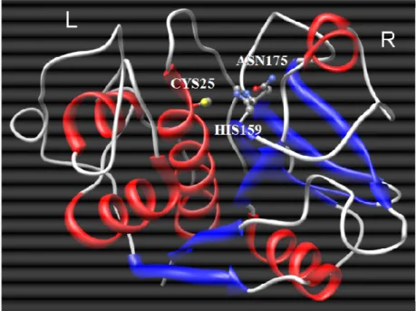
The enzyme is assembled from two domains, each comprising residues from both the N- and C-terminal sections of the polypeptide. One domain consists of a six-stranded antiparallel β-sheet (R-domain) and contains the catalytic residues histidine (His159: papain numbering) and asparagine (Asn175: papain numbering) (Fig. 1) (McGrath, 1999; Sajid & McKerrow, 2002; Barrett et al., 2004).
The L-domain, instead, consists mainly of three α-helices. The cysteine residue (Cys25 based on papain numbering) is embedded in a highly conserved peptide sequence, Cys-Gly-Ser-Cys-Trp-Ala-Phe-Ser (active site cysteine residue in bold) (Fig. 1) (Rawlings & Barrett, 1994; Turk et al., 1997, 1998; Barrett et al., 2004).
Figure 1: Structure of papain (code PDB: 9PAP) (Kamplius et al., 1994).
Molecular graphics images were produced using the UCSF Chimera package.
The catalytic site of papain-like cysteine proteases is highly conserved and formed by three residues: Cys25, His159, and Asn175 (papain numbering). Cys25 and His159 form an ion pair which is stabilized by Asn175 via a hydrogen bond. This triad has some similarities to the active site present in serine proteases (Ser, His, Asp). However, in contrast
to serine proteases the nucleophilic cysteine residue is already ionized prior to substrate binding and thus cysteine proteases can be regarded as a priori activated enzymes (Polgard & Halasz, 1982).
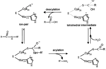
Figure 2: Mechanism of substrate hydrolysis by papain-like cysteine
proteases (Lecaille et al., 2002).
During peptide hydrolysis, the nucleophilic thiolate cysteine attacks the carbonyl carbon of the scissile bond of the bound substrate and forms a tetrahedral intermediate which is stabilized by the so-called oxyanion hole (Ménard et al., 1991). The tetrahedral intermediate transforms into an acyl enzyme (enzyme-substrate thiol ester) with the simultaneous release of the C-terminal portion of the substrate (acylation). This step is followed by the hydrolysis of the acyl enzyme with water, forming a second tetrahedral intermediate which finally splits into the free enzyme and the N-terminal portion of the substrate (deacylation) (Storer & Ménard, 1994, Vernet et al., 1995) (see Fig. 2).
1.1.3. Cruzipain and Brucipain
Trypanosoma cruzi is a protozoan parasite which is transmitted to
humans, as an infectious trypomastigote form, from the bite of a blood-sucking insect. The trypomastigote enters the host bloodstream and ultimately invades a cardiac muscle cell, where it transforms into the intracellular amastigote. Amastigotes replicate within cells, transform back to trypomastigotes, and rupture the cell, releasing the infectious form back into the bloodstream and other cells, amplifying the infection (Duschak & Couto, 2009).
Cysteine proteases appear to be relevant to several aspects of the T.
cruzi life cycle and the parasite-host relationship. In particular, cruzipain
(Fig. 3), the major cysteine protease from T. cruzi epimastigotes, is expressed in all stages of the parasite life cycle, being more abundant in replicating forms and particularly in the insect epimastigote stage. Notably, cruzipain participates in the penetration of T. cruzi trypomastigotes into host cells, in the nutrition of the parasite at the expense of the host, and in the escape mechanisms of the parasite from the immune system of the host (Duschak & Couto, 2009).
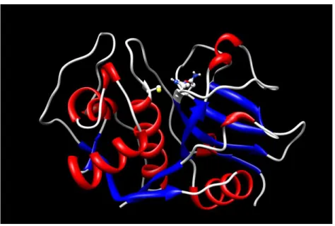
Cruzipain has a structure very similar to that of papain. In fact is composed of polypeptide chain of 215 amino acid residues, folded into two domains (Figure 3). One domain is mainly α-helical (L-domain, residues 12-112 and 208-212), and the other consists of extensive antiparallel β -sheet interactions (R domain, residues 1-11 and 113-207) (Kamphuis et al., 1984, 1985; McGrath et al., 1995). The catalytic residues Cys25, His159 and Asn175, and the extended substrate-binding site, are found in a cleft between the two domains (McGrath et al., 1995).
Figure 3: Structure of Cruzipain (code PDB: 1AIM) (Gillmor et al., 1997).
Molecular graphics images were produced using the UCSF Chimera package.
Trypanosoma brucei is a parasitic protist species that causes
African trypanosomiasis (or sleeping sickness) in humans and in animals in Africa. There are 3 sub-species of T. brucei: T. b. brucei, T. b. gambiense and T. b. Rhodesiense (Barret et al., 2003).
In T. brucei species, the major cysteine protease has primary sequence and biochemical characteristics that are broadly similar to those of mammalian cathepsin L-like (Lonsdale-Eccles & Grab, 1987; Troeberg et
al., 1999; Caffrey et al., 2001), and is encoded by a tandem array of 11
nearly identical gene copies (Berriman et al., 2005). The enzymes in T. b.
rhodesiense and T. b. brucei are termed rhodesain and brucipain (or
trypanopain) respectively (Lonsdale-Eccles & Grab, 1987; Caffrey et al., 2001 Mackey et al., 2004).
Brucipain shows 80% homology and 60% identity with the sequences of cruzipain (Duschak & Couto, 2009).
1.1.4. Protease of type I from L. infantum
The proteases of type I from L. infantum is a glycoprotein, synthesized like pre-pro-enzyme and it has a N-domain of 27 amino acidic residues (aa), a pre-peptidic element of 98 aa, a catalytic domain of 214 aa and a C-domain of 104 aa (Souza et al., 1992; Mottram et al., 1997; Campos-Ponce et al., 2005).
Like cruzipain, the proteases from L.infantum are cathepsins L-like and these proteases are required for parasite replication and virulence (Sakanari et al., 1997; Campos-Ponce et al., 2005; Mundodi et al., 2005).
Sequence analysis showed that they are 90% identical to L. major Cathepsin B, 66% identical to L. mexicana cathepsin B, and 45% identical and 57% homology to cruzipain, indicating high sequence conservation among the trypanosomatid species (Mottram et al., 1997).
1.2. Cystatins family
The inactivation of parasite cysteine proteinase mediated by synthetic inhibitors block replication and differentiation of T. cruzi, T.
Brucei and L. infantum both in vitro and in vivo, providing an alternative to
traditional therapy in drug-resistant parasites (Coombs & Mottram, 1997; Maekawa et al., 1998; McKerrow, 1999; McKerrow et al., 1993, 1995, 1999; McKerrow, 1999; Caffrey et al., 2000; Frame et al., 2000; Lecaille et
al., 2002; Lima et al., 2002; Sajid & McKerrow, 2002; Somanna et al.,
2002; Ascenzi et al., 2004).
Cysteine protease inhibitors (CPI) belonging to the cystatin (super)family inactivate proteases by trapping them in a(n) (ir)reversible, tight equimolar complex (Barrett et al., 1998). The representatives of this group of CPI is characterized by a wide distribution, being present in
mammals, birds, fish, insects, plants and some protozoa (Abrahamson et al., 2003).
The human superfamily of cystatins is divided into three families. Family I, called stefins, comprises intracellular cystatins A and B. Family II includes extracellular and/or transcellular cystatins (cystatins: C, D, E, F, S, SA, and SN). Kininogens, the intravascular cystatins, form family III of cystatins (Barrett, 1987; Rawlings & Barrett, 1990; Turk & Bode, 1991).
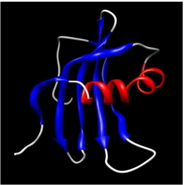
All cystatins have the same structure consisting of a five stranded
β-sheet wrapped around a five turn α-helix. Cystatins contain three segments which are recognized as responsible for the interaction with cysteine proteases. These are the N-terminal fragment and the so-called first and second loops (L1 ans L2, respectively), which are arranged at one edge of the molecule and are believed to directly interact with the catalytic cleft of cysteine proteases (Grzonka et al., 2001; Turk et al., 2008).
These three cystatin regions, containing evolutionarily conserved amino-acid residues, form a wedge-like structure, which interacts with the catalytic cleft of cysteine proteases. These interactions are hydrophobic binding between the regions of cystatins and the corresponding residues forming the binding pockets of the enzyme. In particular, I-loop contains the conserved residues QVVAG, that interact with the active site of cysteine proteases, whereas the N-terminal fragment contains the conserved residues Leu9 and Gly11 (human cystatin numbering) (Grzonka et al., 2001; Turk et
Figure 4: Structure of Stefin A (code PDB: 1DVD) (Gillmor et al., 1997).
Molecular graphics images were produced using the UCSF Chimera package.
Recently a sequence present at the C-terminus of human (hLf) and bovine (bLf) lactoferrin, a mammalian glycoprotein, shows a homology with the sequence of the reactive site of the cystatins (Katunuma et al., 2003). Moreover, this sequence is present in Ovotrasnferrin (Otrf), avian homologous of Lf, and in serum Transferrin (sTf), synthesized in the liver of mammals and secreted into the blood (Anderson et al., 2009).
Table 1: Alignment of Lf sequence with cystatine active site
Cystatin active site LG QVVAG
bLf (680) LGTEYVTA (687)
hLf (683) LGPQYVAG (690)
Otrf (657) LGDKFYTV (664)
sTf (661) LGEEYVKA (688)
1.3. Lactoferrin
Lactoferrin (Lf) is a non-haem iron-binding protein that is part of the transferrin protein family, along with serum transferrin (sTf), ovotransferrin (Otrf), melanotransferrin and the inhibitor of carbonic anhydrase, whose function is to transport iron in the blood serum (González-Chávez et al., 2009).
LF is produced by mucosal epithelial cells in various mammalian species, including humans, cows, goats, horses, dogs and several rodents. Recent studies have shown that Lf is also produced by fish, as it has been identified in rainbowtrout eggs using molecular biology techniques (González-Chávez et al., 2009).
This glycoprotein is found in mucosal secretions, including tears, saliva, vaginal fluids, semen (van der Strate et al., 2001), nasal and bronchial secretions, bile, gastrointestinal fluids, urine (Öztas & Özgünes, 2005) and most highly in milk and colostrum (7 g/L) (Rodriguez et al., 2005) making it the second most abundant protein in milk (Connely, 2001), after caseins. It can also be found in bodily fluids such as blood plasma and amniotic fluid. Lf is also found in considerable amounts in secondary neutrophil granules (15 µg/106 neutrophils) (Bennett & Kokocinski, 1987; González-Chávez et al., 2009), where it plays a significant physiological role.
Lf possesses a great iron-binding affinity and is the only transferring with the ability to retain the metal over a wide pH range (Aisen & Leibman, 1972) including extremely acidic pH. It also exhibits a great
resistance to proteolysis. In addition to these differences, Lf net positive charge and its distribution in various tissues make it a multifunctional protein (Baker & Baker, 2009).
1.3.1 Structure
Lf (Fig. 5) is an80 kDa glycosylated protein of ca. 700 aminoacids (711 aa for hLf and 689 aa bLf) with high homology among species. It is a simple polypeptide chain folded into two symmetrical lobes (N and C lobes) which are highly homologous with one another (33–41% homology) (Anderson et al., 1987, 1989; Baker, 1994; Moore et al., 1997; Sharma et
al., 1998; Baker & Baker, 2009).
Figure 5: Structure of hLf (code PDB: 1FCK) (Baker et al., 2000).
Molecular graphics images were produced using the UCSF Chimera package.
These two lobes are connected by a hinge region containing parts of an α-helix between residues 333 and 343 in human Lf (hLF), which provides additional flexibility to the molecule. The polypeptide chain includes amino acids 1–332 for the N lobe and 344–703 for the C lobe and is made up of α-helix and β-pleated sheet structures that create two domains for each lobe (domains I and II) (Moore et al., 1997). Each lobe can be further divided into two subdomains (N1 and N2 in the N-lobe and C1 and C2 in the C-lobe) that form a cleft inside of which the iron is bound. The subdomain N1 contains residues 1-90 and 251-333, while N2 contains the residues 91-250) (Baker et al., 1987; Moore et al., 1997; Baker & Baker, 2009).
Each lobe can bind a metal atom in synergy with the carbonate ion (CO3
2−
). Lf binds notably Fe2+ and Fe3+ ions, but also Cu2+, Zn2+ and Mn2+ ions (Aisen & Harris, 1989; Baker et al., 1994, 2005; Baker & Baker, 2009).
1.3.2 Functions of Lf
Lf is involved in several physiological functions, including: regulation of iron absorption in the bowel; immune response; antioxidant, anticarcinogenic and anti-inflammatory properties. Protection against microbial infection, whichis the most widely studied function to date (Sanchez et al., 1992; Brock, 1995; Lonnerdal & Iver, 1995; Vorland, 1999; Brock, 2002; Antonini et al., 2005; Baker & Baker, 2009; Leboffe et al., 2009). The antimicrobial activity of LF is mostly due to two mechanisms. The first is iron sequestration in sites of infection, which deprives the microorganism of this nutrient, thus creating a bacteriostatic effect. The other mechanism is the direct interaction of LF with the infectious agent. Positively charged amino acids of LF can interact with anionic molecules on some bacterial, viral, fungal and parasite surfaces, causing cell lysis (Bullen, 1981; Braun & Braun, 2002; Valenti & Antonini, 2005).
Considering the physiological capabilities of Lf in host defence, in addition to current pharmaceutical and nutritional needs, Lf is considered to be a nutraceutical and for several decades investigators have searched for the most convenient way to produce it (González-Chávez et al., 2009).
The antiviral activity of hLf was first demonstrated in mice infected with the polycythemia-inducing strain of the Friend virus complex (FVC-P) (Lu et al., 1987). Since 1994, potent antiviral activity of hLf and bLf has been demonstrated against both enveloped and naked viruses, like
Cytomegalovirus (CMV) (Harmsen et al., 1995; Andersen et al., 2001), Herpes simplex virus (HSV) (Marchetti et al., 1996, 1998; Siciliano et al.,
1999; Valenti & Antonini, 2005), Human immunodeficiency virus (HIV) (Swart et al., 1996 ; Puddu et al., 1998; Berkhout et al., 2004), as well as
Human hepatitis C (HCV) and human hepatitis B (HBV) viruses (Ikeda et al., 1998; Hara et al., 2002).
Molecular mechanims of Lf antiparasitic activity are even more complex. Antiparasitic activities of Lf appear often to involve interference with iron acquisition by some parasites, e.g. Pneumocystis carinii, while Lf appears to act as a specific iron donor in other parasites such as
Trichomonas foetus; in the latter case, Lf could be expected to enhance
infection. It was recently reported that two T. brucei proteins bind human serum transferrin as well as human and bovine Lf. Preincubation of
Toxoplasma gondii and Eimeria stiedai sporozoites with a Lf-derived
peptide, lactoferricin, reduces their infectivity in animal models. Lf antiparasitic activity is also, sometimes, mediated by interaction with host cells. Thus, iron-saturated Lf enhances intramacrophage killing of T. cruzi amastigotes and decreases intra-erythrocytic growth of Plasmodium
falciparum. Lf is able to inhibit the invasion of cultured cells by Plasmodium spp. sporozoites through specific binding to HS. In the case of Plasmodium berghei, Lf reduces invasion by inhibiting the binding of the
plasmodial CS protein, with or without HS, suggesting the possibility that Lf can also bind to the same site on LDL receptor-related protein (LRP) as the CS protein (see Leboffe et al., 2009).
AIM
The protozoan order Kinetoplastida includes the genus
Trypanosoma, members cause some of the most neglected human diseases,
the trypanosomiases. There are many species of trypanosome, and the group infects most vertebrate genera. Several trypanosoma species cause important veterinary diseases, but only two cause significant human diseases (Gilles, 1999). In sub-Saharian Africa, Trypanosoma brucei causes sleeping sickness or human African trypanosomiasis, and in America,
Trypanosoma cruzi causes Chagas’ disease (Gilles, 1999). The
kinetoplastida also contains species of the genus Leishmania that cause a range of diseases in the tropics and subtropics (Gilles, 1999). All microrganisms are transmitted by insect vectors. The transmission in the human host follows blood transfusion, contamined needles, biting insects, vector faeces, and contamined food (Gilles, 1999).
Like all microorganisms, the genus Trypanosoma has an absolute necessity of iron to sustain their growth (Loo & Lalond, 1984; Leboffe et
al,, 2009). In particular, some parasites (e.g. Trypanosoma cruzi) survive in
mammalian blood and could utilize serum mammalian chelates directly. Other parasites, such as leishmania spp., survie intracellularly in mammals (Gilles, 1999). Therefore they have envolved a mechanism to utilize mammalian holo-transferrin (Tf) and/or holo-Lactoferrin (Lf) via several means.
The Tf uptake by T. brucei has been well characterized. In this protozoan, esag6 and esag7 genes encoded for a protein associated to a heterodimeric transferrin-binding protein complex (TFBP) present on the cell membrane (Leboffe et al., 2009). These receptors have the ability to bind Tf from different mammalian host species (Leboffe et al., 2009). Fluorescence and immunoelectron microscopy showed that the T. brucei TFBP–Tf complex is internalized and transported to lysosomes, where Tf is proteolytically degraded to release iron (Leboffe et al., 2009).
On the contrary, the promastigote uptake of iron for Leishmania is different and involves reduction of extracellular iron. Infact holo-Tf binds to a binding protein on the promastigote surface, as well as for Trypanosoma
brucei. One electron is donated to Fe3+ through the action of a membrane-associated or secreted iron reductase complex. The complex could include a cytoplasmic NADPH-requiring reductase which has specificity for NADPH
and which donates an electron to a membrane electron carrier (Leboffe et
al., 2009). Fe2+ has a very low affinity for Tf and therefore dissociates easily (Baker, 2009).
In the last years growing evidences arose pointing to the importance between parasitic cisteine proteases and the virulence and pathogenicity of trypanosomatid parasites. In fact, cysteine proteases belonging to the papain family are relevant to several aspects of the parasite life cycle and of the parasite-host relationship and are thus seen as promising therapeutic target of parasitic diseases (Coombs & Mottram, 1997; Del Nery et al., 1997; McKerrow, 1999; McKerrow et al., 1993, 1995, 1999; Frame et al., 2000; Caffrey et al., 2000; Lecaille et al., 2002; Sajid & McKerrow, 2002; Mottram et al., 2004).
As an example, the major papain-like cysteine proteases from T.
cruzi and L. infantum are mainly expressed in all stages of the parasites life
cycle. Among others, cruzipain participates in the penetration of T. cruzi trypomastigotes into host cells, in the nutrition of the parasite at the expense of the host, and in the escape mechanisms of the parasite from the immune system of the host. Similar biological roles, even though with distinct molecular mechanisms, are been proposed for cysteine proteases from
Leishmania (Coombs & Mottram, 1997; Del Nery et al., 1997; McKerrow,
1999; McKerrow et al., 1993, 1995, 1999; Frame et al., 2000; Caffrey et al., 2000; Lecaille et al., 2002; Sajid & McKerrow, 2002).
Notably, the inactivation of parasite cysteine proteinases by synthetic inhibitors block replication and differentiation of T. cruzi and L.
infantum both in vitro and in vivo, providing an alternative to traditional
therapy in drug-resistant parasites (Coombs & Mottram, 1997; Maekawa et
al., 1998; McKerrow, 1999; McKerrow et al., 1993, 1995, 1999;
McKerrow, 1999; Caffrey et al., 2000; Frame et al., 2000; Lecaille et al., 2002; Lima et al., 2002; Sajid & McKerrow, 2002; Somanna et al., 2002).
Cysteine protease inhibitors (CPI) belonging to the cystatin (super)family inactivate proteases by trapping them in a(n) (ir)reversible, tight equimolar complex (Barrett et al., 1998). The representatives of this group of CPI is characterized by a wide distribution, being present in mammals, birds, fish, insects, plants and some protozoa (Abrahamson et al., 2003; Turk et al., 2008).
Recently it was demonstrated that human and bovine milk has a new function against cysteine proteases (Katunuma et al., 2003). These data strongly support the hypothesis that Lf antiparasitic activity could be due, at least partially, to the inhibition of parasitic proteases.
Therefore, in the present work we report a quantitative investigation on the thermodynamic parameters for Tf inhibition of the cysteine protease from L. infantum, cruzipain and brucipain by hLf and bLf. Moreover, Lf binding to papain has been investigated. Moreover, the influence of the pH on the inhibition constants of Lf with papain and with the protease from L. Infantum have been determinated. The analysis of data allowed the identification of putative aminoacidic residues that may presumably modulate the interaction of the cysteine proteases studied with the Lf. Since this sequence is present also in Otrf (ovotransferrin), avian homologous of Lf, we have investigated the Otrf binding to protozoan proteases.
Results and discussion
3.1. Thermodynamic characterization of papain and
cysteine proteases from L. Infatum, T. cruzi and T. brucei
3.1.1 The catalytic mechanism
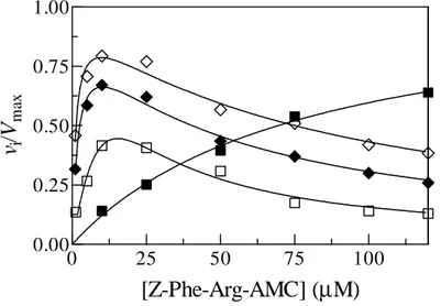
As shown in Fig. 6, the hydrolysis of the fluorogenic substrate Z-Phe-Arg-AMC catalyzed by the papain follows the simple Michaelis-Menten mechanism depicted in Scheme I and defined by equation (1).
Altought similar to the papain, the cysteine proteases from T. cruzi,
T. brucei and L. infantum display the phenomenon of the substrate
inhibition. In fact, as can be seen from the dependence of the relative initial velocity (i.e.: vi/Vmax) of Z-Phe-Arg-AMC hydrolysis on the substrate
concentration, at 25 °C and pH 6.0 (Fig. 6) under steady state conditions (i.e.: [E] < 5 × [S]), the parasite enzymes follow the substrate inhibition mechanism described by Scheme II and defined by equation (2) (Fig.6: theoretical curves as solid lines and experimental data as squares and rumbles for the cysteine proteases from T. Cruzi, T. Brucei and L. infantum, respectively).
Figure 6: Effect of the substrate concentration on the hydrolysis of
Z-Phe-Arg-AMC catalyzed by papain (black squares), cruzipain (black rumbles), the protease from L. infantum (rumbles) and brucipain (squares). Data were obtained at pH 6.0 and 25 °C. Solid lines, representing the theoretical curves, were calculated through an iterative non-linear least squares procedure with sets of parameters given in Table 2 and Table 3, according to Equation (1) for papain and to Equation (2) for cysteine proteases from T.
Cruzi, from T. Brucei and from L. infantum. The standard deviation for each
experimental point was equal to ± 8%. For further details, see text.
0
25
50
75
100
0.00
0.25
0.50
0.75
1.00
[Z-Phe-Arg-AMC] (µM)
v
i/V
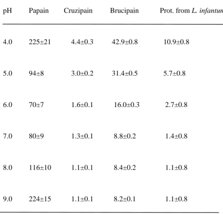
m axTable 2: Values of Km (µM) for the hydrolysis of Z-Phe-Arg-AMC by
proteases from L. infantum, T. cruzi, T . brucei and by papain (25,0°C).
pH Papain Cruzipain Brucipain Prot. from L. infantum
4.0 225±21 4.4±0.3 42.9±0.8 10.9±0.8 5.0 94±8 3.0±0.2 31.4±0.5 5.7±0.8 6.0 70±7 1.6±0.1 16.0±0.3 2.7±0.8 7.0 80±9 1.3±0.1 8.8±0.2 1.4±0.8 8.0 116±10 1.1±0.1 8.4±0.2 1.1±0.8 9.0 224±15 1.1±0.1 8.2±0.1 1.1±0.8
Table 3: Values of Ki (µM) for the hydrolysis of Z-Phe-Arg-AMC by the
proteases from T. cruzi, T. brucei and L. infantum and (25.0°C).
pH Cruzipain Brucipain Protease from L. infantum
4.0 ---* ---* 33±0.8 5.0 48±8 55±0.2 36±0.5 6.0 59±7 68±0.1 40±0.3 7.0 67±9 77±0.1 55±0.2 8.0 73±10 84±0.1 62±0.2 9.0 74±15 85±0.1 57±0.1 *
At such pH value cruzipain and brucipain do not undergo to substrate inhibition phenomenon.
Although the molecular details of substrate inhibition of cysteine proteases are not known, it often occur with synthetic and small substrates (Cazzulo et al., 1990; Lima et al., 1992; Stoka et al., 1998; Venturini et al., 2000; Salvati et al., 2001a). These chemicals are supposed to be able to bind the cysteine proteases interacting with only few residues of the protein and thus leading to the possibility for the synthetic molecule to assume different
configurations in respect with the catalytic Cys25 residue (mature cruzipain numbering). On the geometry of the interaction will then depend the result of the substrate-enzyme reaction (Cleland, 1979). Therefore, Z-Phe-Arg-AMC presumably is capable to bind the active site of parasite enzymes with, at least, two alterative configurations: either productively (Ss, Scheme
II), leading to the hydrolysis of the substrate itself, or following a binding mode that lead to the enzyme inhibition (Si, Scheme II) (Salvati et al.,
2001a; Salvati et al., 2002). Vice versa, papain, although with an active site very similar in the structure to the ones held by the parasite proteases, can interact with Z-Phe-Arg-AMC only through a productive configuration (Ss,
Scheme I) (Szabelski et al., 2001) These considerations agree with the different specificity that characterizes papain, on one side, and parasite cysteine proteases from T. cruzi, T. brucei and from L. infantum, on the other. It has been proposed that the occurrence of the substrate inhibition phenomenon can reflect the presence of Glu residue (Glu205, mature cruzipain numbering) lying at the bottom of the S2 sub-site of the active site of papain-like cysteine proteases (as in the case of cruzipain, brucipain and of the protease from L .infantum) while the corresponding papain residue (Ser) should not be able to permit to the synthetic molecule to assume alternative (and inhibitory) geometries (see Salvati et al., 2001a). Remarkably, Glu205 was found to be capable of adopting different conformations according to the nature of the pairing P2 residue, thus enabling the parasite cysteine proteinase to bind substrates and inhibitors with charged (i.e. Arg) or hydrophobic (i.e. Tyr and Phe) residues at their P2 position (Gillmor et al., 1997; McGrath et al., 1995; Polticelli et al., 2005). Accordingly, if Z-Phe-Arg-AMC binds productively to the cruzipain active centre (i.e. Phe(P2) and Arg(P1) residues fit the S2 and S1 clefts, respectively), then it acts as substrates. On the other hand, if the Arg(P1) residue or the Z and AMC substituents of Z-Phe-Arg-AMC fit the S2 recognition sub-site of cruzipain, the substrate inhibition occurs (i.e. Z-Phe-Arg-AMC acts as an inhibitors) (Salvati et al., 2001a).
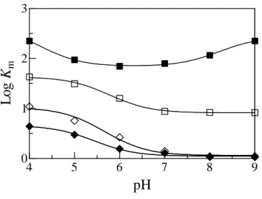
3.1.2 Effect of pH on values of Km and Ki
The analysis of the experimental data according to equation (1) and (2) for papain and parasite cysteine proteases action, respectively, allowed the evaluation, independently from the Vmax estimation, of the Michaelis
constant (Km, for all the four proteases, Fig. 7 and Table 2) and of the
and Table 3) for the hydrolysis of the Z-Phe-Arg-AMC between pH 4.0 to 9.0.
Figure 7: Effect of pH on values of Km for the hydrolysis of
Z-Phe-Arg-AMC catalyzed by papain (black squares), cruzipain (black rumbles), the protease from L. infantum (rumbles) and brucipain (squares). Solid lines represent the best fit of experimental data was calculated, according to Equation (3) for parasite cysteine proteases and according to Equation (4) for papain, through an iterative non-linear least squares procedure with sets of parameters given in Table 1. The standard deviation for each experimental point is given in Table 1. The analysis allowed the determination of the pK’ and pK’’ parameters given in Table 3. For further details, see text.
4
5
6
7
8
9
1.50
1.75
2.00
pH
L
o
g
K
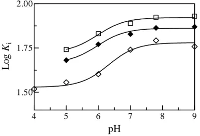
iFigure 8: pH dependence of the substrate inhibition constant Ki (µM) for
the hydrolysis of the Z-Phe-Arg-AMC catalyzed by cruzipain (black rumbles), L. infantum protease (rumbles) and brucipain (squares) at 25,0 °C. Solid lines, representing the best fit of experimental data calculated according to Equation (5) through an iterative nonlinear least squares procedure with sets of parameters given in Table 2. Standard deviation for each of the experimental point is reported in Table 2. For pH values below pH 5 cruzipain does not show the substrate inhibition. The analysis allowed the determination of the pK° parameters given in Table 3. Standard deviation regarding experimental points is given in Table 2. For further details, see text.
4
5
6
7
8
9
0
1
2
3
pH
L
o
g
K
mTable 4: Values of pKa parameters which describe the pH dependence of
the Michaelis constant Km (in black) and the pH dependence of the
substrate inhibition constant Ki (in blue) for the hydrolysis of the
Z-Phe-Arg-AMC by papain and by the cysteine proteases from L. Infantum, from
T. cruzi and from T. brucei (25,0°C).
pKa Values
Papain pK’unl = 4.6±0,1 pK’lig = 3.9±0,2
pK”unl = 7.9±0,1 pK”lig = 8.5±0,1
Cruzipain pK’unl = 5.7±0,1 pK’lig = 5.1±0,1
pK°unl = 5.9±0,2 pK°lig = 6.1±0,2
Brucipain pK’unl = 6.1±0,1 pK’lig = 5.4±0,1
pK°unl = 5.9±0,2 pK°lig = 6.1±0,2
Protease pK’unl = 6.1±0,2 pK’lig = 5.2±0,2
from L. infantum pK°unl = 6.2±0,2 pK°lig = 6.4±0,2
Values of Km for the papain (Fig. 2) depend on the acid-base
equilibrium of two ionising residues. The analysis of data allowed to determine the values of pK’unl (= 4.6±0.1), pK’lig (= 3.9±0.2), pK”unl (=
7.9±0.1) e pK”lig (= 8.5±0.1) (Table 5) and values of pKa reflecte the
equilibrium of two amino acidic residue (Fig. 7 and Table 4), classically recognised as the catalytic residues Cys25 (i.e. pK’unl = 4.6±0.1 and pK’lig =
3.9±0.2) and His159 (i.e. pK’’unl = 7.9±0.1 and pK’’lig = 8.5±0.1) (Hollaway
et al., 1969; Shipton et al., 1976; Ascenzi et al., 1987; Ménard et al., 1990).
Nonetheless, the influence of further residues lying in the active site of the enzyme, like Lys156 and Asp158 can not be excluded and they may account for minor contributions in the enzyme-substrate binding energetic, at least modifying the electric field acting on catalytic residues Cys25 and His159 and thus on their observed pKa values.
On the other hand, the analysis of data concerning cruzipain, brucipain and L. infantum protease action (Fig. 7) allowed the estimation of the pKa values that modulates the protease-substrate binding event, for
cruzipain (pK’unl = 5.7±0.1 e pK’lig = 5.1±0.1), brucipain (pK’unl = 6.1±0.1 e
pK’lig = 5.4±0.1) and for the L. infantum protease (pK’unl = 6.1±0.1 e pK’lig
= 5.2±0.2) (Table 4).
Therefore, while Z-Phe-Arg-AMC binding to papain imply a shift of the apparent pKa of 0.7 unit toward acidic pH values (pK’unl > pK’lig) and
of 0.6 unit toward the alkaline (pK”unl < pK”lig) the interaction of
Z-Phe-Arg-AMC with cruzipain, brucipain and L. infantum protease determines a shift of the apparent pKa to acidic values (pK’unl > pK’lig) of about 0.7 pH
units.
On the other hand, only one apparent amino acidic residue is able to modulate the interaction of Z-Phe-Arg-AMC with cruzipain and with L.
Infantum protease regarding to the productive as well as to the unproductive
binding (SS and Si respectively, see Scheme II). Notably, for all the parasite
enzymes the ligand-independent pKa values which define the pH
dependence of both the productive (pK’unl) and the non-productive binding
process (pK°unl) converge in the limits of the experimental
indeterminateness (i.e. for cruzipain and for L. Infantum protease it results pK’unl ~ pK°unl, see Table 3). These values for cruzipain (pK’unl = 6.2±0.2,
pK°unl = 6.2±0.2) are very close to those that characterize L. Infantum
protease (pK’unl = 5.7±0.1, pK°unl = 5.9±0.2). The main structural
determinant that may account for the observed pH dependence of Z-Phe-Arg-AMC binding to parasite cysteine proteases is probably the Glu205 residue (mature cruzipain numbering). As reported above, this residue, Glu in parasite proteases and Ser in papain, is responsible for the different specificity showed by papain and by parasite cysteine proteases. Notably, previous investigations on cysteine proteases from T. cruzi, L. infantum and
Plasmodium falciparum (Ascenzi et al., 2004). Human cathepsin B (Khouri et al., 1991) and viral cathepsin from the Autographa californica nuclear
polyhedrosis virus (AcNPV) (Bromme & Okamoto, 1995) have shown similar results with reference to the pH dependence of the binding of synthetic ligands to papain-like proteases with a Glu residue equivalent to the Glu205 of cruzipain. It seems to suggest a general model of recognition and of pH-modulation for cysteine proteases with a cathepsin B-like S2 specificity sub-site (Sajid & McKerrow, 2002).
3.2. Inhibition of papain and related proteases by members of the Transferrin family
3.2.1. Tfs are a competitive inhibitors
Lf and Otrf (Tfs) inhibit papain, cruzipain, brucipain and L.
infantum protease (Fig. 9 and Table 5). The inhibition of cysteine proteases
by Lf and Ortf conforms to competitive inhibition according to the classic competitive inhibition mechanism (Scheme III) described by Equations (6), (7) and (8). On the contrary, sTf does not inactive cysteine proteases, according to its different role in mammals (Anderson et al., 2009).
0.000
0.025
0.050
0.075
0.00
0.25
0.50
0.75
1.00
[bLf] (µM)
v
i/v
0Figure 9: Dependence of the initial relative velocity vi/v0 for the hydrolysis
of Z-Phe-Arg-AMC by papain (black squares), cruzipain (black rumbles), L.
infantum protease (rumbles) and brucipain (squares) on the bLf
concentration. Experimental data are been obtained at pH 6.0 and 25 °C. The Z-Phe-Arg-AMC concentration was 50 µM for papain and 5.0 for parasite cysteine proteases. Solid lines represent the theoretical curves, described by Equation (6), obtained through an iterative non-linear least squares procedure with sets of parameters given in Table 5. Standard deviation for each experimental point was equal to ± 8%.
0 25 50 75 100 0.00 0.02 0.04 0.06
[Z-Phe-Arg-AMC] (
µ
M)
K
I ap p 0.0 2.5 5.0 7.5 0.00 0.01 0.02 0.03[Z-Phe-Arg-AMC] (
µ
M)
K
I ap p A BFigure 10: Dependence of the relative inhibition constant KIapp/KI by bLf
binding to papain (Panel A, black squares), with cruzipain (Panel B, black rumbles), L. infantum protease (Panel b, rumbles) and brucipain (Panel B, squares) on the substrate concentration (Z-Phe-Arg-AMC). Experimental data were carried out at 25°C and pH 6.0. Straight lines, representing the best fit of experimental data, were calculated according to Equation (8) through an iterative linear least squares procedure with sets of parameters given in Tables 5-8. Standard deviation for each experimental point was to ± 8%.
Table 5: Values of KI (µM) for bLf, hLf (holo and apo) and Otrf binding to
papain (25.0 °C).
pH bLf hLf-holo hLf-apo Otrf
4.0 (3.5±0.1)x10-1 (7.1±0.6)x10-2 (8.5±0.8)x10-2 (9.8±0.1)x10-1 5.0 (2.2±0.1)x10-1 (7.2±0.6)x10-2 (9.1±0.8)x10-2 (8.6±0.1)x10-1 6.0 (5.1±0.1 )x10-2 (7.6±0.6)x10-2 (9.3±0.1)x10-2 (7.0±0.1)x10-1 7.4 (2.4±0.1)x10-2 (1.6±0.1)x10-1 (2.0±0.1)x10-1 (4.6±0.1)x10-1 8.0 (4.7±0.3)x10-2 (3.4±0.2)x10-1 (4.3±0.1)x10-1 (3.5±0.1)x10-1 9.0 (7.4±0.6)x10-2 (5.1±0.4)x10-1 (6.3±0.1)x10-1 (1.2±0.1)x10-1
Table 6: Values of KI (µM) for bLf, hLf (holo and apo) and Otrf binding to
L. infantum protease (25.0 °C).
pH bLf hLf-holo hLf-apo Otrf
4.0 (2.5±0.1)x10-2 (2.0±0.1)x10-2 (3.5±0.2)x10-2 (9.9±0.8)x10-1 5.0 (2.0±0.1)x10-2 (1.8±0.1)x10-2 (3.3±0.2)x10-2 (8.4±0.1)x10-1 6.0 (1.0±0.1 )x10-2 (1.2±0.1)x10-2 (2.2±0.1)x10-2 (6.9±0.5)x10-1 7.4 (6.0±0.5)x10-3 (9.0±0.8)x10-3 (1.7±0.1)x10-2 (4.5±0.3)x10-1 8.0 (8.8±0.7 )x10-3 (1.2±0.1)x10-2 (2.3±0.1)x10-2 (2.6±0.1)x10-1 9.0 (1.6±0.1)x10-2 (2.3±0.1)x10-2 (4.3±0.3)x10-2 (1.3±0.1)x10-1
Table 7: Values of KI (µM) for bLf, hLf (holo and apo) and Otrf binding to
cruzipain (25.0 °C).
pH bLf hLf-holo hLf-apo Otrf
4.0 (9.7±0.8)x10-2 (3.2±0.1)x10-2 (2.0±0.1)x10-2 (9.8±0.1)x10-1 5.0 (3.3±0.1)x10-2 (2.6±0.1)x10-2 (1.6±0.1)x10-2 (8.9±0.1)x10-1 6.0 (6.8±0.6 )x10-3 (1.3±0.1)x10-2 (8.2±0.7)x10-3 (5.0±0.1)x10-1 7.4 (3.1±0.1)x10-3 (7.5±0.6)x10-3 (4.7±0.1)x10-3 (1.6±0.1)x10-1 8.0 (5.8±0.4 )x10-2 (1.1±0.1)x10-2 (7.0±0.6)x10-3 (6.3±0.5)x10-2 9.0 (2.5±0.1)x10-2 (2.0±0.1)x10-2 (1.3±0.1)x10-2 (4.0±0.3)x10-2
Table 8: Values of KI (µM) for bLf, hLf (holo and apo) and Otrf binding to
brucipain (25.0 °C).
pH bLf hLf-holo hLf-apo Otrf
4.0 (1.1±0.1)x10-1 (9.7±0.1)x10-2 (1.2±0.1)x10-1 (8.1±0.7)x10-1 5.0 (3.5±0.4)x10-2 (5.8±0.1)x10-2 (7.5±0.6)x10-2 (7.9±0.7)x10-1 6.0 (8.7±0.8 )x10-3 (1.8±0.1)x10-2 (2.6±0.1)x10-2 (6.9±0.5)x10-1 7.4 (6.7±0.5)x10-3 (1.5±0.1)x10-2 (2.2±0.1)x10-2 (4.7±0.3)x10-1 8.0 (1.2±0.1 )x10-2 (2.2±0.1)x10-2 (3.1±0.2)x10-2 (4.5±0.3)x10-1 9.0 (3.6±0.2)x10-2 (3.2±0.2)x10-2 (4.4±0.3)x10-2 (4.4±0.3)x10-1
For all the proteases considered in this study, the dependence of the apparent inhibition constant KIapp on the Z-Phe-Arg-AMC concentration,
was linear over the substrate concentration range explored (Fig. 10). These results are compatible only with a complete competitive inhibition mechanism (Scheme III), in fact, the observed relative inhibition constant
KIapp/KI increases linearly with the substrate concentrations (Fig. 10).
Notably, the Y intercept of straight lines is 1. This means that in the absence of the substrate ([Z-Phe-Arg-AMC] = 0, corresponding to the X interpolation value of the Y intercept) the apparent inhibition constant KIapp
corrisponds to the intrinsic ligand-independent equilibrium constant KI
(KIapp= KI).
Moreover, if we compare values the intrinsic substrate-independent constant KI with those cystatins, it appears that Lf a very good inhibitor of
cisteine papain-like proteases (Table 9).
Table 9. Values of the intrinsic ligand-independent constant KI (µM) of
stefin A, of stefin B (cystatins of type I), of chicken egg-white cystatin (CEW Cys, cystatin of type II).
Proteases Papain Cathepsin-B Cathepsin-L Cruzipain
Stefin A 1.8 × 10⁻⁷ a 9.1 × 10⁻⁴ a 1.9 × 10⁻⁵ a 2.1 × 10⁻⁵ c Stefin B 4.9 × 10⁻⁸ b 1.8 × 10⁻² b 3.0 × 10⁻⁶ b 6.0 × 10⁻⁵ c CEW Cys 1.0 × 10⁻⁵ d 8.0 × 10⁻⁵ d 3.0 × 10⁻⁶ d 1.4 × 10⁻⁵ c a Estrada et al. (1998). b Pol & Björk (2001). c Stoka et al. (1995). d Anastasi et al. (1983).
3.2.2. Effect of pH on values of KI
The analysis of the pH dependence of the intrinsic ligand-independent inhibition constant KI (Fig. 11, 12, 13 and 14), according to
Equations (9) and (10) allowed the evaluation of pKa values that define the
acid-base equilibrium of amino acidic residue(s) able to modulate the enzyme(s)-Transferrin recognition event (Tables 10, 11, 12 and 13).
Figure 11: pH dependence of KI (µM) for the binding of bLf (squares),
hLf-holo and -apo (triangles and circles, respectively) and Otrf (reversed triangles) to papain at 25 °C. Solid lines representing the best fit of data, were calculated according to Equation (9) for Otrf, Equation (10) for hLf (apo and holo) and equation (11) for bLf. Continous lines were obtained through an iterative non-linear least squares procedure with sets of parameters given in Tables 5, 6, 7 and 8. For further details see the text.
4
5
6
7
8
9
-2.0
-1.5
-1.0
-0.5
0.0
pH
L
o
g
K
ITable 10: Values of pKa modulating the inhibition constant KI (µM) for
bLf (squares), hLf-apo and –holo (circles and triangles) and Otrf (overturned triangles) to papain (Fig. 6) at 25 °C.
pKa Values
bLf pK’unl = 6.9±0.3 pK’lig = 5.0±0.2
pK”unl = 7.9±0,1 pK”lig = 8.5±0,1
hLf-apo pK’unl = 7.1±0.3 pK’lig = 8.0±0.2
hLf-holo pK’unl = 7.1±0.3 pK’lig = 8.0±0.2
Figure 12: pH dependence of KI (µM) for the binding of bLf (squares),
hLf-holo and -apo (triangles and circles, respectively) and Otrf (reversed triangles) to L. infantum protease at 25 °C. Solid lines representing the best fit of data, were calculated according to Equation (9) for Otrf and according to equation (11) for bLf and for hLf (apo and holo). Continous lines were obtained through an iterative non-linear least squares procedure with sets of parameters given in Tables 5, 6, 7 and 8. For further details see the text.
4
5
6
7
8
9
-2.0
-1.5
-1.0
-0.5
0.0
pH
L
o
g
K
ITable 11: Values of pKa modulating the inhibition constant KI (µM)
regarding for bLf (squares), hLf-apo and –holo (circles and triangles) and Otrf (overturned triangles) to L. infantum protease (Fig. 7) at 25 °C.
pKa Values
bLf pK’unl = 6.4±0.3 pK’lig = 5.6±0.1
pK”unl = 7.7±0.2 pK”lig = 8.3±0.4
hLf-apo pK’unl = 6.2±0.5 pK’lig = 5.8±0.2
pK”unl = 7.9±0.2 pK”lig = 8.5±0.3
hLf-holo pK’unl = 6.2±0.5 pK’lig = 5.8±0.2
pK”unl = 7.9±0.2 pK”lig = 8.5±0.3
4
5
6
7
8
9
-2.0
-1.5
-1.0
-0.5
0.0
pH
L
o
g
K
IFigure 13: pH dependence of KI (µM) for the binding of bLf (squares),
hLf-holo and -apo (triangles and circles, respectively) and Otrf (reversed triangles) to cruzipain at 25 °C. Solid lines representing the best fit of data, were calculated according to Equation (9) for Otrf and according to equation (11) for bLf and for hLf (apo and holo). Continous lines were obtained through an iterative non-linear least squares procedure with sets of parameters given in Tables 5, 6, 7 and 8. For further details see the text.
Table 12: Values of pKa modulating the inhibition constant KI (µM) for
bLf (squares), hLf-apo and –holo (circles and triangles) and Otrf (overturned triangles) to cruzipain (Fig. 8) at 25 °C.
pKa Values
bLf pK’unl = 6.4±0.2 pK’lig = 4.6±0.1
pK”unl = 7.7±0.2 pK”lig = 9.1±0.3
hLf-apo pK’unl = 6.4±0.1 pK’lig = 5.6±0.3
pK”unl = 7.7±0.1 pK”lig = 8.3±0.2
hLf-holo pK’unl = 6.4±0.1 pK’lig = 5.6±0.3
pK”unl = 7.7±0.1 pK”lig = 8.3±0.2
4
5
6
7
8
9
-2.0
-1.5
-1.0
-0.5
0.0
pH
L
o
g
K
IFigure 14: pH dependence of KI (µM) for the binding of bLf (squares),
hLf-holo and -apo (triangles and circles, respectively) and Otrf (reversed triangles) to brucipain at 25 °C. Solid lines representing the best fit of data, were calculated according to Equation (9) for Otrf and according to equation (11) for bLf and for hLf (apo and holo). Continous lines were obtained through an iterative non-linear least squares procedure with sets of parameters given in Tables 5, 6, 7 and 8. For further details see the text.
Table 13: Values of pKa modulating the inhibition constant KI (µM) for
bLf (squares), hLf-apo and –holo (circles and triangles) and Otrf (overturned triangles) to cruzipain (Fig. 9) at 25 °C.
pKa Values
bLf pK’unl = 6.2±0.2 pK’lig = 4.3±0.1
pK”unl = 7.5±0.2 pK”lig = 8.9±0.3
hLf-apo pK’unl = 6.1±0.2 pK’lig = 5.0±0.1
pK”unl = 7.4±0.2 pK”lig = 8.0±0.3
hLf-holo pK’unl = 6.1±0.2 pK’lig = 5.0±0.1
pK”unl = 7.4±0.2 pK”lig = 8.0±0.3
In particular, bLf binding to papain (pK’unl = 6.9±0.3, pK’lig =
5.0±0.2, pK”unl = 7.9±0,1 and pK”lig = 8.5±0,1 ), L.infantum protease (pK’unl
= 6.4±0.3, pK’lig = 5.6±0.1, pK”unl = 7.7±0.2 and pK”lig = 8.3±0.4),
cruzipain (pK’unl = 6.4±0.2, pK’lig = 4.6±0.1, pK”unl = 7.7±0.2 and pK”lig =
9.1±0.3) and brucipain (pK’unl = 6.2±0.2, pK’lig = 4.3±0.1, pK”unl = 7.5±0.2
and pK”lig = 8.9±0.3) is modulated by the acid-base equilibrium of two
amino acidic residues (Fig. 6-9 and Tables 9-12). Inhibitor binding induces a pKa shift of about 1.9 pH units toward acidic values and of about 0.6 pH
units toward the alkaline region for papain (pK*unl > pK*lig and pK**unl <
pK**lig), a pKa shift of about 0.8 pH units toward acidic values and of about
0.6 pH units toward the alkaline region for protease from L. Infantum, a pKa
shift of about 1.8 pH units toward acidic values and of about 1.4 pH units toward the alkaline region for cruzipain and a pKa shift of about 1.9 pH
units toward acidic values and of about 1.4 pH units toward the alkaline region for brucipain.
The interaction of hLf (apo and holo) with papain is modulated by one apparent acidbase ionization (Fig. 6-9 and Tables 9-12) causing a pKa
shift of about 0.9 pH units toward alkaline values (pK’unl = 7.1±0.3 and
pK’lig = 8.0±0.2, both apo-form and holo-form). On the other hand, hLf (apo
and holo) binding to parasitic proteases is modulated by the acid-base equilibrium of two amino acid residues (Fig. 6-9 and Tables 9-12) causing a pKa shift of about 0.4 pH units toward acidic values and of about 0.6 pH
units toward the alkaline region for L. Infantum protease (pK’unl = 6.2±0.5,
pK’lig = 5.8±0.2, pK”unl = 7.9±0.2 and pK”lig = 8.5±0.3), a pKa shift of about
0.8 pH units toward acidic values and of about 0.6 pH units toward the alkaline region for cruzipain (pK’unl = 6.4±0.1, pK’lig = 5.6±0.3, pK”unl =
7.7±0.1 and pK”lig = 8.3±0.2) and a pKa shift of about 1.1 pH units toward
acidic values and of about 0.6 pH units toward the alkaline region for brucipain (pK’unl = 6.1±0.2, pK’lig = 5.0±0.1, pK”unl = 7.4±0.2 and pK”lig =
8.0±0.3).
Instead, the binding of Otrf bLf to papain (pK’unl = 8.2±0.2 and
pK’lig = 7.3±0.2), to protease of type I from L. infantum (pK’unl = 8.5±0.2
and pK’lig = 7.5±0.3), to cruzipain (pK’unl = 7.9±0.2 and pK’lig = 6.6±0.3)
and to brucipain (pK’unl = 7.1±0.1 and pK’lig = 5.9±0.2) is modulated by one
apparent amino acidic residue (Fig. 6-9 and Tables 9-12). It causes a pKa
shift of about 0.9 pH units for papain, 1.0 pH units for protease of type I from L. infantum, 1.3 pH units for cruzipain and 1.2 pH units for brucipain toward acidic values.