Accepted: 2019.03.22 Published: 2019.05.10
Urocortin Induces Phosphorylation of Distinct
Residues of Signal Transducer and Activator of
Transcription 3 (STAT3) via Different Signaling
Pathways
ABCDEF 1
Giovanni Corsetti*
BCD 2
Zhaokan Yuan*
BCDE 3
Claudia Romano*
ABCDEF 2
Carol Chen-Scarabelli*
B 4
Alessandro Fanzani
DE 5
Evasio Pasini
DEF 6
Francesco S. Dioguardi
DE 7
Francesco Onorati
CD 7
Daniele Linardi
DE 8
Richard Knight
CDE 9,10
Hemang Patel
DE 7
Giuseppe Faggian
#DE 11
Louis Saravolatz
# ABCDEF 2Tiziano M. Scarabelli
#* Giovanni Corsetti, Zhaokan Yuan, Claudia Romano and Carol Chen-Scarabelli equally contributed to this work # Giuseppe Faggian, Louis Saravolatz and Tiziano M. Scarabelli equally contributed as senior authors to the manuscript
Corresponding Author: Giovanni Corsetti, e-mail: [email protected]
Source of support: The work was sponsored by a grant provided by the St. John Guild Foundation
Background: Urocortin (Ucn) is a member of the hypothalamic corticotrophin-releasing factor family and has been shown to reduce cell death in the heart caused by ischemia/reperfusion (I/R) injury. Signal transducer and activator of transcription 3 (STAT3) is a transcription factor known to function as a pro-survival and anti-apoptotic fac-tor, whose activation depends on a variety of cytokines, including IL-6. A recent study demonstrated that uro-cortin induced IL-6 release from cardiomyocytes in a CRF-R2-dependent manner, suggesting a possible link be-tween CRF-R2 stimulation and STAT3 activation.
Material/Methods: Experimental work was carried out in HL-1 cardiac myocytes exposed to serum starvation for 16–24 h.
Results: Ucn stimulation led to IL-6 expression and release from mouse atrial HL-1 cardiomyocytes. Ucn treatment led to rapid phosphorylation of JAK2, which was blocked by the protein synthesis inhibitor cycloheximide or the JAK inhibitor AG490. Urocortin treatment induced STAT3 phosphorylation at Y705 and S727 through trans-activation of JAK2 in an IL-6-dependent manner, but had no effect on STAT1 activity. Kinase inhibition exper-iments revealed that urocortin induces STAT3 S727 phosphorylation through ERK1/2 and Y705 phosphoryla-tion through Src tyrosine kinase. In line with this finding, urocortin failed to induce phosphorylaphosphoryla-tion of Y705 residue in SYF cells bearing null mutation of Src, while phosphorylation of S727 residue was unchanged.
Conclusions: Here, we have shown that Ucn induces activation of STAT3 through diverging signaling pathways. Full under-standing of these signaling pathways will help fully exploit the cardioprotective properties of endogenous and exogenous Ucn.
MeSH Keywords: Inflammation Mediators • Myocytes, Cardiac • Urocortins
Full-text PDF: https://www.basic.medscimonit.com/abstract/index/idArt/914611 Authors’ Contribution: Study Design A Data Collection B Statistical Analysis C Data Interpretation D Manuscript Preparation E Literature Search F Funds Collection G
1 Division of Human Anatomy and Physiopathology, Department of Clinical and Experimental Sciences, University of Brescia, Brescia, Italy
2 Center for Heart and Vessel Preclinical Studies, Department of Internal Medicine, St. John Hospital and Medical Center, Wayne State University, Detroit, MI, U.S.A. 3 Department of Clinical and Experimental Sciences, University of Brescia, Brescia,
Italy
4 Department of Molecular and Translational Medicine, University of Brescia, Brescia, Italy
5 Scientific Clinical Institutes Maugeri, Cardiac Rehabilitation Lumezzane Institute, Brescia, Italy
6 Department of Internal Medicine, University of Cagliari, Cagliari, Italy 7 Division of Cardiovascular Surgery, Verona University Hospital, Verona, Italy 8 Medical Research Council (MRC) Toxicology Unit, University of Cambridge,
Cambridge, United Kingdom
9 Department of Internal Medicine, General Medical Education, Ascension St. John Hospital, Detroit, MI, U.S.A.
10 Department of Internal Medicine, Wayne State University – School of Medicine, Detroit, MI, USA
11 Department of Medicine, Ascension St John Hospital and Wayne State University School of Medicine, Detroit, MI, U.S.A
Background
Urocortin (Ucn) is a 40 amino acids peptide, which belongs to the corticotrophin-releasing factor (CRF) family. Besides Ucn, this family includes corticotropin-releasing hormone (CRH) (also known as CRF), urotensin I, sauvagine, Ucn-II, and Ucn-III [1–3]. The hypothalamic neuropeptide CRF is a central regulator of homeostasis by activating hypothalamic-pituitary-adrenal (HPA) axis in response to stress [4]. Although Ucn also contributes to coordination of behavioral and autonomic re-sponses to stressful stimuli by affecting central nervous system like CRF, it has a more significant role in the periphery [5–7]. There are 2 main types of the G-protein-coupled CRF receptors: CRF-R1 and CRF-R2. CRF-R1 is widely expressed in the central nervous system and in the pituitary, and CRF-R2 has 3 differ-ent spliced forms – a, b, and g – of which CRF-R2a is mainly found in the brain, while CRF-R2b is distinctively expressed both in the brain and in the periphery, including cardiac and skeletal muscle [2,8]. Pharmacologic studies have shown that Ucn binds with high affinity to both types of CRF receptors, but it is about 40 times more potent in binding CRF-R2 [2,9]. Ucn has generally been considered to be cardioprotective, as it is released from the heart during ischemia/reperfusion (I/R) in-jury, inhibiting cardiomyocyte death. Ucn expression is upreg-ulated in primary neonatal rat cardiomyocytes exposed to sim-ulated ischemia (hypoxia) [10], as well as in the human heart receiving warm blood cardioplegic arrest and subsequent re-perfusion, where it protects cardiac cells via an autocrine/para-crine mechanism [11]. Furthermore, exogenous Ucn, when ad-ministered both prior to ischemia and during perfusion, reduced infarct size in the rat heart, as well as the occurrence of myo-cyte apoptosis [12,13]. Several early response kinases have been implicated in Ucn-mediated cardioprotection, including ERK1/2 mitogen-activated protein kinase (MAPK), phosphati-dylinositol 3-kinase (PI3K)/Akt, and mitochondrial relocation of protein kinase C-epsilon [9,11,12,14,15]. The non-recep-tor tyrosine kinase, Src, has recently been shown to be phos-phorylated in a CRF receptor-dependent manner after short-term incubation with Ucn and contributes to Ucn-mediated cardioprotection [16].
The signal transducer and activator of transcription (STAT) proteins are a family of transcription factors that can be acti-vated by a wide range of cytokines, growth factors, and hor-mones. Following the binding of cognate ligands to specific cell-surface receptors, sequential tyrosine phosphorylation is triggered by dimerization of the receptor and autophosphory-lation of the receptor-associated Janus kinase (JAK). There are 4 cytosolic members of JAK family – JAK1, JAK2, JAK3, and ty-rosine kinase 2 (TYK2) – all of which transduce extracellular signals to the nucleus through activation of STAT proteins [17]. JAK kinases phosphorylate specific receptor tyrosine residues,
which then serve as a binding site for Src-homology-2 (SH2) domain of STAT proteins.
Recruited STAT3 is phosphorylated on tyrosine 705 residue (Y705) in the transactivation domain by activated JAKs, which leads to STAT3 dimerization, translocation to the nucleus, and increased transcription of target genes [18,19]. In addition to JAKs, receptor tyrosine kinase (RTK) and certain G-protein-coupled receptors (GPCR), together with Src, were reported to directly phosphorylate Y705 residue of STAT3 [18,20]. Binding of IL-6 family cytokines to their receptors can also promote phosphorylation of STAT3 at Serine 727 residue (S727) through MAPK or mammalian target of rapamycin (mTOR), which gen-erally increases transcriptional activity of STAT3 at certain pro-moters [19,21,22]. STAT3 is constitutively active in several hu-man cancers in an IL-6-dependent hu-manner [18,23]. On the other hand, active STAT3 confers beneficial effects and anti-apop-totic protection to cardiomyocytes following I/R injury, while STAT1 is a pro-apoptotic mediator of myocardial cell death [23]. IL-6 is a cytokine produced by macrophages, monocytes, and T cells to promote local immune response, and is a major stim-ulator of a variety of acute-phase inflammatory proteins. IL-6 is generally pro-inflammatory in chronic inflammation; however, at some sites during acute inflammatory response, it plays an anti-inflammatory role mediated through inhibitory effects on TNF-a and IL-1, and activation of IL-10 and IL-1 receptor an-tagonist [24]. As IL-6 interacts with its receptor, it triggers 1 IL-6 receptor and 2 gp130 proteins to form a receptor com-plex, and the activated homodimer of gp130 initiates trans-activation of JAKs and STATs [20]. In cultured rat neonatal car-diomyocytes, but not in cardiac fibroblasts, ischemia (hypoxia) induces synthesis and release of IL-6, which is associated with activation of STAT3 and is involved in late preconditioning via PI3K/Akt, iNOS, and NO-dependent protection. IL-6 family cy-tokines also induce cardiac hypertrophy in rodent and human hearts and decrease contractile function, both acutely and chronically [20,25]. Although not completely understood, IL-6 may have a dual action in the heart; the IL-6-activated JAK/ STAT3 signaling circuit may be acutely cardioprotective, but chronic stimulation may cause maladaptive myocardial hyper-trophy leading to heart failure [20,25].
Similarly, the role of Ucn in inflammation is not yet fully under-stood. Recent studies have started to delineate the pro-inflamma-tory role of Ucn, despite its protective effects against I/R injury. In human rheumatoid synovium, Ucn stimulated the secretion of the inflammatory cytokines IL-1b and IL-6 from mononuclear cells via CRF-R1 [26]. Ucn has also been shown to promote a sig-nificant increase in microvascular permeability in response to inflammation, partially via CRF-R2 [27]. In rodent neonatal car-diomyocytes, Ucn induced IL-6 release in a CRF-R2-dependent manner through activation of ERK1/2, P38, and NF-kB [28].
A connection between Ucn and STAT3 has been previously re-ported by Pan et al. [29]. In both cerebral microvessel endo-thelial RBE4 cells and HEK293 cells co-transfected with CRF-R1 or CRF-R2, Ucn unexpectedly induced significant STAT3, but not STAT1, activation [29].
To verify the mechanism(s) by means of which Ucn induces STAT3 activation, we studied JAK/ERK/Src signaling in mouse HL-1 cardiomyocytes. Our data a) revealed the existence of novel Ucn-stimulated JAK/STAT3 and Src/STAT3 signaling cir-cuits; b) confirmed that Ucn induces the expression and re-lease of IL-6 from cardiac cells; and c) documented that STAT3 phosphorylation at Y705 and S727 is activated by JAK/ERK/ Src signaling cross-talk.
Experimental Procedures
Reagents and antibodies
Products purchased from Sigma (St. Louis, MO) included Claycomb medium, fetal bovine serum, norepinephrine, fibronectin, leukemia inhibitory factor (LIF) and urocortin (rat). Purchases from GIBCO (Invitrogen, Carlsbad, CA) included L-glutamine and Penicillin-Streptomycin. The rabbit polyclonal anti-phospho(P)-Tyr-Src (Y418) antibody was obtained from BioSource (Invitrogen, Carlsbad, CA). The mouse monoclonal Src (B-12) antibody, the monoclonal P-ERK (E-4) anti-body, the rabbit polyclonal anti-ERK1 (C-16) antianti-body, and rab-bit polyclonal anti-IL-6 (M-19) antibody were purchased from Santa Cruz Biotechnology (Santa Cruz Biotechnology, CA). The rabbit polyclonal anti-P-STAT1 (Y701), anti-P-STAT3 (Y705 and S727), anti-STAT1, anti-STAT3 antibodies, and a rabbit mono-clonal anti-P-STAT3 (Y705) antibody were purchased from Cell Signaling Technology (Danvers, MA). The JAK isoforms sampler kit, a rabbit polyclonal anti-JAK2 antibody, and a mouse mono-clonal anti-P-Tyrosine (pY100) antibody were also purchased from Cell Signaling Technology. The specific Src family kinase inhibitor, PP2, 2 MEK1 inhibitors (which can inhibit the activa-tion of downstream ERK1/2 kinases), U126 and PD98059, and AG490 and pyridone 6 (P6, InSolution™) JAK inhibitors were purchased from Calbiochem (La Jolla, CA). The secondary anti-bodies (obtained from Santa Cruz Biotechnology) were conju-gated to horseradish peroxidase. Immunoreactive bands were produced by means of a Western Lightning Chemiluminescence kit (PerkinElmer Life Science, Boston, MA). The Trans-Blot pure nitrocellulose membrane utilized for Western blot trans-fer was purchased from Bio-Rad Laboratory (Hercules, CA), while the protein-G agarose beads was obtained from Upstate Biotechnology (Millipore, Billerica, MA).
Cell preparation and culture
HL-1 cardiomyocytes were grown at 37°C in an atmosphere of 95% air plus 5% CO2, in Claycomb medium complemented with 100 mM norepinephrine, 4 mM L-glutamine, 50 U/ml Penicillin-Streptomycin, and 10% fetal bovine serum (FBS). Following achievement of 80% cell confluence, HL-1 cardiomy-ocytes were serum-starved for a timespan ranging from 16 to 20 h in Claycomb medium, and subsequently utilized for ex-perimentation. Petri dishes and flasks used for culturing HL-1 cells were pre-coated overnight at 37°C with sterile 0.02% gel-atin and 0.1% fibronectin (200: 1).
Western blot analysis
After cell lysis in RIPA buffer [16], lysates were centrifuged at 16 000 g for 10 min at 4°C. Supernatants dissolved in sample buffer were subsequently separated on 10% SDS-PAGE prior to being transferred to a Trans-Blot pure nitrocellulose mem-brane and finally probed for the proteins of interest.
Immunoprecipitation
HL-1 cell lysates were prepared as described above. Supernatants (2 mg) were incubated overnight at 4°C with 2 μg rabbit polyclonal anti-JAK2 antibody. Then, immunopre-cipitates were pulled down with protein-G agarose beads, washed with PBS, and finally used for Western blot analysis, using an anti-phospho-Tyrosine (pY100) monoclonal antibody.
Electrophoretic mobility shift assay (EMSA)
For EMSA, end-labeled [32P]-oligonucleotides probes cor-responding to m67 serum-inducible response element (SIE) gene sequence were used to detect STAT3 bind-ing [30]: 5’-AGCTTGTCGACATTTCCCGTAAATCGTCGAG-3’ and 5’-CTCGACGATTTACGGGAAATGTCGACAAGCT-3’. After labeling and annealing, the double-strand probe was incubated with 5 µg of nuclear extract in 15 µl of binding mixture (50 mM Tis-HCl (PH7.4), 25 mM MgCl2, 0.5 mM DTT, and 50% glycerol) at 4°C for 2 h. For super-shift assay, nuclear extract was pre-in-cubated with 1 µg of either normal rabbit serum or antiserum specific to STAT3 at 4°C for 20 min. The samples were then incubated for an additional 15 min at room temperature. The DNA-protein complexes were resolved on a 5% polyacrylamide gel containing 0.25X TBE buffer that was prerun in 0.25X TBE buffer for 1 h at 100 V. After loading of samples, gel was elec-trophoresed at room temperature for about 2 h at 140 V. The gel was then dried by heating under vacuum and exposed to X-ray film at –80°C overnight.
Preparation of nuclear fraction and cytoplasmic fraction
The nuclear extract was prepared by using “Nuclear Extract Kit” from Active Motif (Carlsbad, CA). HL-1 cells were washed with 1 volume of ice-cold PBS and scraped off gently with a cell lifter. The lifted cells were spun down at 300 g for 5 min at 4°C and then the pellet was re-suspended in 500 µl of hy-potonic buffer and placed on ice for 15 min. We mixed 25 µl of 10% Nonylphenyl-polyethylene glycol solution (Nonidet™ P-40, 0.5% final, from Sigma-Aldrich) with re-suspension by gentle pipetting and then was centrifuged for 30 s at 14 000 g at 4°C. The supernatant (cytoplasmic fraction) was transferred into a pre-chilled microcentrifuge tube. The nuclear pellet was re-suspended in 50 µl Complete Lysis Buffer from the kit by pi-petting up and down, and then vortexed for 10 s at the high-est setting, followed by incubation for 30 min on a rocking platform at 4°C. The suspension was centrifuged for 10 min at 14 000 g at 4°C and the supernatant (nuclear fraction) was subjected to Lowry protein assay before being aliquoted and stored at –80°C.
STAT3 transcription factor assay
TransAM STAT3 assay kit (from Active Motif, Carlsbad, CA), including 96-well STAT assay plates, was used. We added 50 µl of complete binding buffer from the kit or 50 µl of the same buffer containing 5 µg of nuclear extract from each sample to a 96-well assay plate. After incubation for 1 h at room tem-perature, each well was washed 3 times with 200 µl 1× wash buffer. Afterward 100 µl of STAT3 antibody (1: 500) and 100 µl of HRP-conjugated secondary antibody (1: 1000) were added in each well, incubated for 1 h each, followed by washing 4 times. We added 100 µl of developing solution to all wells and incubated them in dark for 10 min until medium blue col-or appeared in the samples and positive control well. We then immediately added100 µl stop solution to turn the color into yellow and the plate was put on a spectrophotometer for the absorbance reading at 450 nM wavelength with a reference wavelength of 655 nM. The plate-reader was blanked by buf-fer in blank wells.
Cytokine enzyme-linked immunosorbent assay (ELISA)
Ucn-induced IL-6 release from HL-1 cardiomyocytes was quanti-fied using a mouse IL-6 ELISA kit from Invitrogen (Carlsbad, CA). Cell cultures were prepared by adding Claycomb medium containing Ucn 10 nM. After incubation at 37°C for 1, 6, 12, 18, and 24 h, the culture supernatants were recovered and stored at –80°C until IL-6 measurements. Concentration data are expressed in pg/mL (sensitivity: 0.55 pg/mL; range: 0.55–400 pg/mL).
IL-6 significantly increased at 1 h and peaked at 12 h, after which it slowly declined, but it was still detectable at 24 h. Data are presented as averages of 3 independent experiments.
Statistical analysis
The differences between unstimulated and stimulated cells were compared using the 2-tailed t test; comparison between different treatment groups was performed by one-way ANOVA. Data are given as mean ± S.E.M., and the significance was ac-cepted at p<0.05.
Results
Urocortin phosphorylates STAT3 in a time- and dose-dependent manner
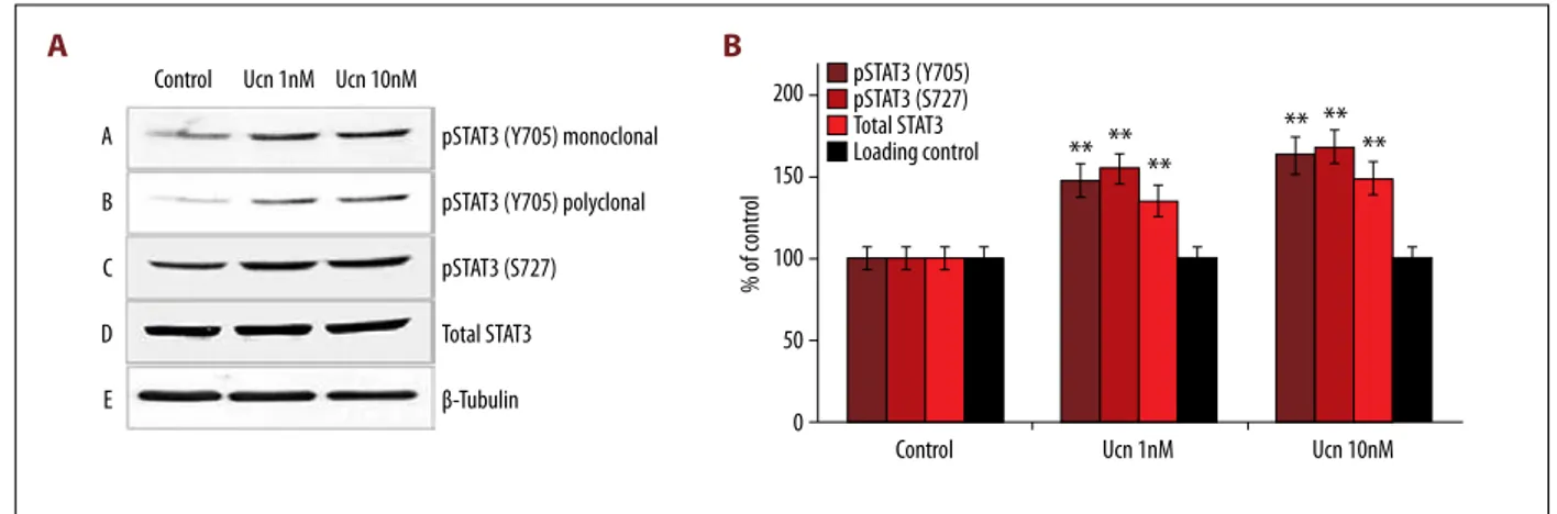
To test whether STAT proteins can be phosphorylated by short-term incubation with Ucn, we used an array of anti-p-STAT an-tibodies and carried out Western blot analysis in Ucn-treated HL-1 cell lysate. Following 10 min of incubation with 1 nM and 10 nM Ucn, STAT3 was phosphorylated at both Y705 and S727, while total STAT3 did not vary (Figure 1A, 1B). Because our re-sults were in line with previously reported data [10,12,13] show-ing that maximal STAT3 phosphorylation (i.e., activation) oc-curred with a concentration of Ucn equal to 10 nM, this dose was selected for use in subsequent experiments.
We next analyzed the time kinetics of Ucn-mediated STAT3 phosphorylation in HL-1 cells incubated for 1, 5, 15, 30, and 60 min and 2 h with 10 nM Ucn (Figure 2A, 2B). Phosphorylation of STAT3 (Y705) was significantly enhanced after 15 min of Ucn incubation, and further increased for up to 2 h. Conversely, Ucn-induced phosphorylation of STAT3 (S727) significantly in-creased after 15 min of incubation, declined after 30 min, and remained practically unchanged for up to 2 h. The expression of total STAT3 protein was unaffected by Ucn incubation. The relative levels of Y705 and S727 phosphorylation, expressed as a percentage of time 0 (control), are shown in Figure 2B. The different kinetics of phosphorylation documented for Y705 and S727 suggests that Ucn activates these 2 STAT3 residues by distinct pathways.
We then analyzed the kinetic of STAT3 phosphorylation in HL-1 cells incubated for 15 min with increasing concentrations of Ucn, ranging from 10–11 M to 10–6 M. Ucn concentrations as low as 10–11 M were sufficient to phosphorylate STAT3 at both Y705 and S727 (Figure 2C–2F). STAT1 (Y701) was not phos-phorylated at any Ucn concentration (Figure 2C, 2D). Likewise, the expression levels of total STAT1 and STAT3 proteins were unaffected by Ucn incubation (Figure 2C–2F). Hence, our data indicate that a) Ucn enhances STAT3 phosphorylation at Y705
and S727 in a time- and dose-dependent manner and that b) this effect is first detected at a concentration as low as 10–11 M.
Urocortin induces DNA binding of phospho-STAT3
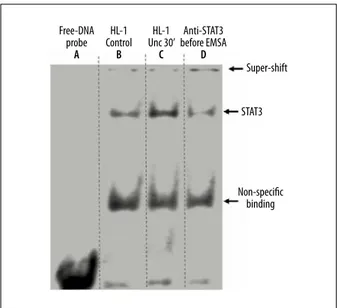
Phosphorylated STAT3 (p-STAT3) proteins form homo- or het-ero-dimers translocate to the nucleus and initiate transcription of target genes. To confirm the specific DNA binding proper-ties of STAT3 after phosphorylation and translocation, elec-trophoretic mobility shift assay (EMSA) and super-shift assay were performed.
A double-stranded radioactive oligonucleotide probe corre-sponding to m67 serum-inducible cis-element (SIE) sequence was used to bind and detect STAT3. As shown in Figure 3, 5 μg nuclear extract from HL-1 cells treated with Ucn (10 nM) for 30 min (lane C) significantly increased DNA binding activity, as compared with the same amount of nuclear proteins from control (untreated) cells (lane B). Moreover, the Ucn-induced DNA-STAT3 complex could be partially super-shifted by pre-incubation with anti-STAT3 antibody before EMSA (lane D). These data show that Ucn-induced STAT3 phosphorylation is accompanied by STAT3 DNA binding.
Urocortin stimulates IL-6 release, leading to STAT3 activation
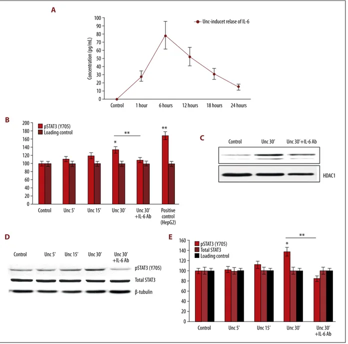
STAT3 can be activated by various factors, including interferons, epidermal growth factor, the IL-6 family of cytokines, and also the hormone leptin [18,20]. Since Ucn was found to induce IL-6 release in rat neonatal myocytes [28], we tested the hypoth-esis that Ucn induces expression and secretion of IL-6, lead-ing to STAT3 activation via an autocrine/paracrine mechanism.
First, we quantified the release of IL-6 in the culture medium of HL-1 cardiac cells incubated with Ucn 10 nM for 30 min us-ing a mouse IL-6 ELISA kit (Figure 4A). IL-6 was not detected in the culture medium of HL-1 cells lacking Ucn (control). The release of IL-6 started at 1 h, increased further for 6 h, peaked at 12 h, and slowly declined thereafter, although it was still detectable at 24 h (Figure 4A).
Second, to test whether IL-6 contributes to activating STAT3 in HL-1 cells, a TransAM STAT3 assay was used in HL-1 nuclear ex-tracts to detect and quantify nuclear translocation of p-STAT3. As shown in Figure 4B, 4C (and in line with the data depicted in Figure 3), incubation of HL-1 cells with Ucn (10 nM) for 30 min significantly increased nuclear STAT3 (Y705) phosphor-ylation as compared with nuclear extracts from control (un-treated) HL-1 cells (100%). Nuclear extracts from HepG2 cells treated with IL-6 (100 ng/ml), used as positive control, showed a drastic increase in nuclear p-STAT3 (Figure 4A right lane). In HL-1 cells treated with Ucn 10 nM for 30 min, the use of an anti-IL-6 antibody (2 μg/ml) to neutralize the effects of Ucn-induced IL-6 release, significantly reduced pSTAT3 (Y705) in the nuclear extracts (Figure 4B, 4C), as well as in whole-cell lysates (Figure 4D, 4E). Hence, Ucn stimulates IL-6 expression in HL-1 cardiomyocytes in a dose-dependent manner.
Urocortin stimulates JAK2/STAT3 signaling in HL-1 cells
Since IL-6 can transactivate JAKs and STAT3 [20], we next ex-amined whether JAKs are activated by Ucn. The JAK isoforms sampler kit (from Cell Signaling Technology) was used to probe JAK1, JAK2, JAK3, and Tyk2 in HL-1 cell lysates. Only JAK2 was expressed at a high level (Figure 5A). Tyrosine phosphory-lation of JAK2 was assessed by immune-precipitation assay and was shown to be increased after 30 min of Ucn (10 nM)
Control pSTAT3 (Y705) pSTAT3 (S727) Total STAT3 Loading control Ucn 1nM Ucn 10nM Control A B C D E
pSTAT3 (Y705) monoclonal pSTAT3 (Y705) polyclonal pSTAT3 (S727) Total STAT3 β-Tubulin Ucn 1nM Ucn 10nM
** ** **
** **
**
200 150 100 50 0 % of cont ro lA
B
Figure 1. Changes in phosphorylation (activation) at the regulatory sites Y705 and S727 of STAT3 in mouse HL-1 cardiomyocytes following 10 min of incubation with different urocortin (Ucn) concentrations (1 and 10 NM). (A) Ucn induced STAT3 phosphorylation at both Y705 and S727 sites, while the expression of total STAT3 protein did not significantly vary. (B) Corresponding clustered column graph of the Western blot shown in Figure 1A. Data are expressed as% of control. * p<0.05, ** P<0.01
Control pSTAT3 (Y705) pSTAT3 (S727) Total STAT3 β-Tubulin 1’ 5’ 15’ 30’ 60’ 120’ Ucn 10 nM
Control Ucn 1’ Ucn 5’ Ucn 15’ Ucn 30’ Ucn 1h Ucn 2h pSTAT3 (Y705) pSTAT3 (S727) Total STAT3 Loading control
*
**
**
*
**
160 140 120 100 80 60 40 20 0 % of contr ol Control pSTAT1 (Y701) Total Stat1 pSTAT3 (Y705) Total STAT3 β-Tubulin 10–11 10–10 10–9 10–8 10–7 10–6 M Ucn concentrationControl 10 to–11 10 to–10 10 to–9 10 to–8 10 to–7 10 to–6 pSTAT1 (Y701) Total STAT1 pSTAT3 (Y705) Total STAT3 Loading control
*
*
*
*
*
*
140 120 100 80 60 40 20 0 % of contr ol Control pSTAT3 (S727) Total STAT3 GAPDH 10–11 10–12 10–10 10–9 10–8 10–7 10–6 M [Ucn] Ucn concentration Control pSTAT3 (S727) Total STAT3 Loading control*
*
**
**
**
**
180 160 140 120 100 80 60 40 20 0 % of contr ol10 to–11 10 to–10 10 to–9 10 to–8 10 to–7 10 to–6
A
C
E
B
D
F
Figure 2. Time-dependent effects of Ucn on STAT3 phosphorylation in HL-1 cells. (A) Representative blots of the time-dependent effects of Ucn [10 nM] using anti-pSTAT3 (Y705 and S727) and total anti-STAT3 polyclonal antibodies. Phosphorylation of STAT3 at both Y705 and S727 sites was significantly enhanced after 15 min. However, while STAT3 phosphorylation at Y705 site further increased at 30, 60, and 120 min, STAT3 phosphorylation at S727 site peaked at 15 min, declined after 30 min, and remained stable after 1 and 2 h. The expression of total STAT3 protein was unaffected by incubation of HL-1 cells with Ucn. (B) Corresponding clustered column graph of the Western blot presented in Figure 1A. Data are expressed as% of control. * p<0.05, ** p<0.01. (C, E) Dose-dependent effects of Ucn (ranging from 10–11 to 10–6 M and from 10–12 to
10–6 M, respectively) after 15 min of incubation using anti-pSTAT1 (Y701), total anti-STAT1, anti-pSTAT3 (Y705), anti-pSTAT3
(S727), and total anti-STAT3 polyclonal antibodies. GAPDH blot is shown as a loading control. Ucn concentrations as low as 10–11 M were sufficient to phosphorylate STAT3 at both Y705 and S727 residues. STAT1 (Y701) phosphorylation was
unaffected at any Ucn concentration. Likewise, the expression levels of total STAT1 and STAT3 proteins were unchanged. (D, F) Corresponding clustered column graphs of the Western blots depicted in Figure 2C and 2D, respectively. Data are expressed as% of control. * p<0.05, ** P<0.01.
incubation (Figure 5B, upper panel). There was no effect on to-tal JAK2 expression (Figure 5B, 5C, lower panel). Pretreatment with cycloheximide (CHX), a eukaryote protein synthesis inhib-itor, demonstrated that protein synthesis is required for Ucn-induced tyrosine phosphorylation of JAK2 (Figure 5B, upper panel). In addition, Ucn-mediated JAK2 phosphorylation was reduced by AG490, a JAK inhibitor (Figure 5C upper panel). Taken together, these data showed that short-term stimulation of HL-1 cardiomyocytes with Ucn activates a JAK2/STAT3 sig-naling circuit via a newly-synthesized protein factor. This also shows that Ucn activates STAT3 in an IL-6-dependent manner (Figure 4), suggesting that IL-6 is necessary for activating the JAK2/STAT3 signaling circuit.
JAK/ERK/Src signal cross-talk activates STAT3 via Y705 and S727 residues
IL-6 has been reported to phosphorylate STAT3 (Y705) via JAK tyrosine kinase, as well as STAT3 (S727) via JAK/PI3K/Akt/mTOR or the MAPK cascade [19–21,25]. Because we have demon-strated that Ucn-induced Y705 phosphorylation is, at least in part, mediated by IL-6, and that Ucn activates JAK2, we next
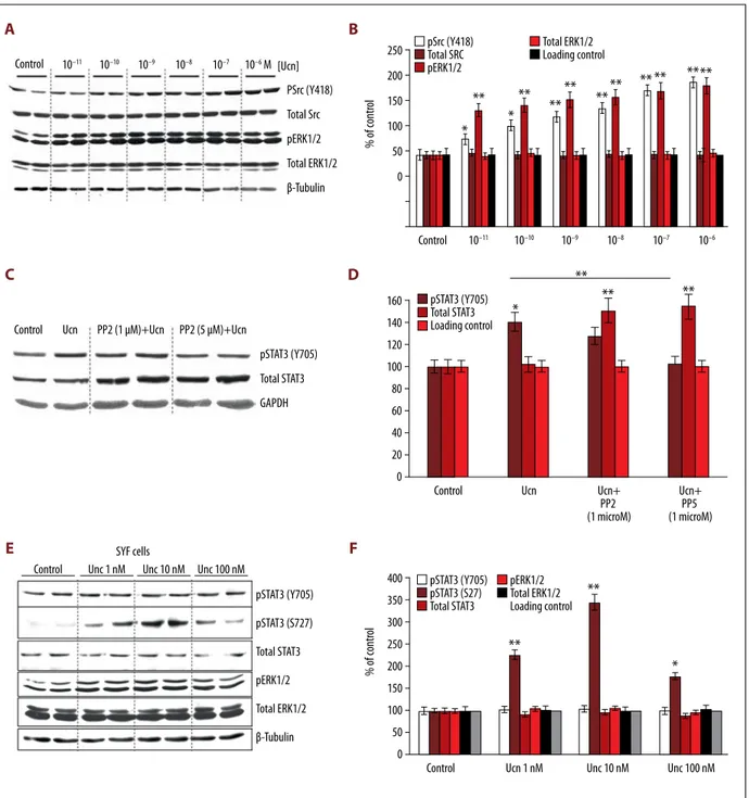
used pharmacological inhibition of these signaling pathways to identify their contribution to Ucn-mediated STAT3 phosphory-lation. To this end, HL-1 cells were incubated with Ucn (10 nM) for 30 min in presence or absence of AG490 (a potent JAK2/ JAK3 inhibitor) and PD98059 (a MEK1 inhibitor), respectively. Western blot analysis showed that the potent JAK2/JAK3 in-hibitor AG490 (50 µM) reduced Ucn-induced phosphorylation of both Y705 and S727 residues of STAT3, while the MEK1 in-hibitor PD98059 (50 µM) abrogated S727 phosphorylation, but only partially reduced Y705 phosphorylation (Figure 6A, 6B). The cell lysate from HL-1 cells treated with leukemia inhibi-tory factor (LIF, 5 ng/ml) for 15 min served as a positive con-trol (Figure 6A, 6B). As expected, Ucn-induced phosphoryla-tion of ERK1/2 was inhibited by the MEK1 inhibitor PD98059 (Figure 6C, 6D). The total expression level of STAT3 and ERK1/2 did not change (Figure 6A–6D). Since AG490 potentially inhib-its EGFR and MAPK besides JAKs, a more sensitive and spe-cific inhibitor of JAK/STAT circuits, tetracyclic pyridone 6 (P6), was used to study the time course of JAK2 activation [32]. HL-1 cells were incubated with Ucn (10 nM) in presence or ab-sence of P6 (0.5 μmol/L). Of note, P6 inhibited Y705 phosphor-ylation after 15 and 30 min of Ucn treatment (Figure 6E, 6F). Collectively, our findings indicate that Ucn-induced JAK2 acti-vation is implicated in the phosphorylation of both Y705 and S727, while Ucn-induced ERK1/2 activation also contributes to phosphorylation of S727.
We recently reported that Src, a non-receptor protein tyro-sine kinase, is involved in Ucn signaling in a CRF receptor-dependent manner and contributes to Ucn-mediated cardiac cell survival [16]. Since Src was shown to directly phosphory-late the Y705 residue of STAT3 to enhance its DNA binding af-finity [18,20], we next tested whether Src is involved in Ucn-induced phosphorylation of Y705. The phosphorylation of Y418 in the kinase domain of Src increased with increasing concen-trations of Ucn (10–10 M to 10–6 M) (Figure 7A, 7B). Likewise, phosphorylation of ERK1/2 progressively increased, while the expression levels of total Src and total ERK1/2 remained un-changed (Figure 7A, 7B). Importantly, in HL-1 cells treated with Ucn (10 nM) for 30 min, a specific Src family kinase inhibitor, PP2 (5 µM), significantly reduced Ucn-induced phosphorylation of STAT3 (Y705) (Figure 7C, 7D). However, since PP2 enhanced the expression of total STAT3 (Figure 7C, 7D), we tested the ability of Src to modulate Ucn-induced STAT3 activation in SYF cells, a fibroblast cell line derived from mouse embryos harbor-ing a null mutation of Src. As shown in Figure 7E, 7F, 15 min of Ucn treatment at any concentration failed to induce phos-phorylation of STAT3 (Y705) in SYF cells, while phosphoryla-tion of S727 residue was maximally stimulated at the 10 nM Ucn concentration. These findings revealed that Src mainly contributes to the phosphorylation of Y705, while JAK/gp130, and likely other signaling proteins, are involved in the phos-phorylation of S727 via activation of ERK1/2.
Free-DNA probe A HL-1 Control B HL-1 Unc 30’ C Super-shift STAT3 Non-specific binding Anti-STAT3 before EMSA D
Figure 3. Electrophoretic mobility shift assay (EMSA) and super-shift assay of STAT3 of 5 μg nuclear extract of each sample. Ucn significantly increased STAT3 binding to DNA probe (lane C). In addition, the Ucn-induced DNA-STAT3 complex could be partially super-shifted by pre-incubation with anti-STAT3 antibody before EMSA (lane D). Lane A shows free DNA probe only. Lane B shows the nuclear extracts from untreated HL-1 cells (Con – Control). Lane C shows nuclear extracts from HL-1 cells treated with Ucn (10 nM, for 30 min). Lane
D shows nuclear extracts from HL-1 cells treated with
Ucn (10 nM, for 30 min) and incubated with an anti-STAT3 antibody before EMSA. See Results section for further description
A
Control 1 hour 6 hours 12 hours 18 hours 24 hours 100 90 80 70 60 50 40 30 20 10 0 Concentration (pg/mL)
Unc-inducet relase of IL-6
Control
Control
HDAC1
Unc 5’ Unc 15’ Unc 30’ Unc 30’ +IL-6 Ab
Unc 30’ Unc 30’+IL-6 Ab
Positive control (HepG2) pSTAT3 (Y705) Loading control
*
**
**
200 180 160 140 120 100 80 60 40 20 0Control Unc 5’ Unc 15’ Unc 30’ Unc 30’ +IL-6 Ab Control Unc 5’ Unc 15’ Unc 30’ Unc 30’
+IL-6 Ab pSTAT3 (Y705) Total STAT3 Loading control pSTAT3 (Y705) Total STAT3 β-tubulin
*
**
160 140 120 100 80 60 40 20 0B
D
C
E
Figure 4. Ucn induced IL-6 release and IL-6-mediated phosphorylation/nuclear translocation of STAT3 (Y705). (A) ELISA test showing the temporal release of IL-6 in the culture medium of HL-1 cells treated with Ucn 10 nM for 30 min. IL-6 release started after 1 h, peaked at 12 h, and declined thereafter, although remaining detectable at 24 h. Data are presented as the average of 3 independent experiments. (B) A TransAM STAT3 assay was used in HL-1 nuclear extracts to detect and quantify nuclear translocation of p-STAT3. Incubation of HL-1 cells with Ucn (10 nM) increased phosphorylation and nuclear translocation of STAT3 (Y705) in a time-dependent manner. Nuclear extracts from HepG2 cells treated with IL-6 (100 ng/ml) were used as positive control. Use of an anti-IL-6 antibody to neutralize the effects of Ucn-induced IL6 release decreased significantly STAT3 phosphorylation at Y705. * p<0.05, ** p<0.01. (C) A representative blot probing pSTAT3 (Y705) in the nuclear extracts from HL-1 cells tested in the TransAM assay is shown. (D) Western blot analysis of whole lysates of HL-1 cells treated with Ucn (10 nM) for increasing incubation times (5, 15, and 30 min) confirmed that neutralization of secreted IL-6 (using an anti-IL-6 antibody) was sufficient to block STAT3 phosphorylation at Y705, which peaked at 30 min. Overall expression of total STAT3 was unaffected. * p<0.05, ** p<0.01. (E) Corresponding clustered column graph of the Western blot shown in Figure 4D. Data are expressed as% of control. * p<0.05, ** P<0.01.
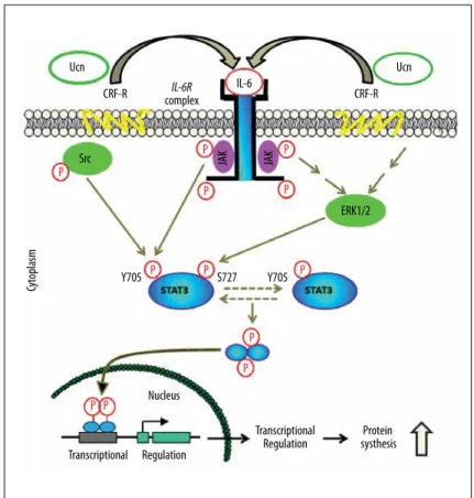
A scheme of Ucn-induced activation of STAT3 via JAK/ERK/Src signal cross-talk is summarized in Figure 8.
Discussion
Our study shows that short-term treatment with Ucn stimu-lates a JAK2/STAT3 signaling circuit in HL-1 cardiomyocytes and phosphorylates distinct residues of STAT3 via different signal pathways.
Previously, we demonstrated that Ucn treatment results in rapid phosphorylation of a non-receptor tyrosine kinase, Src. We also documented that Ucn-activated Src is an upstream modulator of ERK1/2 activation and plays a pivotal role in Ucn-mediated cardioprotection [16]. Besides confirming the above-mentioned findings, our study also showed that Src exclusively contributes to phosphorylation of the Y705 resi-due of STAT3. Furthermore, using the STAT3 transcription fac-tor assay, we showed that Ucn-induced nuclear translocation of STAT3 was partially reduced by a neutralizing IL-6 antibody, indicating that STAT3 activation is mediated by released IL-6 via an autocrine/paracrine mechanism.
IL-6 also activates JAK2. Transactivation of JAK2 by IL-6 is the key modulator in phosphorylation of both residues (Y705
and S727) of STAT3, whereas Ucn-stimulated ERK1/2 mainly phosphorylates the S727 residue of STAT3. The JAK kinases are non-receptor tyrosine kinases generally associated with a number of cytokine receptors also expressed in cardiac my-ocytes [25]. Upon ligand-induced cytokine receptor activation, JAK2 kinase becomes phosphorylated at the tandem tyrosine residues (Y1007/Y1008) in a kinase activation loop and phos-phorylates its associated receptor to provide p-tyrosine for STAT3 and other signaling proteins [20]. In agreement with the literature, tyrosine-phosphorylated JAK2 was markedly in-creased after about 30 min of incubation with Ucn, whereas pretreatment with cycloheximide and AG490 abolished phos-phorylation. This indicates that specific activation of JAK2 by Ucn is mediated through new protein synthesis. Since the re-lease of IL6 in the culture medium of HL-1 cells treated with Ucn (10 nM for 30 minutes) peaked at 12 h, remaining de-tectable even at 24 h, it follows that Ucn-induced phosphory-lation of STAT3 at Y705 is more sustained than that at S727. Considering that Src activation by Ucn lasted only 15 min [16], we have reason to believe that STAT3 phosphorylation at Y705 is slow and long-lasting when triggered by the IL-6/JAK2/STAT3 pathway, and is usually rapid when induced via the CRF-R1/ Src/STAT3 pathway.
Nuclear translocation and DNA binding of STAT3 require phosphorylation of Y705 residue and are independent of
JAK1 NegCon Control Unc
Total JAK2 IP: JAK2 IB (pY100) CHX Unc 25 kDa 150 kDa 100 kDa 75 kDa
JAK2 JAK3 Tyk2
Neg Con Lysate
Control AG490Unc
Total JAK2 IP: JAK2 IB (pY100) Unc
A
B
C
Figure 5. JAK2 expression in HL-1 cells and Ucn-induced tyrosine phosphorylation of JAK2. (A) Western blot analysis of HL-1 cell lysates with antibodies against all 4 members of the Janus family of tyrosine kinases shows that JAK2 is the highly expressed. (B, C) Immunoprecipitation of JAK2 followed by Western blot analysis using an anti-P-tyrosine (pY100) monoclonal Ab and polyclonal anti-JAK2 is shown. In Bupper panels, lysates from HL-1 cells, either untreated (CON) or treated with Ucn 10 nM for 30 min, in presence or absence of cycloheximide 100 μg/ml (CHX). In Cupper panels lysates from HL-1 cells, either untreated (CON), or treated with Ucn 10 nM (with or without pretreatment with AG490 10 μM), prior to being immunoprecipitated with anti-JAK2. Negative control (Neg Con) was prepared as the control, except for use for immunoprecipitation of the anti-JAK2 antibody. In the lower panels, the same membrane from the upper panel was stripped of antibodies and probed for total JAK2, which served as a loading control. A non-precipitated HL-1 cell lysate (non-stimulated control) was used as a positive control in C left column. See Results section for description.
Control pSTAT3 (Y705) pSTAT3 (S727) Total STAT3 β-tubulin Ucn 2 h Control Ucn LIF
Ucn+
PD9809 AG490Unc+ LIF pSTAT3 (Y705)
pSTAT3 (S727) Total STAT3Loading control
*
* *
**
**
**
160 140 120 100 80 60 40 20 0 PD98059Ucn AG490Ucn
pERK1/2 Total ERK1/2 GAPDH Control Ucn PD98059Ucn AG490Ucn LIF
Control Ucn Ucn+
PD9809 AG490Unc+ LIF pERK1/2 Total ERK1/2 Loading control
**
*
**
160 140 120 100 80 60 40 20 0 Control pSTAT3 (Y703) β-Tubulin Ucn 15’ Ucn 15’ +P6 Ucn 30’ Ucn 30’+P6 Control Ucn 15’ Ucn 15’+P6 Ucn 30’ Ucn 30’ +P6 pSTAT (Y703) Loading control
**
350 300 250 200 150 100 50 0*
A
C
E
B
D
F
Figure 6. Ucn-induced phosphorylation of Y705 and S727 residues of STAT3 via JAK and MAPK pathways. (A) Western blot analysis showing the inhibitory effects of AG490 (JAK2/JAK3 inhibitor) and PD98059 (MAPK kinase inhibitor) on Ucn-induced phosphorylation of Y705 and S727 residues of STAT3. HL-1 cells were incubated with Ucn (used at a concentration of 10 nM) for 30 min. The cell lysate from HL-1 cells treated with leukemia inhibitory factor (LIF, 5 ng/ml) for 15 min served as a positive control. (B) Corresponding clustered column graph of the Western blot shown in Figure 6A. Data are expressed as% of control. * p<0.05, ** P<0.01 (C) The inhibitory effects of PD98059 and AG490 on Ucn-induced activation of ERK1/2 are depicted by Western blot analysis. HL-1 cells were treated with Ucn 10 nM for 30 min. A blot of GAPDH is shown as a loading control. (D) Corresponding clustered column graph of the Western blot shown in Figure 6C. Data are expressed as% of control. * p<0.05, ** P<0.01. (E) Western blot of time course study showing the inhibitory effect of P6 (JAK inhibitor) on JAK2-mediated phosphorylation of STAT3 (Y705 residue) in HL-1 cells treated with Ucn (10 nM) for 15 and 30 min. (F) Corresponding clustered column graph of the Western blot presented in Figure 6E. Data are expressed as% of control. * p<0.05, ** P<0.01.
Control PSrc (Y418) Total Src pERK1/2 Total ERK1/2 β-Tubulin 10–11 10–10 10–9 10–8 10–7 10–6 M [Ucn] Control 10–11 10–10 10–9 10–8 10–7 10–6 pSrc (Y418) Total SRC pERK1/2 Total ERK1/2 Loading control
*
*
**
**
**
**
**
**
** **
****
250 200 150 100 50 0 % of contr olControl Ucn PP2 (1 μM)+Ucn PP2 (5 μM)+Ucn
Control Ucn Ucn+ PP2 (1 microM) Ucn+ PP5 (1 microM) pSTAT3 (Y705) Total STAT3 Loading control pSTAT3 (Y705) Total STAT3 GAPDH
**
**
**
*
160 140 120 100 80 60 40 20 0 Control pSTAT3 (Y705) pSTAT3 (S727) Total STAT3 pERK1/2 Total ERK1/2 β-TubulinControl Ucn 1 nM Unc 10 nM Unc 100 nM pSTAT3 (Y705) pSTAT3 (S27) Total STAT3 pERK1/2 Total ERK1/2 Loading control
*
**
**
400 350 300 250 200 150 100 50 0 % of contr ol Unc 1 nM SYF cells Unc 10 nM Unc 100 nMA
C
E
B
D
F
Figure 7. ERK1/2 and Src activate STAT3 through phosphorylation of distinct residues. (A) Western blot showing that Ucn activates Src and ERK1/2 in a dose-dependent manner in HL-1 cells. The expression of total Src and total ERK1/2 remained unchanged. (B) Corresponding clustered column graph of the Western blot presented in Figure 7A. Data are expressed as% of control. * p<0.05, ** P<0.01. (C) Western blot showing the dose-dependent inhibitory effects of PP2 (a specific Src family kinase inhibitor) on Ucn-induced phosphorylation of STAT3 (Y705 residue). PP2 also strongly induced the expression of total STAT3. (D) Corresponding clustered column graph of the Western blot depicted in Figure 7C. Data are expressed as% of control. * p<0.05, ** P<0.01. (E) Western blot of SYF cells incubated with increasing concentrations of Ucn. Cell lysates were prepared for Western blot analysis using anti-pSTAT3 (Y705 and S727), anti-pERK1/2, anti-STAT3, and anti-ERK1/2 antibody. (F) Corresponding clustered column graph of the Western blot presented in Figure 7E. Data are expressed as% of control. * p<0.05, ** P<0.01.
Y705 ERK1/2 JAK JAK Src Cy toplasm Ucn Ucn IL-6 CRF-R complexIL-6R CRF-R Transcriptional Transcriptional
Regulation systhesisProtein Regulation Nucleus S727 P P P P P P P P P P P P Y705
Figure 8. Schematic picture summarizes the effects of Ucn-induced activation of STAT3 via JAK/ERK/Src signal cross-talk.
phosphorylation of S727 residue [19–21]. However, the contri-bution of phosphorylated S727 to regulation of Y705 phosphor-ylation in vivo is still not fully understood. Generally speaking, phosphorylation of a serine residue within the amino acid se-quence PMSP in the C-terminal transactivation domain enhances transcriptional activity of STAT3 when the Y705 residue is phosphorylated, but the regulation is promoter- and cell type-dependent [21]. For example, STAT3-S727-mediated activation of a transfected interferon regulatory factor-1 (IRF-1) promoter in response to interferon-a was reduced by about 50% when the serine was mutated to an alanine (S727A) [33]. On the con-trary, a transfected STAT3-S727A mutant induced an increase in EGF-induced tyrosine phosphorylation of Y705 compared to wild-type STAT3, suggesting that S727 phosphorylation either increases STAT3 tyrosine phosphorylation or inhibits it [21]. The downregulation of STAT3 tyrosine phosphorylation was through a MAPK pathway and might be independent of S727 phosphorylation in some cases. In cardiomyocytes, phosphor-ylation of Y705 residue and activation of STAT3 transcriptional activity by the IL-6 family of cytokines, including IL-6, LIF, and cardiotrophin-1 (CT-1), were attenuated by endothelin-1 and by angiotensin II pretreatment in an ERK-dependent manner, and were relevant to cardiac hypertrophy and acute inflamma-tory responses [34,35]. Temporal profiles of opposite patterns of STAT3 activation were compared with each other: the IL-6 family of cytokines induced rapid and sustained STAT3 (Y705) phosphorylation; endothelin-1 produced a delayed and modest
increase of STAT3 (Y705) phosphorylation, preceded by a short-duration decrease; ERK-mediated S727 phosphorylation and Y705 dephosphorylation increased with time and peaked af-ter 30 min of treatment with angiotensin II; and endothelin-1 and angiotensin II rapidly induced high levels of ERK1/2 acti-vation, which contrasted with the delayed and weak activation of ERK1/2 caused by IL-6 [34–36]. We showed that in HL-1 car-diomyocytes treated with Ucn phosphorylation of Y705 and S727 were both enhanced after 15 min of incubation, without an initial decrease. MEK1 inhibitor (PD98059) significantly duced ERK-mediated S727 phosphorylation, and partially re-duced Y705 phosphorylation. This indicates that ERK1/2 activa-tion and phosphorylaactiva-tion of S727 did not downregulate STAT3 tyrosine phosphorylation. Because Ucn can induce ERK1/2 ac-tivation through multiple signal pathways in a CRF receptor-de-pendent manner [16], in the present study we tested and con-firmed JAK/gp130-mediated ERK1/2 activation.
JAK/STAT signaling has been implicated in cardiac pathophysi-ology, such as pressure overload-induced cardiac hypertrophy and remodeling, ischemic preconditioning, and ischemia/re-perfusion-induced cardiac dysfunction [25,37]. Different mem-bers of the STAT family linked with different receptor-mediated pathways (e.g., STAT1 and STAT3) exert opposite effects on cell growth/survival or apoptosis of cardiomyocytes [17,23]. There are 3 families of proteins that inhibit the JAK/STAT circuit: Suppressor Of Cytokine Signaling (SOCS), Protein Inhibitors
of Activated STATs (PIAS), and SH2-containing Phosphatase (SHP-1). Upon activation, STAT3 binds to the promoter region of SOCS genes and upregulates transcription of the target genes. As a negative feed-back mechanism to IL-6 signaling, SOCS1 and SOCS3 negatively regulate JAK/STAT circuit by ei-ther direct binding to JAK (to inhibit recruitment of ATP and substrate) or by binding to JAK/gp130 signaling complex (to process ubiquitin-mediated degradation), or by competing with STATs for the docking sites at the activated receptors [18,25,38]. Ucn was shown to be released in vitro by rat and human cardiac cells exposed to I/R injury [11,15] and to exert anti-apoptotic effects by binding to CRF receptors [12, 13]. Two CRF receptors – CRF-R1 and CRF-R2 – had been reported to be pres-ent in cardiac muscle [2]. HL-1 cells expressed both CRF-R1 and CRF-R2b as evaluated by Western blot analysis, as well as RT-PCR [16]. SYF cells are a mouse fibroblast cell line derived from mouse embryos harboring null mutations in both alleles of Src family tyrosine kinase Src, Yes, and Fyn [39]. We showed that in SYF cells, Ucn was able to weakly activate ERK1/2. S727, but not Y705 residue of STAT3, was phosphorylated by Ucn in SYF cells, and the peak activation was at 10 nM of Ucn, similar to that of ERK1/2. This indicates that Ucn induced phosphoryla-tion of STAT3 residues (S727 and Y705) through activaphosphoryla-tion of different signaling pathways, and that ERK1/2 mainly contrib-uted to phosphorylation of S727 residue. In the literature, there is evidence of S727-dependent and Y705-independent activa-tion of STAT3, but most studies showed that phosphorylaactiva-tion of Y705 residue is required for nuclear translocation and DNA binding of STAT3 [22,25]. Further investigation is needed to define the mechanism by which STAT3 is activated in a S727-dependent and Y705-inS727-dependent manner in SYF cells, and to explore the role of JAK in mediating S727 phosphorylation via activation of ERK1/2.
Although Ucn was shown to be a potent cardioprotective agent, recent evidence suggests that it might be involved in inflamma-tory response by stimulating the IL-6 family of cytokines and
by increasing microvascular permeability [27,28,40]. Our data showed that Ucn increased the release of IL-6 from HL-1 car-diomyocytes in a time-dependent fashion. IL-6 is an important mediator of the acute inflammatory response, exerting dual effects on inflammation [24]. Cureton et al. demonstrated that Ucn, as a pro-inflammatory agent, caused a significant rise in microvascular permeability (by about 2-fold), which was fur-ther increased (by about 7-fold) in presence of LPS [27]. On the contrary, in 2 mouse models of septic shock, Ucn administra-tion downregulated immune responses in local and systemic inflammation by reducing the release of inflammatory medi-ators from macrophages and preventing infiltration of inflam-matory cells [41]. Likewise, a more recent study showed that Ucn reduced the mortality rate in a mouse model of sepsis by reducing the release, as well as the systemic levels, of the high mobility group box 1 protein (HMGB1), which is a late inflam-matory mediator of severe sepsis [42]. Since the interplay of pro- and anti-inflammatory effects of Ucn is still unclear, the future therapeutic applications of Ucn for diverse heart condi-tions are the focus of several ongoing investigacondi-tions.
Conclusions
In conclusion, we showed that Ucn treatment resulted in rapid phosphorylation of JAK2, which was impeded by the protein synthesis inhibitor cycloheximide, as well as the JAK inhibi-tor AG490. Ucn treatment induced STAT3 phosphorylation at Y705 and S727 through transactivation of JAK2 in an IL-6 de-pendent manner, without affecting STAT1 activity. Ucn caused STAT3 S727 phosphorylation through ERK1/2, while Y705 phos-phorylation was achieved through Src tyrosine kinase. In line with this finding, Ucn failed to induce phosphorylation of Y705 residue in SYF cells carrying null mutation of Src, while phos-phorylation of S727 residue was unchanged. Understanding of the signaling pathways leading to Ucn-induced STAT3 activa-tion is important to exploit the full potential of Ucn-mediated cardioprotection.
References:
1. Basman C, Agrawal P, Knight R et al: Cardioprotective utility of urocortin in myocardial ischemia – reperfusion injury: Where do we stand? Curr Mol Pharmacol, 2018; 11(1): 32–38
2. Perrin MH, Vale WW: Corticotropin releasing factor receptors and their li-gand family. Ann NY Acad Sci, 1999; 885: 312–28
3. Fekete EM, Zorrilla EP: Physiology, pharmacology, and therapeutic relevance of urocortins in mammals: Aancient CRF paralogs. Front Neuroendocrinol, 2007; 28(1): 1–27
4. Axelrod J, Reisine TD: Stress hormones: Their interaction and regulation. Science, 1984; 224(4648): 452–59
5. Vaughan J, Donaldson C, Bittencourt J et al: Urocortin, a mammalian neu-ropeptide related to fish urotensin I and to corticotropin-releasing factor. Nature, 1995; 378(6554): 287–92
6. Díaz I, Smani T: New insights into the mechanisms underlying vascular and cardiac effects of urocortin. Curr Vasc Pharmacol, 2013; 11(4): 457–64
7. Kalantaridou SN, Zoumakis E, Makrigiannakis A et al: Corticotropin-releasing hormone, stress and human reproduction: an update. J Reprod Immunol, 2010; 85(1): 33–39
8. Perrin M, Donaldson C, Chen R et al: Identification of a second corticotro-pin-releasing factor receptor gene and characterization of a cDNA expressed in heart. Proc Natl Acad Sci USA, 1995; 92(7): 2969–73
9. Scarabelli TM, Knight R, Stephanou A et al: Clinical implications of apopto-sis in ischemic myocardium. Curr Probl Cardiol, 2006; 31(3): 181–264 10. Brar BK, Stephanou A, Okosi A et al: CRH-like peptides protect cardiac
my-ocytes from lethal ischaemic injury. Mol Cell Endocrinol, 1999; 158(1–2): 55–63
11. Chen-Scarabelli C, Faggian G, Yuan Z et al: Warm-blood cardioplegic arrest induces selective mitochondrial translocation of protein kinase Cepsilon followed by interaction with 6.1 inwardly rectifying potassium channel sub-unit in viable myocytes overexpressing urocortin. J Thorac Cardiovasc Surg, 2009; 138(5): 1213–21
12. Brar BK, Jonassen AK, Stephanou A et al: Urocortin protects against isch-emic and reperfusion injury via a MAPK-dependent pathway. J Biol Chem, 2000; 275(12): 8508–14
13. Scarabelli TM, Pasini E, Stephanou A et al: Urocortin promotes hemody-namic and bioenergetic recovery and improves cell survival in the isolated rat heart exposed to ischemia/reperfusion. J Am Coll Cardiol, 2002; 40(1): 155–61
14. Brar BK, Stephanou A, Knight R, Latchman DS: Activation of protein kinase B/Akt by urocortin is essential for its ability to protect cardiac cells against hypoxia/reoxygenation-induced cell death. J Mol Cell Cardiol, 2002; 34(4): 483–92
15. Lawrence KM, Chanalaris A, Scarabelli T et al: K(ATP) channel gene ex-pression is induced by urocortin and mediates its cardioprotective effect. Circulation, 2002; 106(12): 1556–62
16. Yuan Z, McCauley R, Chen-Scarabelli C et al: Activation of Src protein tyro-sine kinase plays an essential role in urocortin-mediated cardioprotection. Mol Cell Endocrinol, 2010; 325(1–2): 1–7
17. Knight RA, Scarabelli TM, Stephanou A: STAT transcription in the ischemic heart. JAKSTAT, 2012; 1(2): 111–17
18. Levy DE, Darnell JE Jr.: Stats: Transcriptional control and biological impact. Nat Rev Mol Cell Biol, 2002; 3(9): 651–62
19. Meier JA, Larner AC: Toward a new STATe: The role of STATs in mitochon-drial function. Semin Immunol, 2014; 26(1): 20–28
20. Kurdi M, Booz GW: Can the protective actions of JAK-STAT in the heart be exploited therapeutically? Parsing the regulation of interleukin-6-type cy-tokine signaling. J Cardiovasc Pharmacol, 2007; 50(2): 126–41
21. Decker T, Kovarik P: Serine phosphorylation of STATs. Oncogene, 2000; 19(21): 2628–37
22. Kim JH, Yoon MS, Chen J: Signal transducer and activator of transcription 3 (STAT3) mediates amino acid inhibition of insulin signaling through ser-ine 727 phosphorylation. J Biol Chem, 2009; 284(51): 35425–32 23. Barry SP, Townsend PA, Latchman DS, Stephanou A: Role of the JAK-STAT
pathway in myocardial injury. Trends Mol Med, 2007; 13(2): 82–89 24. Schaper F, Rose-John S: Interleukin-6: Biology, signaling and strategies of
blockade. Cytokine Growth Factor Rev, 2015; 26(5): 475–87
25. Boengler K, Hilfiker-Kleiner D, Drexler H et al: The myocardial JAK/STAT pathway: From protection to failure. Pharmacol Ther, 2008; 120(2): 172–85 26. Kohno M, Kawahito Y, Tsubouchi Y et al: Urocortin expression in synovium
of patients with rheumatoid arthritis and osteoarthritis: Relation to inflam-matory activity. J Clin Endocrinol Metab, 2001; 86(9): 4344–52 27. Cureton EL, Ereso AQ, Victorino GP et al: Local secretion of urocortin 1
pro-motes microvascular permeability during lipopolysaccharide-induced in-flammation. Endocrinology, 2009; 150(12): 5428–37
28. Huang M, Kempuraj D, Papadopoulou N et al: Urocortin induces interleu-kin-6 release from rat cardiomyocytes through p38 MAP kinase, ERK and NF-kappaB activation. J Mol Endocrinol, 2009; 42(5): 397–405
29. Pan W, Tu H, Hsuchou H et al: Unexpected amplification of leptin-induced Stat3 signaling by urocortin: Implications for obesity. J Mol Neurosci, 2007; 33(3): 232–38
30. Wang LH, Kirken RA, Erwin RA et al: JAK3, STAT, and MAPK signaling path-ways as novel molecular targets for the tyrphostin AG-490 regulation of IL-2-mediated T cell response. J Immunol, 1999; 162(7): 3897–904 31. Turner NA, Das A, Warburton P et al: Interleukin-1alpha stimulates
proin-flammatory cytokine expression in human cardiac myofibroblasts. Am J Physiol Heart Circ Physiol, 2009; 297(3): H1117–27
32. Pedranzini L, Dechow T, Berishaj M et al: Pyridone 6, a pan-Janus-activat-ed kinase inhibitor, induces growth inhibition of multiple myeloma cells. Cancer Res, 2006; 66(19): 9714–21
33. Wen Z, Zhong Z, Darnell JE Jr.: Maximal activation of transcription by Stat1 and Stat3 requires both tyrosine and serine phosphorylation. Cell, 1995; 82(2): 241–50
34. Booz GW, Day JN, Speth R, Baker KM: Cytokine G-protein signaling cross-talk in cardiomyocytes: Attenuation of Jak-STAT activation by endothelin-1. Mol Cell Biochem. 2002; 240(1–2): 39–46
35. Bhat GJ, Abraham ST, Baker KM: Angiotensin II interferes with interleukin 6-induced Stat3 signaling by a pathway involving mitogen-activated pro-tein kinase kinase 1. J Biol Chem, 1996; 271(37): 22447–52
36. Booz GW, Day JN, Baker KM: Angiotensin II effects on STAT3 phosphoryla-tion in cardiomyocytes: Evidence for Erk-dependent Tyr705 dephosphory-lation. Basic Res Cardiol, 2003; 98(1): 33–38
37. Wincewicz A, Sulkowski S: Stat proteins as intracellular regulators of re-sistance to myocardial injury in the context of cardiac remodeling and tar-geting for therapy. Adv Clin Exp Med, 2017; 26(4): 703–8
38. Heppler LN, Frank DA: Targeting oncogenic transcription factors: Therapeutic implications of endogenous STAT inhibitors. Trends Cancer, 2017; 3(12): 816–27
39. Parker BM, Wertz SL, Pollard CM et al: Novel insights into the crosstalk be-tween mineralocorticoid receptor and G protein-coupled receptors in heart adverse remodeling and disease. Int J Mol Sci, 2018; 19(12): pii: E3764 40. Janjua S, Lawrence KM, Ng LL, Latchman DS: The cardioprotective agent
uro-cortin induces expression of CT-1. Cardiovasc Toxicol, 2003; 3(3): 255–62 41. Gonzalez-Rey E, Chorny A, Varela N et al: Urocortin and adrenomedullin
pre-vent lethal endotoxemia by down-regulating the inflammatory response. Am J Pathol, 2006; 168(6): 1921–30
42. Chorny A, Delgado M: Neuropeptides rescue mice from lethal sepsis by down-regulating secretion of the late-acting inflammatory mediator high mobility group box 1. Am J Pathol, 2008; 172(5): 1297–307
![Figure 2. Time-dependent effects of Ucn on STAT3 phosphorylation in HL-1 cells. ( A ) Representative blots of the time-dependent effects of Ucn [10 nM] using anti-pSTAT3 (Y705 and S727) and total anti-STAT3 polyclonal antibodies](https://thumb-eu.123doks.com/thumbv2/123dokorg/5555696.66033/6.850.81.781.128.889/figure-dependent-phosphorylation-representative-dependent-effects-polyclonal-antibodies.webp)