123
Key Concepts from the Sixth
THNO Meeting
Jan B. Vermorken
Volker Budach
C. René Leemans
Jean-Pascal Machiels
Piero Nicolai
Brian O’Sullivan
Editors
Critical Issues in Head
and Neck Oncology
Jan B. Vermorken • Volker Budach
C. René Leemans • Jean-Pascal Machiels
Piero Nicolai • Brian O’Sullivan
Editors
Critical Issues in Head and
Neck Oncology
ISBN 978-3-319-98853-5 ISBN 978-3-319-98854-2 (eBook)
https://doi.org/10.1007/978-3-319-98854-2
Library of Congress Control Number: 2018957856 © Springer Nature Switzerland AG 2018
This work is subject to copyright. All rights are reserved by the Publisher, whether the whole or part of the material is concerned, specifically the rights of translation, reprinting, reuse of illustrations, recitation, broadcasting, reproduction on microfilms or in any other physical way, and transmission or information storage and retrieval, electronic adaptation, computer software, or by similar or dissimilar methodology now known or hereafter developed.
The use of general descriptive names, registered names, trademarks, service marks, etc. in this publication does not imply, even in the absence of a specific statement, that such names are exempt from the relevant protective laws and regulations and therefore free for general use.
The publisher, the authors, and the editors are safe to assume that the advice and information in this book are believed to be true and accurate at the date of publication. Neither the publisher nor the authors or the editors give a warranty, express or implied, with respect to the material contained herein or for any errors or omissions that may have been made. The publisher remains neutral with regard to jurisdictional claims in published maps and institutional affiliations.
This Springer imprint is published by the registered company Springer Nature Switzerland AG The registered company address is: Gewerbestrasse 11, 6330 Cham, Switzerland
Jan B. Vermorken
Department of Medical Oncology Antwerp University Hospital Edegem, Belgium
C. René Leemans
Department of Otolaryngology VU University Medical Center Amsterdam, The Netherlands Piero Nicolai
Department of Otorhinolarynology University of Brescia
Brescia, Italy
Volker Budach
Department of Radiation Oncology Charité University Medicine Berlin, Germany
Jean-Pascal Machiels Institut Roi Albert II
Université Catholique de Louvain Louvain-la-Neuve, Belgium Brian O’Sullivan
Department of Radiation Oncology Princess Margaret Cancer Centre University of Toronto
v
Preface
The sixth Trends in Head and Neck Oncology (THNO-6) took place in the Meridien Hotel in Nice, France, November 2–4, 2017, and was organized by the same coor-dinating team as the fifth version with support of Pharma and practical logistical support of Congress Care. This time, the conference was endorsed by the European Head and Neck Society (EHNS) and the European Organization for Research and Treatment of Cancer (EORTC). As on previous occasions, the setup was educa-tional, with a multidisciplinary focus. Case presentations, organized by colleagues from the Centre Antoine Lacassagne in Nice and some members of the coordinating team, induced a lively interaction between faculty and audience and underlined the importance of individualized patient care. Thanks to the dedication of all faculty members this book will be available within a year following the actual meeting, guaranteeing the most up-to-date information in this rapidly evolving field. We are most grateful to all faculty members for their efforts in realizing this important goal.
Edegem, Belgium Jan B. Vermorken
Berlin, Germany Volker Budach
Amsterdam, The Netherlands C. René Leemans
Louvain-la-Neuve, Belgium Jean-Pascal Machiels
Brescia, Italy Piero Nicolai
vii
Part I Epidemiology and Diagnosis
The Role of Vaccination in the Prevention of Head and Neck Cancer. . . . 3
Johannes Berkhof
Cellular and Molecular Pathology in Head and Neck Cancer . . . 15
Phil Sloan and Max Robinson
Part II Prediction of Outcome
Oncogenomics/Proteomics of Head and Neck Cancer. . . 29
Ruud H. Brakenhoff
Targeted Next-Generation Sequencing in Head and Neck Cancer . . . 37
Ingeborg Tinhofer
Homologous Recombination Repair Function as a
Predictor of Treatment Response . . . 51
Kevin J. Harrington
Part III Oral Cavity Cancer
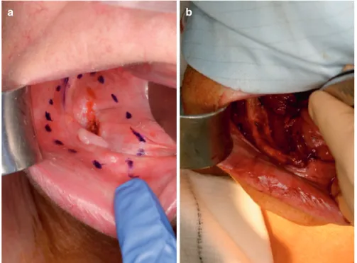
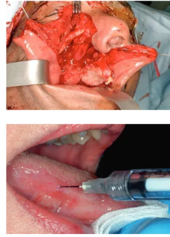
Surgical Management of Oral Cavity Cancer . . . 67
C. René Leemans and Sat Parmar
Reconstruction in the Oral Cavity: When and How . . . 75
Jim Higginson, Prav Praveen, Tim Martin, and Sat Parmar
Induction Chemotherapy:
Does It Have a Place in Oral Cavity Cancer? . . . 99
Jean-Pascal Machiels
Alternative Local Treatment in Oral Cavity Cancer:
Photodynamic Therapy . . . 105
viii
Part IV Oropharynx Cancer
HPV Assessment in Oropharynx Cancer: What is the Gold Standard? . . 119
Panagiota Economopoulou, Ioannis Kotsantis, and Amanda Psyrri
A New Staging System for HPV-Related Oropharynx Cancer:
Rationale, Derivation, Validation and Practical Applications . . . 149
Shao Hui Huang, Zhi-Jian Chen, and Brian O’Sullivan
Should We De-escalate Treatment for HPV
Positive Oropharyngeal Head and Neck Cancer? . . . 165
Hisham Mehanna, Jennifer L. Bryant, and Anthony H. Kong
Is There a New Role for Surgery in Oropharynx Cancer? . . . 171
Yann Litzistorf and Christian Simon
Can We Expect Less Toxicities with Newer Forms of Radiotherapy? . . . . 181
Volker Budach and Alexander Thieme
Part V Miscellaneous Topics
What Is the Optimal Larynx Preservation Approach and
Who Are the Candidates?. . . 215
Jean Louis Lefebvre
Is the Approach to Patients with Unknown Primary
Tumor any Different in 2018? . . . 227
Nausica Montalto, Francesca Del Bon, Alberto Paderno, Riccardo Morello, and Piero Nicolai
Cytotoxic Chemotherapy and Targeted Therapy in
Nasopharyngeal Cancer . . . 251
Jonathan Pan, Jennifer Johnson, and Athanassios Argiris
Are There Alternative Chemotherapy Regimens for
EXTREME in Recurrent/Metastatic Squamous Cell Carcinoma of the Head and Neck (R/M-SCCHN)? . . . 267
Joël Guigay
New Data on Systemic Therapy of Salivary Gland Tumors . . . 277
Salvatore Alfieri and Lisa Licitra
Treatment of Elderly Patients with Head and Neck Cancer . . . 285
Petr Szturz and Jan B. Vermorken
Part VI Thyroid Cancer
Worldwide Thyroid Cancer “Epidemic”: What Is Going On? . . . 311
Salvatore Vaccarella
Surgical Approach to Thyroid Cancer . . . 319
John Cramer and Robert L. Ferris
Part VII Keynote Address
Tumor Immunology, Immunotherapy and Its Application to
Head and Neck Squamous Cell Carcinoma (HNSCC) . . . 341
Jessica M. Moskovitz and Robert L. Ferris
Part I
3 © Springer Nature Switzerland AG 2018
J. B. Vermorken et al. (eds.), Critical Issues in Head and Neck Oncology, https://doi.org/10.1007/978-3-319-98854-2_1
of Head and Neck Cancer
Johannes Berkhof
Introduction
Human papillomavirus (HPV) is the main cause of cervical cancer and also causes a substantial number of cancers at other sites. It was recently estimated that approxi-mately 29,000 oropharyngeal cancers and 8000 oral cavity and larynx cancers, occurring globally in year 2012, could be attributed to HPV and that about 80% of HPV-related head and neck cancer cases occurred in men [1]. Besides, an upward surge in HPV-associated oropharyngeal cancer has been observed in the United States (US) and some European countries in the last years, in particular in males [2–5]. US projections indicate that in 2020, oropharyngeal cancer will occur more frequently than cervical cancer [2]. The disproportionate burden and rising inci-dence of HPV-associated head and neck cancers in men has ignited discussion on the vaccination of boys. So far, most countries with a publicly funded HPV vaccina-tion programme have targeted girls only since the main focus is on prevenvaccina-tion of cervical cancer. The HPV-related burden in men is nowadays being recognized but a long-standing debate exists on whether there is sufficient evidence on the effects of the vaccine against cancer in men and whether the effects are large enough to justify the extra costs of vaccinating boys. In the following, I give an overview of the current evidence on the efficacy and expected impact of HPV vaccination in men and women with a focus on oropharyngeal cancer.
J. Berkhof
Amsterdam UMC, Vrije Universiteit Amsterdam, Epidemiology and Biostatistics, Amsterdam Public Health Research Institute, Amsterdam, The Netherlands
4
HPV Vaccines
There are three HPV vaccines on the market registered for use from the age of 9 years. The vaccines are licensed for the prevention of lesions in the cervix, vulva, vagina, and anus, but not for the prevention of head and neck cancers. Cervarix® (GSK) is a
bivalent vaccine that protects against HPV16 and HPV18 infections and also provides some cross-protection against a few other oncogenic HPV types [6–8]. Cervarix is registered for females and males in Europe and only for females in the US. Gardasil®
(Merck & Co) is a quadrivalent vaccine that protects against HPV16 and HPV18 and also protects against HPV6 and HPV11, responsible for most cases of genital warts and recurrent respiratory papillomatosis. Gardasil is registered for females and males in both Europe and the US. Since 2016, a nonavalent vaccine Gardasil9® (Merck &
Co) has become available with the main purpose to offer improved protection against cervical cancer and high-grade cervical dysplasia. For head and neck cancer preven-tion, the additional benefit of a nonavalent vaccine as compared to a bivalent or quad-rivalent vaccine is limited as HPV16 accounts for about 80% of the HPV DNA positive cases and for about 90% of HPV DNA positive oropharyngeal cancers [9].
Early End-Points
The main reason that the current HPV vaccines are not licensed for the prevention of head and neck cancers is that clinical trials were only able to show an effect against cervical and other anogenital premalignant lesions [10–14]. Unlike anogeni-tal cancers, HPV-positive head and neck cancers have no clearly visible premalig-nant end-point thus their histopathological progression remains poorly defined [15]. Moreover, the mean age of HPV-positive oropharyngeal cancer is above 50 years [16] and this means that if regulatory bodies demand a significant effect on cancer incidences from trials targeting adolescents and young adults, vaccine licensure against oropharyngeal cancer will be postponed for another three to four decades.
The only alternative to showing an effect on dysplasia is to measure oral HPV infections. However, establishing a link from oral HPV infection to cancer and pre-cancer is hard, if not impossible. Studies will never be large enough to show an association between oral infection and invasive cancer. Furthermore, establishing a link between oral infection and subclinical dysplasia in healthy subjects seems ethi-cally unfeasible. Nevertheless, case-control studies have provided strong support that HPV exposure is necessary for HPV-positive oropharyngeal cancer [16, 17] and it is widely accepted that HPV-positive head and neck cancers cannot develop with-out a preceding HPV infection.
The effect of HPV vaccination on the occurrence of oral infections has recently been studied in two populations. Participants in those studies were asked to collect rinse and gargle samples using a mouthwash. The first population consisted of women participating in a randomized trial with the bivalent vaccine [18]. The use of
randomization has the advantage that it minimizes bias related to demographic dif-ferences between the vaccine and the control arm. The effect of vaccination on oral vaccine-type HPV infections was estimated at 93% (1/2910 in vaccinated women versus 15/2924 in unvaccinated women). The second population was the National Health and Nutrition Examination Survey (NHANES), a representative subset of the US population. Two cross-sectional analyses on NHANES indicated that the occurrence of oral quadrivalent vaccine-type HPV infections was about 90% lower in vaccinated as compared to unvaccinated men and women [19, 20]. Limitations of the NHANES population are that vaccine status is self-reported and that subjects are not randomized with respect to vaccination. Regarding the latter, vaccine-associated effects were robust against confounders such as age, sex, sexual behaviour, smok-ing, and race [20]. The decision to get vaccinated may have been influenced by fac-tors that were not measured, but it is unlikely that only unobserved confounders were responsible for the strong association between vaccination and oral infections. In another recent study, HPV16 and HPV18 specific antibodies in the oral mucosa of adult males were induced by vaccination, but the study was not able to demon-strate whether the antibody levels were sufficient to offer protection against incident infections [21]. To conclude, the current evidence on the effect of vaccines on infec-tions and vaccine-induced antibodies in the oral region seems sufficient to include oropharyngeal cancer in the impact and cost-effectiveness assessments of vaccina-tion strategies, but for vaccine licensure there is a need for more data on the effect of vaccination on infections and antibodies in the oral region.
Herd Effects
Most HPV vaccination programmes target girls because women experience the great-est HPV-related disease burden. Of all 630,000 new HPV-related cancers worldwide in 2016, 570,000 cases occurred in women [1]. Nevertheless, exclusion of boys from the programme has raised equity concerns because HPV-related cancers occur in, both, women and men. A widely used argument against sex-neutral vaccination is that vaccination of girls confers indirect protective effects or herd effects to men. This means that heterosexual men would be protected against HPV-associated diseases if the coverage of the girls’ only vaccination programme is high. The required coverage level of a girls’ only programme is, however, difficult to assess because herd effects depend on sexual network features and natural immunity after viral clearance [22].
For estimating herd effects, we usually rely on mathematical HPV infection models that describe the transmission of HPV in sexual networks. HPV infection models require many assumptions and can have a different architecture leading to uncertain and potentially inconsistent results. To study whether predictions pro-vided by independent models were consistent, in a recent study, sixteen independent modelling teams provided estimates of the reduction in HPV16 and HPV18 under different vaccine coverage scenarios [23]. The results from the modelling teams were strikingly consistent despite the fact that models were developed in different
6
settings and calibrated to different data. A main result was that at 80% coverage of a girls’ only programme, the HPV16 prevalence would decrease by 93% in women and by 83% in men. If both girls and boys were vaccinated with a coverage of 80%, HPV16 would virtually be eliminated from the population in most models. At a coverage of 60% among girls and boys, HPV16 would be reduced by 90%. Since the majority of immunization programmes shows coverage levels between 50 and 80%, sex-neutral vaccination is expected to reduce HPV16 and HPV18 prevalence to a very low level. Two important limitations of the models are that they only con-sider heterosexual networks and do not take differences in site-specific transmission into account. Those limitations are not likely to change the general message: sex-neutral vaccination can be important for reducing the prevalence of HPV to a very low level when a girls’ only programme fails to achieve a coverage similar to those observed for paediatric vaccines.
A number of studies have emerged that aim to measure herd effects in real life data. In an Australian study on men attending a sexual clinic after a positive test for
Chlamydia trachomatis [24], a significant reduction in the prevalence of the HPV types targeted by the quadrivalent vaccine from 18 to 7% was observed in Australian-born men before and after the start of the vaccination programme. In the last three calendar years of the study (2013–2015), the prevalence of the HPV types targeted by the vaccine was only 3%. As expected, no decrease in prevalence was observed for the HPV types that were not targeted by the vaccine. Another interesting study is a Finnish randomized trial where communities were either randomized to girls’ only or sex-neutral vaccination with the bivalent vaccine [25]. A herd effect for HPV18 in cervical samples was observed in both study arms. A herd effect was not observed for HPV16 which may be related to the low vaccine coverage of 20% among boys and 45% among girls attending junior high school. The larger herd effect for HPV18 as compared to HPV16 in the Finnish trial concurs with intuition because HPV16 has a higher basic reproductive number than other HPV types [26,
27]. This means that a subject infected with HPV16 infects on average a larger number of susceptible subjects than a subject infected with another HPV type and hence it becomes more difficult to eliminate HPV16 from the population.
In a few years, it will be possible to measure herd effects in nationwide cervical cancer screening registries provided they are linked to vaccination registries. This information will be very important when developing cervical cancer screening algo-rithms for vaccinated cohorts, but its value for head and neck cancer prevention will be limited because herd effects observed in cervical cancer screening are expected to be different from herd effects in future head and neck cancers. The difference will be most pronounced in countries with a girls’ only vaccination programme. Then, herd effects in unvaccinated women will be second-order indirect effects occurring because men have a lower probability of infecting unvaccinated women since they themselves will be indirectly protected by the girls’ only vaccination programme. Therefore, mathematical models will still be needed to estimate the reduction of HPV infections in men and to facilitate decision-making on sex-neutral vaccination.
The Effect of Vaccinating Boys on Cancer in Men
Although herd effects are important to reduce HPV infections in the general popula-tion, the question remains whether vaccination of men would contribute sufficiently to the prevention of cancer in men to justify a sex-neutral vaccination programme. In a Dutch evidence synthesis study conducted in 2015 [28], the effect of vaccinat-ing boys on future cancers in men was calculated. The cancers considered were cancers of the penis, anus, and anal canal, and squamous cell carcinomas of the oropharynx, including the base of tongue and tonsils (international classification of diseases 10th revision code C60, C21, and C01, C09 and C10). HPV aetiological fractions for the different tumour sites were obtained from several sources [29–31] and elevated cancer risks in homosexual and bisexual men (men having sex with men; MSM) as compared to heterosexual men were taken into account [32]. HPV-associated oral cavity and larynx carcinoma were not considered in this study because their burden is low relative to that of HPV-related oropharynx cancer [1]. The herd effects in men achieved when vaccinating girls only were estimated by a mathematical HPV transmission model [33]. The transmission model predicted that a 10% reduction of HPV16 or HPV18 among women would induce an 8% reduc-tion of HPV16 or HPV18 among men. After taking these herd effects into account, the conclusion of the evidence synthesis study [28] was that vaccination of boys would still confer a substantial reduction in future cancer in men. At 60% vaccine coverage among girls, about 800 boys would need to be vaccinated to prevent an additional future cancer in men. Tumour site specific numbers were about 2000 boys for oropharyngeal cancer and anal cancer and 3500 for penile cancer. When the coverage in girls was increased to 90%, tumour-specific numbers were about 6500 boys for oropharyngeal cancer, 2600 for anal cancer, and nearly 30,000 for penile cancer. In the latter situation, the majority of the cancers prevented by vac-cinating boys were anal cancers, which underscores the relevance of HPV vaccina-tion for cancer prevenvaccina-tion in MSM.
In a country with a girls’ only vaccination programme and a coverage of 90%, sex-neutral vaccination can still be motivated as a strategy to prevent cancer in MSM, but targeted vaccination of adolescent and adult MSM has also been sug-gested. Targeted MSM vaccination is less costly than sex-neutral vaccination, but it is not effective in the subset of the HPV-positive MSM. Considering that HPV infections occur soon after the initiation of sexual debut, concerns can be raised with respect to the effectiveness of strategies for early identification of sexually naïve MSMs. Nonetheless, a modelling study indicated that targeted MSM vaccina-tion may be cost-effective up to the age of 40 [34]. It is also important to understand that targeted MSM vaccination does not preclude sex-neutral vaccination and vice versa. After implementation of sex-neutral vaccination, targeted MSM vaccination may still be used as a catch-up for older age groups and as an option for MSM who spent their childhood in a different country.
8
Vaccination Coverage and Programme Resilience
Several modellers have pointed out that even when the coverage of a girls’ only vac-cination programme is low, it is more efficient to increase the uptake among girls than to vaccinate boys in order to reduce the HPV prevalence in the general popula-tion [35, 36]. This argument supports prioritization of efforts to increase the uptake among girls, but it is uncertain whether such efforts would be successful. So far, HPV vaccination programmes in most countries have achieved a coverage far below the 90% target level for paediatric vaccines. A main reason for the limited coverage among girls is that there are recurring concerns about vaccine safety and side effects [37, 38]. An alarming example is the HPV vaccination programme in Denmark where the vaccine coverage decreased from about 80 to 20% in 2015 as a result of an alleged association between HPV vaccine and Postural Orthostatic Tachycardia Syndrome (POTS) [39]. To assess whether these concerns are supported by data, the European Medicines Agency (EMA; www.ema.europa.eu) conducted a large study on the incidence of POTS and Complex Regional Pain Syndrome (CRPS). The EMA compared approximately 60,000 women vaccinated with Gardasil and 40,000 women vaccinated with Cervarix with placebo cohorts but did not find a significant association between adverse events and vaccination status. Besides, the POTS cases in Denmark were mainly observed in one centre suggesting considerable heteroge-neity in the diagnosis of POTS.
The results from the EMA are reassuring, but the Danish example clearly indi-cates that HPV vaccination programmes are vulnerable. The sudden sharp decline in vaccine coverage that has happened in Denmark may happen in any other country as well. Sex-neutral vaccination has been suggested to make programmes more resilient against temporary changes in the vaccination coverage. A recent modelling study predicted that if the vaccine coverage was halved for a period of 5 years, then a sex-neutral vaccination would be about 12-fold more resilient than girls’ only vac-cination in terms of the percentage reduction in HPV prevalence in the female popu-lation [40]. Therefore, as long as HPV vaccine coverage is unpredictable, sex-neutral vaccination may be implemented to stabilize the impact of the programme against temporary variations in coverage.
Economic Considerations
So far, cost-effectiveness studies on sex-neutral vaccination have not yielded consis-tent results. Although some studies were positive, most studies recommended against sex-neutral vaccination [41]. An explanation for this finding is that some economic studies did not consider all non-cervical health outcomes in their main analysis. In a recent review, it was calculated that the standard measure in cost-effectiveness studies, that is the incremental cost-cost-effectiveness ratio, would decrease 3.9-fold if all non-cervical disease had been taken into account, including oropha-ryngeal cancer and genital warts, as compared to cervical cancer only [41]. Of
course, negative results in cost-effectiveness studies are also strongly driven by the HPV transmission dynamics. A Canadian study illustrated that when herd effects from a girls’ only programme are ignored, vaccination of boys is cost-effective even when only prevention of oropharyngeal cancer in men is considered [42]. Another commonly mentioned obstacle for sex-neutral vaccination is the high list price of the HPV vaccine. Sex-neutral vaccination is unlikely to be cost-effective at the cur-rent list price of the vaccine which varies between 100 and 160 euros per dose in high-income countries. For the costs of vaccination, however, a widely used con-tainment strategy is tendering: health authorities use their purchasing power and the competition in the market of the vaccines to perform procurement procedures. This drives down the vaccine cost and enhances the sustainability of a programme. Experience with hepatitis B vaccines suggests that tendering may lead to strong price reductions over time [43]. Besides, in several Italian regions, tender-based HPV vaccine prices in the first 2 years after the vaccine became available were about 50% lower than the list price [44].
In a recent Dutch cost-effectiveness study [45], in which tender-based vaccine costs were set at about 65 euros for a 2 dose schedule and effects on cervical, vulvar, anal, penile, and oropharyngeal cancers were included, it was calculated that sex-neutral vaccination was cost-effective even when the coverage among girls increased up to 90%. Favourable cost-effectiveness results were also obtained in studies eval-uating the vaccination of boys in the Norwegian programme and in the Italian pro-gramme when tender vaccine prices were used instead of list prices [46, 47]. All three studies used local input and therefore conclusions do not have to apply to all high-resource settings. However, altogether these findings at least suggest that countries should re-evaluate their economic argument for adopting a girls’ only vac-cination programme, preferably together with an analysis accounting for country-specific disease burden, country-country-specific and often tender-based vaccine price, achieved vaccine coverage in the girls’ only programme, and the cost of administer-ing vaccines.
Conclusions
The decision to switch from girls only vaccination to sex-neutral vaccination is more difficult to take than the decision to implement a girls’ only vaccination pro-gramme. This is reflected in the speed at which decisions are taken. About 10 years ago, many countries implemented girls’ only vaccination within 1 or 2 years after registration of the vaccine, but only a few of them have switched to sex-neutral vac-cination in the meantime. A main argument used to support sex-neutral vaccination is that the burden of oropharyngeal cancer is disproportionate in men, but hetero-sexual men also benefit from the girls’ only programme via herd effects. To facili-tate decision-making, mathematical models have been used to assess the additional benefit and cost-effectiveness of vaccinating boys. With respect to sex-neutral vac-cination, four conclusions from the models in the literature were that: (1)
10
sex-neutral vaccination may lead to near elimination of HPV16 and HPV18 when coverage levels are about 80%, (2) a girls’ vaccination programme lowers the risk of cancer in men but sex-neutral vaccination still provides substantial extra protec-tion against oropharyngeal and anal cancer when the coverage among girls is mod-erate, (3) sex-neutral vaccination makes a vaccination programme more robust against sudden changes in vaccine coverage, and (4) sex-neutral vaccination is likely to be cost-effective provided a low vaccine price that is negotiated by health authorities.
Models can be criticized and model-based evidence is graded lower than evi-dence from randomized trials and cohort studies. Nevertheless, in the coming years, evidence from cohort studies is unlikely to change our current perspective on the effect of vaccination on disease in men. A decision on sex-neutral vaccination will inevitably be taken under a certain degree of uncertainty, but since the incidence of oropharyngeal cancers is currently on the rise in men, such a decision should be both sound and timely.
Acknowledgement JB has received consultancy fees from Roche, GlaxoSmithKline, and Merck/ SPMSD; these fees were collected by his employer. JB was supported by the Comparing Health Services Interventions for the Prevention of HPV-Related Cancer project, under European Commission FP7 Framework Health 2013 Innovation 1 (grant 603019). JB is grateful to Federica Inturrisi, Thomas Klausch and Venetia Qendri for suggestions and comments.
References
1. de Martel C, Plummer M, Vignat J, Franceschi S. Worldwide burden of cancer attributable to HPV by site, country and HPV type. Int J Cancer. 2017;141(4):664–70.
2. Chaturvedi AK, Engels EA, Pfeiffer RM, et al. Human papillomavirus and rising oropharyn-geal cancer incidence in the United States. J Clin Oncol. 2011;29(32):4294–301.
3. Marur S, D’Souza G, Westra WH, Forastiere AA. HPV-associated head and neck cancer: a virus-related cancer epidemic. Lancet Oncol. 2010;11(8):781–9.
4. McDonald SA, Qendri V, Berkhof J, de Melker HE, Bogaards JA. Disease burden of human papillomavirus infection in the Netherlands, 1989-2014: the gap between females and males is diminishing. Cancer Causes Control. 2017;28(3):203–14.
5. Carlander AF, Gronhoj Larsen C, et al. Continuing rise in oropharyngeal cancer in a high HPV prevalence area: a Danish population-based study from 2011 to 2014. Eur J Cancer. 2017;70:75–82.
6. Wheeler CM, Castellsagué X, Garland SM, et al. Cross-protective efficacy of HPV-16/18 AS04-adjuvanted vaccine against cervical infection and precancer caused by non-vaccine oncogenic HPV types: 4-year end-of-study analysis of the randomised, double-blind PATRICIA trial. Lancet Oncol. 2012;13(1):100–10.
7. Kavanagh K, Pollock KG, Cuschieri K, Palmer T, Cameron RL, Watt C, et al. Changes in the prevalence of human papillomavirus following a national bivalent human papillomavi-rus vaccination programme in Scotland: a 7-year cross-sectional study. Lancet Infect Dis. 2017;17(12):1293–302.
8. Woestenberg PJ, King AJ, van Benthem BHB, et al. Bivalent vaccine effectiveness against type-specific HPV positivity: evidence for cross-protection against oncogenic types among Dutch STI clinic visitors. J Infect Dis. 2018;217(2):213–22.
9. Ndiaye C, Mena M, Alemany L, et al. HPV DNA, E6/E7 mRNA, and p16INK4a detec-tion in head and neck cancers: a systematic review and meta-analysis. Lancet Oncol. 2014;15(12):1319–31.
10. Ault KA. Effect of prophylactic human papillomavirus L1 virus-like-particle vaccine on risk of cervical intraepithelial neoplasia grade 2, grade 3, and adenocarcinoma in situ: a combined analysis of four randomised clinical trials. Lancet. 2007;369(9576):1861–8.
11. Harper DM, Franco EL, Wheeler CM, et al. Sustained efficacy up to 4·5 years of a bivalent L1 virus-like particle vaccine against human papillomavirus types 16 and 18: follow-up from a randomised control trial. Lancet. 2006;367(9518):1247–55.
12. Lehtinen M, Paavonen J, Wheeler CM, Jaisamrarn U, Garland SM, Castellsagué X, et al. Overall efficacy of HPV-16/18 AS04-adjuvanted vaccine against grade 3 or greater cervi-cal intraepithelial neoplasia: 4-year end-of-study analysis of the randomised, double-blind PATRICIA trial. Lancet Oncol. 2012;13(1):89–99.
13. Garland SM, Hernandez-Avila M, Wheeler CM, et al. Quadrivalent vaccine against human papillomavirus to prevent anogenital diseases. N Engl J Med. 2007;356(19):1928–43. 14. Giuliano AR, Palefsky JM, Goldstone S, et al. Efficacy of quadrivalent HPV vaccine against
HPV infection and disease in males. N Engl J Med. 2011;364(5):401–11.
15. Forastiere A, Koch W, Trotti A, Sidransky D. Head and neck cancer. N Engl J Med. 2001;345(26):1890–900.
16. Gillison ML, D’Souza G, Westra W, et al. Distinct risk factor profiles for human papillomavi-rus type 16-positive and human papillomavipapillomavi-rus type 16-negative head and neck cancers. J Natl Cancer Inst. 2008;100(6):407–20.
17. Mork J, Lie AK, Glattre E, et al. Human papillomavirus infection as a risk factor for squa-mous-cell carcinoma of the head and neck. N Engl J Med. 2001;344(15):1125–31.
18. Herrero R, Quint W, Hildesheim A, et al. Reduced prevalence of oral human papillomavirus (HPV) 4 years after bivalent HPV vaccination in a randomized clinical trial in Costa Rica. PLoS One. 2013;8(7):e68329.
19. Hirth JM, Chang M, Resto VA, Group HPVS. Prevalence of oral human papillomavirus by vaccination status among young adults (18-30years old). Vaccine. 2017;35(27):3446–51. 20. Chaturvedi AK, Graubard BI, Broutian T, et al. Effect of prophylactic human papillomavirus
(HPV) vaccination on oral HPV infections among young adults in the United States. J Clin Oncol. 2018;36(3):262–7.
21. Pinto LA, Kemp TJ, Torres BN, et al. Quadrivalent human papillomavirus (HPV) vaccine induces HPV-specific antibodies in the oral cavity: results from the mid-adult male vaccine trial. J Infect Dis. 2016;214(8):1276–83.
22. Garnett GP, Kim JJ, French K, Goldie SJ. Chapter 21: Modelling the impact of HPV vaccines on cervical cancer and screening programmes. Vaccine. 2006;24(Suppl 3):S3/178–86. 23. Brisson M, Bénard É, Drolet M, et al. Population-level impact, herd immunity, and elimination
after human papillomavirus vaccination: a systematic review and meta-analysis of predictions from transmission-dynamic models. Lancet Public Health. 2016;1(1):e8–e17.
24. Chow EPF, Machalek DA, Tabrizi SN, Danielewski JA, Fehler G, Bradshaw CS, et al. Quadrivalent vaccine-targeted human papillomavirus genotypes in heterosexual men after the Australian female human papillomavirus vaccination programme: a retrospective observa-tional study. Lancet Infect Dis. 2017;17(1):68–77.
25. Lehtinen M, Soderlund-Strand A, Vanska S, Luostarinen T, Eriksson T, Natunen K, et al. Impact of gender-neutral or girls-only vaccination against human papillomavirus-results of a community-randomized clinical trial (I). Int J Cancer. 2018;142(5):949–58.
26. Scarbrough Lefebvre CD, Terlinden A, Standaert B. Dissecting the indirect effects caused by vaccines into the basic elements. Hum Vaccin Immunother. 2015;11(9):2142–57.
27. Baussano I, Lazzarato F, Ronco G, Lehtinen M, Dillner J, Franceschi S. Different chal-lenges in eliminating HPV16 compared to other types: a modeling study. J Infect Dis. 2017;216(3):336–44.
12
28. Bogaards JA, Wallinga J, Brakenhoff RH, Meijer CJ, Berkhof J. Direct benefit of vaccinating boys along with girls against oncogenic human papillomavirus: Bayesian evidence synthesis. BMJ. 2015;350:h2016.
29. Heideman DAM, Waterboer T, Pawlita M, et al. Human papillomavirus-16 is the pre-dominant type etiologically involved in penile squamous cell carcinoma. J Clin Oncol. 2007;25(29):4550–6.
30. De Vuyst H, Clifford GM, Nascimento MC, Madeleine MM, Franceschi S. Prevalence and type distribution of human papillomavirus in carcinoma and intraepithelial neoplasia of the vulva, vagina and anus: a meta-analysis. Int J Cancer. 2009;124(7):1626–36.
31. Rietbergen MM, Leemans CR, Bloemena E, et al. Increasing prevalence rates of HPV attribut-able oropharyngeal squamous cell carcinomas in the Netherlands as assessed by a validated test algorithm. Int J Cancer. 2013;132(7):1565–71.
32. Frisch M. Cancer in a population-based cohort of men and women in registered homosexual partnerships. Am J Epidemiol. 2003;157(11):966–72.
33. Bogaards JA, Xiridou M, Coupe VM, Meijer CJ, Wallinga J, Berkhof J. Model-based esti-mation of viral transmissibility and infection-induced resistance from the age-dependent prevalence of infection for 14 high-risk types of human papillomavirus. Am J Epidemiol. 2010;171(7):817–25.
34. Lin A, Ong KJ, Hobbelen P, et al. Impact and cost-effectiveness of selective human papilloma-virus vaccination of men who have sex with men. Clin Infect Dis. 2017;64(5):580–8. 35. Brisson M, van de Velde N, Franco EL, Drolet M, Boily MC. Incremental impact of adding
boys to current human papillomavirus vaccination programs: role of herd immunity. J Infect Dis. 2011;204(3):372–6.
36. Bogaards JA, Kretzschmar M, Xiridou M, Meijer CJ, Berkhof J, Wallinga J. Sex-specific immunization for sexually transmitted infections such as human papillomavirus: insights from mathematical models. PLoS Med. 2011;8(12):e1001147.
37. Krawczyk A, Perez S, King L, Vivion M, Dube E, Rosberger Z. Parents’ decision-making about the human papillomavirus vaccine for their daughters: II. Qualitative results. Hum Vaccin Immunother. 2015;11(2):330–6.
38. Thompson EL, Rosen BL, Vamos CA, Kadono M, Daley EM. Human papillomavirus vac-cination: what are the reasons for nonvaccination among U.S. adolescents? J Adolesc Health. 2017;61(3):288–93.
39. Brinth LS, Pors K, Theibel AC, Mehlsen J. Orthostatic intolerance and postural tachycardia syndrome as suspected adverse effects of vaccination against human papilloma virus. Vaccine. 2015;33(22):2602–5.
40. Elfstrom KM, Lazzarato F, Franceschi S, Dillner J, Baussano I. Human papillomavirus vac-cination of boys and extended catch-up vacvac-cination: effects on the resilience of programs. J Infect Dis. 2016;213(2):199–205.
41. Suijkerbuijk AW, Donken R, Lugner AK, et al. The whole story: a systematic review of economic evaluations of HPV vaccination including non-cervical HPV-associated diseases. Expert Rev Vaccines. 2017;16(4):361–75.
42. Cancer Genome Atlas Research Network, Albert Einstein College of Medicine, Analytical Biological Services, Barretos Cancer Hospital, Baylor College of Medicine, Beckman Research Institute of City of Hope, et al. Integrated genomic and molecular characterization of cervical cancer. Nature. 2017;543(7645):378–84.
43. Andrus JK, Sherris J, Fitzsimmons JW, Kane MA, Aguado MT. Introduction of human papillo-mavirus vaccines into developing countries - international strategies for funding and procure-ment. Vaccine. 2008;26(Suppl 10):K87–92.
44. Garattini L, van de Vooren K, Curto A. Pricing human papillomavirus vaccines. PharmacoEconomics. 2012;30(3):213–7.
45. Qendri V, Bogaards JA, Berkhof J. Health and economic impact of a tender-based, sex-neutral human papillomavirus 16/18 vaccination program in the Netherlands. J Infect Dis. 2017;216(2):210–9.
46. Burger EA, Sy S, Nygard M, Kristiansen IS, Kim JJ. Prevention of HPV-related cancers in Norway: cost-effectiveness of expanding the HPV vaccination program to include pre-adoles-cent boys. PLoS One. 2014;9(3):e89974.
47. Haeussler K, Marcellusi A, Mennini FS, et al. Cost-effectiveness analysis of universal human papillomavirus vaccination using a dynamic Bayesian methodology: the BEST II study. Value Health. 2015;18(8):956–68.
15 © Springer Nature Switzerland AG 2018
J. B. Vermorken et al. (eds.), Critical Issues in Head and Neck Oncology,
https://doi.org/10.1007/978-3-319-98854-2_2
Cellular and Molecular Pathology in Head
and Neck Cancer
Phil Sloan and Max Robinson
Introduction
Advances in technology and the advent of new therapies are driving the transforma-tion of pathology services to provide molecular testing for head and neck cancer. Pathologists are increasingly playing an active role in clinical trials, particularly in the areas of companion biomarker diagnostic development and testing for patient stratification, biobanking and quality assurance. Higher standards and greater con-sistency of reporting in pathology is being achieved through the publication of inter-nationally agreed pathology datasets (https://www.iccr-cancer.org/) and the WHO tumour classification [1]. Advances in computation are enabling the linking of data-sets, so that patients can be tracked and more holistic information about clinical outcomes can be linked to pathological findings, as well as clinical interventional data. Computational biology and advances in molecular techniques are also enabling large scale studies of cancer cell genomes. Increasingly pathology and genomic services are being integrated and the implementation of digital pathology with the use of emerging artificial intelligence algorithms is allowing diagnostic services to be more effectively managed in a way that improves quality. All of these changes will affect the interactions between pathology and other disciplines involved in managing head and neck cancer.
P. Sloan (*)
EPSRC/MRC Newcastle Molecular Pathology Node, Department of Cellular Pathology, Newcastle University, Newcastle upon Tyne, UK
e-mail: [email protected]
M. Robinson
EPSRC/MRC Newcastle Molecular Pathology Node, Department of Cellular Pathology, Newcastle University, Newcastle upon Tyne, UK
Centre for Oral Health Research, Newcastle University, Newcastle upon Tyne, UK
International Datasets and the WHO Classification
The International Collaboration on Cancer Reporting (ICCR) has formulated a new set of guidelines for head and neck cancer that are out to consultation at the time of writing. The aim of the ICCR project is to define Core and Optional items for incor-poration into standards and datasets for histopathology reporting of cancers. At the same time a narrative text provides guidance and clarification for reporting patholo-gists, as well as citing relevant source literature. The ICCR guidelines are endorsed by several international and authoritative organisations and are generally aligned to national datasets such as those produced by the Royal Colleges of Pathologists of Australasia (RCPA) and the United Kingdom (RCP), the College of American Pathologists (CAP) and the Canadian Association of Pathologists-Association Canadienne des Pathologists (CAP-ACP), in association with the Canadian Partnership Against Cancer (CPAC) and the European Congress of Pathology (ECP). An advantage of the ICCR guidelines is that they will be freely available worldwide and if adopted universally will facilitate harmonisation of international reporting standards and clinical trials. There are nine sets of guidelines (Table 1) expected to be published in mid-2018.
Much has been written on the AJCC [2] and UICC TNM8 [3] staging manuals. The alignment between the two systems is a useful step forwards and will ensure greater global uniformity in staging. For the first time in the UICC manual, patho-logical staging and clinical staging are recorded separately for head and neck cancer in certain situations. Further, it is no longer possible to stage patients on the basis of clinical examination alone, as molecular testing for p16 is required for oropharyn-geal cancer staging, for example. There are also changes in neck staging that are determined by histologically demonstrated extra-nodal extension. The ICCR guide-lines on nodal dissection referred to above will be helpful in providing pathologists with exemplar images and descriptions that will assure higher consistence in patho-logical staging.
The World Health Organisation classification of head and neck tumours [1] was published towards the end of 2017 and provides an international gold standard that defines and describes the pathology and genetics of disease entities. In several instances diagnosis mandates the use of biomarkers, for example p16 immunohisto-chemistry for HPV associated oropharyngeal squamous carcinoma, and definitive
Table 1 ICCR head and neck datasets Nasal cavity and paranasal sinuses
Major salivary gland Oral cavity
Nasopharynx and oropharynx Larynx, hypopharynx and trachea Odontogenic tumours
Ear
Nodal excisions and neck dissection Mucosal melanoma
17
pathological diagnosis can no longer be based on morphology alone. Several new entities have been accepted into the classification. Clinical experience of biological behaviour, responses to therapy and clinical outcomes for such rare entities can now be accrued. The most significant change to the 2017 edition is the recognition of human papillomavirus related squamous cell carcinoma as a distinct entity, which closely aligns with the latest UICC and AJCC staging systems. Other major changes involve descriptions of new entities in the sinonasal tract and salivary glands, as well as introduction of a new chapter on tumours and tumour like lesions of the neck. Odontogenic cysts are now included in the WHO classification and some controver-sial entities have been discarded or more logically classified throughout the text.
Molecular Sequencing
During the last 5 years, several studies have been published that have begun to define the molecular landscape of head and neck cancer. Several studies employing next generation sequencing (NGS), often involving whole exome sequencing have been reported. These studies have identified driver mutations in head and neck squa-mous cell carcinoma that could be potential targets for therapy. The Cancer Genome Atlas (TCGA) consortium [4] published a comprehensive molecular catalogue on head and neck squamous carcinoma in 2015. Frequent mutations of novel onco-genes that are targets for therapy were not, however, identified [5]. On the other hand, head and neck squamous cell carcinoma is characterized by numerous muta-tions that create neo-antigens, providing a rationale for the development of immu-notherapeutic approaches.
Interestingly, analysis of TCGA data showed a relationship with patient age. Distinct mutational clusters were found in very young (19–40 years) as well as very old (>80 years) patients. In older patients four enriched pathways (Axon Guidance, ECM-Receptor Interaction, Focal Adhesion and Notch Signalling) that are only sporadically mutated in the other age groups were identified. By analogy to biologi-cal function the four pathways are supposed to regulate cell motility, tumour inva-sion and angiogenesis and may lead to less aggressive tumours in older age [6]. However, a disadvantage of NGS is that the mean sequencing coverage of ~80-fold results in limited sensitivity for the detection of tumour subclones. The value of NGS studies is greatly enhanced if clinical cohorts are selected that aim to address specific oncological issues such as the identification of tumours that are chemo/ radio-resistant [7]. More studies are needed where the clinical cohorts are precisely defined by stage, subsite, aetiology and therapeutic response in order to elucidate molecular profiles that have clinical utility.
Whole genome sequencing has become progressively less expensive and is an attractive methodology because of the comprehensive genomic coverage that it allows. In the UK, the 100,000 Genomes project is an ambitious programme that aims to create a resource for research that potentially could make a step change in our understanding of the molecular basis of cancer, including head and neck cancer.
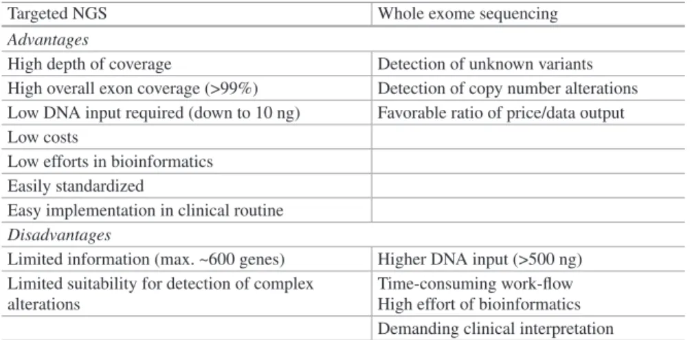
Studies involving whole genome sequencing in head and neck cancer have already been published by Keck et al. 2015 [8] and these allowed head and neck cancer to be stratified into three subtypes (Table 2).
In the same year, a meta-analysis of whole genome sequencing data published by De Cecco et al. [9] separated head and neck cancer into six subtypes. Analysis was performed using different criteria to those of Keck et al. and was based on the tumours biological characteristics and de-regulated signalling pathways. De Cecco et al. designated the subtypes as immunoreactive, inflammatory, human papilloma-virus (HPV)-like, classical, hypoxia- associated, and mesenchymal. Interestingly, adverse behaviour was associated with the hypoxia-associated and mesenchymal subtypes. These publications used differing but to some extent overlapping criteria. One of the limitations of the computational biology approach to analysis of large genomic datasets is that groups are defined by assumed biological behaviour and validation will be required before application of such data can be extended to indi-vidual patients. Nevertheless, WGS studies do indicate that that head and neck can-cer has molecular subgroups and it is likely that the heterogeneity observed will be refined and ultimately will allow stratification for prognosis and therapy in the future.
Further analysis and validation studies are required to understand better the data from WGS platforms to allow the findings to translate into precision therapy for individual patients. Once a fuller understanding of the molecular pathology of head and neck cancer is gained, it is likely that the WGS will inform the development of gene panels that would have the advantages of greater sensitivity and specificity, lower cost, better turnaround time, reproducibility and the possibility of testing using formalin fixed paraffin embedded tissue. Alternatively, the cost of whole genome testing is falling year by year and turnaround times are shortening. It may be possible to introduce WGS into routine clinical services in the future. A current limitation is that fresh tissue must be used. Until this is resolved, biopsy samples would have to include adequate representative tissue for conventional histopatho-logical sectioning as well as sufficient fresh tissue for WGS. This is against the trend for smaller biopsies preferred for clinical reasons, and would be highly prob-lematic for laryngeal biopsies or small mucosal cancers, for example those arising in oral potentially malignant disorders. There are many other challenges to the implementation of WGS, not least the development of the bioinformatics analysis algorithms and expertise necessary to interpret sequence data for individual patients. Further, analysis of TCGA data relating to head and neck cancer to date has not identified obvious targets for specific drugs and currently there is no convincing
Table 2 Head and neck squamous
carcinoma subsets [8] Basal subtype—HPVHER, hypoxia −, high expression of EGFR/
Classical (CL subtype)—low expression of EGFR/HER HPV+ CL
HPV− CL
Immune/mesenchymal (IM subtype) CD8+ infiltration HPV+ IM
19
case for introduction of WGS for head and neck cancer into pathology services [5]. The most promising use of sequencing for head and neck cancer is in the identifica-tion of neo-antigens that could be used to develop personalised T- lymphocyte tar-geted therapy, but more research is needed to demonstrate efficacy.
Stromal Factors
Interplay between the cancer cells and the adjacent stroma is a significant determi-nant of behaviour and outcomes. Fibroblast heterogeneity is a poorly understood process but single cell genomic studies of head and neck cancer reveal two sub-populations of fibroblasts, one of which expresses smooth muscle actin (SMA) and one of which does not. The SMA expressing fibroblasts represent myofibroblasts whilst the other population represents normal and senescing fibroblasts. In vitro studies show that fibroblasts can be induced to express smooth muscle actin by TGF beta, indicating a reversible phenotype [10]. Furthermore in vitro, three fibroblast subpopulations can be identified that have distinctive genetic profiles. These are fibroblasts, myofibroblasts and senescent fibroblasts [11]. Importantly, fibroblast populations appear to have prognostic value [12]. Further studies of the tumour microenvironment are likely to provide insights into complex biological interac-tions that underpin cancer invasion and metastasis.
Matrix macromolecules are also an important determinant of cancer cell behav-iour. In tongue carcinoma, the abundance of the tenascin C has been shown to be a significant prognostic factor [13]. In contrast to fibronectin which mediates fibro-blast adhesion, tenascin C has been shown to have an anti-adhesive effect, facilitat-ing cell migration in vitro [14]. In normal murine and human dorsal lingual epithelium, tenascin C has a distinctive pattern of distribution being located at the tips of the connective tissue papillae but not along the bases of the rete processes [15]. This distinctive pattern of distribution may relate to epithelial stem cell distri-bution and amplification divisions, facilitating cell flow along basement membrane or though cell signalling mechanisms. In a cohort of early stage tongue cancers, poor cumulative survival was associated with the presence of abundant stromal tenascin and fibronectin, whereas cellular tenascin did not distinguish the groups. This might be explained by the assembly and accumulation of tenascin in the extra-cellular matrix [13]. In addition to the matrix factors, epithelial-mesenchymal tran-sition is a recognised process in head and neck cancer biology and elucidation of the pathways involved may lead to identification of future therapeutic targets.
Immunological Landscape
The introduction of immunotherapy into head and neck cancer practice offers a new range of therapeutic options. There is considerable interest in the use of programmed cell death 1 (PD-1)/programmed cell death ligand 1 (PD-L1) inhibitors in head and
neck cancer. The concept of precision medicine is that targeted therapies should be delivered with a rational biological basis and consequently regulatory bodies (e.g. FDA, USA; EMA, Europe; MHRA, UK) recommend the development of compan-ion and complementary biomarkers for these class of drugs. Compancompan-ion biomarkers act as ‘gate-keepers’ and govern the use of the drug, whereas, complementary tests guide clinical decisions, but not access to the treatment. For example, in non-small cell lung cancer the PD-1 inhibitor, pembrolizumab, has companion immunohisto-chemical tests for assessing PD-L1 expression in formalin-fixed paraffin-embedded tissue sections (Clones: Dako 22C3; Ventana SP263). Detection of PD-L1 on malig-nant cells, with a ‘cut off’ of 50% allows first line treatment with pembrolizumab, whereas, only 1% of malignant cell need to be positive for the treatment of recurrent disease (second line treatment; [16]. Nivolumab, another PD-1 inhibitor used in non-small cell lung cancer, has complementary immunohistochemical tests for PD-L1 expression (Clones: Dako 28-8; Ventana SP263), expression guides treat-ment decisions, but there are no absolute ‘cut offs’ that determine clinical utility [16]. In head and neck cancer, pembrolizumab and nivolumab have both been evalu-ated in clinical trials [17–20]. The manufacturers recommend complementary tests (pembrolizumab, Dako 22C3; nivolumab, Dako 28.8), however, there are issues around interpretation of the tests: should the pathologist report PDL-1 expression on malignant cells or malignant cell and immune cells? What are the optimal cut offs for drug efficacy? Checkmate 141 demonstrated that PDL-1 expression by at least 1% of malignant cells is associated with improved overall survival [18], whereas the Keynote trials showed that PDL-1 expression by tumour cells and immune cells is more effective at identifying the patients who are ‘responders’ [17, 19]. The reproducibility of scoring systems in head and neck cancer are yet to be established, but in lung cancer inter-laboratory variability is known to be problem-atic and the use of ‘in vitro diagnostic devices’ (IVD), as opposed to laboratory- developed assays, is recommended (NordiQC, 2018 (http://www.nordiqc.org/ epitope.php?id=102)). The IVD manufacturers of PDL-1 tests are supporting pathologist training by ‘face to face’ engagement and online training modules. Participation in external quality assurance schemes is also recommended (http:// www.nordiqc.org/epitope.php?id=102). In the future, it is likely that the delivery of immuno-onocology drugs, such as the PDL-1/PD1 inhibitors, will be supported by multiplex tests assessing broader ‘immune activation’ with complex algorithms pre-dicting clinical efficacy [19].
Recently, immunogenomic studies have demonstrated heterogeneity between tumour types. In a large-scale study Thorsson et al. [21] identified six immune sub-types that are hypothesized to define immune response patterns impacting on prog-nosis. Immune subtypes differ by somatic aberrations, microenvironment, and survival characteristics.
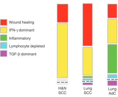
Multiple control modalities of molecular networks have been shown to affect tumour-immune interactions. In a cohort of 10,000 cancers, six immune subtypes were identified. These are: wound healing, IFN-γ dominant, inflammatory, lympho-cyte depleted, immunologically quiet, and TGF-beta dominant. Cancers were char-acterized by differences in macrophage or lymphocyte signatures, Th1:Th2 cell
21
ratio, extent of intra-tumoural heterogeneity, aneuploidy, extent of neo-antigen load, overall cell proliferation, expression of immunomodulatory genes, and prognosis. Interestingly, head and neck squamous cell carcinoma shows two main immune pat-terns (>90%); most are IFN-γ dominant with wound healing slightly lower and other types rarely represented. The immune pattern is similar to lung squamous cell carcinoma but differs from lung adenocarcinoma significantly (Fig. 1). Interestingly, subpopulations of resident memory T cells (CD103) as tumour infiltrating lympho-cytes have been demonstrated to regulate the magnitude of cytotoxic T cell responses in lung cancer [22]. It is likely that further research will elucidate the role of immune cells in head and neck cancer opening the door to the development of novel indi-vidualised T cell therapies.
Digital Pathology and Image Analysis
Digital pathology is increasingly being adopted for reporting worldwide, as it offers the advantages of developing larger hub laboratories that can offer a quality assured service with rapid turnaround times covering a wide geographical area, whilst allowing pathologists to remain close to the clinical teams in their hospital. At the same time, digital pathology is thought to be a solution to the shortfall in the pathol-ogy workforce, allowing clinical demand to be better matched to reporting capacity.
Lung AdC Lung SCC H&N SCC IFN-γ dominant Inflammatory Lymphocyte depleted TGF-β dominant Wound healing
Fig. 1 Immune types of head and neck and lung cancer. The immune landscape of squamous carcinoma of lung is broadly similar to that of squamous cell carcinoma of the head and neck, whereas adenocarcinoma of the lung shows marked differences. Adapted from: Thorsson V, Immune Landscape of Cancer. Immunity. 2018;48:812–30
Further work is needed in the expanding field of digital pathology, particularly in the training of pathologists, validation of systems in the workplace, health econom-ics, integrated LIMs systems and governance relating to the sharing and use of images. Image analysis is a logical extension of digital pathology and it offers the possibility of producing quantitative results on individual biopsies in a reproducible and inexpensive way.
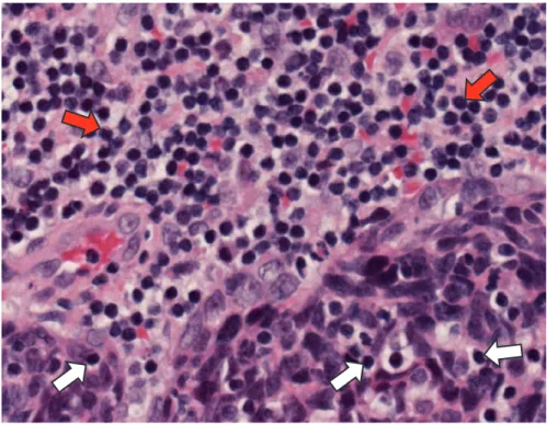
The presence of tumour infiltrating lymphocytes (TILs, Fig. 2) has been shown to be prognostic in a variety of tumours including head and neck squamous carci-noma [23]. Numerous artificial intelligence systems are being developed currently for image analysis and these could potentially produce quantitative data for TILs using multiplex immunohistochemical imaging. It may be possible to use machine learning to identify TILs using routine haematoxylin and eosin stained sections, with validation by the pathologist. Alternatively, immunohistochemistry can be used to identify immune cell populations in an individual tumour and it may be easier and more accurate to quantitate such stained sections without pathologist annotation. Although still expensive and technically challenging, multiplexing allows a panel of immunomarkers to be used on a single section. In that way, back-ground immune cells in a tumour can be quantitated and their spatial relationships can also be measured. Numerous combinations of immunomarkers are possible by
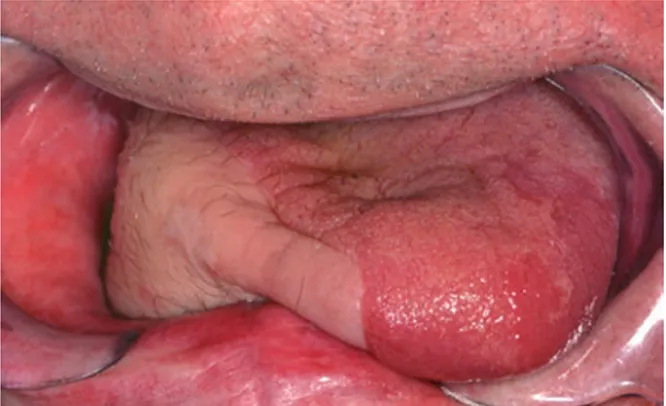
Fig. 2 Tumour infiltrating lymphocytes (white arrows) in HPV associated oropharyngeal carci-noma. Background mucosa associated lymphocytes (red arrows) are present in the top part of the photomicrograph
23
multiplexing and research is needed to evaluate these and develop suitable panels with diagnostic utility. Currently, multiplex immunohistochemistry is expensive, time consuming and technically challenging.
Biomarkers in Current Practice
For squamous cell carcinoma of the head and neck, HPV and EBER testing now form part of routine practice. Current guidelines mandate the use of p16 immuno-histochemistry for oropharyngeal cancer. A number of laboratory protocols have been validated for HPV specific testing and these are discussed in chapter “HPV Assessment in Oropharynx Cancer: What is the Gold Standard?”.
Molecular Pathology and Salivary Diagnostics
The 2017 WHO classification [1] now includes secretory carcinoma, characterised by the ETV6 rearrangement. Most malignant salivary tumours have been shown to possess characteristic molecular abnormalities, most frequently characteristic fusion genes (Table 3). Whilst for the most salivary tumours, diagnosis is based purely on histological appearances, increasingly a morpho-molecular approach is being used. It is important to sample salivary tumours thoroughly in the pathology laboratory because they can exhibit marked heterogeneity, sometimes with only one part showing characteristic diagnostic features. Immunohistochemistry is increas-ing applied to salivary tumours reflectincreas-ing a change since the WHO 2005 classifica-tion where immunohistochemistry was regarded as generally unhelpful. Immunohistochemical identification of SOX 10 is useful for acinic cell and secre-tory carcinomas. However, positive staining may be seen in other salivary tumours
Table 3 Molecular pathology of selected salivary tumours
Salivary tumour Molecular pathology
Adenoid cystic carcinoma MYB(L1)-NFIB
Secretory carcinoma NTRK3,
ETV6-Mucoepidermoid carcinoma CRTC1- or CRTC3-MAML2
Polymorphous carcinoma PRKD1-3, PRKD1
Myoepithelial carcinoma FGFR1-PLAG1, TGFBR3-PLAG1, ND4-PLAG1
Acinic cell carcinoma HTN3-MSANTD3
Hyalinising clear cell carcinoma EWSR1-ATF1
Salivary duct carcinoma NCOA4-RET, HER2 gene amplification, TP53, PIK3CA,
HRAS mutation PTEN loss/mutation
Carcinoma ex PSA PLAG1, HMGA2
Others Actionable genomic alterations
and SOX 10 should not be used as a sole diagnostic marker. DOG1 is expressed on the brush borders of acinic cell carcinoma cells and is highly specific in our hands, being able to distinguish between acinic cell carcinoma and secretory carcinoma. Where the diagnosis of secretory carcinoma is suspected on morphology, a panel showing SOX10 (+), DOG1 (−) and S100 (+) is useful to identify tumours for molecular testing. A combination of Cytokeratin7 and p63 is useful for identifica-tion of the luminal and myoepithelial layers in a variety of salivary tumours that exhibit double layered ductal differentiation. Other immunohistochemical markers may be useful, and those used for salivary lymphomas are outside the scope of this chapter. Molecular diagnostics are particularly useful in salivary carcinomas where high grade transformation has occurred and when the histological pattern conse-quently may be less clear. Molecular testing is also of value where a therapeutic target is identified. In this regard, the finding of the ETV6-NTRK3 fusion that char-acterises most secretory carcinomas is an essential perquisite to therapy targeting NTRK positive tumours. Although most secretory carcinomas are small and can be treated successfully by surgical removal, it is recognised that high grade transforma-tion can occur and then NTRK targeted therapy, at present available as part of a global basket trial, STARTRK-2 (https://www.ignyta.com), may be an option.
The wide availability of next generation sequencing and fusion gene discovery platforms is likely to lead to more detailed morpho-molecular correlates that have prognostic value and can be used to guide therapy. Recently, for example, genomic differences were found that distinguish myoepithelial carcinoma arising de novo from myoepithelial carcinoma arising in pleomorphic adenoma. It was found that TGFB3- PLAG1 fusions characterise myoepithelial carcinoma de novo (a good prognosis group), whereas FGFR1-PLAG1 fusions identify myoepithelial carci-noma arising in pleomorphic adecarci-noma which has a poorer prognosis. A diagnosis of invasive myoepithelial carcinoma arising in pleomorphic adenoma may indicate a more aggressive surgical and radiotherapy approach than would be used for myo-epithelial carcinoma de novo.
References
1. El-Naggar AK, Chan JKC, Grandis JR, Takata T, Slootweg PJ, editors. WHO classification of tumours, vol. 9. 4th ed. Lyon: IARC; 2017.
2. Amin MB, Edge S, Greene F, Byrd DR, Brookland RK, Washington MK, Gershenwald JE, Compton CC, Hess KR, Sullivan DC, Jessup JM, Brierley JD, Gaspar LE, Schilsky RL, Balch CM, Winchester DP, Asare EA, Madera M, Gress DM, Meyer LR, editors. AJCC cancer stag-ing manual. 8th ed. New York: Sprstag-inger; 2017.
3. Brierley JD, Gospodarowicz MK, Wittekind C, editors. TNM classification of malignant tumours. 8th ed. Chichester: Wiley; 2017.
4. The Cancer Genome Atlas. 2015. https://cancergenome.nih.gov/.
5. Leemans CR, Snijders PJF, Brakenhoff RH. The molecular landscape of head and neck cancer.
Nat Rev Cancer. 2018;18:269–82. https://doi.org/10.1038/nrc.2018.11.
6. Meucci S, Keilholz U, Tinhofer I, Ebner OA. Mutational load and mutational patterns in
relation to age in head and neck cancer. Oncotarget. 2016;7(43):69188–99. https://doi.
25 7. Niehr F, Eder T, Pilz T, Konschak R, Treue D, Klauschen F, Bockmayr M, Türkmen S, Jöhrens
K, Budach V, Tinhofer I. Multilayered omics-based analysis of a head and neck cancer model of cisplatin resistance reveals intratumoral heterogeneity and treatment-induced clonal
selec-tion. Clin Cancer Res. 2018;24(1):158–68. https://doi.org/10.1158/1078-0432.CCR-17-2410.
8. Keck MK, Zuo Z, Khattri A, Stricker TP, Brown CD, Imanguli M, Rieke D, Endhardt K, Fang P, Brägelmann J, DeBoer R, El-Dinali M, Aktolga S, Lei Z, Tan P, Rozen SG, Salgia R, Weichselbaum RR, Lingen MW, Story MD, Ang KK, Cohen EE, White KP, Vokes EE, Seiwert TY. Integrative analysis of head and neck cancer identifies two biologically distinct HPV and
three non-HPV subtypes. Clin Cancer Res. 2015;21(4):870–81.
https://doi.org/10.1158/1078-0432.CCR-14-2481.
9. De Cecco L, Nicolau M, Giannoccaro M, Daidone MG, Bossi P, Locati L, Licitra L, Canevari S. Head and neck cancer subtypes with biological and clinical relevance: meta-analysis of gene-expression data. Oncotarget. 2015;6(11):9627–42.
10. Melling GE, Flannery SE, Abidin SA, Clemmens H, Prajapati P, Hinsley EE, Hunt S, Catto JWF, Coletta RD, Mellone M, Thomas GJ, Parkinson EK, Prime SS, Paterson IC, Buttle DJ,
Lambert DW. A miRNA-145/TGF-β1 negative feedback loop regulates the cancer-associated
fibroblast phenotype. Carcinogenesis. 2018. https://doi.org/10.1093/carcin/bgy032 [Epub
ahead of print].
11. Mellone M, Hanley CJ, Thirdborough S, Mellows T, Garcia E, Woo J, Tod J, Frampton S, Jenei V, Moutasim KA, Kabir TD, Brennan PA, Venturi G, Ford K, Herranz N, Lim KP, Clarke J, Lambert DW, Prime SS, Underwood TJ, Vijayanand P, Eliceiri KW, Woelk C, King EV, Gil J, Ottensmeier CH, Thomas GJ. Induction of fibroblast senescence generates a non- fibrogenic myofibroblast phenotype that differentially impacts on cancer prognosis. Aging.
2016;9(1):114–32. https://doi.org/10.18632/aging.101127.
12. Almangush A, Heikkinen I, Bakhti N, Mäkinen LK, Kauppila JH, Pukkila M, Hagström J, Laranne J, Soini Y, Kowalski LP, Grénman R, Haglund C, Mäkitie AA, Coletta RD, Leivo I, Salo T. Prognostic impact of tumour-stroma ratio in early-stage oral tongue cancers.
Histopathology. 2018. https://doi.org/10.1111/his.13481 [Epub ahead of print].
13. Sundquist E, Kauppila JH, Veijola J, Mroueh R, Lehenkari P, Laitinen S, Risteli J, Soini Y, Kosma VM, Sawazaki-Calone I, Macedo CC, Bloigu R, Coletta RD, Salo T. Tenascin-C and fibronectin expression divide early stage tongue cancer into low- and high-risk groups. Br J
Cancer. 2017;116(5):640–8. https://doi.org/10.1038/bjc.2016.455.
14. Adams JC, Chiquet-Ehrismann R, Tucker RP. The evolution of tenascins and fibronectin. Cell
Adhes Migr. 2015;9(1-2):22–33. https://doi.org/10.4161/19336918.2014.970030.
15. Sloan P, Schor SL, Lopes V, Chiquet-Ehrismann R. Immunohistochemical study of the het-erogeneity of tenascin distribution within the oral mucosa of the mouse. Arch Oral Biol. 1990;35(1):67–70.
16. Büttner R, Gosney JR, Skov BG, Adam J, Motoi N, Bloom KJ, Dietel M, Longshore JW, López-Ríos F, Penault-Llorca F, Viale G, Wotherspoon AC, Kerr KM, Tsao MS. Programmed death-ligand 1 immunohistochemistry testing: a review of analytical assays and clinical
imple-mentation in non-small-cell lung cancer. J Clin Oncol. 2017;35(34):3867–76. https://doi.
org/10.1200/JCO.2017.74.7642.
17. Chow LQM, Haddad R, Gupta S, Mahipal A, Mehra R, Tahara M, Berger R, Eder JP, Burtness B, Lee SH, Keam B, Kang H, Muro K, Weiss J, Geva R, Lin CC, Chung HC, Meister A, Dolled-Filhart M, Pathiraja K, Cheng JD, Seiwert TY. Antitumor activity of pembrolizumab in biomarker-unselected patients with recurrent and/or metastatic head and neck squamous cell carcinoma: results from the phase Ib KEYNOTE-012 expansion cohort. J Clin Oncol.
2016;34(32):3838–45. https://doi.org/10.1200/JCO.2016.68.1478.
18. Ferris RL, Blumenschein G Jr, Fayette J, Guigay J, Colevas AD, Licitra L, Harrington K, Kasper S, Vokes EE, Even C, Worden F, Saba NF, Iglesias Docampo LC, Haddad R, Rordorf T, Kiyota N, Tahara M, Monga M, Lynch M, Geese WJ, Kopit J, Shaw JW, Gillison ML. Nivolumab for recurrent squamous-cell carcinoma of the head and neck. N Engl J Med. 2016;375(19):1856–67.
19. Seiwert TY, Burtness B, Mehra R, Weiss J, Berger R, Eder JP, Heath K, McClanahan T, Lunceford J, Gause C, Cheng JD, Chow LQ. Safety and clinical activity of pembrolizumab Cellular and Molecular Pathology in Head and Neck Cancer