UNIVERSITÀ DEGLI STUDI DI SALERNO
Dipartimento di Farmacia
Dottorato di ricerca
in
Scienze Farmaceutiche
Ciclo XIV N.S. – Anno di discussione 2016
Coordinatore: Chiar.mo Prof. Gianluca Sbardella
Study of the mechanism of action of
bioactive plant terpenoids
settore scientifico disciplinare di afferenza
:
Chim/09
Dottorando Tutore
Index
Chapter 1
-Introduction ... 1
1.1 Importance of plant secondary metabolites in drug discovery ... 1
1.2 Terpenes ... 4
1.3 Drug discovery today ... 5
1.4 Proteomic in drug discovery: the state of the art ... 9
1.5 Scope of thesis ... 14
1.5.1 Outline of the thesis ... 15
References ... 16
Chapter 2 -Introduction ... 20
2.1 15-ketoatractyligenin methyl ester (SR2017) ... 20
2.2 Aims of the project ... 21
Results and discussion ... 23
2.3 Identification of the putative 15-ketoatractyligenin methyl ester targets by chemical proteomics... 23
2.4 Targets validation by Surface Plasmon Resonance ... 25
2.4.1 Selectivity of 15-ketoatractyligenin methyl ester towards different isoform of PPARγ ... 26
2.5 Structural characterization of SR2017/PPARɣ complex ... 27
2.6 15-ketoatractyligenin methyl ester is a bona fide PPARγ ligand and requires the binding to C285 for its transcriptional activity ... 28
2.8 Screening of 15-ketoatractyligenin methyl ester analogous using SPR analysis
... 36
2.9 Discussion ... 38
Materials and methods ... 40
2.10 Chemical proteomics ... 40
2.11 Surface plasmon resonance (SPR) ... 42
2.12 PARPγ/LBD peptide mapping ... 43
2.13 Computational chemistry ... 43
2.13.1 Protein and Ligand Preparation ... 43
2.13.2 Docking Simulations ... 44
2.14 Plasmids and transient transfection experiments ... 45
References ... 46
Chapter 3 -Introduction ... 52
3.1 Oridonin: insight its multifunctional effects ... 52
3.2 Aims of the project ... 54
Results and discussion ... 58
3.3 Synthesis and characterization of fluorescent oridonin companion imaging drug ... 58
3.4 Oridonin uptake in live cells ... 61
3.5 Optimization of DARTS protocol ... 62
3.6 Identification of molecular targets for oridonin using DARTS ... 64
3.6.1 Validation of selectivity of oridonin towards identified molecular targets using DARTS approach ... 67
3.6.2 Western Blot analysis to validate some of the molecular targets of oridonin ... 68
3.7 “Complementary” chemical proteomic approach: affinity purification and mass spectrometry analysis of oridonin partners ... 69
3.9 Monitoring of nucleolin engagement by oridonin in live cells using CETSA
assay ... 74
3.10 Oridonin/HSP70 interaction validation in cell system using CESTA ... 79
3.11 In vitro imaging of nucleolin colocalization with oridoninBODIPY FL2 ... 80
3.12 Oridonin biological effect on nucleolin into the cell ... 82
3.12.1 Levels and post translational modifications of nucleolin ... 82
3.12.2 Regulation and influence of target mRNAs of nucleolin by oridonin ... 86
3.12.3 Inhibition of protein synthesis by oridonin ... 87
3.13 Screening in vitro of new potential nucleolin inhibitors using CETSA approach ... 90
3.14 Discussion ... 93
Materials and methods ... 97
3.15 Synthesis and Characterization of Oridonin-BODIPY FL ... 97
3.15.1 Synthesis and charactherization of ent-1a,6b-Dihydroxy-7,14-isopropylidene ketal-15-oxo-7,20-epoxy-16-kaurene (compound 1)... 97
3.15.2 Synthesis of compound 2 ... 98
3.15.3 Synthesis of compound 3 ... 98
3.15.4 Synthesis of compound 4 ... 98
3.15.5 Stability assay ... 99
3.16 Biological characterization of oridonin BODIPY FL ... 99
3.16.1 Cell Viability Assay ... 99
3.16.2 Covalent binding Oridonin BODIPY FL/ HSP70 ... 100
3.17 Live cell fluorescence microscopy in cell of oridonin BODIPY FL ... 100
3.18 Target identification approaches ... 101
3.18.1 Optimization of DARTS protocol ... 101
3.18.2 Evaluation of oridonin interference with subtilisin using DARTS approach ... 102
3.18.3 DARTS experiment on whole cell lysate ... 102
3.18.4 DARTS experiment on intact cell ... 103
3.18.6 Samples preparation for mass spectrometer analysis ... 104
3.19 SPR analysis of nucleolin complexes ... 105
3.20 CETSA approach ... 106
3.20.1 Determination of the apparent melting curve of nucleolin by CETSA .. 106
3.20.2 Determination of EC50 of complex nucleolin/oridonin by CETSA ... 107
3.21 Biological effect of oridonin in cell ... 108
3.21.1 RNA isolation and quantitative real-time RT-PCR (qRT-PCR) ... 108
3.21.2 Protein Synthesis assay ... 109
3.21.3 Cell viability and cell cycle analysis ... 109
3.22 Synthesis and characterization of oridonin BODIPY FL2 ... 110
3.22.1 Synthesis and charactherization of compound 1 ... 110
3.22.2 Synthesis and charactherization of compound 2 ... 110
3.22.3 Immunofluorescence and co localization of oridonin BODIPY FL2 .... 111
3.23 Screening of terpenes using CETSA approach ... 111
3.24 Western blotting analysis ... 111
3.25 Statistical analysis ... 112
References ... 113
Chapter 4 -Introduction ... 119
4.1 Background and aims of the projects ... 119
4.2 Fluorescence polarization applied to invitral microscopy analysis ... 120
4.3 A new method based on CESTA approach ... 124
Materials and methods ... 129
4.4 Live cell fluorescence microscopy in cell of doxorubicin ... 129
4.5 CETSA approach ... 129
4.5.1 Determination of the apparent melting curve of Btk by CETSA ... 129
4.5.2 Determination of EC50 of Btk/IbrutinibSirCOOH complex by CETSA .. 131
4.7 Statistical analysis ... 132
Abstract
Natural products are small-molecule secondary metabolites displaying considerable structural complexity and “privileged scaffolds”. They are able to bind several endogenous targets eliciting biological effects as chemical weapons or to convey information from one organism to another.
Nowadays, medicinal plant drug discovery continues to provide new and important leads against various pharmacological targets. Therefore, the primary purpose of this PhD thesis has been a comprehensive characterization of the interactome profile and then the molecular mechanism of action of bioactive natural molecules. Achieving this in an effective, unbiased and efficient manner subsists as a significant challenge for the new era in drug discovery and optimization. Indeed, the full understanding of the mechanism of action of natural molecules could lead to a number of advantages: first of all, exploit their full therapeutic potential, the identification of side effects or toxicity, or the ability to set up target-based assays and to allow structure activity relationships studies to guide medicinal chemistry efforts towards lead optimization.
In my research project, the attention was paid on ent-kaurane diterpenes, a class of natural terpenoids with a great structural variability and a wide spectrum of biological activities. Firstly, I focused on the determination of the interactome of a semi synthetic compound 15-ketoatractyligenin methyl ester. This compound has been previously reported to possess high antiproliferative activity against several solid tumor-derived cell lines. In this regard, I decided to investigate the mechanism of action of this actratylignin derivative researching first of all its molecular targets, responsible for the biological activity. In order to achieve this goal, I used a chemical proteomic approach first. This study led to the identification of PPARγ as the main cellular partner
of this compound; achieved results were supported and validated through different biological assays.
Subsequently, I studied another diterpene: oridonin. This molecule has been shown to have multiple biological activities. Among them, the anticancer activity has been repeatedly reported by many research groups. With the aim of expanding and validate our knowledge about this molecule, also seen the limitations of the fishing for partners method, I decided to use two orthogonal compound-centric proteomics approaches to define the possible protein target(s) of oridonin. Using this strategy HSP70 and nucleolin were identified. Therefore, several in vitro and in cell tests have been performed to validate the interaction of oridonin with these proteins, and to evaluate its effect on their activity. Some of these tests were developed and optimized during my period of research abroad at the Massachusset General Hospital- Center for System Biology -Harvard Medical School; in that twelve months period I expanded my knowledge into the techniques useful for the study of the mechanism of action of a small molecule, also applying experimental methods complementary to proteomics and focusing on the use of high-resolution intravital microscopy imaging for drug pharmacology.
-1
Introduction
1.1 Importance of plant secondary metabolites in drug discovery
The use of natural substances, particularly plant small molecules, to control human diseases is a centuries old practice that has led to the discovery of more than half of all “modern” pharmaceuticals (Katz L. & Baltz R.H. 2016). Thus, there is increasing interest in the clues from traditional uses of plant extracts to guide new drug discovery;to have an idea of the sigfnificance of hetnobotanic use of plant drugs it is sufficient just to consider the wide consumption of medicinal plants in the traditional Chinese medicine to cure and prevent diseases. Besides, more than half of humanity does not have access to modern medicine they rely on traditional treatments (Cordell & Colvard, 2012).
Actually, natural products play a significant role in the drug discovery and development process and this is particularly evident in the areas of cancer and infectious diseases, where over 60% and 75% of therapeutic agents have to be considered of natural origin. This contribution seems impressive, but it can be easily explained in the light of the chemical and evolutionistic proprieties shown by natural products. First of all, they exhibit a wide range of pharmacophores and a high degree of stereochemistry. Therefore, they have been - and they are still - an invaluable source of inspiration for organic chemist to synthesize novel drug candidates (Beghyn T. et al. 2008; Hunter P. 2008; Koehn Koehn F.E. & Carter G.T. 2005), since they provide a new starting point for new synthetic compound with diverse structures and often with multiple stereocenters that can be challenging synthetically (Clardy J. & Walsh C. 2004; Nicolaou K.C. & Snyder S.A. 2004; Peterson E.A. & Overman L.E. 2004). Indeed, many structural features common to natural products (e.g., chiral center, aromatic rings, complex ring system, degree of molecule saturation and number and ratio of heteroatoms) are expected to contribute to their ability to provide hits even against the more difficult
2
screening targets, such as protein-protein interactions (Balunas M.J. & Kinghorn A.D. 2005). Moreover natural products may have the additional advantage of being natural metabolites: compounds that are efficient as drugs have been suggested to have the propriety of ‘metabolite-likness’(Herty J. et
al. 2009). Then, natural compounds are a good starting point for the setting up
of libraries to test for drug discovery, not only for their complex and diversified chemical space but also for their ability to interact with biomolecules.
Although natural products have not been developed to bind to human proteins, they can do it very well. There are two main theories to explain this phenomenon: the first, widely accepted, is that it is the result of long-term co-evolution within biological communities; interacting organism, that evolved in close proximity to one another, developed compounds that could influence the biological process of neighboring species (Ji H.F. et al. 2009). The second theory, advanced by Konrad T. et al. and called xenohormesis, (Figure 1) is based on the hypothesis that there have been common ancestors of plants and animals able to synthesize a large number of stress- induced secondary metabolites; animals and fungi, that feed on plants, gradually lost the capacity to synthesize these low-weight molecular compounds, but retained the ability to sense these chemical cues in plants, possibly in order to detect when plants were stressed and gain an early warning of changing environmental condition (Konrad T.H. & David A.S. 2008).
3
Fig.1: The Xenohormesis hypothesis.
Adopted from Konrad T.H. & David A.S. 2008
In fact, natural products are evolutionary preselected thus representing structural requirements for the binding to proteins. Their structural scaffolds represent the biologically relevant and prevalidated fractions of chemical
structure space explored by nature so far (Koch M.A. et al. 2005).Moreover,
recently Stuart Schreiber, Paul Clemons and their colleagues at the Broad Institute in Boston performed a bioinformatics analysis of natural product targets, thus demonstrating that natural molecules statically tend to target proteins with a high number of protein– protein interactions that are particularly essential to an organism (Dancík V. et al. 2010). This observation is consistent with the common role played by natural products as chemical weapons against predators or competitors.
4
Despite the success of plant molecules as drugs and the large number of benefits they show, they are losing favor among drug developers. One reason is the perceived “dirtiness” of plant molecules: a molecule is considered “dirty” if it interacts with numerous endogenous proteins. Such compounds presumably are more likely to have negative “off target” effects than a molecule that specifically targets a single protein. Flying in the face of this dogma, there are examples of plant molecules that, despite interacting with multiple human enzymes and receptors are surprisingly safe (Prasad S. & Tyagi A.K. 2015). Take for example salicylic acid or curcumin, are surprisingly powerful and nontoxic although they are two multitarget molecules. In fact, having multi-target molecules could be a winning strategy to face various diseases. It has been increasingly recognized that, in several pathologies, there are a large number of mutated genes and/or modified proteins that disrupt multiple pathways, which normally exhibit extensive biological cross-talk and redundancy. Therefore, the development of ‘magic bullet’ drugs that bind selectively to single protein targets also appears less clinically useful, since, interfering with a single target and/or pathway may not abrogate the disease. Moreover, a promising strategy for mitigating an acquired drug resistance or suppress disease, is to simultaneously inhibit multiple molecular pathways, either by using several agents in combination or by using a single agent that concurrently blocks multiple targets or pathways.
1.2 Terpenes
The terpenes are an important class of plant secondary metabolites which constitute a vast family of natural substances structurally different from each
other, whose starting elements for the biosynthesis are the isoprene units.
They are produced by diverse organisms to perform an assortment of
biological functions in varying ecological contexts. They are derived
5
C5H8. Isoprene itself does not undergo the building process, but rather
activated forms, isopentenyl pyrophosphate (IPP or also isopentenyl diphosphate) and dimethylallyl pyrophosphate (DMAPP or also dimethylallyl diphosphate), are the actual components in the biosynthetic pathway.
Although all terpenoids are synthesized from two five-carbon building blocks, the structures and functions vary widely. Many terpenes have shown several biological activities. Isoprenoids and derivatives play a critical role in all living systems: the cell structure, systems of electron transport in cell-cell signals (steroids, abscisic acid, gibberellic acid, phytol ecc.), in the structure of organisms and interactions between them. Some isoprenoids play a role in plant defense systems against attack by micro-organisms and insects, act as allelopathic compounds in plant-insect and plant-environment (Lange B.M. et
al., 2000).Furthermore, they are used for the treatment of human diseases. In
fact, there is a wide spectrum of biological and pharmacological activities for these types of substances such as antibacterial, antifungal, antiparasitic, anti-inflammatory, cytotoxic and antitumoral (Coll J. et al. 2007, Evans F.J. & Taylor S.E. 1983). Although they shown all of these activities, approximately 35,000 terpenes have been identified and the majority of possible functions of these molecules are unknown. Then, it would be interesting to study this class of natural molecules to find out its potential biological effects and uses in therapy.
1.3 Drug discovery today
Drug discovery is an inherently complex process with an industrial base, through which potential new medicines are identified and it involves a wide range of scientific disciplines, including biology, chemistry and pharmacology. When chemistry had reached a degree of maturity that allowed its principles and methods to be applied to problems outside of chemistry itself and when pharmacology had become a well-defined scientific discipline in its
6
own right, drug discovery as we know it today began its career; in fact, this discipline is not much older than a century. Drug research has contributed more to the progress of medicine during the past century than any other scientific factor. During the first half of last century, drug research was shaped and enriched by several new technologies, all of which left their imprint on drug discovery and on therapy. Genomic era and then rapid DNA sequencing, combinatorial chemistry, cell-based assays, and automated highthroughput screening (HTS) has led to a “new” concept of drug discovery. Therefore, in the last decade dramatic changes in the approaches to biomedical discovery occurred, also due to the advent of massively parallel sequencing technologies to sequence genomes, the ability to characterize the transcriptome and an improved ability to evaluate the proteome (Roti G. & Stegmaier K. 2012). The development of a new drug can generally be divided into phases (Figure 2). The first is the preclinical phase, which usually takes 3 to 4 years. If successful, this phase is followed by an application to national or international drug agencies (DA) as an investigational new drug (IND). After an IND is approved, the next steps are clinical phases 1, 2 and 3, which require approximately 1, 2, and 3 years, respectively, for completion. Importantly, throughout this process the DA and investigators leading the trials communicate with each other, so that such issues as safety are monitored. The manufacturer then files a new drug application (NDA) with the DA for approval. This application can either be approved or rejected, or the DA might request further study before making a decision. Following acceptance, the DA can also request that the manufacturer conduct additional post-marketing studies. Overall, this entire process, on average, takes between 8 to 13 years.
7
Fig.2: Overview of drug discovery process.
Adopted from: Beom-Su Jang 2013
Generally, drug development often fails at late stages, after significant cost and time investments (Paul S.M. et al. 2010). It’s enough to consider a current price tag of over US $1 billion per drug and an average of 13 years of investment (Paweletz C.P. et al. 2011; Kola I. & Landis J. 2004; Collins I. & Workman P. 2006). Furthermore, only three out of ten approved drugs manage to recover their respective development costs. Therefore, the large upfront time and monetary expenditures limit the number of drugs that can be moved from the bench to the clinic. Actually, drugs fail in the clinic for two main reasons; the first is that they do not work and the second is that they are not safe. To accelerate drug development, and subsequently reduce exorbitant costs and high failure rates, the pharmaceutical industry needs to increase its
overall R&D efficiency, not just productivity (Paul S.M. et al. 2010). In order
to solve this problem, participation of academic centers in aspects of drug discovery and development beyond target identification and clinical trials is always been significant contributors to the discovery of new drugs in therapies (Shamas-Din A. & Schimmer A.D. 2015).
In the typical drug development process, small-molecule ligands are identified by screening compound libraries against purified targets, using suitable biochemical assays. Hits derived from such screens are then further improved in an iterative medicinal chemistry process that includes profiling and validation in cell-based in vitro assays. This approach relies heavily on
high-8
throughput screening methods, empirical and experimental compound selection and optimization, and the development of relevant animal models of disease. Additionally, computational techniques, including drug docking simulations and quantitative SAR (QSAR), are increasingly employed to improve the efficiency in lead compound optimization (Vinegoni C. et al. 2015). In this regard, academic institutions developed several techniques and skills to evaluate the best candidate in the drug discovery process: they have established high-throughput screening platforms, formed medicinal chemistry teams, and built capabilities in pharmacokinetic studies with the aim of developing small-molecule drugs. Thus, the aim of these institutions is to identify and validate new therapeutic targets through biological studies and testing new drugs developed by the pharmaceutical industry in clinical trials. That’s why drug discovery in academic centers represent a unique opportunity for the industry. Therefore, since they do not consider necessarily issues related to market share and profitability, investigators can pursue drug candidates for targets of scientific interest. In addition, chemical probes that do not reach the stage of clinical trials are important experimental tools, since the probes can be used to reveal new biological insights. Finally, academic investigators can make important contributions to the rationale for development of new therapeutic agents, even if the intellectual property rights of the drug are held by others.
However, advancing drugs from academic institutions into clinical trials remains challenging, and these institutions face common obstacles. In fact, heavy investment in money and time is the major obstacle to be overcome. One solution to this problem is for academic groups to replace “old” drug allowing the rapid evaluation of them into the clinical trial phase. The repositioning of thalidomide for myeloma, for example, is a dramatic success but there will likely be few examples of this strategy that show similar impact (Shamas-Din A. & Schimmer A.D. 2015).
9
An important challenge now is how to best exploit these new capabilities for
therapeutic benefit. Thus, it is very important to know not only
pharmacokinetic and pharmacodynamic parameters of a drug but above all its mechanism of action. Target identification and confirmation for small molecules is often the rate limiting step in drug discovery. Although drugs can be approved without a clear knowledge of their target and/or mode of action, the full characterization of the protein binding profile of a small molecule is an important prerequisite for a complete picture of the biology behind it. In fact, complete comprehension of a drug leads many advantages in terms of improvement and potential of the molecule itself and in terms of versatility of the same, also allowing its possible use against more pathologies. In this regard proteomic approaches fits very well in the macro complex of drug discovery. Proteomics is an essential and crucial method to discover and explain drugs, not only because of the knowledge of the effects of drug candidates on their protein targets, but also to shed light on the cellular mechanisms behind the observed phenotype (Guo S. et al. 2013).
1.4 Proteomic in drug discovery: the state of the art
The publication of the full human genome sequence in 2003 by the International Human Genome Sequencing Consortium is a crucial milestone in the history of genetic research (Yan S.K. et al. 2015). The completion of human genome has created much excitement from the impact that this wealth of information is likely to have on the process of drug discovery and development (Reiss T. 2001; Lander E.S. et al., 2001). It has been postulated that scientists could use genomic information to identify and validate a host of new drug targets and tailor specific drugs based on an individual’s detailed genetic makeup (Cockett M. et al., 2000). Although this new field of genomics holds many promises, it is clear that analysis of DNA and/or RNA content alone is not sufficient to understand cell biology and disease. However,
10
analysis of the information produced by genomics, when measured against comparable information regarding protein expression, has led to the conclusion that message abundance fails to correlate with protein quantity (Nutall M.E. 2001). Further, post-translational processes such as protein modifications or protein degradation remain unaccounted for in genomic analysis (Anderson L & Seilhamer J 1997; Mann M. 1999). Since both cell function and its biochemical regulation depend on protein activity, and the correlation between message level and protein activity is low, the mere measurement of expression has proven to be inadequate. Consequently, the development of drug-discovery technologies has begun to shift from genomics to proteomics (Simpson R.J. & Dorow D.S. 2001).
Proteomics, as a scientific field, is defined as the study of the protein products of the genome, and their interactions and functions. Hence, the proteins expressed at a given time in a given environment constitute a proteome (Dove A. 1999). From a technology viewpoint, traditional proteomics involves separation of proteins in a proteome, coupled to a means of identification. This science is very challenging since protein levels vary widely with both cell type and environment (Bichsel V.E. et al., 2001). Second, unlike genomics, which can amplify benefits from the amplification of single genes using the polymerase chain reaction (PCR), protein science has no comparable amplification method (Blackstock W.P. & Weir M.P. 1999). Third, proteomics is complicated by the fact that the absolute quantity of protein is of limited interest to drug discovery, because protein activities are highly regulated post-translationally (Srinivas P.R. et al. 2001). Therefore, proteins can be abundant, yet possess little activity. Finally, because proteins interact functionally in
vivo, protein–protein and protein–small molecule interactions need to be
evaluated in processes of interest (Cravatt B.F. & Sorensen E.J. 2001).
The metabolism of a cell or of an entire organism is mainly regulated by proteins, acting individually and, more frequently, in pathways. In particular,
11
the function of a protein can be defined on the basis of its interactions, and pathways are cascades of specific protein interactions that are necessary to activate distinct cellular functions. Genetic mutations or environmental factors deregulate these pathways, leading to disease conditions. A detailed knowledge of the pathways active inside the cell and of how they are deranged in a particular pathology is fundamental for drug discovery as it allows the identification of new drug targets. Since the pharmacologic effects of a drug can only be appreciated when its interactions with cellular components are clearly delineated, an integrated deconvolution of drug target interactions for each drug is necessary. The wide use of proteomic in drug target identification has enhanced our confidence in improving our understanding of the molecular
mechanisms of these drugs. In fact, with the development of related
high-throughput analytical technologies and mass spectrometry, proteomics has been rapidly developed in various research fields. Thus, an unprecedented number of biological targets have been tested, and various technologies emerging today provide us with a superior platform to further investigate drug targets.
12
Fig.3: Main applications of functional, chemical and clinical proteomics in drug discovery.
Adopted from Savino R. et al. 2012
As mentioned before, proteomic is spread in different fields, namely functional, chemical and clinical proteomics, all going to somehow impact on biomarker and drug discovery processes (Figure3).
The proteome records the flow of information that starting within the cells, through the intercellular protein network, goes beyond the extracellular microenvironment up to come to the blood microenvironment (Matta A. et al., 2010). Accordingly, the proteome may reflect immediate and characteristic changes in response to disease processes and external stimulation. By the means of proteomic tools such as mass spectrometry (MS), it is possible to qualitatively and quantitatively reveal molecular profiles contained in healthy or clinical samples. Consequently, MS technologies offer the opportunity to screen and discovery simultaneously multiple biomarkers, which consist of a pattern of up- or down-regulated molecules (proteins, peptides, metabolites,
13
Basically, clinical proteomics covers all MS-based preclinical and basic science studies aimed at discovering and understanding the role of proteins in pathological processes in order to facilitate the early diagnosis of disease, the prognosis prediction, the identification of new therapeutic targets and the evaluation of treatment response (Beretta L. 2007; Matt P. et al. 2008).
Another emerging research area in the proteomic is functional proteomics, aimed to monitor and analyze the spatial and temporal properties of the molecular networks and fluxes involved in living cells (Godovac-Zimmermann J. & Brown L.R. 2001). It is focused on the generation of information about proteins, such as expression levels, interacting partners, post-translational modifications (PTMs) and activity, which all contribute to elucidate pathways active inside the cells and, ultimately, to a functional understanding of biological systems.. In recent years, functional proteomics has been used to analyze not only the formation of specific protein-protein interactions, but alsoto understand how these interactions lead to the assembly of macromolecular protein complexes that are regulated by PTMs and which
affect pathway functions.In particular, these approaches are addressed towards
two major targets: the elucidation of biological functions of unknown proteins and the definition of cellular mechanisms at the molecular level. In the cells, many proteins display their biological functions through the rapid and transient association within large protein complexes. Understanding protein functions as well as unraveling molecular mechanisms within the cell then depend on the identification of the interacting protein partners. The association of an unknown protein with partners belonging to a specific protein complex involved in a particular mechanism would be strongly suggestive of its biological function (Gavin A.C. et al. 2002; Ho Y. et al. 2002). Furthermore, a detailed description of the cellular signaling pathways might greatly benefit from the elucidation of protein–protein interactions in vivo (Lewis T.S. et al., 2000).
14
Finally a powerful weapon to profile previously uncharacterized proteins via identifying drug target interactions is chemical proteomics: a multidisciplinary research area integrating biochemistry and cell biology with organic synthesis
and MS.Chemical proteomics comes in two different flavors: (i) activity based
probe profiling (ABPP), which focuses on the enzymatic activity of a particular protein family, and (ii) a compound-centric approach, which focuses on characterizing the molecular mechanism of action of an individual bioactive small molecule (Rix U. & Superti-Furga G. 2009). Thus, they also serve different purposes. ABPP detects members of a defined class of enzymes that are active under certain conditions-for example, in a disease. This method can lead to the identification of new proteins with the respective biochemical activity, or it can be applied to determine the selectivity profile of drugs targeting an enzyme family via pretreatment of the lysate with the drug of interest and subsequent labeling and identification of the remaining enzymes using appropriate reactive probes. Compound centric chemical proteomics consists of classical drug affinity chromatography that is similar to the method used for decades but that is now performed in combination with modern high-resolution MS analysis and statistics or bioinformatics for subsequent identification of binding proteins (Savino R. et al., 2012).
Finally, understanding protein function and unraveling cellular mechanisms at the molecular level constitute a major need in modern biology. Therefore, chemical proteomics is an initial technology that is useful in clinical testing and drug development.
1.5 Scope of thesis
Naturally occurring secondary metabolites are small molecules displaying considerable structural complexity and “privileged scaffolds”. They are able to bind several endogenous targets, eliciting biological effects as chemical weapons or as information vectors from one organism to another. Concerning
15
the human use, medicinal plant drug discovery continues to provide new and important leads against various diseases including cancer, HIV/AIDS, Alzheimer’s, malaria, and pain. Therefore, the overall aim of this Ph.D. study was to set-up, optimize, develop, and apply proteomics methods to elucidate the molecular mechanisms of action of bioactive natural molecules. It is indeed evident that the full understanding of these mechanisms could lead to a number of advantages: first of all, exploit the full therapeutic potential of promising compounds, the identification of their side effects or toxicity, or the ability to set up target-based assays. Moreover, it would allow performing structure activity relationships studies to guide medicinal chemistry efforts towards lead optimization. Achieving this in an effective, unbiased and efficient manner subsists as a significant challenge for the new era in drug discovery and optimization. Therefore, protein sample preparation, fractionation, high-throughput LC-MS/MS, data analysis, and, in particular, label free and chemical proteomic approaches were used.
1.5.1 Outline of the thesis
This research paid attention on diterpenes, a class of natural terpenoids with a wide structural variability and known spectrum of biological activities. Firstly, (Chapter 2) the determination of the interactome of a semi synthetic compound 15-ketoatractyligenin methyl ester is described. This compound has been previously reported to possess high antiproliferative and cytoxocitity activities against several solid tumor-derived cell lines. Therefore, we select it to investigate its mechanism of action, through the identification of the molecular targets responsible for its biological activity. In order to achieve this goal, a chemical proteomic approach was used; the emerged results were subsequently validated by biophysics, biological and bioinformatic assays. In the Chapter 3 the chemical-biological study of oridonin is discussed. This diterpene has been shown to have multiple biological activities. Among them,
16
the anticancer activity has been repeatedly reported by many research groups. With the aim of expanding and validate our knowledge about this molecule, we decided to use several approaches for the identification of its possible targets in cell. More in details, two orthogonal compound-centric proteomics approaches were used. Moreover, to validate the interaction of oridonin with these proteins, and to evaluate its effect on their activity several in vitro and in cell tests have been performed.
Finally, to expand my knowledge into the techniques useful for the study of the mechanism of action of a small molecule, methods complementary to proteomics have been applied. In particular, in Chapter 4 new orthogonal techniques useful for the study of the mechanism of action of drugs and the use of high-resolution intravital microscopy imaging for drug pharmacology were discussed.
References
Anderson L. & Seilhamer J., (1997), A comparison of selected mRNA and protein abundances in human liver., Electrophoresis., 18:533-537.
Balunas M.J. & Kinghorn A.D., (2005), Drug discovery from medicinal plants., Life Sci.,22;78(5):431-41.
Beghyn T, Deprez-Poulain R, Willand N, Folleas B, Deprez B, (2008), Natural compounds: leads or ideas? Bioinspired molecules for drug discovery, Chem Biol Drug Des, 72(1):3-15.
Beretta L., (2007), Proteomics from the clinical perspective: Many hopes and much debate. Nat. Methods., 4(10):785-6.
Bichsel V.E., Liotta L.A., Petricoin E.F., (2001), Cancer proteomics: from biomarker discovery to signal pathway profiling., Cancer J.,7:69-78.
Blackstock W.P. & Weir M.P., (1999), Proteomics: quantitative and physical mapping of cellular proteins., Trends Biotechnol., 17:121-127.
17
Cockett M., Dracopoli N., Sigal E., (2000), Applied genomics: integration of the technology within pharmaceutical research and development., Curr Opin Biotechnol., 11(6):602-9.
Coll J., Tandrón Y.A., Zeng X., (2007), New phytoecdysteroids from cultured plants of Ajuga nipponensis Makino., 72(3):270-7.
Collins I.& Workman P., (2006), New approaches to molecular cancer therapeutics., Nat. Chem. Biol., 2, 689–700.
Cordell G.A. & Colvard M.D., (2012), Natural products and traditional medicine: turning on a paradigm. J Nat Prod.,23;75(3):514-25.
Cravatt B.F. & Sorensen E.J., (2000), Chemical strategies for the global analysis of protein function., Curr Opin Chem Biol .,4:663-668.
Dancík V., Seiler K.P., Young,D.W., Schreiber, S.L., Clemons,P.A., (2010), Distinct biological network properties between the targets of natural products and disease genes., J. Am.Chem.Soc., 132(27):9259-61.
Dove A., (1999), Proteomics: translating genomics into products?., Nat Biotechnol., 17:233-236.
Evans F.J. & Taylor S.E., (1983), Pro-inflammatory, tumour-promoting and anti-tumour diterpenes of the plant families Euphorbiaceae and Thymelaeaceae., Fortschr Chem Org Naturst.,44:1-99.
Gavin A.C., Bösche M., Krause R., Grandi P., Marzioch M., Bauer A., Schultz J., Rick J.M., Michon A.M., Cruciat C.M., Remor M., Höfert C., Schelder M., et al., (2002)., Functional organization of the yeast proteome by systematic analysis of protein complexes., Nature., ;415(6868):141-7.
Godovac-Zimmermann J. & Brown L.R., (2001), Perspectives for mass spectrometry and functional proteomics., Mass Spectrom Rev.20(1):1– 57.
Guo S. Zou J., Wang G. (2013), Advances in the proteomic discovery of novel therapeutic targets in cancer, Drug. Des. Ther. 24;7:1259-71.
Hert J., Irwin J.J., Laggner C., Keiser M.J., Shoichet B.K., (2009), Quantifying biogenic bias in screening libraries.,Nat Chem Biol., 5(7):479-83.
Ho Y., Gruhler A., Heilbut A., Bader G.D., Moore L., Adams S.L., Millar A., Taylor P., Bennett K., Boutilier K., Yang L., Wolting C., Donaldson I., Schandorff S., Shewnarane J., Vo M., et al .,(2002), Systematic identification of protein complexes in Saccharomyces cerevisiae by mass spectrometry., Nature., 415(6868):180-3. Howitz K.T. & Sinclair D.A., (2008), Xenohormesis: sensing the chemical cues of other
species., Cell., 133(3):387-91.
Hunter P., (2008), Harnessing Nature's wisdom. Turning to Nature for inspiration and avoiding her follies, EMBO Rep, 9(9):838-4.
18
Ji H.F., Li X.J., Zhang H.Y. (2009), Natural products and drug discovery. Can thousands of years of ancient medical knowledge lead us to new and powerful drug combinations in the fight against cancer and dementia? EMBO RepMar; 10(3): 194–200.
Ji H.F., Li X.J., Zhang H.Y., (2009), Natural products and drug discovery. Can thousands of years of ancient medical knowledge lead us to new and powerful drug combinations in the fight against cancer and dementia?., EMBO RepMar; 10(3): 194–200.
Katz L & Baltz R.H., (2016), 76 Natural product discovery: past, present, and future., J Ind Microbiol Biotechnol.;43(2-3):155-59.
Koch M.A., Schuffenhauer A., Scheck M., Wetzel S., Casaulta M., Odermatt A., Ertl P., Waldmann H., (2005), Charting biologically relevant chemical space: a structural classification of natural products (SCONP)., Proc Natl Acad Sci U S A. 2005 Nov 29;102(48).
Koehn F.E. & Carter G.T., (2005), Rediscovering natural products as a source of new drugs. Discov Med., 5(26):159-64.
Kola I. & Landis J., (2004), Can the pharmaceutical industry reduce attrition rates?., Nat Rev Drug Discov., Nat Rev Drug Discov., 3(8):711-5.
Konrad T.H. & David A. S., (2008), Xenohormesis: Sensing the Chemical Cues of Other Species Cell. 2; 133(3): 387–391.
Lander E.S., Linton L.M., Birren B., Nusbaum C., Zody M.C., Baldwin J., Devon K., Dewar K., Doyle M., FitzHugh W., Funke R., Gage D., Harris K., Heaford A., Howland J., Kann L., et al.., (2001), Initial sequencing and analysis of the human genome., Nature., 15;409(6822):860-921.
Lange B.M., Rujan T., Martin W., Croteau R., (2000), Isoprenoid biosynthesis: the evolution of two ancient and distinct pathways across genomes., Proc Natl Acad Sci U S A;97(24):13172-7.
Lewis T.S., Hunt J.B., Aveline L.D., Jonscher K.R., Louie D.F., Yeh J.M., Nahreini T.S., Resing K.A., Ahn N.G., (2000), Identification of novel MAP kinase pathway signaling targets by functional proteomics and mass spectrometry, Mol Cell, 6:1343– 54.
Mann M., (1999), Quantitative proteomics?., Nat Biotechnol,17:954-955.
Matt P., Fu Z., Fu Q., van Eyk J.E., (2008), Biomarker discovery: Proteome fractionation and separation in biological samples.,Physiol., Genomics.,14, 33,(1) 12–17.
Matta A., Ralhan R., DeSouza L.V., Siu K.W., (2010), Mass spectrometry-based clinical proteomics: Head-and-neck cancer biomarkers and drug-targets discovery. Mass Spectrom. Rev., 29(6)945–961.
Nicolaou K.C. &Snyder S.A., (2004), The essence of total synthesis., Proc Natl Acad Sci U S A., 17,101(33):11929-36.
19
Nutall M.E., (2001), Drug discovery and target validation., Cells Tissues Organ., 169:265-271.
Paul S.M, Mytelka D.S., Dunwiddie C.T., Persinger C.C., Munos B.H., Lindborg S.R., Schacht A.L., (2010), How to improve R&D productivity: the pharmaceutical industry's grand challenge., Nat Rev Drug Discov., 9(3):203-14.
Paweletz C.P., Andersen J.N., Pollock R., Nagashima K., Hayashi M.L., Yu SU., Guo H., Bobkova E.V., Xu Z., Northrup A., Blume-Jensen P., Hendrickson R.C., Chi A., (2011), Identification of direct target engagement biomarkers for kinase-targeted therapeutics., PLoS One., 6(10):e26459.
Peterson E.A. & Overman L.E., (2004), Contiguous stereogenic quaternary carbons: a daunting challenge in natural products synthesis., Proc Natl Acad Sci U S A., 101(33):11943-8.
Prasad S. & Tyagi A.K., (2015), Curcumin and its analogues: a potential natural compound against HIV infection and AIDS. Food Funct;6(11):3412-9.
Reiss T., (2001), Drug discovery of the future: the implications of the human genome project.,Trends Biotechnol.,19(12):496-9.
Rix U. & Superti-Furga G., (2009), Target profiling of small molecules by chemical proteomics, Nature Chemical Biology ,5, 616 – 624.
Savino R, Paduano S, Preianò M, Terracciano R, (2012), The Proteomics Big Challenge for Biomarkers and New Drug-Targets Discovery, Int. J. Mol. Sci, 13(11):13926-48. Shamas-Din A. & Schimmer A.D., (2015), Drug discovery in academia., Exp Hematol.,
43(8):713-7.
Simpson R.J. & Dorow D.S., (2001), Cancer proteomics: from signaling networks to tumor markers.,Trends Biotechnol., 19:S40-S48.
Srinivas P.R., Srivastava S., Hanash S., Wright G.L., (2001), Proteomics in early detection of cancer., Jr. Clin Chem., 47:1901-1911.
Vinegoni C., Dubach J.M., Thurber G.M., Miller M.A., Mazitschek R., Weissleder R., (2015), Advances in measuring single-cell pharmacology in vivo., Drug Discov Today., 20(9):1087-92.
Yan S.K., Liu R.H., Jin H.Z., Liu X.R., Ye J., Shan L., Zhang W.D.,Chin J.,(2015), "Omics" in pharmaceutical research: overview, applications, challenges, and future perspectives., Nat Med. 13(1):3-21.
20
Introduction
2.1 15-ketoatractyligenin methyl ester (SR2017)
Ent kaurenes represent one of the most important classes of diterpenoids and they are constituted by a perhydrophenantrene unit fused with a cyclopentane unit forming a bridge between carbons C-8 and C-13 (Hanson J.R. 2009). These diterpenes have been shown to possess several biological activities and they are present in different plant species belonging to several families such as Asteraceae, Euphorbiaceae, Apiaceae, Lamiaceae and other families. Thus, they can be used as drug or lead compounds (Sun H.D. et al. 2006; García P.A. et al. 2007; Wang L. et al. 2011).
Atractyligenin, the nor-ent-kaurane diterpene aglycon of the glycoside atractyloside originally extracted from the roots of Atractylis gummifera L. (Popat A. et al. 2001), exerts a weak hibition of oxidative phosphorylation in the mitochondria of hepatocytes and some anti-proliferative effects towards several cancer cell lines, but it also shows a significant toxicity for normal cells. Therefore, atractylgenin is considered an unsafe compound, and its high toxicity of this compound is due to its strychnine-like action, producing convulsions of a hypoglycemic type 2 (Santi R. & Luciani, S.1978; Klingenberg, M. 1989). Atractyligenin and other related compounds are quite common in nature (Piozzi F. et al. 1967). They are present in different species:
Wedelia glauca (Schteingart C.D. & Pomilio A.B. 1984), Iphinona aucheri
(Roeder E. et al. 1994), Drymaria arenariodes (Vargas D. et al. 1988) and were implicated in the death of cattle, camels, and other livestock that accidentally ingested these plants. Moreover, they are present in Coffea beans (arabica and to a lesser extent robustica). It has been suggested that atractylosides may possibly be responsible for the statistical link between coffee drinking and pancreatic cancer.
21
In order to overcame the high toxicity of atractylgenin, several semisynthetic analogues were synthesized. Among these the 15- ketoatractyligenin methyl ester, here named SR2017 (Figure 1), showed the higher potency as anticancer drug, having anti-proliferative and pro-apoptotic activity towards different
cancer cell lines with IC50 in the order of nano molar (Rosselli S. et al. 2007).
Studies performed in vitro and in vivo, revealed that treatment of cancer cells with SR2017 affects Akt activation inhibiting the PI3K pathway (Cotugno R.
et al. 2014). Even if in literature there are several papers regarding
bio-pharmacological and toxicological effects of this molecule, few data are available concerning the identification of the SR2017 molecular target(s). Therefore, the molecular mechanism of action of this drug is still not clear.
Fig.1: Chemical structure of SR2017
2.2 Aims of the project
In the attempt to discover and develop new drugs, the full understanding of the mechanism of action at a molecular level of bioactive compounds is an essential step. Actually, the mere observation of the effects of chemical entities on cells, tissues or organisms is not enough to consider them for further uses or optimizations. Therefore, unbiased approaches aimed to the identification of the molecular target(s) of action of promising molecules are emerging as a required starting point for many pharmaceutical and
22
biochemical researches. In that field, proteomic-based studies play a central role, potentially allowing to describe all the possible interactors of a selected compound (Rix U. & Superti-Furga G. 2008). In this regard in the present study, to define the possible protein target(s) of the semi synthetic diterpene SR2017 we used a chemical proteomic approach. This study allowed us to identify a list of possible targets, that were confirmed and validate using orthogonal experimental approaches. Moreover, the effects of SR2017 on the biological activity of its target(s) were investigated, as well as the binding mode of this diterpene to its protein partner(s). The achieved results would permit to further optimize the compound for a possible therapeutic use.
23
Results and discussion
2.3 Identification of the putative 15-ketoatractyligenin methyl ester targets by chemical proteomics
To attempt the identification of the molecular target(s) of 15-ketoatractyligenin methyl ester (SR2017), responsible for its activity, a chemical proteomics approach was used. This is one of the most versatile methods to profile cellular targets of selected drug candidates, and it is based on compound-immobilized affinity chromatography (Katayama H. & Oda Y.
2007; Dal Piaz F. et al. 2013). The chemical immobilization of a small
molecule ligand is usually achieved through suitable functional group. For the immobilization of SR2017, the hydroxyl group at position C-2 of the diterpene, not fundamental to its biological activity (Rosselli S. et al. 2007), was modified by an epoxy-activated sepharose resin. Reaction conditions were selected in order to prevent the modification of the α,β-unsaturated carbonyl group, essential for the activity of this class of diterpenes (Lee I.S. et al. 1996; Wijeratne E.M. et al. 2012). All the modification steps were monitored by HPLC; the whole procedure led to 80% of immobilization of SR2017 on the solid support used.
The obtained drug-linked beads were incubated with protein extracts from Jurkat cells (Human T cell lymphoblast-like cell line), selected for their previously demonstrated susceptibility to SR2017 (Cotugno R. et al. 2014). After 30 minutes of incubation, the beads were extensively washed to remove any non-specifically interacting protein. Negative control experiments were simultaneously performed, using the same resin capped with ethanolamine to distinguish between specifically bound components and background contaminants (Figure 2).
24
Fig.2: Workflow of chemical proteomic approach used for SR2017
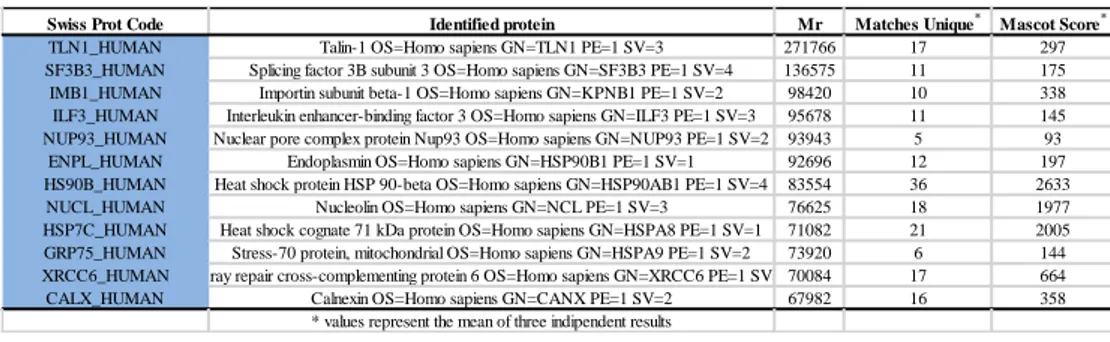
The proteins tightly bound to each resin were eluted and resolved by SDS-PAGE; the gel lanes containing the proteins eluted from the beads modified with SR2017 and the control ones were cut in 10 pieces, digested with trypsin, and analyzed by high resolution nanoLC MS/MS. Resulting MS and MS/MS data were analyzed by Mascot Search Engine software. Chemical proteomic experiments were performed in triplicate and only proteins identified in all the experiments were taken into account; moreover, proteins identified from both SR2017-modified and control beads were excluded. Using this strategy, it was possible to identify the molecular chaperone heat shock protein 60 (Hsp60), and the nuclear receptor peroxisome proliferator-activated receptor gamma (PPARɣ) as putative protein targets of our diterpene (Figure 3).
Fig.3: Proteins identified by chemical proteomic experiments as putative SR2017 molecular
25
2.4 Targets validation by Surface Plasmon Resonance
The ability of SR2017 to interact with each of the proteins identified in the chemical proteomics experiments was assayed by surface plasmon resonance (SPR). SPR is an optical technique, based on the evanescent wave phenomenon, able to measure changes in refractive index onto a sensor surface, and it is suitable for characterizing macromolecular interactions. The binding between a compound in solution and its ligand immobilized on the sensor surface results in a change of the refractive index, that could be monitored in real time allowing the measurement of association and dissociation rates; therefore, different concentration of the diterpene were injected on each of the putative protein target, singularly immobilized on sensor chips (Dal Piaz F. et al. 2009). Obtained sensorgrams (Figure 4A) clearly showed a significant interaction of SR2017 with PPARɣ (Festa C. et
al. 2012); a software aided elaboration of SPR results allowed measuring an
equilibrium dissociation constants (KD) of 89.3 ± 5.5 nM for the
SR2017/PPARɣ complex. Conversely, no affinity of the diterpene towards Hsp60 was observed (Figure 4B).
Fig.4: A) Sensograms obtained from the binding of SR2017 to PPARɣ; B) Sensograms
obtained from the binding of SR2017 to Hsp60.
Therefore, considering all these data we decided to perform an in-depth investigation on the biological role of SR2017 in the interaction with this protein. PPARɣ is a member of the PPARs subfamily (peroxisome
26
proliferator-activated receptors) of the nuclear receptor of ligand-inducible transcription factors (Laudet V. et al. 1992). It probably is the main regulator of adipocites differentiation in human, but also plays a key role in lipid and glucose metabolism equilibrium, thus controlling cell proliferation (Tontonoz P. & Spiegelman B.M. 2008). Therefore, this protein is considered a pharmacologic target for metabolic dysfunctions (Choi J.H. et al. 2010), and for cancer (Reka A.K. et al. 2010). Several thiazolidinediones have been identified as PPARɣ agonists, and some of them have be approved for type 2 diabetes therapy (Meggs D.G. et al. 1998; Kumar S. et al. 1998); however, some problems concerning the cardiovascular safety and possible hepatotoxicity of these drugs were observed (Watkins P.B. & Whitcomb R.W 1998; Nissen S.E. & Wolski K 2010). Therefore, there is still a significant requirement of new types of PPARɣ ligands, possibly acting by a mechanism different from that of thiazolidinediones. Recently, some bioactive compounds derived from plants have been described as promising activators of PPARɣ (Wang L. et al. 2014). On these basis, the identification of PPARɣ as a possible molecular target of SR2017 was interesting, since modulation of PPARɣ activity could account for many of the cellular effects previously described for this compound (Cotugno R. et al. 2014). Indeed, a pro-apoptotic activity mediated by inhibition of the PI3K/Akt system was reported for several PPARɣ activators (Moon L. et al. 2011; Kulkarni A.A. et al. 2011; Honda A. et al. 2009). Therefore, we performed different experiment in order to get more details.
2.4.1 Selectivity of 15-ketoatractyligenin methyl ester towards different isoform of PPARγ
PPAR nuclear receptor is a big family including three different isoforms, PPARɣ, PPARα and PPARδ encoded by different genes. Therefore, to evaluate the selectivity of SR2017 towards PPARγ, this compound was also
27
subjected to SPR analyses on PPARα and PPARδ, two proteins structurally and functionally related to PPARγ (Fruchart J.C. 2009; Reilly S.M. & Lee C.H. 2008). All the SPR experiments were performed using the full length proteins.
PPARδ and PPARα were immobilized on a sensor chip and increasing concentration of our diterpene were injected. No binding occurred between SR2017 and PPARδ; conversely, some interaction of this compound with
PPARα was observed, but the KD measured (1.32 ± 0.08 µM) was about 50
time higher than that measured for the PPARγ/SR2017 complex. A comparison between the sensorgrams acquired for the interaction of SR2017 with PPARα and PPARγ reveals that the binding phases are similar, but there are clearly different kinetics of dissociation: PPARα/SR2017 complex was completely dissociated after less than 50 s, whereas PPARγ/SR2017 complex dissociation required long times and remains uncompleted.
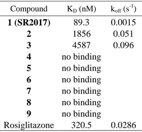
2.5 Structural characterization of SR2017/PPARɣ complex
The SR2017/PPARɣ complex appeared to be very stable, as inferred by the
measured low koff; (see Table 1) moreover, SPR sensorgrams showed that the
dissociation of this complex required long times and remains uncompleted. This data, and the presence in the PPARɣ amino acidic sequence of a cysteine residue (Cys285), highly reactive towards nucleophile groups such as a α,β-unsaturated ketones (Shiraki T. et al. 2005), prompted us to investigate the possible formation of a covalent bond between our diterpene and the nuclear receptor. Such bond could indeed be almost partially responsible for the high stability of the complex. In that aim, we performed a classic MS-based peptide mapping on the SR2017/PPARɣ complex using trypsin as proteolytic agent, to investigate the presence of covalently modified peptides. This analysis allowed us covering most of the protein sequence. Besides, a doubly charged ion at m/z 659,8481 was observed suggesting the presence of the peptide
281-28
288 covalently bound to SR2017; this hypothesis was confirmed by MS/MS analysis of this ion, thus reveling the modification occurring to Cys285 via Micheal addition (Figure 5).
Fig.5:High resolution mass spectrum and MS/MS data of peptide 281-288 covalently bound to SR2017
2.6 15-ketoatractyligenin methyl ester is a bona fide PPARγ ligand and requires the binding to C285 for its transcriptional activity
To verify if SR2017 is a bona fide PPARγ ligand able to activate PPARγ-mediated transcription, we transiently transfected a PPRE-TK-luciferase-reporter plasmid in HEK293 cells (Human Embryonic Kidney 293) that stably express an exogenous PPARγ (in addition to the endogenous protein). In collaboration with Prof. Vittorio Colantuoni from the University of Sannio, it was possible to evaluate the biological relevance of SR2017 in vitro. More in details, HEK293 cells were selected as they express a fixed and known amount of PPARγ, so that the differences in luciferase activity are due to the different ligand used.
First, we tested the antiproliferative effect of SR2017 on HT-29 colon-cancer derived cells and compared the achieved results with those observed for rosiglitazone, a fully characterized PPARγ agonist (Hong G. et al. 2003). Cells
29
were cultured for 24 and 48 hs in the presence of increasing amounts of SR2017 or rosiglitazone, and counted (Figure 6). 15-Ketoatractyligenin methyl ester displayed a dosage-dependent inhibition of cell growth that was more pronounced than that produced by rosiglitazone. This stronger effect suggests that it likely activates specific antiproliferative and/or proapoptotic pathways.
Once established the cytotoxic effect of SR2017 also on this cell model, trasfected HEK 293 cells were treated with increasing concentrations of rosiglitazone or SR2017 for 24 h and harvested for luciferase activity 48 h later. Rosiglitazone induced luciferase activity in a dose-dependent manner reaching a peak at 0.8 µM and declining at higher concentrations as illustrated in Figure 7. Therefore, it was possible to validate the interaction of SR2017 with PPARγ. The obtained results displayed a transactivation activity of SR2017 that was about 40% lower than the full agonist rosiglitazone, with a peak at 1 µM. These results demonstrate that SR2017 can be considered a PPARγ partial agonist compared to the full agonist.
30
Fig.6: To assess the effect of SR2017 on exponentially growing HEK293 cells, cells were
treated with increasing doses of SR2017 or rosiglitazone (1,5 and 10 µM). Cells were collected and counted after 24 and 48h.
Finally, we asked whether the binding of SR2017 to Cys285 in the ligand-binding pocket is required for the PPARγ transcriptional activity. In fact, previous results (see 2.5) shown the covalent interaction of SR2017 with this cysteine and its implication on the stabilization of the complex. To this goal we transiently transfected basal HEK293T cells with the PPRE-TK-luciferase-reporter plasmid along with the expression vectors for the wild-type PPARγ1 or a mutant version carrying the Cys285Ala substitution (Waku T. et al. 2009).
31
Fig.7: Transient transfection assay of the PPRE-luciferase reporter gene in human HEK293T
cells stably expressing an exogenous Flag-tagged wild type PPARγ1. After transfection, cells were treated for 24 h with rosiglitazone or SR2017 respectively, at the indicated doses. Luciferase activity is reported as fold induction after normalization to β-galactosidase activity
used as transfection control.
Cells were exposed for 24 h either to the vehicle alone or two concentrations of rosiglitazone and SR2017 shown to produce the highest induction; luciferase activity was measured in the cell extracts 48 h later. Rosiglitazone was able to bind both the wild type and mutant receptor with similar affinity and stimulate equivalent luciferase activity. SR2017 interacted with wild type PPARγ and stimulated 40 % less luciferase activity than rosiglitazone, in line with being a partial agonist, as reported above. Strikingly, the interaction of SR2017 with the C285A mutant resulted in an even lower luciferase activity (Figure 8). These results show that the C285A mutation does not influence rosiglitazone-induced PPARγ transcriptional activity, while it impairs the induction elicited by SR2017, clearly demonstrating that the binding to this amino-acid residue is absolutely required for stimulating PPARγ transcriptional activity.
32
Fig.8: Human HEK293T cells were cotransfected with the PPRE-luciferase reporter gene
along with wild-type PPARγ1 or its (Cys285Ala) mutant version. After transfection, cells were treated for 24 h with rosiglitazone or SR2017 at the indicated concentrations. Luciferase
activity is reported as fold induction after normalization to β-galactosidase activity used as transfection control.
2.7 Molecular docking of SR2017/PPARγcomplex
SR2017 is determined to be a partial agonist covalently bound to PPARγ in the current work (Figure 5). The covalent coupling of the ligand to Cys285 of PPARγ is the result of a Michael addition (conjugate addition) for which the organic reaction is well established and illustrated in Figure 9. The Michael acceptor contains an electron withdrawing group conjugated to an activated carbon that is then subject to attack by the nucleophilic Cys285.
33
Fig.9: Mechanism of covalent coupling of SR2017 to Cys285 of PPARγ
Thus, in collaboration with Prof. Antonio La Vecchia from the University of Naples Federico II, we chose to employ the covalent docking protocol CovDock ( Zhu, K. et al. 2014; Toledo-Warshaviak D. et al. 2014.) implemented in the in the Schrödinger Suite (Schrödinger, LLC, New York, NY, 2015) in order to visualize possible conformational states of the Cys285 thioether resulting from a reaction with compound SR2017, assuming that its chemical reactivity (vide supra) is relevant in the context of the protein. CovDock uses different tools of the Schrödinger Suite to mimic distinct stages of covalent inhibitor binding. The first step is a classical noncovalent docking with an alanine mutation of the nucleophilic side chain followed by an automated bond formation and a second docking step with the covalent bond in place. The basic concept of the software is that a covalently bound ligand has to adopt an energetically favorable unbound pose before bond formation occurs and that these unbound poses do not change dramatically during the reaction pathway, because conformational sampling is done solely prior the
noncovalent docking step. Docking was carried out employing the publicly
available X-ray crystal structure of PPARγ in complex with the partial agonist LT175 (PDB code: 3B3K) (Montanari R. et al. 2008).
A low-energy pose of SR2017 covalently bound to Cys285 was predicted by CovDock, and this conformation, which adopts a R configuration at position 16 of the Michael acceptor, is stabilized by several H-bonds with the key
34
Unlike rosiglitazone, which takes a U-shape conformation in the ligand-binding pocket and wrap around H3 to directly contact the AF-2 helix (H12),
SR2017 occupies the region of PPAR delimited by the H3 and the -sheet (
-sheet sub-pocket) and makes no contact with H12 or residues involved in co-activator recruitment (Figure 10a,c), as already observed in other structures of complexes with partial agonists, such as the complexes with BVT.13, MRL-24, and nTZDpa (Bruning, J. B. et al. 2007). In contrast, the full agonist rosiglitazone occupies roughly 40% of the ligand-binding site of PPARγ in a
U-shaped conformation and consists of a polar head and hydrophobic tail.The
polar head makes a net of the H-bonds with Ser289, His323, His449, and Tyr473 PPARγ side chains, while forming a hydrophobic region with Phe363,
Gln286, Phe282, and Leu469. Despite the fact thatSR2017 binds in a different
mode than rosiglitazone, occupying the -sheet sub-pocket, the ent-kaurane
skeleton of SR2017 overlaps with the hydrophobic region of rosiglitazone
when the two structures are superposed (Figure 10b).
The carbonyl and the ethereal oxygens of the ester group at position 4 of the ligand form two H-bonds, one with the NH backbone of Ser342 (located at the
-sheet) and one with both NH2 and Nε of Arg288 side chain on H3. The
hydroxyl group at position 2 establishes a H-bond with the C=O backbone of
Ile281 (dOH-O = 2.8 Å) as well as a very weak H-bond (dOH-N = 4.1 Ǻ) with the
side chain of His266, located at the Ω loop that links H2 to H3. Non-polar contacts are observed along the full extension of the SR2017 molecule. These contacts start at the Ω-loop of the protein, and extend all the way through the ligand binding pocket. Residues involved in these interactions include Phe264 (part of the Ω-loop), Ile281, Gly284, and Phe287 on H3, Val339, Ile341 and