Indications and Results of Endovenous Laser Treatment of Saphenous Incompetence
64
0
0
Testo completo
(2) INDICE. 1. INTRODUCTION....................................................................................... 3 2. MATERIALS AND METHODS ............................................................. 12 2.1 Patient Selection ....................................................................................................... 12 2.1.1 Role of Duplex Ultrasound (DUS) evaluation in patient selection ............. 12 2.1.1.1 Venous Anatomy ..................................................................................... 13 2.1.1.2 DUS Technique ....................................................................................... 15 2.1.2 Selection Criteria......................................................................................... 18 2.2 Patient population..................................................................................................... 20 2.3 Description of Technique ......................................................................................... 21 2.4 Study Endpoints and Definitions ............................................................................ 28. 3. RESULTS................................................................................................... 31 4. DISCUSSION ............................................................................................ 33 4.1 Improved patient comfort using Endovenous Ablation compared to Traditional Surgery ....................................................................................................................... 34 4.2 Occlusion rates, complications, importance of tumescent anaesthesia in EVLT and RFA and reasons to choice EVLT. .................................................................... 35 4.3 Choice of best EVLT parameters ............................................................................ 41 4.4 Comparison of recurrence between traditional surgery and EVLT ..................... 45 4.5 Learning curve and proper EVLT indications ........................................................ 49 4.6 Use of concomitant phlebectomy ............................................................................. 50 4.7 Small Saphenous Vein experience......................................................................... 51. 5. CONCLUSION.......................................................................................... 53 6. REFERENCES .......................................................................................... 54. 2.
(3) 1. INTRODUCTION LOWER extremity venous insufficiency is a common medical condition afflicting 25% of women and 15% of men in the United States and in Europe 1. Gender, pregnancy, hormones, aging, and gravitational forces from prolonged standing or sitting are the most common factors that influence the appearance or worsening of primary varicose veins 2,3. Leg symptoms of primary venous insufficiency include aching pain, fatigue, burning, itching, heaviness, night cramps and restless limbs. Symptoms arise from pressure on somatic nerves by dilated veins and are typically worsened with prolonged standing, during the premenstrual period, or in warm weather 4. Left untreated, nearly 50% of patients with significant superficial venous insufficiency will eventually experience chronic venous insufficiency characterized by lower-extremity swelling, eczema, pigmentation, haemorrhage, and skin ulceration 5,6. During the past decade, increased interest in venous disorders and the development of new non-invasive diagnostic tests and minimally invasive treatment options have led to tremendous advancement in the understanding and management of varicose vein. Great saphenous vein (GSV) reflux is the most common underlying cause of significant varicose veins. The impact of incompetence of the small saphenous vein (SSV) system reflux is also significant 7. Reflux secondary to an incompetent GSV and SSV may give rise to clusters of varicosities located in multiple areas of the lower extremities. Treatment of superficial venous disease is directed toward excluding the defective vein(s) so that venous blood returns to the right heart through normal deep veins. Therefore, when it is determined that GSV and SSV reflux is the principal underlying problem, treatment should involve eliminating the highest point of reflux with ablation of any associated incompetent venous segments. This kind of treatment has undergone. 3.
(4) important changes during the past 8 years. Prior to this period, elimination of saphenous vein reflux was either accomplished surgically (ligation and stripping) or chemically (sclerotherapy).. Traditional treatment of GSV reflux has been surgical removal of the vein. Although surgical ligation and stripping has proven to be the most successful treatment method for truncal varicosities, when the sapheno-femoral junction (SFJ) and the GSV are incompetent. 8-11. , it is associated with high recurrence rate and significant peri-operative. morbidity. Surgical ligation of the SFJ, with or without GSV stripping has been associated with quite high recurrence rates. The figures vary from series to series, ranging from 18 to 62 % at 10 years 12-14. The reported risk of saphenous nerve injury is up to 40 % with fulllength GSV stripping 15. Even partial stripping down to knee level is associated with a 7 % risk of saphenous nerve damage 16. In addition, traditional surgical approach carries risk of complications such as bleeding, unsightly scarring, haematoma formation, wound infection and deep vein thrombosis (DVT) and it’s often performed under extensive anaesthesia ( general or subaracnoid) with its associated risks. Some of these drawbacks have been minimized by today’s ligation and stripping, performed on day surgery basis with groin to knee invaginating stripping and concomitant branch miniphlebectomy under a minor form of anaesthesia with the aim of avoiding the trauma of conventional surgery 17-19(Figure 1).. 4.
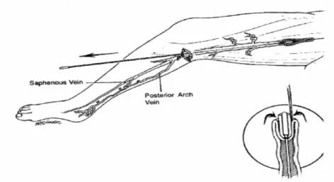
(5) Figure 1. Surgical removal of the saphenous vein as practiced today consists largely of performing the inversion stripping manoeuvre as illustrated here. The inguinal incision is usually 2 cm in length. The incision at the knee is 3 mm in length (enlarged in the figure for illustrative reasons).. Even so surgical stripping has not been well accepted by patients who perceive the procedure as risky, disfiguring, requiring hospitalization, and requiring lengthy convalescence. Sclerotherapy, on the other hand, is performed commonly throughout the world with minimal risk, but with high failure rates. Less invasive surgical treatments including high ligation of the GSV at the SFJ have been attempted with the hope that gravitational reflux would be controlled while the vein is preserved for possible use as a bypass graft. Unfortunately, ligation of the GSV alone usually results in recurrent varicose veins. 20. . Even when high ligation has been combined with phlebectomy of varicose. tributaries or retrograde sclerotherapy, recurrence has been the rule 21,22. Traditional treatment of SSV reflux is ligation of the sapheno-popliteal junction (SPJ) with or without stripping of the SSV. However recurrence rate following surgery may be as high as 50% at 3 years23. In many instances this is the result of inaccurate ligation of the SPJ. In addition neovascularisation, which is considerable cause for. 5.
(6) recurrence following SFJ ligation and GSV stripping may have a role. 24,25. . The frequency. with which this occurs following SSV surgery has not been investigated but when present it allows further reflux into the SSV, which is often still present. Also the risk of nerve injury (sural nerve) following SSV stripping is not neglectable 26. In an attempt to reduce morbidity and improve recovery time, several minimally invasive techniques have been developed and proposed as alternatives to surgery. In recent years, the introduction of new endovenous ablation technologies, namely the VNUS Closure® endovenous radiofrequency ablation (RFA) procedure (VNUS Medical Technologies, San Jose, CA) and endovenous laser treatment (EVLT) have stimulated much interest in venous disease management and has rejuvenated the field. RFA and EVLT achieve thermal ablation of refluxing saphenous veins and were primarily developed to treat varicose veins due to SFJ and GSV reflux. The goal of these new endovenous techniques is to avoid the drawbacks associated with traditional surgical treatment for varicose veins and due to their minimally invasive nature they have been widely adopted by practitioners and accepted by patients. These minimally invasive therapies employ both the delivery of thermal energy to the vein wall (via intraluminal means) to destroy the intima and denature collagen in the media resulting in” pan-mural” damage associated to intraluminal minimal thrombosis in order to achieve subsequent fibrous occlusion of the vein. General procedure techniques are very similar between RFA and EVLT. Both procedures involve ultrasound imaging guidance, vein access and catheter navigation, tumescent fluid infiltration, and continuous pullback of the catheter or laser fiber to deliver energy along the vein. However, the mechanism of action is different between the two technologies.. 6.
(7) RFA closure was the first technique to reach the level of widespread clinical use for treating the GSV. It leads to occlusion of the vessel by means of a disposable endovenous catheter, introduced in the vein, delivering thermal energy from a bipolar radiofrequency source (Figure 2-3) through the direct contact of its electrodes with the vein wall. RFA has a temperature-controlled feedback loop that monitors the vein wall contact with the catheter and controls energy delivery. The contact between the catheter electrodes causes endothelium denudation, heat-induced venous spasm with immediate vein shrinkage and collagen destruction, which produces maximal physical contraction, followed by inflammatory response and fibrosis of the treated vein. Clinically the device produces precise tissue destruction with minimal formation of thrombus. The bipolar endovenous catheter system, with a feedback mechanism, uses a 460-kHz current to heat the vein wall to a target temperature with power typically in the range of 2 to 4 W. Polar molecules, such as water, transfer electrical energy to heat energy by current-induced molecular movement resulting in temperatures between 85ºC and 90ºC at the vein wall. To avoid drainage of heat energy by intravascular blood flow, the vein must be emptied as much as possible by means of placing the patient in Trendelenburg position and the use of external compression (Esmarch bandage). These manoeuvres result in full contact between the vein wall and the catheter tip electrodes. Together with a slow initial catheter pullback rate in the range of 1 cm/min, followed by 2 to 4 cm/min thereafter, the vein wall collagen undergoes a relevant shrinkage process that is more pronounced than observed with laser and is almost absent with sclerotherapy.. 7.
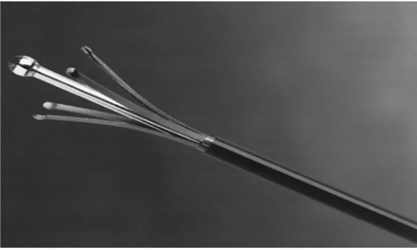
(8) Figure 2. The intravenous catheter consists of sheathed electrodes specifically designed for treatment of GSV and a thermocouple for temperature monitoring. It is available in sizes of 6 and 8F.. Figure 3. The RF generator shown here connects directly to the intraluminal catheter placed in the GSV and allows monitoring of electrode temperature (85°C).. EVLT is based on a thermal process too, allowing delivery of laser energy directly into the blood vessel lumen. In 1999 Boné 27 first reported on delivery of endoluminal laser energy. Subsequently, in 2001, Navarro et al.. 28. published the first report of a minimally. non invasive method of treating the entire incompetent GSV using an 810-nm diode laser. This relatively new minimally invasive technique, which received approval from the US. 8.
(9) Food and Drug Administration in January 2002, is nowadays commonly applied to the GSV with high success rates (88%-100%) 29-31 and seems to be the most promising among the two endovenous procedure 32,33. The treatment is based on the conversion of light into thermal energy through the absorption of laser-light by the haemoglobin or by haemoglobin and water (depending on the diode laser wavelength) within the vein. Because haemoglobin (or haemoglobin and water) acts as a chromophore, local presence of blood is required to conduct laser energy. Proebstle has demonstrated that endoluminal heat damage is caused by steam bubbles originating from boiling blood at the tip of the laser fiber. 34. . Steam bubbles transfer heat to the vein wall resulting in damage to the. endothelial and sub-endothelial tissues. Proebstle is convinced, in contrast to the mode of action of RFA where a significant shrinkage of the vessel wall is observed, that EVLT heat-mediated damage is directed mostly to the inner vein wall (the endothelium is destroyed by a process known as selective photothermolysis) leading to a thrombotic occlusion of the treated vein34-35 with a kind of thrombus more stable than in a spontaneous thrombophlebitis of the proximal GSV, where partial or total recanalization is the rule, probably due to the density of laser-induced damage. This observation was supported by a histological study performed by Corcos et al. that showed that when permanent occlusion was observed, the endothelium and intima were always damaged and that success was independent of the vessel wall thickness 36. A more accreditate theory, confirmed from recent anatomo-pathological studies. 37. supports the concept that the pathological lesion of the vein is not restricted to the tunica intima but is deeper and extensively affects the tunica media and adventitia, which differentiates endovenous laser treatment from sclerotherapy. The effect caused by EVLT is an extensive and in-depth damage with alteration of the proteins constructing the entire. 9.
(10) vein wall (pan-mural coagulative necrosis), which triggers the process of sclerosing of the wall itself, associated to a laser induced thrombosis. This is the “primum movens” of the subsequent process of shrinkage and sclerosis of the vein. Demonstration of the denaturation of the collagen of the media is proof of in-depth penetration of the heat in the vein wall and can explain the reduction of its diameter, as observed in the post-operative ultrasound, though the mechanism of shrinkage of the collagen . Technically vein occlusion is accomplished by heating the vein wall with different wavelength diode laser energies delivered via a 600-µm fiberoptic. The most frequently used lasers are diode lasers, with wavelengths between 810 nm and 980 nm ( 810-940-880 nm.). The 810 and 940 nm wavelengths are predominantly absorbed by hemoglobin, which is needed intravenously to a small extent to guarantee successful laser treatment, whereas the 980 nm wavelength is absorbed both by hemoglobin and water. The treatment is usually performed using local anaesthesia. Access to the great or small saphenous is achieved by either needle puncture or the stab wound–Mueller hook approach. A guide-wire is introduced into the vein and manoeuvred towards its junction with the deep vein under ultrasonographic guidance. A catheter is then introduced over the guide-wire, which is removed. A diode bare-tipped optical laser fiber is inserted into the catheter, exposed distally for 3 cm out of the catheter and positioned approximately 2-3 cm below the sapheno-femoral (usually behind epigastric superficial vein) or saphenopopliteal junction (behind gemellar tributaries, if joint in a common trunk with SSV, or competent Giacomini vein), the position being confirmed by ultrasonography. Perivascular tumescent infiltration of local anaesthetic along the length of the vein to be treated aside from its anaesthetic function absorbs and dissipates the heat generated during the procedure, preventing injury to the surrounding tissues, and compresses circumferentially. 10.
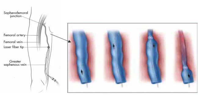
(11) the vein around the laser fiber. The laser is turned on, and the fiber is slowly pulled back through the vessel, thus allowing delivery of laser energy to the vessel lumen to produce entire vein wall damage with subsequent fibrosis (Figure 4).. Figure 4. Diagram of the laser fiber being slowly withdrawn within the GSV as laser energy is delivered endovenously. Sufficient heating of the vein wall (ranging from 55° C to 85 °C) is necessary to cause collagen contraction and denudation of endothelium. This stimulates vein wall thickening and eventual luminal contraction with subsequent fibrosis of the vein. We report our preliminary experience of EVLT of GSV and SSV reflux.. 11.
(12) 2. MATERIALS AND METHODS This prospective, non-randomized, consecutive-enrolment study included 24 patients who underwent endovenous laser treatment of incompetent GSV or SSV segments with 940-nm diode laser energy delivered intraluminally for treatment of primary varicose veins.. 2.1 Patient Selection Directed history and physical examination, including duplex ultrasound (DUS) evaluation of the superficial venous system, was performed on limbs of subjects with varicose veins. DUS has demonstrated that saphenous veins are often not the refluxing vessel causing varicosis. Anterolateral tributary veins, posteromedial tributary veins, and even small groin veins such as epigastric veins can be the source of abnormal venous hemodynamics. If a surgeon identifies the correct refluxing vein(s) prior to treatment, the outcome will be favourable. The treating physician must therefore be self-sufficient with regard to handling an ultrasound probe and recognizing the nuances of venous anatomy.. 2.1.1 Role of Duplex Ultrasound (DUS) evaluation in patient selection DUS is an essential component of EVLT. Clinical evaluation based solely on the distribution of a venous abnormality can suggest a pattern of incompetence. Unfortunately, different patterns of incompetence can result in a similar visual appearance of abnormalities. Therefore, treatment decisions based solely on the clinical evaluations, even. 12.
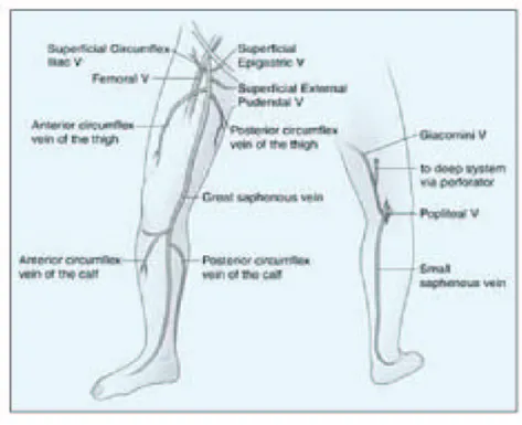
(13) those supplemented by a handheld directional Doppler, are often fraught with errors. DUS is required to evaluate patients with superficial venous insufficiency. It is advisable that all patients undergoing evaluation for varicose veins, oedema, or venous skin changes undergo an ultrasound of the superficial venous system to determine the pattern(s) of incompetence prior to making treatment recommendations.. 2.1.1.1. Venous Anatomy. The superficial venous system is composed of the saphenous veins, their primary branches, and their tributaries. 38,39. . All of these veins are superficial to the deep or. muscular fascia. The main trunks of the saphenous system are the GSV and small saphenous vein SSV. These trunks and some of the named tributaries of the GSV are actually intrafascial, deep to the superficial but still superficial to the deep fascia. The GSV begins on the dorsum of the foot, ascending first anterior to the medial malleolus, and then along the inner thigh to ultimately join the femoral vein at the fossa ovale a few centimeters below the inguinal ligament. The GSV has two important tributaries in the calf and in the thigh. In addition, there are three smaller tributaries that join the GSV just prior to entering the fossa ovale (Figure 5).. 13.
(14) Figure 5. Frontal and posterior diagrams of the lower extremity demonstrating the great and small saphenous veins and their named tributaries. The two saphenous systems can be connected via the vein of Giacomini.. The saphenous veins and these important tributaries are connected to a complex network of collecting veins that drain the skin and subcutaneous tissues, as well as innumerable perforating veins that normally drain blood from the saphenous veins and their tributaries through the muscular fascia to the deep veins. The SSV begins on the lateral foot, passes posterior to the lateral malleolus, and ascends the posterior calf. 40. . This intrafascial vein will drain into the popliteal vein just. above the popliteal crease in approximately 70% of patients. In most of the other patients, there is a cephalad extension of the SSV that usually drains to the deep veins through a posterior thigh perforating vein or to the GSV via a connection known as the vein of. 14.
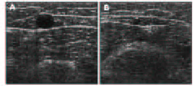
(15) Giacomini. On axial ultrasound, the SSV and GSV will have the appearance of Cleopatra’s eye (Figure 6 A and B).. Figure 6. Axial DUS appearance of truncal veins. Two-dimensional DUS image of the thigh portion of enlarged refluxing GSV within the saphenous space. The anechoic central circle is the lumen of the GSV surrounded by fat in the saphenous space, which is demarcated by the curvilinear anterior and posterior portions of the saphenous fascia (A). DUS image of a normal diameter SSV at the level of the gastrocnemius muscles. The anechoic vein is also found in a saphenous space demarcated anteriorly and posteriorly by portions of the sheath (B).. The vein will appear as the pupil outlined by the curvilinear saphenous space, which is separated from the surrounding tissues by the echogenic superficial and deep fascia. This appearance is helpful in distinguishing the truncal veins from more superficial tributaries.. 2.1.1.2. DUS Technique. The equipment required to perform the examination of the superficial venous system is a linear 7.5 MHz to 10 MHz transducer capable of displaying greyscale two-. 15.
(16) dimensional and pulse-wave Doppler (PWD) images. Color-Doppler is a helpful feature that can make the examination more efficient, but it is not required. When evaluating patients for reflux, the examination should be performed in the standing position. The reflux that leads to venous pathology can be reliably documented in this position. The examination begins at the SFJ. The GSV is followed from its junction down beyond the level of any visible varicose veins. The relationship of the GSV to any abnormal veins is assessed by tracing its course and the course of any tributaries that might lead to the abnormal veins. It is important to be aware of the standard tributary anatomy of the GSV and to recognize the frequent variations that are found 38,39. The caliber of the GSV is then assessed. Normally, the vein is 4 mm in diameter. Veins > 7 mm have a high incidence of reflux. Reflux can occur in smaller veins, but even if found, it is usually clinically unimportant. Peripheral to the take off of incompetent tributary veins, the caliber of the vein often decreases. Any vein segment suspected of having reflux by size or by relationship to varicose veins is then evaluated with ColorDoppler and PWD to directly visualize the direction of flow. All suspicious segments should be examined with PWD. Reflux can be easily documented by looking for antegrade flow followed by retrograde flow after a quick, firm compression of a peripheral segment of the GSV. Generally, when evaluating the GSV, compression of the calf should lead to augmentation of antegrade venous flow. Upon release of compression, little if any retrograde flow should be noted. Reflux is documented when a significant amount of retrograde flow is found (Figure 7).. 16.
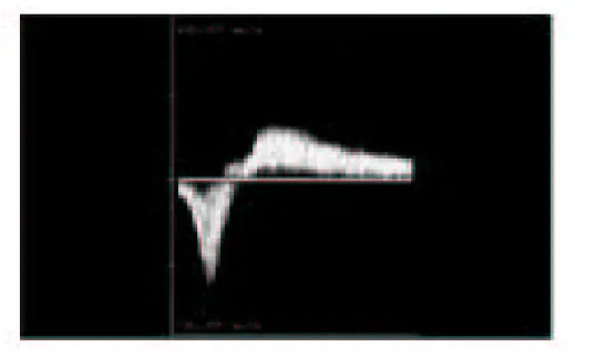
(17) Endovenousaricose Veins Figure 7. Pulse-wave Doppler imaging of an incompetent GSV segment is performed during and immediately after external compression of the extremity at a more peripheral location. Blood velocities on the y-axis are plotted against time on the x-axis. Augmentation of flow toward the heart is normally seen with calf compression (velocities mapped below the x-axis). However, upon release of external compression, flow directed toward the feet is seen in incompetent segments (velocities above the x-axis). A normal vein may have 0.5 second of retrograde flow.. Although the criteria of 0.5 second of retrograde flow has been used to identify pathological reflux, several seconds of retrograde flow is usually found in patients with incompetence. Next, the patient is turned away from the examiner and the SSV is evaluated. The process of evaluation is similar to that of the GSV. The SSV normally measures 3 mm; enlarged veins are frequently incompetent. Finally, an evaluation of the femoral vein and the popliteal veins for reflux and obstruction should be included in the examination. In some cases, DUS in patients with varicose veins will not identify truncal vein incompetence. These non-truncal pathways are much more common in multiparous women and include pudendal and gluteal vein incompetence. 41. . Other important sources of non-. 17.
(18) truncal reflux include incompetent perforating veins in the medial and lateral thigh and popliteal fossa, which can usually be identified with DUS. Occasionally, these sources, especially the pudendal source, can lead to GSV incompetence more peripherally in the leg, so it is worth completely examining the saphenous trunks in all patients.. 2.1.2 Selection Criteria Certain patients are not suitable for endovenous therapy. Patients with multiple comorbidities are not good candidates. Furthermore, patients with known allergy to local anaesthesia, thrombophilia, prior DVT with incomplete recanalization, and active superficial phlebitis are best treated conservatively with compression. There are two anatomical considerations that make endovenous therapy undesirable. Veins located just below the surface of the skin are best removed surgically. An endothermally treated vein immediately below the skin will result in an unsatisfactory cosmetic result because patients often develop a stain and/or palpable cord on the skin of the medial thigh and leg. Although resolution of these problems is often spontaneous, they may persist for more than 12 months. Second, vein tortuosity can be a challenge because guide-wire navigation is difficult. In some cases, by employing multiple entry sites, these veins can be satisfactorily treated. Experience and careful clinical judgment are essential in these cases. Our Inclusion and Exclusion criteria are reported in the next Table 1.. 18.
(19) Table 1. EVLT Inclusion and Exclusion criteria used at Policlinico Tor Vergata Inclusion criteria: • Varicose veins caused by SFJ incompetence with GSV reflux or by SaphenousPopliteal Junction ( SPJ) incompetence with SSV reflux as demonstrated by DUS • Age of at least 18 years • Completed written informed consent from acknowledging awareness of the alternative treatments available, risk involved, and other issues that conform to the standard of care for informed consent practices • Ability to return for scheduled follow-up examinations for at least 12 months after endovenous laser treatment. Exclusion criteria: • Not favourable vein anatomy - vein diameter in standing position > 20 mm - vein distance from the skin < 4 mm ( vein superficiality) - multiple big refluxing collateral branches or perforating veins on saphenous course - short but excessive dilatations of GSV and SSV - reflux from junction vessels in particularly from epigastric veins - duplications, anatomic malformations or extreme tortuousity of GSV or SSV that would not allow endovenous catheterization and passage of the laser fiber as identified on pre-treatment DUS mapping. • Previous sclerotherapy of saphenous vein • Non palpable pedal pulses • Inability to ambulate • Deep vein thrombosis • General poor health • Pregnancy, nursing, or plans to become pregnant immediately after the treatment. After initial examination and consultation, patients meeting the selection criteria were offered the choice of surgery or EVLT. Nearly all patients chose EVLT over surgical stripping. All patients were treated after extensive informed consent and according to the ethical guidelines of the 1975 Declaration of Helsinki. We thoroughly informed patients about the procedure operation risks, possibility of disease recurrence in case of recanalisation of the vein and the limited amount of available data on the long term efficacy of this technique and the patients signed the written informed consent form.. 19.
(20) 2.2 Patient population Twenty-one incompetent GSVs and three SSVs were treated with EVLT over a 5 months period from January 2006 to May 2006. No patients were lost to follow-up (FU). The population treated was 82% (20 of 24) female and 18% (4 of 24) male, with a mean age of 52.8 (range, 18-80 years, SD 13,2 years). The severity of venous insufficiency was categorized according to CEAP classification (Porter) (Table 2 and 3). The preoperative CEAP Clinical Stage was 2 in all the patients. Pre-treatment GSV diameter, measured in the upright position approximately 2 cm below the SFJ, ranged from 5 mm to 16 mm (mean, 8,6 mm; SD, 2,8 mm). Pre-treatment SSV diameter, measured in the upright position approximately 2 cm below the SPJ, ranged from 4 mm to 8 mm (mean, 5,7 mm; SD, 2,1 mm). FU ranged from 12 month to 16 months with a mean FU period of 13,8 months and an SD of 1,5 months.. Table 2. CEAP Classification of chronic lower extremity venous disease (Porter & Moneta 1995). Mark Definition C Clinical signs (grade0-6), supplemented by (s) for symptomatic and (a) for asymptomatic presentation E Etiologic Classification (Congenital, Primary, Secondary) A Anatomic Distribution (Superficial, Deep, or Perforator, alone or in combination) P Pathophysiologic Dysfunction (Reflux or Obstruction, alone or in combination). 20.
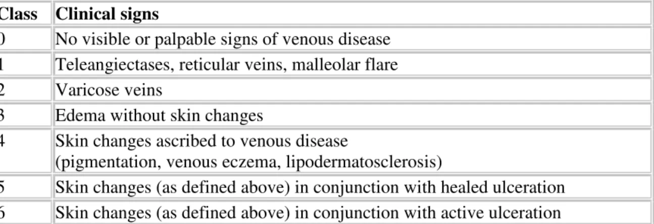
(21) Table 3. CEAP Clinical classification of chronic lower extremity venous disease (Porter & Moneta 1995).. Class 0 1 2 3 4 5 6. Clinical signs No visible or palpable signs of venous disease Teleangiectases, reticular veins, malleolar flare Varicose veins Edema without skin changes Skin changes ascribed to venous disease (pigmentation, venous eczema, lipodermatosclerosis) Skin changes (as defined above) in conjunction with healed ulceration Skin changes (as defined above) in conjunction with active ulceration. 2.3 Description of Technique Preoperative hemodinamic DUS was performed in the upright position to map incompetent sources of venous reflux and then to mark with indelebile skin marker, the skin overlying the incompetent portion of the GSV starting at the SFJ or of the SSV starting at the SPJ. Transverse measurements of the GSV were made at the upper, medium lower part of upper leg. After DUS mapping, an entry point was chosen. Entry was chosen at the lowest level of the primary incompetent segment or where the vein becomes too small to access, usually at the level where the last incompetent tributary vein joins the truncal vein, below which point the vein is normal in calibre and regains competence. This point for GSV is frequently around knee level or on the lower thigh and for SSV at the median calf. The patient was then positioned on operating table, betadine skin cleansed and draped in the usual sterile fashion. The first 6 patients underwent local assisted anaesthesia, without performing tumescent anaesthesia (TA) and using sedation with Propofol during the pullback action time of the laser probe. All the others were treated under local. 21.
(22) anaesthesia with TA. With use of local anaesthesia (2 ml Lidocaine 1%) access was obtained in the GSV or SSV via a percutaneus needle puncture (19G needle) using ultrasound guidance or a stab wound-Muller hook approach. Stab wound-Muller hook approach was our preferred, performed in 14 on 24 limbs treated (58,3%). The decision for percutaneous or stab wound entry was made depending on the quantity and size of leg branch varicosities dictating a need for phlebectomy near the entry point. The GSV entry point was in 15 cases below the median condilus of the knee and in 6 cases about 5-10 cm above the knee. The SSV entry point was always at the medium leg. A 5-F introducer catheter was advanced into the GSV o SSV over a 0,035-inch guide-wire and positioned around 2-3 cm past the SFJ, usually behind the superficial epigastric tributary, or the SPJ (or behind gemellary tributaries, if joint in a common trunk with SSV, or competent Giacomini vein if present). Intraluminal position within the vein was confirmed by aspiration of non-pulsatile venous blood and visualization with DUS (this is usually best done with a longitudinal projection). The catheter was flushed and a 600-µm laser fiber, connected to a 940 nm Diode Laser Equipment (Medilas Plus Dornier), was inserted and advanced up to the distal tip of the catheter (Figure 7-8). The catheter was then withdrawn exposing the distal 3 cm of the bare-tipped laser fiber and blocked with the fiber. The sheath and fiber were pulled back together and positioned in the proper position under DUS guidance. The laser fiber is depicted as an intraluminal echogenic line. The correct position was confirmed by direct visualization of the red aiming beam of the laser fiber through the skin.. 22.
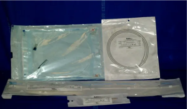
(23) Figure 7. Diode laser Medilas Plus Dornier using a 940-nm wavelength. Figure 8. The material necessary for the procedure (19 G needle for percoutaneous introduction mode, dilatator and catheter complex 5 F 55 cm, 0,035” “J”guide-wire, 600 µm laser probe ) can be assembled in dedicated kits.. 23.
(24) With the exception of the first group of 6 patients, treated under local assisted anaesthesia, tumescent local anaesthesia consisting of 100–200 mL of 0.2% lidocaine neutralized with sodium bicarbonate, was administered along the perivenous space with use of DUS guidance. Both EVLT and RFA are performed using tumescent anaesthesia (TA). In the early days of radiofrequency ablation, patients were sometimes left with skin burns or paresthesias. After the advent of subfascial, perivenous tumescent anaesthesia, those complications rarely arise. Using real-time DUS guidance, a 25-gauge needle is placed in the saphenous canal, in multiple points along the course of the vein, and the fluid is injected surrounding the entire vein wall in the saphenous compartment (Figure 9 - 10 11). This accomplishes three things: 1) Creates a reservoir of fluid surrounding the vein that dissect and separate the vein from perivenous tissue and acts as a “heat sink” minimizing the possibility of heatrelated damage to adjacent tissues (prevention of skin burns and nerve damage). When heat is placed inside the vein during the venous ablation, the heat is quickly dissipated through the wall of the vein precluding any heat-related injury of surrounding tissue. As a result, the rate of skin burns and the paresthesias has been reduced to less than 1% in experienced hands. 2) The tumescent fluid compresses and reduces the diameter of even the largest veins to provide vein wall apposition around the fiber tip with subsequent circumferential heating of the vein wall. 3) Effective analgesia. The patient should experience a painless procedure, and postoperatively, most patients are comfortable with a daily non-steroidal antiinflammatory. The tumescent technique eliminates the hemodynamic risks of. 24.
(25) sympathectomy associated with a conduction block (epidural or spinal anaesthetic), and the cardiac and pulmonary risks associated with general endotracheal anaesthesia. DUS guidance of the needle used to inject the TA fluid in the perivenous saphenous sheath is essential to maximize the effectiveness and efficiency of its delivery. A successful TA results in a perivenous hypoechoic halo that obliterates the vein along its course.. Figure 9. Duplex US (transverse view) demonstrating appearance of the GSV before and after proper delivery of tumescent anaesthesia. (a) Intraluminal position of laser fiber and catheter within an enlarged GSV located in the saphenous space; (b) proper and adeguate deliver of tumescent anaesthesia results in fluid surrounding a compressed GSV. 25.
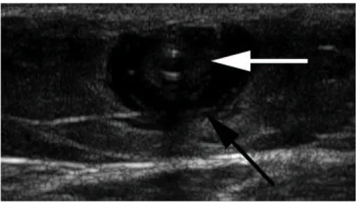
(26) Figure 10. Fluid surrounding the GSV after injection of tumescent anaesthesia in the saphenous compartment (black arrow) with collapsed wall (white arrow) of the GSV around the catheter.. Figure 11. Ultra sound image in axial view recorded after tumescent anaesthesia. The position of the fiber at the center of the vein is clearly seen. 26.
(27) After TA the patient was placed in mild Trendelemburg position, thus decreasing venous pressure and the tip of the laser fiber was repositioned within the GSV behind epigastric vein or within SSV 2 cm distal to the SPJ or behind a common trunk with gemellar tributaries, or a competent Giacomini vein if present. Tip position was checked by DUS and direct visualization of the red aiming beam through the skin. Laser energy was delivered pressing the pedal of the 940-nm diode laser equipment using a power of 15 W in continuous pullback mode at a rate of 1-3 mm/sec in order to deliver between 60 and 80 J/cmL in the GSV and between 40 and 60 J/cmL in the SSV. The vein was treated from the positioning point below the SFJ/SPJ to approximately 1 cm above the skin entry site. Length of GSV treated with endovenous laser ranged from 25 cm to 50 cm (mean, 37,6 cm; SD, 6,7 cm). The laser fiber was withdrawn at an average rate of 2-3 mm per second (14-18 cm per minute). The total energy delivered ranged from 870 J to 3540 J, with a mean of 2715,7 J ( SD, 565,9 J). The average energy ranged from 55 J/cm to 90 J/cm, with a mean of 71 (SD,12,4). Length of SSV treated with endovenous laser ranged from 20 cm to 30 cm (mean, 23,3 cm; SD, 5,8 cm). The laser fiber was withdrawn at an average rate of 3 mm per second (18 cm per minute). The total energy delivered ranged from 980 J to 1250 J, with a mean of 1103,3 J ( SD, 136,5 J). The average energy ranged from 42 J/cm to 49 J/cm, with a mean of 46,7 (SD,4,0). Immediate collapse of the GSV or LSV and absence of flow was confirmed after the procedure by DUS study. All patients in this series underwent concomitant miniphlebectomy using a crochet device done through a 2-mm skin cut to remove the dilated collateral veins immediately after the endovascular laser ablation was completed.. 27.
(28) Usually ½-inch Steri-Strips cut into thirds were placed over the previous entry site and phlebectomy incisions and covered with sterile gauze pad. A full-thigh class II (30-40 mmHg), compression stocking or pantyhose was worn for at least 1 week at all times, except to sleep or shower. Patients were instructed to walk immediately following the procedure and to resume their normal daily activities immediately with the exception of vigorous gym workouts. All patients received low molecular weight heparin (2000 IU of enoxaparine subcutaneously) once daily for 1 week and were also instructed to take a nonsteroidal anti-inflammatory medication once daily for the first 3 days after the procedure. Clinical and duplex US follow-up was obtained at 1 week, 1, 3, 6, 9, 12 and 16 months.. 2.4 Study Endpoints and Definitions. DUS criteria for successful treatment were considered the following as described by Min 31: •. At 1 week, minimally decreased in diameter and incompressible vein, with hyperechoic and thickened walls, and no flow seen within the occluded lumen on Color Doppler interrogation.. •. At 3 and 6 months evaluation, an incompressible, occluded vein, substantially reduced in diameter (at least 50%), and without flow on Color Doppler interrogation.. •. At 9 months and 1 year, a minimal residual hyperechoic fibrosis cord without detectable flow or complete disappearance of the vein. It is important to note that the expected appearance 1–2 weeks after endovenous. laser is a slightly smaller GSV/SSV demonstrating wall thickening with absence of flow within the treated vein segment. The vein lumen is usually obliterated by the thickened. 28.
(29) wall, which has low level echoes and is incompressible. This wall thickening should be differentiated from acute GSV thrombosis wherein the vein is also incompressible but the lumen is filled with anechoic acute thrombus. Several weeks after successful endovenous laser treatment, resolution of the acute inflammation in the vein wall should result in reduction in vein diameter. After several months, most of the treated vein segments will fibrose and be difficult to identify. Alternatively, superficial thrombophlebitis with GSV thrombus would result in recanalization of the vein. A longitudinal view of an incompetent GSV is seen in Figure 8a. Figure 8b demonstrates the typical ColorDoppler appearance of a successfully treated GSV 3 months after EVLT.. (a). (b). Figure 8. Color-Doppler examinations (longitudinal views) of the GSV at the SFJ demonstrating successful occlusion after endovenous laser treatment. (a) Pre-treatment evaluation demonstrates an enlarged GSV with reflux after distal calf compression; (b) 3-months follow-up examination shows typical appearance of the proximal GSV with occlusion of the treated segment.. Clinical evaluation was performed on all subjects at 1 week, 1, 3, 6, 9, 12 and 16 months by the same physician (G.V.) who participated to all the endovenous laser. 29.
(30) procedures. Patients were queried about symptomatic relief at follow-up visits, particularly improvement or resolution of lower-extremity pain believed to be associated with venous insufficiency. Improvement in the appearance of the leg, including swelling, pigmentation, or other skin changes secondary to chronic venous insufficiency were assessed by the patient. Patients were evaluated for possible adverse reactions caused by endovenous laser treatment at each follow-up visit. Minor complications were defined as those that had no significant clinical sequelae, such as ecchymosis, pain, induration of treated vein, superficial phlebitis. Major complications were defined as those necessitating an increased level of care, surgery, hospitalization, or permanent adverse sequelae such as DVT, paresthesia from nerve injury, skin burns. .. 30.
(31) 3. RESULTS Successful access and endovenous placement of the laser fiber was achieved in all patients (100%). FU results ranging from 1 month to 16 months (mean, 13,8 months; SD, 1,5 months) were obtained in all the limbs treated with EVLT during the study period. The procedure was a technical success in all patients (100%) of cases. Successful endovenous laser treatment, as defined by complete occlusion of GSV and SSV (no flow detectable by ColorDoppler interrogation), was seen in 24 of 24 limbs (100%) at the 1 week follow-up. At 12-18 months follow-up all veins treated remained closed. There has been 1 recurrence on 21 GSV treated (4,8%) noticed at 3 month FU from secondary incompetence of a tributary vessels of the SFJ. No new recurrences were seen later during FU that were not present at 3 months. No recurrence were observed in the 3 SSV treated. After one month all patients noted relief of symptoms and showed significant improvement in the appearance of the limb without evident residual scars. By 3 months after treatment, pain was greatly improved or resolved in all treated limbs. Ecchymosis and mild-to-moderate thigh or leg pain were the most common presenting symptoms during the first week and were easily treated with medication. Ecchymosis, present in 8 of 24 ( 33,3%) of patients resolved in all subjects before 1-month FU. 20 of 24 (83%) of subjects felt a delayed tightness, described as “pulling” along the course of the treated GSV or SSV, peaking 4–7 days after laser treatment and lasting 3–10 days. This sensation as described by Min. 31. is not felt in the treatment failures and may. correlate with expected acute inflammation resulting in vein wall thickening and shortening as seen on DUS. All minor complications listed earlier resolved without sequelae.. 31.
(32) There have been no skin burns, cases of deep or superficial vein thrombosis, or other major complications with the exception of 4 paresthesias at tight level on 21 GSV treated (19 %) resolved in 6 months. These paresthesias occurred only in the first group of 6 GSV treated under local assisted anaesthesia without using TA. No paresthesia were observed in the 3 SSV treated. The procedure was well-tolerated by all the subjects treated. Overall treatment satisfaction was determined by asking subjects if they would recommend the procedure to a friend with similar leg vein problems, and 24 of 24 subjects (100%) indicated they would recommend the procedure.. Table 4. Occlusion rate, recurrence and complications after EVLT of GSV and SSV Occlusion Rate Recurrence Minor Complications. - Superficial Thrombophlebitis - Ecchymosis - Excessive pain -“Pulling” sensation. Major Complications - DVT - Skin Burns - Paresthesias. EVLT GSV % ( n = 21 ) 100% 4,8% (1/21). EVLT SSV % ( n = 3 ) 100% 0%. 0% 33,3% (7/21) 0% 90,4% (19/21). 0% 33% (1/3) 0% 33% (1/3). 0% 0% 19% (4/21). 0% 0% 0%. 32.
(33) 4. DISCUSSION Traditional treatment for GSV and SSV insufficiency and resulting varicosities has involved surgical ligation and stripping of the vessel segments. However, the associated morbidity (post-operative haematoma, paresthesias, wound complications especially in the groin), the need of more extensive anaesthesia and patient dissatisfaction with the procedural results have led to the development of alternative techniques for treating these vessels. Less invasive alternatives to open surgery seek to reduce risk, morbidity, while leading to acceptable short and long-term results. Endovenous methods for treating incompetent GSV are not new. Early attempts to selectively damage saphenous veins wall to occlude them by electro-coagulation involved creation of a thrombus within the vessel lumen, ultimately resulting in recanalization. 43,44. .. The search for less invasive treatment alternatives has more recently led to the development of DUS guided foam-sclerotherapy that has gained popularity and has the advantage of significantly lower cost compared to all others techniques. This technique appear to be useful in expert hands but long-term studies have failed to prove durability comparable to surgery, with GSV occlusion rates of 90% at 28 days but only 81% at 3 years 45-46. EVLT and RFA are the two newest endovenous techniques, introduced in the last 7 years, both determining an endovenous obliteration of the incompetent troncular vein through a thermal injury, providing patients with effective alternatives to ligation and stripping. Surgical treatment is still considered the “gold standard” for varicose veins and has stood the test of time. In the early years of endovenous laser treatment of varicose veins, many surgeons were loath to embrace the new technology because vein stripping had been. 33.
(34) proven safe and effective, and data supporting endovenous treatments were sparse. Nevertheless the merits of conventional surgery have to be judged against these rapidly evolving minimally invasive endovenous techniques. As a matter of fact, as we can see from the review of literature, during the past 5 years, we have seen dramatic improvements of these procedures. Recent data have demonstrated the safety and efficacy of these techniques, as well as their superiority to venous stripping in areas of neovascularization and improved patient comfort. We report next a review of literature on the vary aspects of endovenous procedure and a comparison with our preliminary experience with EVLT.. 4.1 Improved patient comfort using Endovenous Ablation compared to Traditional Surgery The advantages of RFA over traditional vein stripping surgery are demonstrated in three randomized comparative trials 54-57. Rautio et al randomized 28 patients to receive either radiofrequency obliteration or vein stripping and reported significantly less postoperative pain, less postoperative analgesia requirements, and faster recovery in the radiofrequency group 54. The EVOLVeS study was a multicenter, prospective, randomized study, comparing quality-of-life factors between radiofrequency ablation and vein stripping. In all outcome variables, radiofrequency ablation superceded venous stripping: faster recovery, less postoperative pain, fewer adverse events, and superior quality-of-life score. 55. .The most. significant differences seen between RFA and vein stripping surgery were patient recovery, postoperative morbidities, and patient quality of life. The mean time to return to normal. 34.
(35) activity was 1.15 days versus 3.89 days (P= .02), and the percentage of patients returning to routine activities within 1 day was 80.5% versus 46.9% (P<.01) for the RFA and vein stripping surgery groups, respectively55. Similar findings were reported by the Stötter group in Germany in their own randomized trial 57. Use of RFA resulted in significantly less postoperative pain, either measured with visual analog scale or quality-of-life instruments, reduced need for analgesics, and fewer complications and adverse events in these studies. 54-57. . Patients treated with RFA. experienced superior quality of life in both the short term and out to 2 years after treatment compared to those treated with vein stripping surgery. 55-57. . When comparing the two. procedures on 16 patients with bilateral recurrent disease, RFA was faster, resulted in less postoperative pain and bruising, and had higher patient preference than vein stripping surgery. The only randomized trial comparing EVLT to vein stripping showed that patients experienced the same level of postoperative pain between the two groups, but EVLT patients had less bruising and swelling than did patients who underwent vein stripping surgery 96.. 4.2 Occlusion rates, complications, importance of tumescent anaesthesia in EVLT and RFA and reasons to choice EVLT. Review of literature reports on endovenous saphenous ablation demonstrates quite similar occlusion rates but different patterns of complications related to the two endovenous techniques.. 35.
(36) Occlusion rates have been high with both techniques, but they have been somewhat higher after EVLT (88% to 100%) 29-31,47-52 than after RFA (83% to 100%) 53-61. EVLT clinical success rates greater than 90%, with a low incidence of complications, have been reported with use of various wavelengths (810-940-980 nm) and energy setting 29-31,47-52 . Min et al. reported on 499 limbs treated a recurrence rate of less than 7 % at 2 years’follow-up, with all recurrences occurring before 9 months and most by 3 months 31. Proebstle et al. 49 reported early recanalization requiring treatment in fewer than 10 % of treated GSVs at 12 months’ follow-up after EVLT. A recent systemic review of EVLT 29 has described 13 case series with encouraging early and mid-term results (occlusion rate ranging from 88% to 100%). Navarro et al at the 2003 UIP World Congress in San Diego reported a success rates approaching 95% after 4-year follow-up on 200 limbs treated with endovenous laser. The investigators noted the recurrences were due to recanalization and not to neovascularization. There was also progression of disease secondary incompetence of sapheno-femoral junction branches, which were not treated 47. Sharif et al reported a complete occlusion rate of 94% at the end of 1 year followup on 143 limbs treated 51. A recent cooperative, Italian multicenter clinical study. 52. on 1076 limbs as shown. impressive occlusion rate in the immediate post-operation (99%) and after 36 months follow-up (97%). In the first large RFA series to be reported 58, acute closure was achieved in 93% of 141 saphenous veins and the 2-year continued closure rate exceeds 90% with only a small fraction of the original anatomic failures requiring repeat treatment.. 36.
(37) Follow-up at 2 years on EVOLVeS patients demonstrated the same treatment efficacy between the radiofrequency ablation and the vein stripping groups with 91.2% versus 91.7% of limbs free of reflux, respectively. In addition, the patient quality-of-life scores and pain scores were significantly better (P<.05) at 2 years for radiofrequency ablation over vein stripping, demonstrating lasting benefit for the patients 56. Merchant et al. reported long-term rates of great saphenous vein occlusion at 5 years after treatment in the range of 85% to 90% and, unlike after surgical treatment of the SFJ, no neovascularization could be observed 97.. Reported complication rates range between 4% and 23% after RFA. 53-61. and. between 0% and 10% after EVLT 29-31,47-52. The most serious complication of varicose vein treatment is DVT with possible pulmonary embolization. A less serious but troublesome complication is nerve injury with resultant paraesthesia. In a number of early reports of traditional varicose vein surgery, the incidence of pulmonary embolization has ranged from 0.4% to 0.6% and paresthesias have occurred in 10%-20% of cases. Methods of detecting deep venous thrombosis have been cumbersome in the period in which varicose vein surgery has been reported, but it is acknowledged that approximately 1% of such patients will encounter deep venous thrombosis 62. Despite the encouraging results of endovenous procedure, in early reports there has been mounting anxiety by clinicians over the incidence of DVT following endothermal vein ablation and the potential for fatal pulmonary embolism. The initial enthusiasm for RFA has been tempered by a high incidence of deep vein thrombosis (16 %) showed in one study. 63. . In the Closure registry, three of 522 patients. were found to have deep venous thrombosis (0.57%). Pulmonary embolus was encountered. 37.
(38) in 0.17% of cases. During initial experience of EVLT at the Mayo Clinic they encountered three cases on 56 limbs (7,7%) with thrombus extension into the common femoral vein resolved in 1 month under anticoagulation therapy without any sintomatology. The high incidence of DVT in this study, that doesn’t find a corrispective in other reports, could find a reason in the lack of DUS guide during the procedure confirmed from the authors. Review of the literature revealed that the incidence of thrombus extension into the common femoral vein or deep vein thrombosis in published clinical series is 0,3% after EVLT and 2,1% after RFA. 64. . More recent data from a large cooperative study using a. homogenous standardized technique documented no DVT on 1076 limbs treated. 52. .. Authors are firmly convinced that to avoid thrombotic extension to the femoral vein the laser obliteration should start behind the superficial epigastric vein in order to obtain a “flushing stump”. So it’s very important the exact positioning of laser probe behind the superficial epigastric vein under DUS control. Others sound advices that we consider important in thrombotic prevention are: -. to use energy doses suggested in literature about the setting of the laser without increase too much the power. -. to perform the procedure always under DUS control. -. to prefer the local tumescent anaesthesia and mobilize the patient as soon as possible with elasto-compression and “to force” him/her to be fully ambulant. -. to screen the patients for thrombotic risks and do thrombotic prophylaxis with low dose heparin in patients at risk. -. to do always a duplex check on day 2nd/3rd post-operatively.. As regard post-procedure paraesthesia, a dysfunction in the territory of the greater saphenous nerve is often reported with RFA also if in more recent reports the effective use. 38.
(39) of tumescent anaesthesia appears to have reduced the incidence of this kind of heat-related complications. In the Closure registry this was found in 12.5% of limbs treated at 1 week, 2.75% at 12 months, and 3.6% at 24 months (data on file, VNUS Technologies, Inc.). With EVLT of the GSV, mild ecchymosis and a “pulling” sensation in the thigh are seen frequently after treatment, however, complication of paresthesia is considered very rare compared to open surgery. In OUR EXPERIENCE we had no major complication but 4 paresthesias at tight level on 21 GSV treated (19%) resolved in 6 months. These paresthesias occurred only in the first group of 6 GSV treated under local assisted anaesthesia without using TA. It confirms the fact that Tumescent anaesthesia is essential in this procedure. In addition to its role as an anaesthetic, delivery of tumescent fluid in the surrounding perivenous space will: (1). provide a fluid barrier protecting adjacent non-target structures from heat-related damage reducing dramatically the possibility of paresthesias . In any case although the temperature at the laser fibre tip exceeds 720 C, that of surrounding tissue reaches a median of 34.5 C (maximum 43,3 °C) when using a 12 W power source67,68. Since irreversible nerve damage only occurs at temperatures exceeding 45 °C the proximity of these to the saphenous veins should not be a cause for concern also if TA is not well done. That is confirmed by our early experience of 4 paresthesias on 6 patients (67%) treated without TA where the complication was transitory, resolving in 6 months time.. (2). compress even the largest diameter veins to ensure circumferential contact between the laser fiber and vein walls maximizing energy transfer. A recent study by. 39.
(40) Desmyttere et al. has demonstrated that after tumescent anaesthesia, the vein diameter was usually reduced down to 5 mm or less 66. Proper tumescent anaesthesia has made complications rare and since tumescent anaesthesia is always delivered, patients feel no pain during EVLT at the suggested or commonly used laser parameters. The pain that patients feel occurs 5–8 days following the procedure and is related to the inflammation resulting from a successful endovenous ablation. It is not related to the presence or degree of ecchymosis nor is it the result of nontarget laser damage to perivenous tissue. According to recent reports both RFA and laser catheters seems to be effective methods of endovenous saphenous ablation and both can be used safely with local anaesthesia in experienced hands. Even if RFA seems to be slightly less painful in the recovery period and patients present with fewer ecchymoses postoperatively 69 our choice of using EVLT instead of RFA is due to following technical and economic reasons, confirmed by the experience of single institutions that compared the two procedures 64,65: 1. RFA catheters are approximately sevenfold more expensive than EVLT fibers. 2. EVLT pullback rates are rapid and treatment time is much shorter 3. EVLT is associated with a low risk of heat-related complications in comparison to RFA. This is explained by the faster rate of withdrawal and the shallower depth of penetration by laser. Laser energy avoids widespread dissemination of heat that causes damage to the non-target tissues. Because the potential for heating of adjacent perivenous tissue is high, safe treatment with RFA depends on proper delivery of adequate tumescent anaesthesia. 4. EVLT saphenous obliteration rates are likely higher and there’s non need for continuous monitoring of catheter temperature and the need for an irrigation system. 40.
(41) used for the ablation. In fact in RFA intraoperatively, residue build-up at the catheter tip can prompt a delay in the procedure because of catheter cleaning and re-entry into the vein. RFA catheters require infusion of intraluminal, heparinized saline to minimize residue build-up at the catheter tip, but if heparinized saline is infused too vigorously, the treated vein segment adjacent to the catheter tip will cool excessively and may retard closure. On the contrary during EVLT fiber removal for cleaning during treatment is never required, and infusion of heparinized saline solution is also not required. 5. EVLT includes an aiming beam for improved transdermal identification of the tip during placement and pullback. 6. EVLT is a more versatile device because the smaller-profile fibers readily enter smaller target veins. Transmission of energy through a small-diameter, flexible fiber allows minimal access site. In fact the 600-µm diameter fibers can enter micropuncture sheats, thus facilitating the treatment of small tributaries and perforators in the same setting. 7. RFA treatment is limited to saphenous veins with diameters (supine position) of 2-12 mm. 8. Patients with pacemakers are not excluded from EVLT. 4.3 Choice of best EVLT parameters PARAMETERS for safe and successful EVLT have been identified. 31. . These. parameters include wavelength, laser power and mode of energy administration (pulsed or continuous energy delivering mode, fiber pullback rate). Several wavelengths have been. 41.
(42) proposed, respectively 810, 940, 980, 1064 and 1320 nm. 30,35,70,71. .. Three Laser. wavelengths (810, 940, and 980) are currently FDA-approved and most commonly used. The results of a mathematical modeling for 810 and 980 nm. 72. shows that 980 nm. leads to a slightly greater damage of the tunica intima when compared to 810 nm. This is owed to its better absorption by blood. However, because of the inaccuracy on vein diameter, the authors consider that the choice of the wavelength between 810 and 980 nm has no influence on the results. As a matter of fact there is no difference between laser wavelengths with respect to mode of endovenous action, the steam bubble-mediated heat damage to the inner vein wall. Wavelengths of 810 nm, 940 nm, and 980 nm have demonstrated identical results in generating steam bubbles in heparinized blood in an in vitro study designed to evaluate the role of intravascular blood for the effective transfer of thermal damage to the vein wall through absorption of the three different wavelengths. 35. .. Furthermore, multiple clinical studies have documented equivalent vein closure rates for all three laser wavelengths, suggesting that are equivalent in efficacy and safety. 35,73,74. .. Kabnick compared two wavelengths in a randomized and single-blinded fashion to determine if there were differences between the 810-nm laser and the 980-nm laser. The 810-nm wavelength is specific for haemoglobin absorption, whereas the 980-nm wavelength is specific for haemoglobin and water. Overall, the results of the study showed that both the 810-nm and 980-nm wavelengths were effective in closing the GSV. Few untoward events resulted with either device; no deep vein thrombosis, pulmonary embolism, skin burns, or paresthesia were reported 75. Excluding wavelength’s influence on final result specialists in EVLT field are nowadays turned toward standardizing energy dosages and mode of energy administration (pulsed or continuous mode of delivering , pullback rate).. 42.
(43) The product of pullback rate and power yields the total dose of energy delivered to a vein during treatment. When normalized to the length of vein treated, the average energy dose is expressed as joules per centimetre (J/cm). At the wavelengths mostly common used, laser power is usually set between 10 and 15 W and the energy is administered endovenously, either in a pulsed fashion (generally 1second pulse duration, 1- second off-phase of the laser with fiber pull back in 2 to 3 mm increments during each 1 second interval)28,30 or continuously with a constant pullback of the laser fiber (pullback velocity ranging from 1 to 3 mm)79. Only one article reported pulse energies as high as 100 to 150 J; however, this was associated with an extraordinarily high rate of nerve and skin damage 76. Unfortunately, the published data regarding early success or failure of EVLT cannot be linked directly to the administered laser fluencies because this parameter is often not reported. However, recent data suggest a relationship between higher laser energies delivered per vein length (cm) and improved closure rates 73,74,77. Literature suggests that average energy doses ranging from 60 J/cm to 100 J/cm are required for effective ablation. These doses induce a heating of the vein wall which is necessary to cause collagen contraction and destruction of endothelium. This stimulates vein wall thickening leading to luminal contraction, venous thrombosis and vein fibrosis. Proebstle et al. showed that the administered laser fluence, as calculated by cylindrical approximation of the proximal GSV segment, proved to be the most significant predictor of early EVLT failure in a multivariate statistical analysis 77. When using the continuous mode (810 nm, 14 W), Timperman, has treated 100 veins with an average energy of 95 J/cm (range, 57–145 J/cm; SD: 16 J/cm). Follow-up and success at 1 week was 100%, 96% at 3 month follow up and finally 95% at 9 month. 43.
(44) follow-up. 74. . Using 58 J/cm, in another series, Timperman reported only a 76% complete. vein ablation rate. Similarly, Theivacumar has confirmed that, of all parameters, energy per cm was the main determinant of successful GSV ablation by laser treatment 78. Despite Proebstle and Timperman have advocated the use of higher energy for greater clinical response, the ideal laser energy to achieve permanent vein occlusion without unwanted thermal injury to surrounding structures is currently unknown. Anyway it seems important to give the necessary fluency of energy (60-80 J/cmL) into the treated vein in order to reach the goal of the treatment: reabsorption of the vein. As regard the administration mode of energy continuous pullback of the laser fiber accelerates the EVLT procedure, and theoretically avoids perforation of the vein wall. In fact postoperative discomfort and bruising are often seen with EVLT which act by causing boiling blood to injure and possibly perforate the vein wall. Vein wall perforation with associated extravasation of blood has been proposed as the cause of patient discomfort after “pulsed” endovenous laser therapy. Min et al. compared their initial experience using pulsed mode. 30. with the continuous-mode. 79. . To evaluate the safety and. efficacy of continuous-mode endovenous laser therapy and its effect on post-procedure bruising and discomfort, 150 incompetent GSVs were treated in 131 patients with the following parameters: 14W continuous-mode at a pullback rate of 3 to 5 mm/s. Occlusion of the GSV was achieved in 99% of the cases after initial treatment. Non-entry site bruising was noted in 24% of the subjects at the 1-week follow-up visit, 86% of which felt tightness. There were no skin burns, parasthesias, or incidence of DVT. Less bruising was noted with continuous-mode, however, the degree/absence of bruising did not correlate with the degree of patient discomfort. Therefore EVLT related side effects such as ecchymosis and phlebitis should be reduced with continuous fiber pullback. A recent study. 44.
(45) based on mathematical modeling. 72. has clearly demonstrated that both pulsed and. continuous mode operations of the laser can be efficient. An interesting observation in this model is that less amount of energy is required in pulsed mode than in continuous mode. Damaging the vein sequentially along its entire length may lead to permanent occlusion. However, the pulsed mode requires a very precise positioning of the fiber after each pullback and the duration of the treatment is much longer. For these reasons, continuous irradiation seems to be preferred nowadays by most clinicians. In OUR EXPERIENCE the delivering of 60-80 joules/cm of energy in a continuous mode along the treated veins leads to optimal results (100% of occlusion rate) with little patient’s postoperative discomfort and with acceptable percentage of mild ecchymosis (33,3%) resolved in all subjects before 1-month FU.. 4.4 Comparison of recurrence between traditional surgery and EVLT Although most surgeons have reduced traditional surgical complications by becoming devotees of invagination techniques, the big non solved problem of open surgery is the high recurrence rate. Sarin et al.. 10. reported an 18% rate of recurrent GSV reflux after ligation and. stripping and a 45% rate of recurrence after high ligation alone, appearing as early as 3 months after treatment. Similarly, Dwerryhouse et al.. 12. found a recurrence rate of 29%. after ligation and stripping of the GSV and 71% after high ligation alone. The reported rate of clinical recurrence ranges from 20% to 80% after an interval of between 5 and 20 years 80. post-operatively. Jones. 81. investigated patients by colour flow duplex scanning and. showed recurrence in 43% of patients 2 years after surgical ligation and in 25% after. 45.
(46) ligation and stripping of the saphenous trunk. Couffinhal. 82. reported clinical and DUS. evaluation and showed a recurrence rate at 2 years of 16%. Creton. 83,84. showed more. frequent recurrence in the small saphenous vein (50% at 6 years) compared to the great saphenous vein (50% at 12 years). The rate of recurrence increases with time, probably because of progression of the disease. It is recognised in recent publications. 85. that there are three major sources of. recurrence following varicose vein surgery. The first group of causes is attributable to inadequate or incomplete initial treatment and result in recurrence in 55-70% of cases. These arise either due to a tactical error resulting in failure to identify all incompetent veins or due to failure to carry out technically adequate primary treatment (despite a correct preoperative diagnosis). The second group of causes arises from the progression of venous disease resulting in development of varices in previously normal veins. This accounts for about 20-25% of recurrences. The third cause of recurrence is neovascularisation where varices arise in the track of previously stripped or ligated veins and account for about 5-25% of recurrences. Allegra et al., in a recent study has shown that after 5 years post surgical flush ligation and stripping varicose veins recur despite technically correct surgery confirmed on post-operative duplex ultrasonography and that the likelihood of recurrence increase in the case of SSV treatment group (30% vs. 12,6% of GSV treatment group) 86. As we can see from literature one important issue that affects long-term recurrence in GSV open surgery is neovascularization. Neovascularization refers to the growth of new blood vessels in the groin, often after vein stripping, resulting in high recurrence rates. One of the main advantages of endovenous procedures emerging from literature is the reduced incidence of neovascularization seen after treatment. Performing endovenous ablation of. 46.
(47) the GSV without dissection of the SFJ violates a cardinal rule in saphenous vein surgery that each of the tributaries must be individually divided. Surprisingly, the combined experiences with trans-catheter endovenous ablation procedures have shown lower recurrence rates than with surgical ligation and stripping. In Fischer’s study carried out to 39 years, neovascularization was seen in 60% of groins after surgical ligation and stripping, of which 30% required additional treatment. 87. . Pichot et al reported on clinical. and duplex findings on 63 limbs 2 years after great saphenous vein radiofrequency endovenous obliteration. Neovascularity was not identified in any groin. 88. . One theory. behind the cause of neovascularization is the concept of “frustrated venous drainage”. When performing saphenous ligation and stripping, surgeons are trained to sweep, or eliminate, all vessels in the groin. New endovenous techniques, avoid the groin and preserve small venous tributaries (superficial epigastric and sometimes the superficial pudendal, and superficial iliac circumflex veins) that drain the lower abdomen; physiologic tributary flow is relatively undisturbed (does not incite groin neovascularity), and the GSV is eliminated as the refluxing conduit. Perhaps minimizing dissection in the groin and preserving venous drainage in normal, competent tributaries while removing only the abnormal refluxing segments does not incite neovascularization. Although early studies suggested that neovascularization was absent after thermal ablation 88 recent studies demonstrate that it does occur in a small percentage of cases 89. The early literature reports endovenous failure rates of 2-12 %, after 1-3years from endovascular treatment. 29,31,90. . Failure for endovenous treatments is defined as any. recanalization (segmental or full length) of any ablated vein based on examination by ultrasound imaging (it doesn’t coincide to clinical evident recurrence). In most reported series, the failures seem to occur during the first 12 months. The reason for this variable. 47.
(48) percentage of failure rate ranging around 10% is presently unclear. Parameters influencing failure and recanalization rates of EVLT of GSV are still to be determined. Factors proposed are: -. Diameter of the vein before treatment vein size.. -. Leaving large tributaries or perforating veins untreated. -. Obesity ( excess body mass index). -. Patient receiving anticoagulation or platelet inhibiting therapy. -. Technical error. -. Energy low rates. Proebstle et al. have demonstrated that the more relevant risk factor for EVLT failure or recanalization is the administration of low laser fluences 77. But anyway the most important advantage of thermal and chemical ablation over surgical stripping is the ease associated with assisted closure. Specifically, failed (i.e., recanalized) treatment segments are easily rescued (i.e., closed) with a single session of ultrasound-guided sclerotherapy. Failures after surgical stripping require a more complex combination of therapy. In this regard, patients need to be counseled preoperatively, which will provide them with realistic expectations. It is also important to notice that some open GSVs after the EVLT procedure described in literature were noticeably smaller, and patients were symptom-free without recurrent varicose veins 65. As a possible explanation, it could be that shrinking a refluxing vein to a smaller diameter allowed valves to completely or partially close, thus decreasing or eliminating reflux. In our series during the follow-up with DUS we had no recanalization but we observed 1 recurrences. The pattern of recurrence was secondary incompetency of a tributary vessel of the SFJ ( lateral accessory saphenous vein) occurring in 1 of 21 GSV. 48.
(49) treated (4,8 %) with DUS evidence at the third month and clinical at the sixth month. Other authors describe pattern of new reflux from the lateral accessory saphenous vein. 49. detected within formerly competent lateral accessory veins during 12-month follow-up. One potential problem of EVLT is that it does not obliterate the groin tributaries of the GSV, which are traditionally ligated during surgical treatment. However, as already discussed before, recurrence after surgery is often attributed to neovascularization following ligation of these competent tributaries. 91-93. which may best be left intact during. laser treatment, focusing on obliteration of the incompetent main GSV trunk alone. In our experience recurrence from groin’s tributary is possible and that’s why we advocate very strict anatomical inclusion criteria. To achieve optimal effects, the selection of patients is very important. We can consider our recurrence as an error of inclusion, probably because pre-existing incompetence of the tributary branch, not identified at the DUS mapping, with a position close to the epigastric vein and thus not obliterated during the procedure. Anyway, should a tributary develop reflux and prove to be a source of recurrent varicosities, the problem can be easily managed without further surgery by using ultrasound-guided sclerotherapy.. 4.5 Learning curve and proper EVLT indications Learning curve is easy but to ensure the best results and patient safety, it is imperative that physicians considering performing EVLT are very well trained in the complexities of venous anatomy and proper administration of minimally invasive techniques. Most important is the need for training in the use of DUS as a diagnostic tool. Optimal DUS knowledge is essential for a careful patient selection leading to best results. In our experience almost 50% of patients are eligible for EVLT but only 20-30% present. 49.
Figura
+7

Documenti correlati
But using a Stakeholder Management Theory perspective the performance of the company will be different for the various stakeholder groups the company is related to, since
The present study highlights that the technology of manufacture of plasters and mortars at Erimi- Laonin tou Porakou from EC III to LC IA was based on the use of
For each valid feature configuration ψ of a strongly unambiguous product line, we write CST ψ to denote the class signature table obtained by applying the signatures of the
Based on this background, the objectives of the present study were: (1) To develop and validate new predictive equations for REE specific for subjects with obesity, especially
( 2014 ) we directly cross-matched their lists of probable mem- bers with the Gaia DR2 data with no magnitude restriction, and found that although their stars are consistent groups
For a clean sample of extragalactic QSO-like sources one expects that the distribu- tions of the normalised parallaxes and proper motions are Gaus- sian distributions with (almost)
In recent years, a trend in AI research has started to pursue human-level, general artificial intel- ligence (AGI). Although the AGI framework is char- acterized by different
326 Loftus E.F., Eyewitness Testimony, Harvard University Press, Cambridge (Mass.) 1979 Lone G., La memoria autobiografica-Conoscenza di sé e appartenenze sociali, Carocci, Roma
