Università degli studi di Roma “Tor Vergata”
Facoltà di Scienze Matematiche, Fisiche e Naturali
Tesi di Dottorato in Scienze Chimiche
XIX CICLO
“Synthesis and Characterization of New
Porphyrin-Fullerene Architectures and Their Potential Applications for
Photocurrent Generation”
Supervisore: Dottorando:
Prof. Pietro Tagliatesta
Angelo Lembo
I would like to thank the professor Pietro Tagliatesta, who gave me hospitality in his laboratory during these three years of my Ph.D. thesis and who offered me the possibility to learn a lot about chemical synthesis and also because he believed in all my ideas even if they appeared impossible to realize and above all when such ideas failed he continued to trust in me.
I also would like to thank Mr. Alessandro Leoni, who helped me to solve all the technical problems in the lab and who never failed in giving me good advices to do right my work, he was for me more than a technician. I must not forget to say “thank you” to Mr. Giuseppe D’Arcangelo for GC and FAB Mass analyses and Dott. Marzia Nuccetelli for the MALDI-TOF analyses. I am very grateful to professor Dirk M. Guldi, who gave me the opportunity to study the photophysical properties of my compounds in his laboratories at the “Friedrich-Alexander University” in Erlangen (Germany) where I have started to learn something about the fluorescence studies; in particular I am also very grateful to all of the guys of the research group of prof. Guldi who were very kind with me during my periods in Erlangen, their kindness and helpfulness made me feel at home. A particular thank to Mateusz Wielopolski, who performed the computational studies on the HOMO-LUMO orbitals of the porphyrin-fullerene compounds reported in this thesis and to Vito Sgobba who never said “no” to all my demands for help. Last, but not least, I would like to thank my family: my mother Luisa, my father Michele and my brother Alessandro. They tolerated all my groundless complaints during these years even that they always supported me in doing everything and they never had doubts about my capabilities. To conclude, as Latins said: “dulcis in fundo” (sweet at the end) I have to be very grateful to my girlfriend Valentina, she gave me the biggest help that I needed, with her constant and not intrusive presence she was always able to find the right words to support me in all the moments of my work, especially in the difficult ones. She was always with me, contributing to make more beautiful this adventure, also making renunciations to share with me every moments and I have only one regret: the impossibility to find the right words to say thank her.
1. Natural Photosynthetic Systems Pag. 1
1.1. Introduction Pag. 1
1.2. Natural Photosynthetic Systems Pag. 2
1.3. Energetic Aspects of Photosynthesis Pag. 11
2. Electron Transfer Theory Pag. 14
2.1. Introduction Pag. 14
2.2. General Observations Pag. 14
2.3. Classical Marcus Theory Pag. 16
2.4. Electronic Factor, Solvent and Bridge Effect on
Electron Transfer Reactions Pag. 23
3. Systems Able to Reproduce Natural Energy and Electron
Transfer Reactions Pag. 27
3.1. Introduction Pag. 27
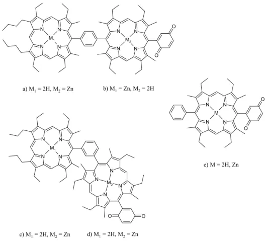
3.2. Porphyrin-Quinone Systems Pag. 28
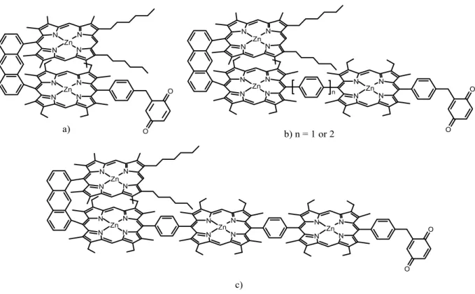
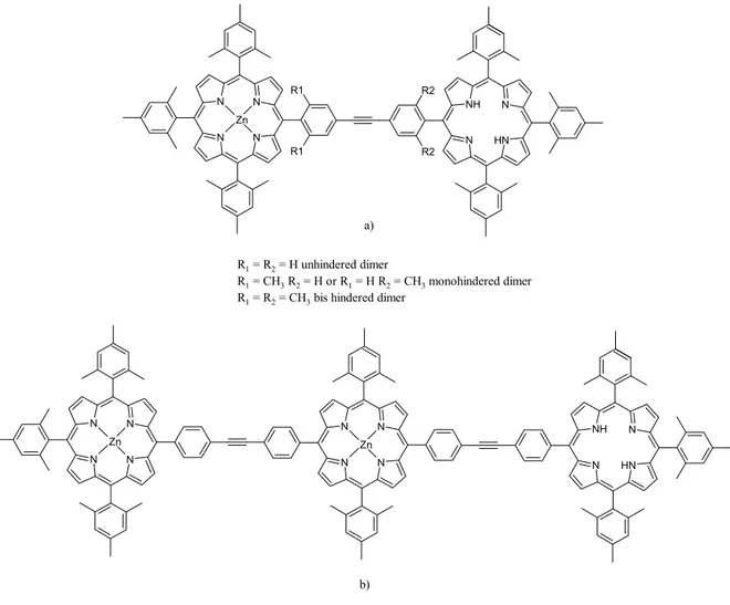
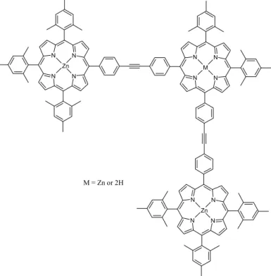
3.3. Porphyrin Light Harvesting Systems Pag. 39
3.4. Porphyrin-[60]Fullerene Systems Pag. 56
3.4.1. Covalently Linked Porphyrin-Fullerene
Systems Pag. 57
3.4.2. Self-Assembling Porphyrin-Fullerene
Systems Pag. 68
3.4.3. Porhyrin-“Wire”-Fullerene Systems Pag. 74
4. New Porphyrin-Fullerene Compounds Pag. 79
4.1. Introduction Pag. 79
4.2. Synthetic Strategy Pag. 80
4.3. Complete Study of a New β-Ethynyl Linked
Porphyrin-Fullerene Dyad Pag. 105
4.4. Preliminary Studies on New
4.6. Conclusions and Potential Applications Pag. 141
5. Synthetic Methodologies Pag. 145
5.1. General Methods Pag. 145
5.2. Chemicals Pag. 146
5.3. Syntheses Pag. 146
Chapter 1
“Natural Photosynthetic Systems”
1.1. Introduction
One of the most studied and appealing cycle of reactions in natural systems is the photosynthetic cycle; because its peculiar and fascinating aspects, involving light harvesting, energy and electron transfer and above all, water splitting and conversion of carbon dioxide into carbohydrates, the photosynthesis is one of the most articulated enzymatic catalyzed reaction path on the Earth. The most relevant aspect of the photosynthesis is the fact that all the energy necessary to the whole process is supplied by the sunlight. All the photosynthetic organisms in fact, are able to convert the sunlight radiation into chemical potential useful for the carbohydrates synthesis1. It is possible to understand the importance of this aspect considering that the enormous quantity of energy, coming from the sun, that during the daylight hours reaches the Earth surface is a thousand time higher than that produced by all of electrical power stations and this is true considering even only the ground surface of the U.S.A2.
More or less 50% of that radiation, roughly in the visible, is of a frequency useful for photosynthetic organisms. Moreover it has not be neglected that the by-product of photosynthetic activity of green plants, algae and cyanobacteria is the molecular oxygen; so the photosynthesis, expelling oxygen as by-product of reaction and assimilating carbon dioxide in organic matter, determines the composition of our atmosphere and sustains almost all life of our planet providing a readily usable carbon source1.
For these reasons the photosynthesis was one of the most studied phenomena of our times since the isolation and characterization of chloroplasts by Robert Hill in the 1939. Especially during the last decade a lot of scientists have perceived the powerful aspects and potential applications of the photosynthetic processes, in particular, the possibility to use the sunlight as a renewable energy source has produced a very large number of studies concerning the photophysical characterization and X-Ray structure determination of different photosynthetic natural systems. It is important to notice that only if we understand how sunlight is used by nature we will be able to reproduce some aspects of the photosynthesis that help us to obtain energy without using oil or in general fossil fuel, that is a consuming energy source.
The capability to develop processes that use the sunlight to produce energy, hold also a social implication: the possibility to fill the existing economic gap between the northern and southern region of the world3 as was outlined by Giacomo Ciamician in his lecture “The Photochemistry
in the 1912: “Solar energy is not evenly distributed over the surface of the earth. There are
privileged regions, and others are less favoured by the climate. The former ones would be the prosperous ones if we should become able to utilize the energy of the sun. The tropical countries would be conquered by civilization which would in this manner return to its
birth-place”.3,4
The study of a photosynthetic apparatus requires a large effort between different disciplines, such as quantum mechanics, biochemistry, biophysics, molecular and structural biology and so on1. One of the most important data that we have to know to elucidate the photosynthesis mechanism is the crystal structure of the system, in order to understand how the different chromophores work and how they can interact with each-other. Starting from this point it is possible to shed light into the physical laws that govern processes like energy and electron transfer and trying to set up a synthetic apparatus able to mimic the photosynthetic reactions. Obviously since nature has an enormous advantage, consisting in million of years of evolution, it will be impossible to reproduce a wholly working photosynthetic system, but what we have to do is to find out the principal rules that will allow us to simulate the meaning aspects of the entire photosynthesis.
In the following chapters the green plant and bacterial photosynthesis will be discussed, including some structural aspects concerning the spatial disposition and energetic interaction of the main chromophores, in order to point out how nature regulates a very complicate process fine tuning every single aspect.
1.2. Natural Photosynthetic Systems
The green plants photosynthesis is accomplished by several reactions occurring in the chloroplasts1. These reactions are catalyzed by a series of protein complexes hosted in the lipid membrane bilayer. Beyond the simple aspect of the chemical reaction for the glucose synthesis, depicted in the Scheme 1.2.1, are hidden complex and articulated bio-machines able to produce carbohydrates starting from water and carbon dioxide.
6CO
2+ 6H
2O
C
6H
12O
6+ 6O
2hν
The principal actors of this history are the Photosystem I (PSI) and Photosystem II (PSII) that capture the light energy to funnel it towards the photosynthetic reaction centers (RC) where the resonant energy produces the first step of the electron transfer chain: the expulsion of one electron. In the subsequent steps a series of protein complexes such as cytochrome (Cytb6f
complex), plastocyanine and ferredoxin are involved; all these proteic complexes have the task to bring electrons up to the ferredoxin-NADP reductase for the reduction of NADP to NADPH. The last part of the work, but not less important, is performed by the ATP-Synthase, that produce ATP exploiting the chemical potential of proton gradient, previously formed through the thylakoid membranes, as consequence of the electrons transport along the photoactive subunits (Figure 1.2.1)1.
Figure 1.2. 1 Structures of the membrane- protein complexes that drive oxygenic photosynthesis1
To conclude the photosynthethic cycle the ATP and NADPH produced in the “bright phase” of the process were used in the “dark phase” to fix the CO2 in sugar. The two photosystems, PSI
and PSII, hold a key role in the whole process and they belong to the family of the photosynthetic reaction centers, that is divided in two classes according to the terminal electron acceptor: PSI belongs to the type I that presents iron-sulphur clusters as electron acceptor, while PSII belongs to the type II having a quinone as the final electron acceptor5. Both systems share some structural and functional aspects such as the presence of a certain number of chlorophylls as antenna system, the existence of a reaction center containing a special pair of chlorophylls (P680 for PSII and P700 for PSI) that act as first electron donor. Moreover both reaction centers
Furthermore some similarities have been also found in the helical arrangements of the central protein core, that harbours the reaction centers of the two systems and the capability to drive the light energy toward the chlorophyll special pair. All these aspects induce to consider that there was a common ancestor both for PSI and for PSII6. Anyway their resemblance is only apparent because, if we focus our attention to the role that they cover in the photosynthesis, it is clear how the nature, through the evolution, has gave them very different works to do. The PSII (P680) can be considered as the strongest oxidant in nature able to oxidize a tyrosine residue,
that in turn takes an electron from the manganese ions cluster. After several cycles it generate a high potential (> 1 V) sufficient to split water into oxygen and protons. On the other hand the PSI (P700) carries out the opposite work and could be considered the strongest reductant in
natural organisms. The reducing equivalents produced by P700 are used by the photosynthethic
organisms to reduce the iron-sulphur protein Ferredoxin1. Such opposite functions imply also fundamental differences between the two systems, not only under a structural point of view. One macroscopic difference is the number of chlorophylls that constitute the antenna system. In PSI we find a larger number of pigments, not only in the core of the system but also in the extrinsic protein complex called Light Harvesting Complex (LHC) that helps the photosystem to capture the photons6,7. This implies different quantum yield: almost 100% for PSI and 85% for PSII. The minor content of chromophores may be also attributed to the destructive oxidant potential produced by P680 that can oxidize every pigment in proximity of the special pair. In
fact the loss of quanta in P680 can cause damages in the surrounding protein and a fact that
corroborates this hypothesis is the fast turn over in the synthesis of the subunit D1 (see Figure 1.2.2) of PSII. Because its proximity to the special pair, its synthesis represents the 50% of the total protein synthesis in the chloroplast even if the D1 subunit constitutes only 0.1% of the total protein1. To demonstrate that the photosynthesis is fine tuned in all its aspect we have to underline that the nature has supported such type of consideration. In fact when the extraction of water is impaired (for example at low temperature) the P680+ can receive electrons from a
secondary pathway involving Cytb559, a carotenoid and a chlorophyll located at the periphery of
the PSII reaction center (Figure 1.2.3); this prevents serious damage to the protein bulk of PSII itself1,8.
For a better understanding of the functionalities of the two photosystems, we will discuss separately some aspects concerning PSII and PSI, despite the fact that they act in series. Even if there are many differences between a plant and a photosynthethic bacterium, their relative reaction centers and photosystems show a certain degree of similarity, except for some structural and regulating tools, that will be outlined later. At this point of discussion the generic
description of one photosystem could be considered of general validity. The structure of PSII is taken from crystallographic data coming from a study on cyanobacterium
Thermosynecochoccus Elongatus8. The PSII is a dimer with the two monomer subunits almost
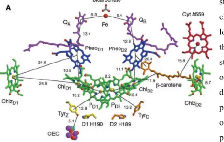
identical and containing 19 protein subunits. Each monomer also includes one oxygen evolving center (OEC), one heme b, one heme c, two plastoquinones, two pheophytins, one nonheme Fe and two bicarbonate ions (Figure 1.2.3). After photon excitation, one electron is extracted from the excited state of P680 and passing from a
second chlorophyll (ChlD1) and a
pheophytin (PheoD1), it arrives to the
plastoquinone QB. After that with the help
of the nonheme Fe, the electron is transferred to the plastoquinone QA, that
after a second reduction accepts two protons and releasing into the membrane matrix the electrons toward the Cytb6f. During the last
passage protons are pumped from the stroma in the inner space of the tylakoid lumen. It is possible to see in the Figure 1.2.2 that also the monomer has a pseudo twofold-symmetry that relates the D1, CP47 and PsbI subunits to D2, CP43 and PsbX subunits. The pseudo C2 element of
symmetry is a constant that we can find in all the photosystem subunits and about this aspect there are some discussions regarding the photoactivity of the two branches of the reaction center (Figure 1.2.3). For example in the PSII reaction center only the A branch is active toward the electron transfer while the B branch participates apparently giving only the contribution of quinone QB8. This evidence comes from
the first published crystallographic data of PSII reaction center where only one quinone (QA) is
present9. Other proofs for this argument could be found in the structural data regarding the two branches. Indeed on the B branch there is a larger distance between the chromophores and differences in the nature of local amino-acids: in the A branch more polar amino-acid side chains can stabilize better the charge separate states such as: P+Chl-, P+Pheo- and finally P+Q
A
-(where P indicates the generic chlorophyll special pair)1,8. Some experiments have
Figure 1.2. 2 (A) View of PSII dimer perpendicular to
the membrane. (B) View of PSII monomer along the membrane normal from the luminal side8
demonstrated that the disposition of the phytyl chains in the chromophores of the A branch are well arranged to get in close contact the cofactors, especially chlorophyll and pheophytin, in order to have a better electronic coupling10. Some performed calculations, taking in consideration the structural aspect of PSII reaction center, have shown that even if the B branch is inactive it has a fundamental role in the process. The excitonic states calculated by Nikolay Ivashin and Sven Larsson10, using time dependent functional theory (DFT) to obtain the Qy
states and an effective Hamiltonian for interactions between them, underline how the trap state is localized on the B side. This is due to the fact that the coplanar orientation of the chlorophyll ring 1 vinyl group with the tetrapyrrole macrocycle, generates a lower excited state, mainly distributed on the B branch. From crystallographic data this group is perpendicular to the chlorophyll ring in the A side. For the same reason it is possible to assume that also the triplet
state, coming from the decay of charge separated state, is localized on the B branch. On the other hand having a triplet state on the A branch, where oxygen evolves, could be very destructive because of the possibility to generate singlet oxygen. So the nature has placed the probable source of the singlet oxygen near the oxygen quencher, the carotenes on the B side. Around the PSII reaction center many β-carotene molecules are arranged, mainly on the B side, all of them having a trans conformation. Some of these β-carotene groups have the head group in direct contact with a chlorophyll and this suggests that they can facilitate the long distance electron flow, in particular from Cyt b559 in case of risk for self-photooxidation. Another peculiar aspect
of the PSII is the presence of the Mn cluster ions, called oxygen-evolving center (OEC)8, that is capable to carry out the oxidation of water. It works as an electrical accumulator repeatedly oxidized by the P680+ up to four time through the “S cycle”2, where S indicates one oxidation
state of the cluster, being the S0 the most reduced state and S4 the most oxidized one. In the S4
state the OEC can split the water into dioxygen and protons, coming back to its natural reduced state. The geometrical disposition of the Mn ions in the cluster recalls a cubane like structure2,8
Figure 1.2. 3 Electron tansfer cofactors of PSII reaction center, with
with 3 µ-oxo bridges connecting three Mn ions in the cluster plus a further µ-oxo bridge binding an external Mn ion that, with the help of the Ca ion present in the cubane like structure, is believed to be the active site for the oxidation of water. The oxidation mechanism of water has still to be clarified, but it is possible to assume that an high electrophilic species, like Mn(V)oxo or Mn(IV)oxyl radical, generated in the active site, can undergo to the attack of a water molecule bound in the active site. Obviously in this description the local amino-acid side-chain plays a key role modulating the distances and interactions between every components and favouring the water binding8. To conclude the description of PSII it should be mentioned the presence of an additional extrinsic protein complex harvesting system: Light Harvesting Complex II (LHC-II). As it was pointed out before, the PSII is poor of chlorophylls in the proximity of the reaction center, so the nature has provided an auxiliary apparatus to modulate the energy absorption. The crystal structure of LHC-II was isolated in a trimeric arrangement from Spinacea oleracea by Zhenfeng Liu, Kebin
Wang and co-workers7. Each monomer contains 14 chlorophylls, 8 chlorophylls a and 6 chlorophylls b located around the interface between each monomer. In the study was outlined how a net of hydrogen bonds brings three chlorophylls b in close contact leading a sort of cluster and how this disposition can facilitate the energy transfer. Besides the pseudo circular disposition of pigments, forming an inner ring (6 Chla) and an outer one (8Chla + 8Chlb) at the monomer-monomer interface in the trimeric structure, allows a very efficient absorption of incident light from all directions. From the crystallographic data a
consistent number of carotene molecules emerges, most of them are in all trans conformation; such molecules not only cover a structural role but together with xanthophyll molecules are involved in a non radiative dissipation of excess energy, that is one of the photoprotective strategies evolved by the plants. LHC-II has another important role: it takes part to the distribution of excitation energy between the two photosystems7,11. This aspect depicts the two photosystems as two dynamic machines capable to adapt themselves to the environmental changes in order to optimize the photosynthesis efficiency.
The description of plant PSI will be discussed in a parallel way with that of photosynthetic bacteria because there are many differences that must be outlined. One of the best refined
Figure 1.2. 4 Crystallographic model of
OEC. Mn cyan, O red, C grey, N blue, Ca green, BCT denotes bicarbonate. Unless otherwise indicated, amino acids belong to the D1 subunit2
structure of plant PSI was obtained by Adam Ben-Shem, Felix Frolow and Nathan Nelson from
Pisuvum sativum6. The PSI was crystallized together with Light Harvesting Complex I (LHC-I).
The first evidence that emerges from the data is the fact that the plant PSI is monomeric while the PSII is dimeric and furthermore the cyanobacterial PSI is trimeric5. While such difference between PSI and PSII can be justified considering the differences previously described, the trimeric structure of PSI in cyanobacteria arises from the different environmental conditions in which bacteria must live. Despite this fact, also in the protein core of PSI, where we find the reaction center, again a pseudo two-fold symmetry can be found. In the Figure 1.2.5 is clearly visible how the LHC-I is loosely bound to PSI and forms a sort of belt surrounding one side of the photosystem. At the same time between them there is a cleft that is filled with chlorophylls that cover an important role for the energy migration. In fact considering the distance between the antenna system and the core of PSI, the energy transfer from the periphery of LHC-I should be very much lower without the so called “gap” chlorophylls (dark pink in the Figure 1.2.5). A similar work is done by the “linker” chlorophylls (red in the Figure 1.2.5) that guarantee a good energy transfer along the dimeric subunit (Lhca1-Lhca4 and Lhca2-Lhca3) of the LHC-I. The main difference in term of absorption between LHC-II and LHC-I is the longer wavelength absorption spectrum, mainly due to the tighter interaction between chromophores in the LHC-I. An interesting study of Robert C. Jennings, Giuseppe Zucchelli and co-workers12 had shown how the fluorescence decay of the PSI-LHC system is wavelength dependent in the region that goes from 690 to 770 nm. A comparison between fluorescence steady state data and time resolved measurements elaborated with the Arrenius-Eyring theory confirms that the “wavelength dependent” activation energy is due to a different thermal activation energy for energy transfer from the low energy chlorophyll states (red chlorophyll states on the LHC-I) to the bulk chlorophyll state (in the core of PSI). The biological role of this low energy chlorophylls is to supply a better
Figure 1.2. 5 A view from the stroma of the plant PSI. The
subunit of LHC-I are indicated as Lhca1, Lhca2, Lhca3, Lhca4 and relative Chls are in blue. “Gap” chlorophylls in dark pink and “ linker” chlorophylls in red6
light absorption in dense vegetation where the ambient light is enriched by the wavelength above 690 nm respect to the normal daylight. What we have just described underlines one more time the dynamism of plant photosynthetic apparatus that is able to adapt itself in real time to the fickle environmental conditions, in order to have always the best performance in the photosynthesis. In the plant PSI crystal structure we can find at least other two evidences of this ability: the LHC-I is composed by four monomers bound together giving two dimeric units, but only the subunit Lhca1 is tightly bound to the core of PSI through the proteic subunit indicated in the Figure 1.2.5 as PsaG. This suggests that the Lhca1 could be thought as an anchor point for the LHC-I, while the remaining subunit are bound at varying stoichiometries depending on environmental conditions6. In the PSI it has been seen that the protein subunit named PSaH is necessary as docking site for LHC-II, in fact as we have
pointed out before, when an excess of light was captured by PSII a small population of LHC-II trimers dissociates from PSII and associates with PSI, in order to redistribute the excitation energy11. Finally another evolutive improvement in the plant PSI consists in the presence of a longer amino terminal domain in the PsaF subunit respect to that in cyanobacteria, that allows a better binding of plastocyanin, giving as a result a two order of magnitude faster electron transfer from the copper atom of that protein to P700 (Figure 1.2.6). The reaction center of PSI
is composed by six chlorophylls, two of them forming the special pair (P700), two philloquinones plus different
iron-sulphur clusters, all these cofactors are arranged in a two-fold symmetry like that seen in the PSII reaction center, but in this case both branches are photoactive towards the electron transfer, even if the A side presents higher rate
constant values. In the crystal structure of PSI from cyanobacterium Thermosynecochoccus
Elongatus published by Patrick Jordan, Petra Fromme and co-workers5 is outlined how this
dissymmetry in the electron transfer could be explained taking in consideration the hydrogen bonding net present in the A branch, and absent in the B branch; this net can facilitate the stabilization of the different excited states. Despite the fact that this was seen in a cyanobacterium, we can assume that it is valid also for plants and green algae because the structural organization of chromophores and amino-acids environment is preserved, even if we
Figure 1.2. 6 P700 of green plants. The
arrow indicates the two tryptophan residues necessary for electron transfer from copper ion of Plastocyanin to the oxidized P700. The
interaction of PsaF subunit with plastocyanin is also shown6
can find different quaternary structures (e.g. plant PSI monomer versus cyanobacterium PSI trimer). In the Figure 1.2.6 the two tryptophan residues necessary for electron transfer from copper ion of Plastocyanin to the oxidized P700 are also shown and the electron flow through the
pigments of PSI reaction center is almost similar to that we have seen for PSII reaction center. Excited P700 donates one electron to the adjacent chlorophylls that in turn pass the electron to
the phylloquinone and the electron arrives to the Ferredoxin, passing through the iron-sulphur clusters. P700+ is finally reduced by Plastocyanin with electron coming from PSII. To conclude
the description of PSI we should not forget to mention that the PSI can perform also cyclic electron transfer (see Figure 1.2.1), having as a partner the Cytb6f1. This path is used only to
pump protons within the lumen of tylakoid and most probably is connected to the fact that are necessary more than 12 protons, for plants, to perform the synthesis of 3 ATP molecules and with this alternative path the organism is able to recover the right quantity of protons: in fact the proton number for the ATP synthesis is species dependent. For example the yeast mitochondria need 10 protons to synthesize 3 ATP while plants 14 protons and since with the linear electron flow from PSII to PSI only 12 protons enter in the tylakoid lumen, the remaining 2 recovered with cyclic electron transfer. In principle this can also justify the higher quantum yield of PSI respect to PSII.
We have seen that the entire photosynthesis is fine tuned with a lot of tools such as: hydrogen bonds, protein structures, distances between the chromophores, amino-acids surrounding, electronic coupling between chlorophylls and we have also
seen that the plants are able to adjust the stoichiometry of their active systems as answer to the environmental changes. This is true even for the cyanobacteria that under stress conditions (iron deficiency) produce a giant ring chlorophyll-protein complex around the trimeric PSI13 (Figure 1.2.7). All these aspects point out how it is impossible to reproduce synthetically such type of systems and also understand the physical laws that govern the phenomena it is not so easy when we have to deal with hundred chromophores that interact between them in different way as we can see in the subsequent paragraph.
Figure 1.2. 7 PSI trimer structure
and giant chlorophylls ring obtained from X-Ray crystallographic data plus electron microscopy13b
1.3. Energetic Aspects of Photosynthesis
In this brief section the coupling of chromophores, the rising transients after reaction center photoexcitation and the electron transfer mechanism will be discussed. Our aim is not to clarify completely all the energetic aspects but to show how much is complicate to find unique theory that can explain in a simple manner the energetic interaction between the single chromophores. For our purpose it is useful to take as example the bacteria reaction center that shows a simpler absorption pattern due to the fact that in the photosynthetic purple bacteria is possible to obtain the crystals of the reaction center separated from the light harvesting complex. This reduces the difficulty in the interpretation of the data that come out from the experiments. In the Figure 1.3.1 the absorption features in the visible and near IR of Rhodobacter Sphaeroides reaction center is reported, with relative chromophores colours code14. Since the structural analyses have revealed high similarity between the pigment arrangement of PSII and that of bacteria, the bacterial reaction center can serve as a realistic model for PSII. In the reported spectrum the absorption of the special pair bacteriochlorophylls is located at higher wavelength as result of the high level of electronic coupling between the two pigments14. This allows to the special pair to act as a sink of energy, that, once it was captured, is transferred to the near chromophore which shows an absorption band at higher energy and so on. One of the first debate, on the electron transfer in the reaction center, was focalized on the existence of the P+BA- charge separated state (where BA
indicates the bacteriochlorophyll in the A branch); the first ultrafast studies of J. L. Martin and J. Breton15 showed that the lifetime of special pair excited state (P*) was more or less 2.8 ps and that the rising time for the reduced bacteriopheophytin (HA) had the same value, so the authors
concluded that the first electron transfer step occurs between the special pair and bacteriopheophytin in one step, leaving to the bacteriochlorophyll BA the only role to increase
the electronic coupling in order to design a superexchange electron transfer mechanism. In 1989 Kirmaier and Holten16, and later (1991) Zinth and Wachtveitl17 published papers where new
transient absorption data, with a time costant of 0.9 ps, were reported around 1020 nm, where the absorption of BChl- (BA-) is located. This was a proof for a “two steps” electron transfer
Figure 1.3. 1 Absorption spectrum of
reaction center from Rhodobacter Shpaeroides
mechanism. Other evidences for the existence of P+BA- come from transient dichroism
experiments that reveal an angle between the transition moment dipole of the special pair and that of the subsequent intermediate in electron transfer reaction, of 26°±8°, that corresponds to the angle between Qy transitions of the special pair P and those of the accessory
bacteriochlorophyll BA14. At the moment a more recent investigation performed by A. Dobek,
M. Ziolek18 and co-workers outlines again the presence of a “two-steps” electron transfer mechanism in the primary electron transfer reaction. Recently another study of Kirmaier and Holten19 was published to evaluate which is the origin of the two different electron transfer rate in the two branches of reaction center. Using the DNA site directed mutagenesis, different
Rhodobacter Capsulatus reaction centers were prepared, in these centers it is possible to find
modified energetic level of the radical pairs in order to evaluate how much the electronic coupling participate to the difference in electron transfer rate constant along the two branches. The results indicate that almost the 30% of the discrepancy in the rate constant has to be attributed to the electronic coupling, the remaining 65%-70% due to other factors such as free energy, reorganization energy and one or two step electron transfer mechanisms in the two sides of reaction center. An interesting study about the energy transfer interaction in the LHC of purple bacteria was published by V. Sundström, T. Pullerits and R. van Grondelle20. They have analyzed the nature of energy transfer in purple bacteria LHC-II. They found that the reaction center is arranged in a circular structure composed by 9 subunits, each subunit presents an αβ helices-heterodimer that binds three bacteriochlorophylls a, two of them very close to each other, plus carotenoid molecules. In the purple bacteria LHC the looser bound BChls a constitute the B800 system, while the tightly bound ones constitute the B850 system, both of these systems presenting a circular arrangement of the pigments. The energy transfer between this two systems and between each pigments within one system, was studied by applying the incoherent hopping transfer, when very weak interactions are present, and exciton theory when the interactions between the chromophores are much stronger. The principal outcome of this study is the difficulty to describe every part of the system with only one theory. When the authors tried to calculate the lowest exciton state of LHC, applying the perfect exciton model, they obtained a lowest excitonic state with very little dipole strength, since all dipoles were considered to be in the plane of the ring. This implies a low-temperature non radiative state. The experimental data showed the contrary, with a dipole strength localized on 2-3 monomer. Moreover the dipole moment of emitting state was more or less temperature independent. Further refinements of calculations, introducing disorder elements, allowed them to obtain a result much more closer to the experimental data; in conclusion this means that the disorder
(such as spectral inhomogeneity) destroys the “delocalized exciton” picture that could be applied to a maximum of 4 monomer. We conclude that in some cases we have to adopt exciton theory, while in other cases the electron hopping theory. Another outcome of the previous investigation was to show a dense interaction between the carotenoid molecules and B850, in fact almost all energy absorbed by these molecule is transferred to the B850 system, the 75% in a direct way, while the remaining 25% passing before through the B800 system. In the work is also showed how carotenoids are able to give ultrafast response to local electrical field generated by excited chlorophylls and how this response is different when the excitation is located on B800 or B850. This feature of the carotenoid molecules is good candidate as probe of local dynamic and structural changes.
In the short picture of photosynthesis given in this paragraph, a very high level of complexity about energetic interaction emerges, but it is possible to get into account some parameters such as: electronic coupling, distance and relative orientation between chromophores and also temperature dependent electron transfer rate, adopting the simpler Marcus theory.
Chapter 2
“Electron Transfer Theory”
2.1. Introduction
In order to simulate the natural electron and energy transfer reactions is necessary to have a theoretical background that allows us to take into considerations many, the principal ones, of the aspects that characterize such type of reaction. We have to be grateful to the work of many scientists such as Libby, Taube, Hush and first of all Marcus if at the moment it is possible to conceive and build up synthetic systems that mimic some of the aspects of natural photosynthesis. In particular the theory developed by Rudolph A. Marcus21, as consequence of his studies and conclusions on experiments carried out by Libby22 on isotopic electron transfer in metal complexes, is one of the best full-comprehensive and well organized theory. Such theory is suitable to explain a lot of experimental data and it also foresaw the existence of a particularly interesting region in the electron transfer free energies, the so called “inverted region”, before that such region was found in the experimental analyses.
This theory gained to Marcus the Nobel Prize in Chemistry in the 199223 and in this chapter some of its features will be summarized.
2.2. General Observations
To point out the main points of the Marcus theory is useful to start from general observation on a bimolecular electron transfer reaction with a donor (D) and an acceptor (A), both the molecules being in the fundamental state or in the respective excited states. In any case the
principles are the same and no modifications are necessaries. Usually the Marcus electron-transfer theory is applied for outer sphere electron-transfer. Considering the three steps depicted in the Scheme 2.2.1, to have an electron transfer reaction both donor and acceptor must form a precursor complex21 (2.2.1a), after that the reorganization of nuclear coordinates and electron energy levels leads the precursor complex in a transition state in which the electron transfer takes place, generating the following complex (2.2.1b) that, for the Franck-Condon principle, has the same nuclear coordinates of precursor complex. The last step is the dissociation in the products (2.2.1c).
Figure 2.2. 1 Rudolph
2.2.1), and to make some experimental considerations about the phenomenon: if ks is much
more higher than k-ET the equation for kobs becomes simpler (Expression 2.2.2a) and when the
dissociation rate constant (kd) is much higher than the constant for the forward electron transfer
(kET) the expression of kobs is that reported in the Expression 2.2.2b and we are still able to
obtain information on ket from experimental measurements; on the contrary if kET is much
higher than kd we are in the diffusion-controlled limit and we could not obtain any type of
information about the electron transfer rate. Obviously if the donor and acceptor molecules are hold together by covalent bonds the only meaning step reported in the Scheme 2.2.1 is described by the equation 2.2.1b.
D + A
D/A
D
+/A
-D
++ A
-D/A
D
+/A
-ka kd kET k-ET ks 1a 1b 1cScheme 2.2. 1 Three steps in a bimolecular electron-transfer reaction
or
Expression 2.2. 1 Electron transfer rate constant for a bimolecular reaction from steady state analysis
2a 2b 2c ET s ET d ET d a obs
k
k
k
k
k
k
k
k
−+
+
=
1
+
+
=
− s ET ET a d a obsk
k
k
k
k
k
k
1
1
1
+
=
ET d a obsk
k
k
k
1
1
1
d ET a obsk
k
k
k
≅
k
obs≅
k
aExtremely important it is to know the different state energies respect to the ground state, especially for the photoinduced electron-transfer. In this latter case we should know both the singlet and triplet excited state energies and first of all the free energy of reaction (∆G°, Expression 2.2.3) that could be obtained from cyclic voltammetry measurements since it is given by the difference between the standard reduction potentials of donor and acceptor; the terms ωP and ωR correct for the work of bringing D+ and A- and D and A, respectively, together.
2.3. Classical Marcus Theory
21,24,25Despite the fact that the Marcus theory presents a quantum mechanical aspect, in a first step it is possible to calculate the electron transfer rate applying a classical method based on the simple transition state theory. To simplify the problem, the multinuclear coordinates dependence of the reactant and product energy potential surfaces could be reduced to a mono-dimensional dependence, introducing the reaction coordinate, that is the path along which the electron transfer takes place. In general if all the nuclear modes, including solvent molecules, are considered, the potential energy surface is not parabolic, but describing this potential surface as a Gibbs free energy, we can still use a parabolic approximation.
In the subsequent discussion the letters R and P will be used to indicate respectively the reactant (D/A) and the products (D+/A-). Taking into consideration what we have just reported, the electron transfer rate constant can be expressed as a product of three factors (Expression 2.3.1), kel, that indicates the electronic transmission coefficient usually considered to be unitary in the
classical approach, νn, the frequency of passage through the transition state and finally an
exponential term that takes into account the activation energy (∆G*).
R P A A D D
E
E
e
G
+
ω
−
ω
−
=
∆
° ° ° − +Expression 2.2. 3 Free energy of electron transfer reaction
where P and R indicate products and reagents respectively being e the electronic charge
∆
−
=
∗T
k
G
k
k
B n el ETν
exp
Expression 2.3. 1 Electron transfer
An important quantity in the theory is the reorganization energy λ defined as the change in the Gibbs energy when the reactant state (D/A) are distorted in the equilibrium coordinates of product state (D+/A-) without electron transfer (see Figure 2.3.1). When the vibrational modes of both reactant and product states are assumed to be parabolic it is possible to find a correlation between ∆G° and ∆G* in which is involved also λ (Expression 2.3.2).
One of the most relevant consequence of the relation described in the Expression 2.3.2 is the existence of the “inverted region” in electron transfer reaction. In general we expect that an increasing in the free energy ∆G° should favour the formation of the products but in this case is not so obvious. It is possible to analyze the fact considering the four cases depicted in the Figure 2.3.1.
(
)
λ
λ
4
2 ° ∗=
+
∆
∆
G
G
Expression 2.3. 2 Relation between activation free
energy and free energy of electron transfer reaction
Figure 2.3. 1 Four cases for electron transfer reactions depending on free energy
values related to the reorganization energy. For comments see text.
∆G* -∆G° λ P R -∆G° λ ∆G* = 0 P R ∆G* -∆G° λ P R ∆G* λ ∆G°= 0 P R a) b) d) c) ∆G* -∆G° λ P R -∆G° λ ∆G* = 0 P R ∆G* -∆G° λ P R ∆G* λ ∆G°= 0 P R a) b) d) c)
In the case a) the reaction is isoergonic (∆G° = 0) and there is an appreciable value of activation energy (∆G*); when there is a gain in the free energy, ∆G° becomes more negative, b) case. We
have an increment in the kET and in this condition we are in the “normal region” where
0 ≤ -∆G° ≤ λ. The electron transfer rate constant can reach a maximum value when there are no activation energy barriers, c) case, where the activation energy is equal to zero. This happens if -∆G° = λ and in a such situation kET = kelνn and since kel is assumed to be unitary in a classical
approach, kel = νn ≈ 1013 s-1. At this point if the free energy starts to become much more
negative (-∆G° > λ) the intersection between the two surfaces is shifted towards left, determining an increasing ∆G*, that leads to a decrease in k
ET. This is the so called “inverted
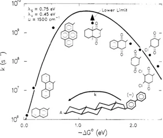
region”. By a physical point of view this means that the products are formed in a high-distorted and high-energy state. This was one innovating point of the Marcus theory, even because the first experimental data about the inverted region came out only in the 1984 when Miller, Closs and Calcaterra26,27 published their contributions on electron transfer reaction in molecules that hold together, through a rigid carbon system, biphenyl donor moiety and different acceptor molecules. Their work shows clearly the pseudo parabolic dependence of kET from free energy
of the reaction (Figure 2.3.2). From such considerations an important role emerges for λ: in fact this energetic parameter could be thought as an arbiter respect to the rate constant of electron transfer reactions. For this reason is necessary to have a model that makes explicit some aspects of such reorganization energy. Remaining in the classical approach, λ may be split in two contributions: the first deriving from the nuclear vibrational modes elaborated with an harmonic potential having a reduced force constant for each vibration. This component
is independent from the solvent and is indicated as λin (Expression 2.3.3). The other
component, λout, is called the solvent reorganization energy since it arises from different
orientation and polarization of solvent molecules around the reactant state (D/A) and product state (D+/A-). While in most cases the λ
in value is quite small, the respective value for λout
presents consistent entity. The solvent reorganization energy could be modelled considering the solvent as a dielectric continuum and in this case λ is described by the Expression 2.3.4.
Figure 2.3. 2 Dependence of kET from the free energy
of electron transfer reaction (-∆G°). Inverted region is
Solving the integral in Expression 2.3.4 means to adopt a more elaborated model for the reactants and products and in this case is possible to consider the reagents as spheres. In such a way the λout is described by the equation reported in Expression 2.3.5, where the aD and aA are
the radii of the donor and acceptor molecules, rDA is the center-to-center distance and ∆e is the
charge transferred in the reaction.
The reorganization of the solvent molecules is a question that has an enormous importance since many electron transfer reactions cannot occur in some solvents and can occur in others. This is mainly true for the photoinduced electron transfer when donor and acceptor are chemically bound together through a “linker”. In such a case the starting state may be uncharged while the final products are charged and the choose of the right solvent, for having the longest lifetime for the charge separate state, becomes very important. In fact very polar media can stabilize the charged final products but, due to its polarity, will be affected by high value of λout, on the contrary very apolar solvent will have low values for λout, but high
(
)
∑
−
=
i eq P eq R i inf
r
r
2 __2
1
λ
Expression 2.3 3 Equation for inner reorganization energy, being
fi a reduce force constant and req the equilibrium bond length of
reactant (R) and products (P). The sum is taken over the significant intramolecular vibrations.
(
E
RE
P)
dV
s op out∫
−
−
=
ε
ε
ε
λ
1
1
2
1
0Expression 2.3 4 Equation for solvent reorganization energy, where εs and
εop indicate respectively the static and optical dielectric constant of the
medium and ε0 the permittivity of the vacuum. ER and EP are the electric
fields generated by reactants and products
( )
−
−
+
∆
=
s op DA A D outr
a
a
e
ε
ε
πε
λ
1
1
1
2
1
2
1
4
0 2Expression 2.3 5 Equation for solvent reorganization
activation energy for electron transfer reaction, due to its inefficiency in solvating the activated complex with a partial charge separation. We should reach a compromise. Another further evidence of the importance of λ is given by the studies carried out by Libby who pointed out how small ions, such as Fe2+ or Fe3+, had lower kET respect higher ions22,23, such us [Fe(CN)6]
3-, [Fe(CN)6]4- or MnO4-, being equal the electron transfer process. Libby explained this fact
assuming that the solvent molecules could not rearrange their relative orientation when an almost instantaneous electron transfer occurs, generating an activation energy barrier, that is higher for small ions compared with less solvated complex. In his study Libby also found a sort of exception: the isotopic electron transfer in the pair [Co(NH3)6]3+ and [Co(NH3)6]2+ showed
small kET value, even if these ions are big complexes like [Fe(CN)6]3- or [Fe(CN)6]4-. This
discrepancy was justified by Libby adducing a large difference in the Co-N bond length in the two different oxidized complexes23, but this aspect didn’t sound good to Marcus who was moving his first steps towards the electron transfer theory just from this point.
In the classical approach the factor kel in the Expression 2.3.1, is assumed to be equal to one,
but this is not even the truth; we can divide the electron transfer reactions in two classes, according to the relative electronic coupling between the reactants and products state: adiabatic electron transfer
reactions with high electronic coupling that have a kel ≈ 1, and non
adiabatic electron transfer reactions with a low value of electronic
coupling and in this case kel will be much more less than 1. An
exemplification of this difference is given in the Figure 2.3.3: in the
adiabatic reaction the electronic coupling is enough strong to split in
two, at the intersection, the two potential surfaces. In this case the electron transfer remains mainly in the lower surface and the transmission coefficient is more or less equal to one. In non
adiabatic reaction the system usually stays on the reactant potential
surface and only occasionally crosses over the product potential surface. To take into consideration this aspect we need to modify the classical vision of the Marcus theory and introduce the quantum mechanical aspect.
Before to show the equation for kET in the non adiabatic reactions it
is necessary to underline the key role played by the nuclear motions in this circumstance. In particular the vibrational function overlap of both products and reactants is important. In fact the electron transfer can take place when such a overlap is good enough in proximity of the
D/A D+/A -D/A D+/A -a) b) D/A D+/A -D/A D+/A -D/A D+/A -D/A D+/A -a) b) Figure 2.3. 3 Adiabatic a) and nonadiabatic b) electron transfer
intersection point, even if the system does not have the right activation energy. This is due to tunnelling effect which can be distinguished in electron and nuclear tunnelling, the latter is more important because of the very little shifts of the nuclei from reactants to products state for generating the effect.
Looking at the Figure 2.3.4 we have three possibilities to see an electron transfer: electron transfer for electron tunneling at the transition state, represented by the wave functions in the case 1), where the systems are at the crossing point with the same nuclear configuration (overlap between the vibrational wave functions) and so there is a finite possibility to have electron transfer although the electronic coupling (in general indicated as HRP) is very small.
Since it is necessary to reach the excited vibrational state the whole reaction will be temperature dependent. In the case 2) is represented the activated nuclear tunneling that allows the electron transfer reactions to take place even if the two systems are below the intersection point, also in this case there will be a dependence from the temperature. As last, temperature independent
nuclear tunnelling is depicted in the case 3), where there is again a finite possibility to have
electron transfer even at very low temperature, because there could be very imperceptible overlap between vibrational wave functions and
in such a case the reaction is temperature independent.
To formulate the new expression for the electron transfer rate constant in the non adiabatic condition it is possible to start from the Fermi “Golden Rule” that expresses such a constant as a product between the electronic coupling (HRP)
and the Condon factor (F.C.W.D. Franck-Condon weighted density of states) that calculates the overlap of all the vibrational modes, including those of the solvent molecules, each weighted for the relative probability (Expression 2.3.6). To elaborate the general expression it is necessary to solve the
Franck-Condon factor and for this purpose there is a semiclassical approach that treats quantum mechanically only the vibrational modes of the systems while those of the solvents are treated with a classical method. Adopting further exemplifications24 the non-adiabatic kET can be well
defined by the equation in the Expression 2.3.7. Such equation works very well in the normal
D/A D+/A -1) 2) 3) D/A D+/A -1) 2) 3)
Figure 2.3. 4 Energy surfaces for quantum
mechanical electron transfer theory. The wave functions illustrate symbolically the importance of vibrational overlap.
region but not in the inverted region. In fact Marcus and Siders had shown28 that the falloff of kET in the inverted region is not parabolic but almost linear because the nuclear tunneling is
very active, due to a good overlap of the vibrational wave functions.
The inverted region gains a very high importance in the photoinduced electron transfer, as Kakitani and Mataga revealed in their work29. Adopting their modifications to the Marcus theory, an uncharged starting state should have a larger value of λ comparing to an ion pairs. In this case they predicted that a charge separation reaction should enter in the inverted region only at high value of –∆G°, while the charge recombination reaction should present a smaller value of –∆G° to enter in the inverted region. This aspect is really useful for obtaining high lifetime of charge separation state.
In this paragraph all the aspects that influence the electron transfer were presented by a theoretical point of view. In the further paragraph the experiments, carried out to shed light into such type of reaction, will be discussed in order to outline in a brief manner how the different factors influencing the electron transfer act.
.
.
.
.
2
H
2F
C
W
D
k
ET
⋅
RP⋅
=
h
π
° °Ψ
Ψ
=
R el P RPH
H
ˆ
( ) (
)
∑∑
−
=
° ° j i Rj Pi Rj Rj PiP
D
W
C
F
.
.
.
.
χ
χ
2ε
δ
ε
ε
Expression 2.3 6 Expression of kET by a quantum
mechanical point of view. Ψ indicates the electronic wave function of reactants (R) and products (P); χPi the
vibrational wave function of the level i for the product and χRj the vibrational wave function of the level j for the
reactants.
(
)
(
)
+
∆
−
=
− °T
k
G
T
k
H
k
B B RP ETλ
λ
πλ
π
4
exp
4
2
2 12 2h
Expression 2.3 7 Semiclassical expression for the electron
2.4. Electronic Factor, Solvent and Bridge Effect on Electron Transfer
Reactions.
To well understand how the electronic factor changes within different systems, is useful to take in consideration some elementary models and for this aim is also useful to rearrange the expression of kET, that will be divided into three parts according to the treatment made by
Marcus, Sutin and Newton25,30,31. The electron transfer rate constant could be expressed as the
product of the afore mentioned three factors: electronic transmission coefficient kel, effective
nuclear vibration frequency νn that tends to destroy the right configuration at the parabolas
intersection and the nuclear factor kn. These three factors are well expressed in the equation
reported below32:
An important aspect is how the electronic coupling changes with the distance between the redox sites. To simplify the model is possible to assume30,31,32 that Hrp decreases exponentially with
the distance, such decrease being measured by the parameter β in the Expression 2.4.2, where r0
is the van der Waals contact distance between the redox sites and Hrp0 is the electronic coupling
for such distance.
Obviously the kET decrease with the distance interposed between the donor and acceptor site,
could be different if such distance is simply filled with solvent molecules or with a “molecular n n el ET
k
k
k
=
ν
−
−
−
−
=
n el n el elk
ν
ν
ν
ν
2
exp
2
2
exp
1
2
2 1 2 = RT HRP elλ
π
ν
h
∆
−
=
∗RT
G
k
nexp
Expression 2.4. 1 The different factors taken in consideration in this
chapter.
(
)
−
−
=
2
0 0r
r
H
H
rp rpβ
Expression 2.4. 2 Distance dependence of
bridge”. In this latter case it is possible that the donor and acceptor orbitals can interact with the bridge orbitals. If the bridge is composed by different units is possible to estimate the electronic coupling between the donor and the first unit, the acceptor and the last unit and also between the different units. If such coupling is not high, through the perturbation model32,33,34,35 , it is possible to write the whole electronic coupling as reported in the Expression 2.4.3.
The last equation reported for the electronic coupling can be used if the interaction with both L.U.M.O. and H.O.M.O. of the bridge is present. In the first case we will have an electron exchange while in the second an hole exchange. An important aspect must be underlined: the electronic behaviour of the molecule that acts as a “bridge” is not an absolute property, but will be dependent upon the donor and acceptor energy levels and also upon the symmetry of the whole system. One of the first study which has furnished data about the “bridge” behaviour, involves metal-to-metal charge transfer, in particular diruthenium complexes where the bridge is formed by polyene subunit32 (Figure 2.4.1). Such studies were carried out in different
solvents36. The most important results, considering as solvents nitrobenzene and water, show a value for β equal to 0.2 Å
-1, that underlines how the polyene acts as
a “molecular wire” due to the extended π conjugation system. Also the behaviour of nuclear and electronic factor were calculated and in general an almost adiabatic reaction (kel ≈ 1) was found for the polyene bridge even in the presence of a change of
metal ions. Due to large value of λout for such a bridge, the nuclear factor was found to be much
more less than one and the distance dependence much more pronounced for the nuclear factor than for the electronic factor. For other bridges such as poly-phenyl bridged ruthenium complex, the β value is higher, most probably due to the lack of coplanarity between the phenyl rings37. Changing the nature of the bridge, polyproline instead polyene, has revealed less efficiency in mediating the electron transfer. In fact in the poly-proline bridged system like
∏
− = + + − − = 1 1 1 1 , 1 1 n i d i i i D nA D rp E E H E E H H HExpression 2.4. 3 Electronic coupling in a Donor-Bridge-Acceptor
unit, where ED indicates the donor or acceptor energy and Eithe
energy of the unit i.
N N n Ru Ru NH3 NH3 NH3 NH3 NH3 NH3 NH3 NH3 NH3 NH3 II III
Figure 2.4 1 Polyene bridged di-ruthenium complex, one
Os(II)-Ru(III), both nuclear and electronic factor are less than one32 with higher value of β (≈ 0.7 Å-1); this aspect is probably due to the higher energy of the L.U.M.O. in the polyproline bridge than in the polyene bridge. Differences within the same polyproline systems come out varying the metal, so the couple (bpy-)RuII-CoIII has a value for β twice smaller than the system Os(II)-Ru(III), because the redox potentials of the first system are smaller than those of the second one, allowing a better mixing with the orbitals of the bridge32.
Another useful study on the parameters that influence the electron transfer is available in the works published by James R. Bolton, John A. Schmidt, Mary D. Archer, Jacquin H. Wilford and coworkers about photoinduced electron transfer in porphyrin-quinone dyad connected with different bridge unit38. In these studies particular attention was given to nature of the linkage, solvent and temperature effects. It has been
reported that for the molecule depicted in the Figure 2.4.2, a porphyrin linked to a quinone by an amino-acid unit, the electron transfer is faster, in general, in those solvents that present an intermediate value of polarity or more precisely a lower value of εs such as 1,2-dibromoethane,
chloroform ad chlorobenzene39. Moreover it was found that, for the porphyrin-quinone dyad reported in the figure, the electronic coupling
between the two moieties is solvent dependent, most probably because conformational effects due to the different dielectric constant of the solvents. Also different bridges were tested, in particular poliaminoacid bridge was found to be very effective because the kET drops slowly
increasing the distance. When an unsaturated bridge is introduced between porphyrin and quinone, the electron transfer goes faster comparing with a saturated ring, but noteworthy a constrained ring such as cyclobutane shows a faster electron transfer than bicyclooctane ring. The strained aliphatic orbitals probably mediate more efficiently the electron transfer reaction. In these two last examples it has shown how some parameters can influence both the simple electron transfer reaction and photoinduced electron transfer. In the next chapter will be discussed in more details all the systems that are able to reproduce the primary electron transfer reaction which occurs in the natural photosystems, with particular attention to the systems having porphyrin moiety. These chromophores present photochemical properties very similar to those of chlorophylls and bacteriochlorophylls; moreover the chemical synthesis of porphyrin allows to introduce different chemical groups that lead to a very wide type of interactions
N N H N NH N H O O O
Figure 2.4 2 Porphyrin-Quinone dyad model,
between donor and acceptor moiety, useful to obtain long lifetime of the charge separated state. To reach this aim it would be useful to have a very fast forward charge separation and a back-electron transfer deeply inside in the Marcus inverted region.
Chapter 3
“Systems Able to Reproduce Natural Energy and Electron Transfer Reactions”
3.1. Introduction
Once that the Marcus theory became very popular and very useful to understand the influence of different aspects on the electron transfer reactions, the works about systems able to reproduce the primary electron transfer step occurring in the natural photosystems, underwent an enormous increase, especially for those systems that present porphyrin moieties, quinone and carotenoid molecules. Such a systems were deeply studied in order to find a favourable spatial disposition of single chromophores and also a favourable electronic interaction in order to reproduce the longest lifetime of the charge separated state. Many attempts were carried out in such direction and one considerable progress was achieved when Kroto and Smalley discovered the fullerene compounds, in particular the [60]fullerene (C60); for their discovery they were
awarded with Nobel prize in the 199640. This molecule shows particular and interesting features and first of all it has been demonstrated its ability to be good electron acceptor41. This last characteristic makes the C60 suitable for electron transfer reactions. Chemically speaking the
C60 behaves as an electron poor compound, so it rapidly undergoes to the electrophilic attack;
taking into consideration this aspect different functionalizations of C60 were developed, in
particular the two most famous are the Bingle-Hirsch reaction42 and the Prato, Maggini and Scorrano reaction43. Thank to these studies it was possible to synthesize many porphyrin-fullerene dyads that gave good results in terms of electron transfer.
The use of fullerene has opened a sort of new era for the electron transfer reaction and it made possible to build up also devices for the storage of the solar energy in order to convert it into chemical potential. In the following paragraphs different systems will be discussed, all able to mimic the electron transfer reactions occurring in the natural photosystems, with particular attention to the interaction typology between the chromophores, the nature of the electron transfer, the yield of the reaction and also the lifetime of the charge separated state. Finally some indications will be suggested on how these systems could be used to build organic solar cells, in order to confer also a practical aspect to all the studies carried out on this argument.