UNIVERSITY OF PISA
School of Graduate Studies
“Scienza del Farmaco e delle Sostanze Bioattive
”
PhD THESIS
2006-2008
“Natural and Synthetic Polysaccarides for the
Promotion of Ocular and Oral Drug Absorption”
Chiara Zaino
DIRECTOR OF THE SCHOOL: SUPERVISOR: Prof. Claudia Martini Prof. Giacomo Di Colo
CONTENTS
INTRODUCTION 1
SECTION I - Novel quaternary ammonium chitosan derivatives for the promotion of intraocular drug absorption
3
I.1 Introduction 3
I.2 Materials and methods 5
I.2.1 Materials 5
I.2.2 Diethylaminoalkylation of chitosan 5
I.2.3 Permeation measurements across excised rabbit cornea 6 I.2.4 Gravimetric evaluation of corneal hydration levels 6
I.2.5 Preparation of ophthalmic drops 7
I.2.6 Assessment of NaFlu-polymer interactions 7
I.2.7 Permeation data treatment 8
I.2.8 In vivo tests 9
I.3 Results and discussion 10
I.3.1 Permeation studies 10
I.3.1.1 Permeation of DMS 11
I.3.1.2 Permeation of NaFlu 12
I.3.2 Formulation of solutions for in vivo tests 13
I.3.3 In vivo tests 14
I.4 Conclusions 16
Tables 17
Figures 20
Publications 25
SECTION II – Improved synthesis of quaternary
ammonium-chitosan conjugates (N+-Ch) for enhanced intestinal drug
permeation
26
II.1 Introduction 26
II.2 Materials and methods 28
II.2.1 Materials 28
II.2.2 Controlled synthesis of N+-Ch polymers 28
II.2.3 NMR studies 29
II.2.4 Permeation experiments 30
II.2.6 Test of viability of epithelial cells 31
II.3 Results and discussion 31
II.3.1 Control of the aminoalkylation reaction 31
II.3.2 Permeation experiments 33
II.3.2.1 Data for NaFlu 33
II.3.2.2 Data for FD4 33
II.3.2.3 Data for Rh-123 34
II.4 Conclusions 35
Tables 38
Figures 43
Publications 44
SECTION III – A Novel Polyelectrolyte Complex (PEC) Hydrogel for Controlled Drug Delivery to the Distal Intestine
45
III.1 Introduction 45
III.2 Materials and methods 47
III.2.1 Materials 47
III.2.2 Preparation and Characterization of PEC Microcapsules
(MCPS) 47
III.2.3 MCPS Size 48
III.2.4 MCPS Morphology 48
III.2.5 Solid State NMR Measurements 48
III.2.6 Drug Release Kinetics, Drug Content, and Entrapment Efficiency
49
III.2.7 HPLC Analysis 49
III.2.7.1 FD4 49
III.2.7.2 DMS 50
III.2.8 Drug Release from Monolithic Matrices Prepared from PEC MCPS
50
III.2.8.1 Preparation of Matrices 50
III.2.8.2 Swelling Kinetics of Matrices 50
III.2.8.3 Enteric Coating of Matrices 50
III.2.8.4 Composition of Simulated GI Fluids 51 III.2.8.5 Preparation of Simulated Ascending Colon Environment
(SACE) 51
III.2.8.6 Measurement of Release Kinetics 52
III.2.8.7 Release Data Treatment 52
III.3 Results and discussion 53
III.3.2 Drug Release from PEC MCPS 57 III.3.2.1 Swelling and Release Properties of Tablet Matrices
Based on PEC MCPS
57 III.3.2.2 Realizing Controlled Drug Release to Proximal Colon 59
III.4 Conclusions 60
Tables 62
Figures 65
Publications 71
SECTION IV - Novel Multifunctional Chitosan Conjugates as enhancers of intestinal drug permeability
72
IV.1 Introduction 72
IV.2 Materials and method 72
IV.2.1 Materials 72
IV.2.2 Synthesis and characterization of multifunctional derivatives of chitosan
73
IV.2.3 Permeation experiments 74
IV.2.4 Measurement of drug-polymer interactions 74 IV.2.5 Test of viability of epithelial cells 74
IV.3 Results and discussion 75
IV.3.1 Synthesis of multifunctional derivatives of chitosan 75
IV.3.2 Permeation experiments 76
IV.4 Conclusions 77
Tables 79
Figures 82
Publications 84
SECTION V - Selected Polysaccharides at Comparison for their Mucoadhesiveness and Effect on Precorneal Residence of Different Drugs in the Rabbit Model
85
V.1 Introduction 85
V.2 Materials and method 87
V.2.1 Materials 87
V.2.2 FITC-Labeling of Polysaccharides 87
V.2.3 Preparation of Ophthalmic Drops 87
V.2.5 In Vitro Comparative Evaluation of Polysaccharide
Mucoadhesiveness 89
V.2.6 Measurement of Elimination Kinetics from Tear Fluid of Rabbits
89 V.2.6.1 Elimination Kinetics of Polysaccharides 89 V.2.6.2 Elimination Kinetics of Drugs in the Presence of
Polysaccharides
90
V.2.6.3 Elimination Data Treatment 91
V.2.7 Measurement of Drug-Polymer Interactions 92
V.3 Results and discussion 92
V.3.1 Comparative Assessment of Polysaccharide
Mucoadhesiveness 92
V.3.2 Polysaccharide Effect on Drug Residence in Tear Fluid of Rabbits
94 V.3.2.1 Case of KF 94 V.3.2.2 Case of DS 95 V.4 Conclusions 96 Tables 98 Figures 101 Publications 104 REFERENCES 105
INTRODUCTION
Administering a drug by the parenteral route has the advantages of bypassing the absorption process and the first-pass metabolism, thereby maximizing the bioavailability. For this reason, such a route is still the most used for administration of macromolecular drugs.
Parenteral administration, however, has the disadvantage of being invasive, and hence, hardly accepted by the patient. Also, with cancer drugs it generates serious toxicity problems. On the other hand, the oral route, which is the most accepted by the patients as noninvasive and practical, has the inconveniences of requiring a prompt absorption of the drug, and of exposing it to the harsh conditions of the stomach and to metabolic degradation processes, including hepatic firstpass metabolism, which limits the bioavailability of many drugs, such as cardiovascular drugs, analgesics, and peptides.
Drug administration via the nasal, buccal, vaginal, and ocular mucosae is noninvasive, is not subjected to hepatic first-pass metabolism and harsh environmental conditions, and further, mucosal surfaces are readily accessible. These benefits have prompted the numerous studies that have been carried out in recent years on transmucosal drug absorption. Generally, hydrophilic drugs that cross the epithelia via the paracellular route, such as, for example, peptides and proteins, have a poor permeability across the membrane, resulting in insufficient bioavailability. Therefore, reversible modifications of the drug molecule, for example, by making a permeable prodrug, or of the epithelial barrier structure, for example, by permeation enhancers, are required.
The approaches that have been, and are currently being investigated for transepithelial drug delivery have very recently been discussed by Majumdar and Mitra [1]. Other recent reviews deal with the issues relevant to the optimization of absorption across specific mucosal surfaces, such as the intestinal [2], nasal [3,4], buccal [5-8] and ocular [9] ones, or to the paracellular penetration enhancement via modulation of the
tight junctions connecting epithelial cells [10-12]. Low molecular weight enhancers have been studied and applied for the last two decades (fatty acids, glycerides, chelators) or for longer (bile salts, surfactants). More recent are the studies on high molecular weight enhancers, in fact, the first report on the transmucosal absorption-enhancing properties of a semisynthetic polymer, chitosan, dates back to 1994 [17]. Since then, numerous studies have been carried out to evaluate the efficacy of polymeric absorption enhancers, their mechanisms of action, their structure-activity relationships and their safety. In regard to the last of these issues, it is observed that, unlike the low molecular weight enhancers, the physicochemical characteristics of which favor their own absorption, high molecular weight polymers are generally not absorbed, and this minimizes the risk of systemic toxicity. Another important advantage of macromolecular enhancers over small molecular weight compounds resides in the fact that most of the former have mucoadhesive properties, which often act in synergism with the permeability-enhancing ones by facilitating and prolonging contact of the polymer molecule with the epithelium surface.
The enormous potential of the enhancer approach to an effective, comfortable, and safe transmucosal administration of challenging drugs has stimulated an ever-increasing interest in putting to test new prospective polymeric enhancers.
SECTION I - Novel quaternary ammonium chitosan
derivatives for the promotion of intraocular drug
absorption
I.1. Introduction
Topical, non-invasive drug treatment of the anterior chamber of the eye requires an effective intraocular drug absorption. Hence, the importance of non-irritant, non-toxic and efficient transcorneal drug penetration enhancers is clearly apparent. Chitosan, a biocompatible and biodegradable polymer obtained by deacetylation of chitin, has increased the absorption of a number of hydrophilic compounds and peptide drugs across the intestinal [13-16], nasal [17], and buccal [18] epithelia. Also, the ability of chitosan hydrochloride to facilitate the buccal and vaginal absorption of acyclovir [19] and the transcorneal penetration of the poorly water-soluble antibiotic ofloxacin into the aqueous humour of rabbit eyes [20] was demonstrated. From here it descends that chitosan can enhance drug absorption across not only epithelial cell monolayers, but also stratified epithelia, such as the cornea. The effectiveness of chitosan as an absorption enhancer, however, is severely limited by its insolubility at the neutral pH of most physiological environments [22]. This inconvenience was circumvented by synthesizing the partially quaternized derivative N-trimethylchitosan chloride (TMC), which is soluble irrespective of pH. Indeed, it has proved an effective permeation enhancer of hydrophilic molecules and macromolecules across intestinal and buccal epithelia [23-26] as well as of ofloxacin and dexamethasone across the cornea [20, 27] in neutral environments. It was reported that chitosan and TMC act on the epithelium through an interaction of the polycationic polymers with the negatively charged sites on the cell membrane and/or in the tight junctions joining epithelial cells [13, 23, 24, 28-30]. Consequently, it was thought that the absorption-enhancing efficacy of TMC could depend on its charge density which, in neutral or alkaline environments, is determined by its quaternization degree [30]. In the light of the above information, novel
chitosan derivatives with pendant quaternary ammonium groups were prepared by reacting chitosan with 2-diethylaminoethyl chloride (DEAE-Cl) under different conditions [31]. The general structure of these derivatives, assessed by NMR analysis, is depicted in Figure 1. Their substitution degree and number of adjacent quaternary ammonium groups in the pendant short chains was shown to depend on the molar ratio between the reactant DEAE-Cl and the chitosan repeating unit, used in the synthesis. The ability of such derivatives to promote the penetration of hydrophobic (rhodamine 123) as well as hydrophilic (fluorescein sodium) drug models across the excised porcine buccal mucosa was evidenced [31]. Hence, these polymers show promise of behaving as penetration enhancers of different drugs across different epithelia.
The aim of the present work has been the study of the potential of the above quaternary ammonium derivatives of chitosan to enhance intraocular drug absorption via either the transcellular or the paracellular transport route. To this purpose, the hydrophobic dexamethasone (log P = 1.95, according to Leo et al. [32]) was chosen as the marker of the transcellular transcorneal absorption route, and also because it is commonly used to treat the inflammation of the ocular anterior segment, while the polar fluorescein sodium was chosen as the marker of the paracellular route. The latter molecule has already been used for the above function, although the epithelium under study was the intestinal [33] or the buccal [31] one. The effects of chitosan derivatives having different degrees of substitution and numbers of quaternary ammonium groups in the pendant chains, on the permeability of each marker across the excised rabbit cornea have been assessed and compared with the effect of the known penetration enhancer, TMC. A confirmation of the permeability enhancing effect has been sought in vivo, by comparing the pharmacokinetics in the precorneal area and in the aqueous of the rabbit eyes following instillation of isoviscous medicated eyedrops containing or not the more representative enhancer.
I.2. Materials and methods I.2.1 Materials
Fluorescein sodium salt (NaFlu) and dexamethasone (DMS) (Sigma); 2-diethylaminoethyl chloride (DEAE-Cl) hydrochloride and poly(vinyl alcohol) 72000 (PVA) (Fluka); chitosan (minimum 90% deacetylated) from shrimp shell (ChS) (Chito-clear FG90, Primex, Drammen, Norway) were used. The commercial ChS had an average viscometric molecular weight of 590 kDa. The deacetylation degree determined by IR or NMR was 90% or 82%. TMC with a quaternization degree of 46% was prepared from ChS by an already described method [20].
I.2.2 Diethylaminoalkylation of chitosan
The diethylaminoalkylation of chitosan was carried out as described in a previous paper [31]. Two grams of the commercial ChS powder were dissolved in 80 ml of 0.11 M hydrochloric acid (pH 4.7). For the reaction, 4 or 8 g DEAE-Cl HCl, and 11 ml of 15% sodium hydroxide were added in sequence to the chitosan solution, under vigorous stirring at 60-65°C. The molar ratio between DEAE-Cl and the ChS repeating unit was 2, or 4. Following the addition of the sodium hydroxide solution, a mucilage was formed, which dissolved in about 5 min. Stirring and heating were continued for a total of 2 h, during which the pH was kept at about 8 by repeatedly adding sodium hydroxide pellets. Then, the reaction mixture was made to pH 7, by adding 1 M hydrochloric acid, and clarified by filtration through filter paper under vacuum. Then it was purified by dialysis (Spectra/Por, cut-off 3500, Spectrum Laboratories Inc., Rancho Dominguez, CA, USA) under sink conditions. The dialysis was continued until no chloride was found in the external phase, then the internal solution was lyophilized. According to the NMR analysis described in the previous paper [31] the derivative obtained from the reaction of ChS with DEAE-Cl in the 1:2 molar ratio had a degree of substitution of 40% and an average of 1.6 quaternary ammonium groups in the pendant chains (N+-ChS-2),
while the derivative resulting from the 1:4 molar ratio between the ChS repeating unit and DEAE-Cl had a degree of substitution of 132% and an average of 2.5 quaternary ammonium groups in the pendant chains (N+ -ChS-4).
I.2.3 Permeation measurements across excised rabbit cornea
Male, New Zealand albino rabbits of 2.5-3.0 kg were used. They were treated as prescribed in the publication ‘Guide for the care and use of laboratory animals’ (NIH Publication No. 92-93, revised 1985). All experiments were carried out under veterinary supervision, and the protocols were approved by the Ethical-Scientific Commitee of the University.
The rabbits were euthanized with intravenous pentobarbital (Pentothal sodium, Farmaceutici Gellini, Aprilia, Italy). The eyes were proptosed, and the corneas, with a 2 mm ring of sclera, were immediately excised and mounted in perfusion cells fabricated according to Camber [34]. The corneal area available for diffusion was 0.78 cm2. The cell was maintained at 35 ± 1°C. Preheated glutathione bicarbonate Ringer buffer pH 6.8 (GBR) was added to both the donor (1.0 ml) and the receptor (3.0 ml) compartment. To ensure oxygenation and agitation, an O2-CO2 (95:5) mixture was bubbled through each compartment at a rate of 3-4 bubble/s. After 10-min equilibration, the solution in the donor side was replaced with 1.0 ml of a solution (NaFlu) or a suspension (DMS) of the test substance 0.3% w/v in GBR (control), or of such a mixture containing 1% w/v of the polymer under test. At appropriate time intervals, 100 µl of the receptor solution was withdrawn for analysis, and replaced with an equal volume of fresh preheated buffer. Each experiment had a 4.0-h duration, and was repeated at least 6 times.
I.2.4 Gravimetric evaluation of corneal hydration levels
At the end of each permeation run the cornea was removed from the perfusion apparatus and the percent corneal hydration level was
evaluated by measuring the total water content of the cornea by desiccation. After carefully removing the remaining sclera the trimmed cornea was gently blotted dry and the wet corneal weight (Ww) was determined (10-5 g). The sample was then desiccated in an oven at 100°C for 6 h after which it attained a constant weight (dry corneal weight, Wd). The percent corneal hydration level (HL%) was calculated as [1−(Wd/Ww)]100.
I.2.5 Preparation of ophthalmic drops
For in vivo tests, the following ophthalmic drops were prepared: 0.3% w/v NaFlu, 1% w/v N+-ChS-4, 4.3% w/v poly(vinyl alcohol) (PVA) in phosphate buffer pH 7.4, 0.0375 M made isotonic with sodium chloride (PB) (code, NaFlu Sample); 0.3% w/v NaFlu, 5.5% w/v PVA in PB (code, NaFlu Reference); 0.3% w/v DMS, 1% w/v N+-ChS-4, 4.3% w/v PVA in PB (code, DMS Sample); 0.3% w/v DMS, 5.5% w/v PVA in PB (code, DMS Reference). The DMS drops contained a dispersion of drug particles (< 1.5 µm) obtained by spray-drying a 0.1 mg/ml drug solution (Mini Spray Dryer BÜCHI B-191, inlet and outlet air temperatures, 150°C and 60°C, respectively; spray nozzle, 0.7 mm; feed flow, 8 ml/min). The osmolality of the drops, measured by a microosmometer (Hermann Roebling, Berlin) ranged between 398 and 412 mOsm/Kg. Rheograms of the drops were recorded at 35°C with a Haake RS1 rheometer, equipped with the coaxial cylinders Z40 (rotor) and Z41 (stator). Data were acquired and analyzed by Rheo Win Pro software (Haake).
I.2.6 Assessment of NaFlu-polymer interactions
A previously described method, based on the dynamic dialysis technique [20], was used to determine the NaFlu-polymer interactions in the donor phases of the permeation measurements and in the ophthalmic drops, at 35°C. Permeant flux through a porous cellulose membrane (Spectra/Por
, molecular weight cutoff, 3500 Da, Spectrum Laboratories Inc., Rancho Dominguez, CA) under quasi-steady state conditions was
measured in the presence or absence of the polymer in the donor phase. The NaFlu initial concentration was equal to that used in the ex vivo and in vivo experiments, i.e., 0.3% w/v. Sink conditions were ensured in the receptor medium, which contained the same solutes at the same concentrations as the donor, except for the permeant and the polymer, in order to prevent volume variations due to osmosis. The receptor was analysed for NaFlu by an already described HPLC method [27]. The regression for the fitting of dialysis data, expressed as drug concentration in the donor vs. time, to 1st order kinetics was always significant (r2≥0.987, n≥8). This allowed calculation of the dialysis rate constant. Under the above experimental conditions, a reduction of the dialysis rate constant caused by the polymers was considered a sign and a measure of permeant-polymer interactions. The fraction of non-interacting permeant,
fF, was expressed by the following equation [35]:
a p F k k f = Eq. I.1.
where kp and ka represent the dialysis constants in the presence and in the
absence of polymer, respectively.
Differences between the determined values of kp and ka were considered
significant, on the basis of the Student's t-test, at P < 0.05.
I.2.7 Permeation data treatment
For each permeation run a value of the apparent permeability coefficient, P*
app, of permeant across the cornea was calculated from the
following equation, assuming passive diffusion under steady-state conditions: F 0 app * f AC 1 dt dM P = Eq. I.2. where: A 1 dt dM
, the permeation flux, is the slope of the linear portion of the cumulative amount permeated per unit surface area vs. time plot, C0 is the
has already been defined in Eq. I.1. In the case of DMS, which was present in the donor as a suspension, the product C0fF is theoretically
equal to the drug solubility in the donor phase (0.12 mg/ml), whatever the DMS-polymer binding. For each plot, the linear regression analysis was extended to the set of data points that gave the best fit, as judged from the r2 value. Also the lag time, L*, that is the time axis intercept of the regression line, was calculated for each plot. The single P*
app and L*
values were averaged to calculate the mean apparent permeability, Papp
and the mean lag time, L (n=6). The mean cumulative amount permeated per unit area at any given time was calculated to plot each permeation profile and to determine T4h, i.e., the cumulative transport over the whole time of experiment. The significance of the difference between two Papp, or
L, or T4h values was assessed by the Student’s t-test (P < 0.05). For the chitosan derivatives that produced a significant Papp increase, this was
measured by the enhancement ratio (ER), defined as the ratio between the Papp values obtained in the presence and in the absence of the
enhancer.
I.2.8 In vivo tests
Male, New Zealand albino rabbits of 2.5-3.0 kg were used. To evaluate the rabbit eye irritation caused by the ophthalmic drops under study, a modified Draize test was carried out [36]. Two drops (50 µl each), corresponding to 0.3 mg of NaFlu or DMS, were instilled at 1 min intervals into the lower conjunctival sac of the rabbits, with care to avoid spillage. For determination of the kinetics of NaFlu or DMS disappearance from tear fluid, at intervals tear fluid samples were collected from the lower marginal tear strip using 1.0 µl disposable glass capillaries (Microcaps, Drummond Scientific Co., U.S.A.) flushed with 1.0 µl water. After further dilution with 100 µl water the samples were directly injected for analysis by HPLC [27, 31]. Data were obtained from six eyes. For measurement of NaFlu or DMS transcorneal penetration, after pre-established times from instillation the rabbits were anaesthetized, then 60-80 µl of aqueous humour were
aspirated from the anterior chamber, using a 1.0 ml insulin syringe fitted with a 29 gauge needle. At least six animals were used for each time point. For analysis, each sample was mixed with an equal volume of acetonitrile, then it was centrifuged and 20 µl of the supernatant were analized by HPLC [27, 31]. The area under the concentration in aqueous humour versus time curve (AUC), was calculated by the linear trapezoidal rule, between 0 and 180 min for NaFlu, and between 0 and 150 min for DMS. Reported statistical methods were used in comparing AUC values [37]. The significance of differences between pharmacokinetic parameters was evaluated by the Student's t-test (p < 0.05).
I.3 Results and discussion
I.3.1 Permeation studies
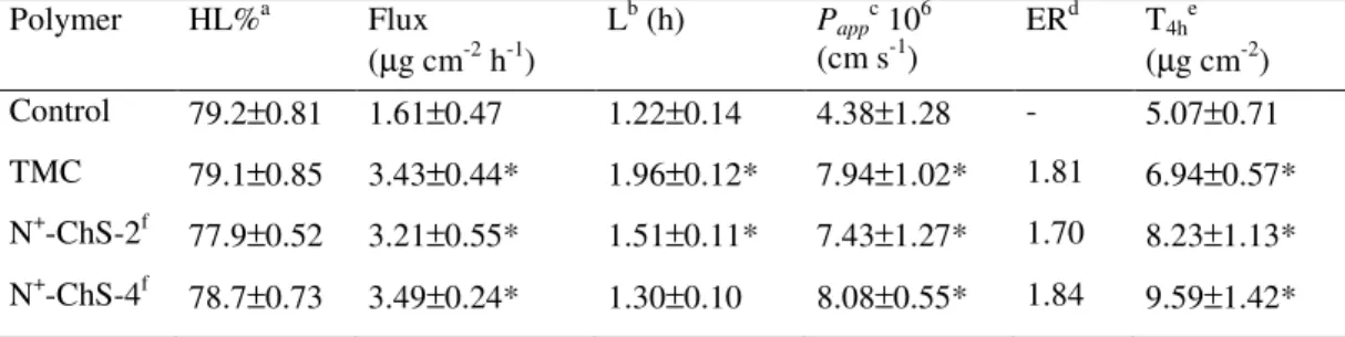
The experimental model used to determine the corneal permeability has been demonstrated by previous reports to be valid [34, 38]. The hydration level (HL%) of the corneal tissue has been considered a sensitive indicator of tissue integrity. According to the literature, the normal water content of the rabbit cornea ranges between 75% and 78% (see, e.g., ref. 39) a corneal damage being indicated by hydration levels increased to 83% or more [38]. In the present work the corneal hydration was determined, at the end of each permeation run, by the usual gravimetric method [38, 40-41]. The resulting HL% values are listed in
Tables I.1. and I.2. None of them is higher than the value of 80.1±0.69%,
reported by Monti et al. [38] for freshly excised rabbit corneas, which indicates that neither the permeants nor the chitosan derivatives produced any substantial damage to the tissue in the perfusion apparatus. The effects of chitosan derivatives on the epithelium permeability may stem from polymer interactions not only with the epithelium structures, but also with the permeant in the donor solution. Indeed, the latter interaction type might depress the permeability in that the permeant fraction bound to the polymer can be considered impermeable. The concentration of the
permeant fraction free from interactions with the polymer, represented by the product C0fF, was introduced into Eq. I.2., so that the calculated
permeability values are independent of binding and only reflect polymer interactions with the epithelium.
I.3.1.1 Permeation of DMS
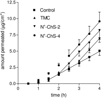
The data on DMS permeation in the absence (control) or in the presence of each of the chitosan derivatives under study (1% w/v) are presented in Figure I.2., while the relevant parameters are listed in Table
I.1. All regressions, applied to the linear portion of each plot, are significant
(r2 > 0.99). As appears from the values of Papp, calculated as illustrated in
Section I.2.7, this property was significantly enhanced by the chitosan
derivatives with respect to the control, the difference in the extent of enhancement between different polymers being not significant. Also the lag time preceding the attainment of steady-state permeation was significantly increased by the polymers, which suggests that a time longer than the control lag time was required for the permeability enhancing effect to reach its maximum intensity. The L values seen in Table I.1. for the different chitosan derivatives are significantly different from one another, the value for N+-ChS-4 being the shortest. In fact, with this polymer the total transport over the whole time of experiment is the maximum of all cases. Then, although the chitosan derivatives tested produced about the same enhancement of DMS corneal permeability at steady state, N+ -ChS-4 has shown the best potential as an intraocular DMS absorption promoter in virtue of the comparative rapidity of its effect. This is particularly important if the short contact times usually allowed by eyedrops is considered. Since DMS is supposed to cross the cornea via the transcellular route, the enhancement effect may be thought to result from a strong interaction between the pendant quaternary ammonium groups of N+-ChS-4 and the negatively charged cell surface, which would perturb the order of the membrane lipid bilayers.
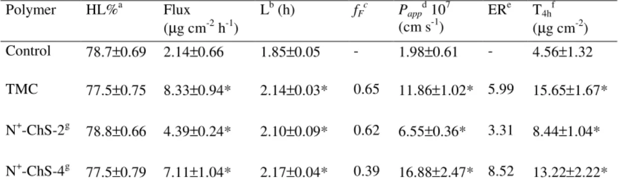
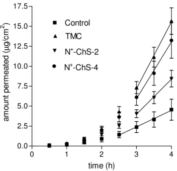
I.3.1.2 Permeation of NaFlu
The data on NaFlu permeation in the absence or in the presence of each of the chitosan derivatives under study (1% w/v) are presented in
Figure I.3., while the relevant parameters are listed in Table I.2. All
regressions, applied to the linear portion of each plot, were significant (r2 > 0.99). This allowed calculating flux and Papp values for each experiment
type, according to the criteria illustrated in Section I.2.7. The Papp value for
the NaFlu control (absence of chitosan derivatives from the donor) is one order of magnitude lower than that for the DMS control, as results from a comparison of relevant data in Tables I.1. and I.2. If NaFlu is supposed to cross the cornea via the paracellular route, these data would indicate that this route is much less pervious to the polar NaFlu than the transcellular route is to the non-polar DMS. The fF values in Table I.2. testify to a
significant NaFlu binding by the chitosan derivatives, ascribed to an ionic interaction between the anionic NaFlu and the polycationic polymers. This was accounted for by calculating the relevant Papp values by means of Eq.
I.2. Such values, listed in Table I.2., along with the corresponding ones for
DMS, seen in Table I.1., indicate that the present chitosan derivatives are able to effectively enhance the corneal permeability of highly polar molecules, such as NaFlu, as well as that of non-polar ones, such as DMS. Unlike in the case of DMS, where the different chitosan derivatives exerted much the same effect on corneal permeability, the enhanced Papp
values for NaFlu appear in Table I.2. to be significantly different from one another, the enhancers ranking in the following order of effectiveness: N+ -ChS-4 > TMC > N+-ChS-2. This difference between DMS and NaFlu could be explained by assuming that the polymers would enhance the permeability of NaFlu by acting on corneal structures (i.e., the tight junctions) different from those supposed to control the permeation of DMS (i.e., the cell membranes). Although the relevant ER values indicate that the enhancing effects on NaFlu permeability are always remarkably stronger than those exerted on DMS, yet in no case did the resulting Papp
suggest that the transcellular route across the cornea is much more pervious to non-polar drugs than the paracellular route is to highly polar ones, despite the enhancing effect exerted on the latter by the present chitosan derivatives. As stated in the foregoing discussion, the Papp values
for NaFlu were calculated by Eq. I.2. in order to separate the effects of the enhancers on the corneal structures from those exerted on permeability by the NaFlu-polymer binding. Such a binding would counteract the Papp
enhancing effect, and therefore, the flux and T4h values produced by N+ -ChS-4 were not significantly different from those produced by TMC, despite the significantly higher Papp value for the former, because N+
-ChS-4 exerted a stronger binding and allowed a lower fF compared to TMC.
The L values for the chitosan derivatives, seen in Table I.2., are close to the value for the control, which indicates that the time required by the polymers to exert their enhancing effects was hardly longer than that required for attaining steady-state permeation conditions. This prevents any evaluation of the promptness of the effects, based on the L value.
I.3.2 Formulation of solutions for in vivo tests
The corneal permeability enhancing effects of the chitosan derivatives, evidenced by the ex vivo permeation experiments discussed in the preceding section, are per se insufficient to anticipate effective transcorneal absorption enhancements in vivo, because some time may be required for the enhancers to be effective, while the contact times allowed by eyedrops are rather short. Thus, in vivo tests were carried out to verify, in the rabbit model, the ocular tolerability and the efficacy of N+ -ChS-4, which has proved the more effective permeability enhancer. The viscosity of the ophthalmic drops containing the polymer under test was increased by adding PVA, a polymer known to be inert on the cornea. By this means the residence of model drug and enhancer in the precorneal area was prolonged by reducing tear fluid drainage. The relevant rheograms (not shown) exhibited a pseudoplastic behaviour, appropriate for ophthalmic solutions. The viscosity of Samples and respective
References was made similar in order to equalize the residence time of the permeants in the precorneal area. Under this condition the N+-ChS-4 effect on the pharmacokinetics in the aqueous could solely be ascribed to a corneal permeability enhancement [20]. The viscosity values at 35 °C and 100 s-1 were: 28.28 mPas, for NaFlu Sample; 27.50 mPas for DMS Sample; 20.05 mPas for NaFlu Reference; 19.75 mPas for DMS Reference.
I.3.3 In vivo tests
The modified Draize test [40] revealed a slight conjunctival discharge and a very slight reddening of the conjunctiva, but no chemosis, during the first 30 min after application of the Samples. In none of the six rabbits did the Iirr score exceed 3. Then, on the whole N+-ChS-4 exhibited
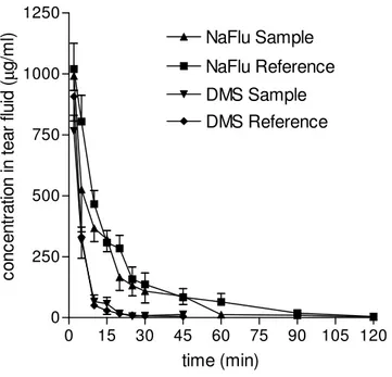
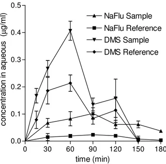
a fair ocular tolerability. As appears from Figure I.4. the lacrimal clearance was similar for each Sample and the respective Reference. This elicited evidencing the transcorneal penetration enhancing effect of the polymer via its effect on the pharmacokinetics in the aqueous. The lachrimal clearance of DMS was faster than that of NaFlu, possibly because the suspended DMS particles caused a more frequent lachrimal turnover. The concentration vs. time profiles in the aqueous are compared in Figure 5 while the pharmacokinetic data are listed in Table I.3. DMS Reference shows a much higher intraocular availability than NaFlu Reference, essentially due to a much more favourable partitioning of the lipophilic DMS into the corneal cell membrane. DMS Sample produced a significant enhancement of DMS intraocular absorption, in agreement with the significant increase of DMS permeability across the excised cornea produced by N+-ChS-4 in the course of the ex vivo experiments discussed in Section 1.3.1.1. The polymer is unlikely to produce this effect by improving drug partitioning into the cornea, but rather, it is likely to facilitate transcellular DMS transport by perturbing the lipid bilayers of the corneal cell membranes. The data show that NaFlu transcorneal penetration from NaFlu Reference is almost insignificant, mostly because
of an unfavourable distribution of the very hydrophilic permeant into the cornea, further depressed by the binding of 52% NaFlu molecules by the PVA contained in NaFlu Reference. The presence of N+-ChS-4 in NaFlu Sample increased the NaFlu bound fraction to 76%, as shown by the dynamic dialysis measurements. Nevertheless, N+-ChS-4 in NaFlu Sample remarkably enhanced intraocular absorption, as shown by the relevant Cmax, AUC and AUCrel values. This result confirms the N+-ChS-4 ability to enhance NaFlu corneal permeability, evidenced by the ex vivo experiments and ascribed to a loosening of the tight junctions between corneal cells. In fact, there is an overall agreement between the results of the ex vivo experiments, found in Tables I.1. and I.2. and those of the in vivo ones, found in Table I.3.. The enhancement effect exerted on corneal permeability by N+-ChS-4 is seen from the relevant ER values to be remarkably stronger for NaFlu than for DMS. Yet, the enhanced Papp of
NaFlu remains lower than the not enhanced (control) Papp of DMS. In
agreement with these results, the AUCrel value, which measures the N+ -ChS-4 effect on the intraocular availability of DMS or NaFlu, is remarkably higher for the latter than for the former, yet the enhanced AUC and Cmax values for NaFlu remain lower than the respective not enhanced values for DMS. It should be recognized, in this respect, that different AUC and Cmax values for two different permeants are suggestive of differences in corneal permeability, even though other pharmacokinetic parameters may be different. It may be noticed that the concentration vs. time curves in the aqueous, seen in Figure I.5., show an initially rapid absorption with no evident lag time, in apparent contrast with the ex vivo data presented in
Figures I.2. and I.3. and in Tables I.1. and I.2.
Such an apparent contradiction can be solved as follows. A lag time is needed to attain steady-state flux across the excised cornea in the ex vivo experiments, where steady-state conditions are ensured by a time-constant permeant concentration in the donor. On the other hand, in the in vivo tests the permeant concentration at the absorption site is maximal at
the beginning of contact, and declines rapidly, as appears from the concentration vs. time plots in tear fluid, seen in Figure I.4.
I.4 Conclusions
The ex vivo and in vivo experiments have concurrently put into evidence the potential of substituted chitosans of the N,O-[N,N-diethylaminomethyl(diethyldimethylene ammonium)nmethyl] type to enhance the corneal permeability, and hence, the intraocular penetration of either non-polar or highly polar drugs. N+-ChS-4, the more substituted derivative with the higher positive charge density has resulted more effective than TMC, the well known transepithelial absorption enhancer, and has shown a fair ocular tolerability. Because DMS, taken as a model of non-polar drugs, and NaFlu, model of highly polar ones, are supposed to cross the cornea via the transcellular and the paracellular route, respectively, then N+-ChS-4 is believed to act both by perturbing the order of the lipid bilayers of the cell membrane and by loosening the tight junctions joining the corneal cells. The paracellular route remains comparatively difficult to penetrate, despite the enhancing effect of N+ -ChS-4. Indeed, although the effect on NaFlu corneal permeability and intraocular availability is stronger than that exerted on the respective properties of DMS, still the enhanced permeability and intraocular availability of the polar molecule have resulted lower than the corresponding, not enhanced properties of the non-polar one. It must be considered, nevertheless, that the anionic NaFlu was partially bound by the polycationic N+-ChS-4. Although the relevant permeability value is independent of the binding, since it was calculated on the free NaFlu fraction basis, the transcorneal flux, and hence, the intraocular availability could have been depressed by the binding. Then, the effect of N+-ChS-4 on the intraocular transport of a polar drug not interacting with the polymer (e.g., a cationic one) may be expected to be stronger than that evidenced in the present work with NaFlu.
TABLES
Table I.1. - Data on DMS permeation across excised rabbit cornea from GBR containing
0.3% w/v DMS and 1% w/v of different chitosan derivatives (mean ± SD, n = 6).
Polymer HL%a Flux (µg cm-2 h-1) Lb (h) Pappc 106 (cm s-1) ERd T4he (µg cm-2) Control 79.2±0.81 1.61±0.47 1.22±0.14 4.38±1.28 - 5.07±0.71 TMC 79.1±0.85 3.43±0.44* 1.96±0.12* 7.94±1.02* 1.81 6.94±0.57* N+-ChS-2f 77.9±0.52 3.21±0.55* 1.51±0.11* 7.43±1.27* 1.70 8.23±1.13* N+-ChS-4f 78.7±0.73 3.49±0.24* 1.30±0.10 8.08±0.55* 1.84 9.59±1.42* a
Corneal hydration level.
b Lag time. c Apparent permeability. d Enhancement ratio. e
Cumulative transport over the whole time of experiment (4 h).
f
See Figure I.1. and Section I.2.2. for polymer structure and explanation of polymer code. The data marked by * are significantly different from the respective controls (P < 0.05).
Table I.2. - Data on NaFlu permeation across excised rabbit cornea from GBR containing
0.3% w/v NaFlu and 1% w/v of different chitosan derivatives (mean ± SD, n = 6).
Polymer HL%a Flux (µg cm-2 h-1) Lb (h) fFc Pappd 107 (cm s-1) ERe T4hf (µg cm-2) Control 78.7±0.69 2.14±0.66 1.85±0.05 - 1.98±0.61 - 4.56±1.32 TMC 77.5±0.75 8.33±0.94* 2.14±0.03* 0.65 11.86±1.02* 5.99 15.65±1.67* N+-ChS-2g 78.8±0.66 4.39±0.24* 2.10±0.09* 0.62 6.55±0.36* 3.31 8.44±1.04* N+-ChS-4g 77.5±0.79 7.11±1.04* 2.17±0.04* 0.39 16.88±2.47* 8.52 13.22±2.22* a
Corneal hydration level.
b
Lag time.
c
NaFlu fraction free from polymer binding.
d
Apparent permeability.
e
Enhancement ratio.
f
Cumulative transport over the whole time of experiment (4 h).
g
See Figure I.1. and Section I.2.2. for polymer structure and explanation of polymer code. The data marked by * are significantly different from the respective controls (P < 0.05).
Table I.3. - Pharmacokinetic data for the aqueous (mean ± SD, n ≥ 6)a.
Vehiclea Cmax (µg ml-1) tmax (min) AUC (µg ml-1 min) AUCrel
DMS Reference 0.214±0.030 60 17.13±2.17 1
DMS Sample 0.407±0.052* 60 28.27±2.88* 1.65
NaFlu Reference 0.023±0.006 90 2.37±0.56 1
NaFlu Sample 0.102±0.021* 90 10.80±1.27* 4.56
a
See Sections I.2.5., I.2.8. and I.3.3. for vehicle composition, in vivo tests and data discussion.
FIGURES
Figure I.1. - Structure of N,O-[N,N-diethylaminomethyl(diethyldimethylene
ammonium)nmethyl] chitosans.
O NHR O O CH2OR O H CH2 ( CH2 N CH2CH3 CH2CH3 CH2)n + CH2 N CH2CH CH2CH3 3 R= H;
Figure I.2. - Effects of chitosan derivatives on DMS permeation across excised rabbit
cornea. Means ± SD of 6 runs. Regression lines are reported (r2 > 0.99). Explanation of polymer codes is found in Section I.2.2.
0 1 2 3 4 0.0 2.5 5.0 7.5 10.0 12.5 Control TMC N+-ChS-4 N+-ChS-2 time (h) am ou nt p er m ea te d (µ g/ cm 2 )
Figure I.3. - Effects of chitosan derivatives on NaFlu permeation across excised rabbit
cornea. Means ± SD of 6 runs. Regression lines are reported (r2 > 0.99). Explanation of polymer codes is found in Section I.2.2.
0 1 2 3 4 0.0 2.5 5.0 7.5 10.0 12.5 15.0 17.5 Control TMC N+-ChS-2 N+-ChS-4 time (h) am ou nt p er m ea te d (µ g/ cm 2 )
Figure I.4. - Kinetics of DMS and NaFlu disappearance from tear fluid following
instillation of ophthalmic drops (see Section I.2.5. for vehicle composition). Means ± SD of at least 6 values obtained with different animals.
0 15 30 45 60 75 90 105 120 0 250 500 750 1000 1250 NaFlu Reference NaFlu Sample DMS Sample DMS Reference time (min) co nc en tr at io n in te ar fl ui d (µ g/ m l)
Figure I.5. - Pharmacokinetics in the aqueous following instillation of ophthalmic drops
(see Section I.2.5. for vehicle composition). Means ± SD of at least 6 values obtained with different animals.
0 30 60 90 120 150 180 0.0 0.1 0.2 0.3 0.4 0.5 DMS Reference DMS Sample NaFlu Reference NaFlu Sample time (min) co nc en tr at io n in a qu eo us ( µ g/ m l)
PUBLICATIONS
Zambito Y., Zaino C., Carelli V., Serafini M.F., Di Colo G., A novel
quaternary ammonium chitosan derivative for the promotion of intraocular drug absorption, 33rd Annual Meeting & Exposition of the Controlled
Release Society, pp 940-940, Wien, Austria, 2006.
Zambito Y., Zaino C., Burchielli S., Carelli V., Serafini M.F., Di Colo G.,
Novel quaternary ammonium chitosan derivatives for the promotion of intraocular drug absorption, Journal of Drug Delivery Science and
SECTION II - Improved synthesis of quaternary
ammonium-chitosan conjugates (N
+-Ch) for enhanced intestinal drug
permeation
II.1 Introduction
The ability of chitosan to promote drug absorption across different epithelia (intestinal, nasal, buccal, corneal) is now well documented [13-20]. Chitosan has been partially quaternized to yield the watersoluble N-trimethylchitosan chloride (TMC) [21] in order to circumvent the limits of chitosan insolubility at neutral pH. TMC has proved an effective drug permeation enhancer across intestinal, nasal, buccal, corneal epithelia in neutral environments [20, 23-27, 30, 46]. It was reported that the absorption-enhancing efficacy of TMC could depend on its charge density which, in neutral or alkaline environments, is determined by its degree of quaternization (DQ) [29, 30]. However, other structural properties of polymer must concur in determining its effectiveness, in fact, methyldiethylaminoethyl dextran, the fully quaternized derivative of diethylaminoethyl dextran [54], failed to enhance ofloxacin permeability across the reconstituted cornea [20]. It was also reported that mucoadhesion is a key element of TMC polymers for being effective as absorption enhancers at mucosal surfaces and that the mucoadhesivity of these polymers increases with increasing MW [52]. Then, on the basis of literature information it could be hypothesized that the transmucosal drug absorption enhancing properties of quaternized chitosans depend on MW, DQ and other structural features. In this light, novel chitosan derivatives with pendant quaternary ammonium groups were prepared by reacting chitosan with 2 diethylaminoethyl chloride (DEAE-Cl) under different conditions [31]. These derivatives were shown by NMR analysis to have the structure of quaternary ammonium-chitosan conjugates (N+-Ch), as depicted in Figure I.1. The ability of N+-Ch polymers to promote the penetration of hydrophobic (rhodamine 123) as well as hydrophilic (fluorescein sodium) drug models across the excised porcine buccal
mucosa was evidenced [31]. Such polymers have also shown the ability to enhance the permeability of the hydrophobic dexamethasone and that of the hydrophilic fluorescein sodium across the excised rabbit cornea, and to promote the intraocular absorption of these drug models in vivo, in the rabbit model [55]. The enhancing effects of N+-Ch polymers were found to be stronger than those of the known penetration enhancer, TMC [31, 55]. Hence, these polymers show promise of behaving as effective penetration enhancers for different drugs across different epithelia. The present work aims at extending the knowledge of the potential of N+-Ch polymers to safely promote transepithelial drug absorption. For the first time ever, in the present work selected polymers of the N+-Ch type have been tested on excised rat intestinal epithelium for their ability to promote penetration of two hydrophilic drug models, i.e., fluorescein sodium (low MW) and fluorescein isothiocyanate dextran (MW 4400 Da), and a lipophilic one, i.e., rhodamine 123, without causing any significant damage to the tissue. Fluorescein sodium and fluorescein isothiocyanate dextran are transported via the paracellular route, while rhodamine 123 is transported via both the paracellular and the transcellular routes [50]. In our previous work [31, 55] N+-Ch polymers, each obtained and characterized from a single batch, were studied. However, we subsequently verified that the reproducibility of the synthesis reaction was unsatisfactory, probably because the pH of the reaction mixture was not controlled strictly, in fact, it was let to vary during the reaction. Therefore, in the present development of the work the pH has been strictly controlled at 5, 6, 7, 8 or 9 over the reaction time, and the pH dependence of the DS and n values of product and process reproducibility have been assessed. By these means the polymer structure could be predetermined through a strict control of the synthesis conditions, namely, molar ratio between reactant DEAE-Cl and chitosan repeating unit, reaction temperature and pH.
II.2 Materials and methods
II.2.1 Materials
Fluorescein sodium salt (NaFlu), rhodamine 123 (Rh-123), fluorescein isothiocyanate dextran, MW 4400Da (FD4), trypan blue (Sigma); 2-diethylaminoethyl chloride (DEAE-Cl) hydrochloride (Fluka); chitosan (minimum 90% deacetylated) from shrimp shell (Ch) (Chito-clear FG90, Primex, Drammen, Norway) were used. The commercial Ch had an average viscometric molecular weight of 590 kDa. The deacetylation degree, determined by IR or NMR, was 90% or 82%. TMC with a DQ of 46% was prepared from Ch by an already described method [20].
II.2.2 Controlled synthesis of N+-Ch polymers
Two grams of commercial Ch powder were dissolved in 80 ml of 0.11M hydrochloric acid (pH 4.7). Eight grams of DEAE-Cl HCl and 11 ml of 15% sodium hydroxide were added in sequence to the Ch solution, under vigorous stirring at 65°C. The molar ratio between DEAE-Cl and the Ch repeating unit was 4. Stirring and heating were continued for a total of 2h during which the pH of the reaction mixture was kept under strict control at a pre-established value (5, 6, 7, 8 or 9) by keeping the electrode of a pH-meter immersed in the reaction mixture and adding concentrated aqueous NaOH when needed. Then the mixture was made to pH 7 with 1M hydrochloric acid, and clarified by filtration through filter paper under vacuum. Next it was purified by dialysis (Spectra/Por®, cut-off 3500, Spectrum Laboratories Inc., Rancho Dominguez, CA, USA) under sink conditions. The dialysis was continued until no chloride was found in the external phase, then the internal solution was lyophilized to obtain the purified product. Each chitosan derivative is coded N+-Ch-x, where x is a figure indicating the pH at which the synthesis was carried out. The property of N+-Ch-7 or N+-Ch-8 to dissolve in neutral aqueous environment was investigated by progressively adding the polymer portionwise to
phosphate buffer pH 7.4 under stirring, and observing the aspect of the resulting mixture.
II.2.3 NMR studies
The effect of the synthesis conditions on polymer MW was studied by NMR using the Diffusion Ordered SpectroscopY (DOSY) technique. This allows measurement of diffusion coefficients (D) in solution, which depend on molecular hydrodynamic radius (RH), and hence, on molecular size, according to the following Stokes–Einstein equation [48]:
where k is the Boltzmann constant, T is the absolute temperature, and
η
isthe solvent viscosity.
On this basis, also the diffusion coefficients of polymeric materials have been correlated with their molecular weights [44]. 1H NMR measurements were performed on a NMR spectrometer operating at 600 MHz. The temperature was controlled to ± 0.1°C. DOSY experiments were carried out by using a stimulated echo sequence with self-compensating gradient schemes, a spectral width of 5000 Hz and 32K data points. Typically, a value of 700ms was used for ∆, 1.5ms for δ, and g was varied in 20 steps (four transients each) to obtain the better decrease in the resonance intensity at the largest gradient amplitudes. The baselines of all arrayed spectra were corrected prior to data processing. After data acquisition, each FID was apodized with 1.0 Hz line broadening and Fourier transformed. The data were processed with the DOSY macro (involving the determination of the resonance heights of all the signals above a pre-established threshold and the fitting of the decay curve for each resonance to a Gaussian function) to obtain pseudo two-dimensional spectra with NMR chemical shifts along one axis and calculated diffusion coefficients along the other.
6 R kT D H πη =
II.2.4 Permeation experiments
For permeation studies, the intestinal mucosa was excised from non-fasting male Wistar rats weighing 250–300 g. After sacrificing the rats, the first 20cm of jejunum were immediately removed. The excised intestine was cut into strips of 1.5 cm, rinsed free of luminal contents and mounted in Ussing type chambers (0.78cm2 exposed surface area) without stripping off the underlying muscle layer. The pre-thermostated donor medium, containing 250 mM NaCl, 2.6 mM MgSO4, 10 mM KCl, 40 mM glucose and 50 mM NaHCO3 buffered with 50 mM Hepes (N-[2-hydroxyethyl] piperazine-NI-[2-ethanesulfonic acid]) pH 6.8 was added to the apical side
and the same salt composition buffered with 40 mM Hepes pH 7.4 was added to the basolateral side. In order to ensure oxygenation and agitation, a mixture of 95% O2 and 5% CO2 was bubbled through each compartment. The Ussing chambers were then placed in a water bath at 37 °C. After a 20-min equilibration period, a solution of NaFlu (0.2%, w/v), or FD4 (0.1, 0.2 or 0.5%, w/v), or Rh-123 (0.001%, w/v) in the donor medium was added to the apical chambers. The apical to basolateral transport of the drug models was investigated in the absence or presence of 0.5% (w/v) of the polymer under test in the donor (TMC, N+-Ch-7 or N+ Ch-8). At 30-min intervals over 180 min, 100µl samples were withdrawn from the acceptor chambers and replaced by the same volume of fresh medium. The amount of permeated drug model was determined by HPLC, as already described [26, 55].
II.2.5 Measurement of drug–polymer interactions
A previously described method, based on the dynamic dialysis technique [20], was used as described in Section I.2.6 to determine the drug-polymer interactions in the donor phase of the permeation experiments referred to in the preceding section. Drug flux through a porous cellulose membrane (molecular weight cut-off, 3500 Da, with NaFlu or Rh-123, or 10000-12000 Da, with FD4) under quasi-steady state conditions was measured in the presence or absence of the polymer in the
donor phase. The polymer and initial drug concentrations in the donor were equal to those used in the ex vivo permeation experiments, referred to in the preceding Section. The receptor was analysed for the drug by HPLC, as already described [26, 55].
The regression for the fitting of dialysis data, expressed as drug concentration in the donor vs. time, to 1st order kinetics was always significant (r2
≥ 0.97, n≥ 8). This allowed calculation of the fraction of free, non-interacting drug, fF, as described in Section I.2.6.
Permeation data were treated as previously described in Section I.2.7.
II.2.6 Test of viability of epithelial cells
Epithelial cell viability following contact of intestinal mucosa with N+-Ch-7 or N+-Ch-8 was checked by the trypan blue test [45, 53].
After the permeation experiments, the donor medium was replaced by 1ml trypan blue die and the mucosa was incubated for 30 min. Microscopy demonstrated that the mucus was still present. No blue colour was detectable within the cells, thus testifying to their viability. These observations were in agreement with the results of viability studies previously published by others [45, 53].
II.3 Results and discussion
II.3.1 Control of the aminoalkylation reaction
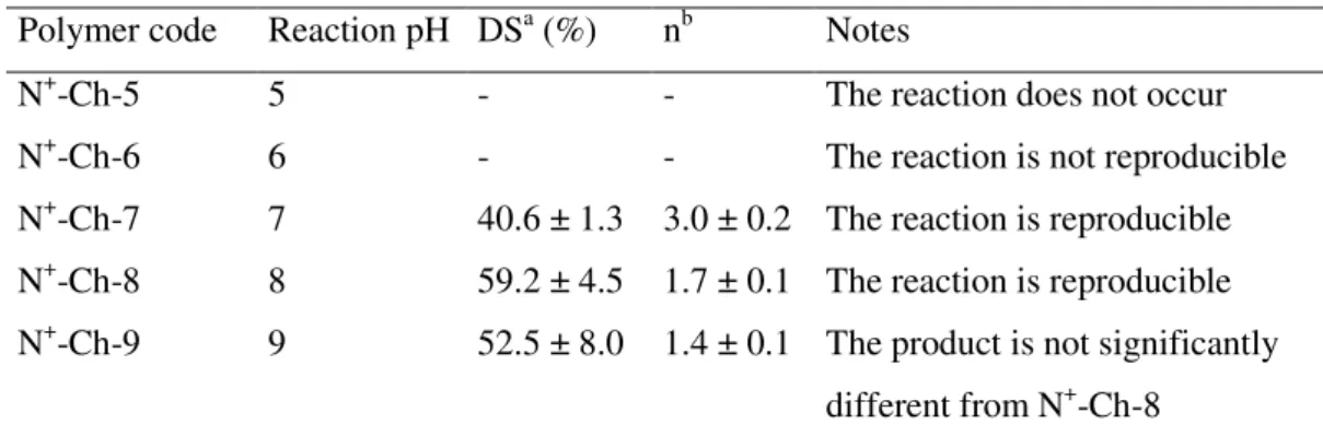
We have verified that the synthesis reaction of the N+-Ch conjugates, if performed as in our previous work [31, 55], has an unsatisfactory reproducibility of the structural parameters DS and n. This would imply a difficulty in predetermining these values and, consequently, the permeability-enhancing properties of the polymer. It was thought that this was due to the lack of a strict control of the reaction mixture pH. Therefore, in the present work such a pH has been controlled strictly at 5, 6, 7, 8 or 9 over the reaction time. The characteristics of the derivatives
obtained, determined by NMR as described in the previous paper [31], are reported in Table II.1. It was observed that at pH 5 the reaction did not occur, at pH 6 it was not reproducible, at pH 7 or higher the reaction was reproducible. In particular, as the pH was increased from 7 to 8 the DS value increased while the n value decreased. A pH increase from 8 to 9 exerted no further effect. Thus, the control of the reaction pH succeeded in making both the reaction reproducible and the structural parameters modulatable. When N+-Ch-7 or N+-Ch-8 was progressively added to phosphate buffer pH 7.4 a clear solution was initially obtained. This became a clear gel, which blocked the stirring, when the polymer concentration reached 0.4–0.5 g/ml. At thermodynamic equilibrium the system always appeared to be composed of a single, clear phase. Then it can be stated that both derivatives are freely water-soluble in neutral environment. As stated in the Introduction section, the MW of polymer could influence its enhancing effect on drug transport across the intestinal epithelium [51]. This prompts the question whether the synthesis conditions could cause some fragmentation of the Ch chain. In principle, no important fragmentation was expected in the cases where a pH close to neutrality (e.g., 7 or 8) was maintained throughout the synthesis. Nevertheless, NMR studies using the DOSY technique were carried out to confirm this assumption. The starting Ch is characterized by a high MW, and hence, by a diffusion coefficient in the limit region of determination by DOSY technique. Nevertheless, fragmentation should result in a significant increase of diffusion coefficient. In fact, it appears from the DOSY maps presented in Fig. II.1. that the diffusion coefficients of the Ch derivatives N+-Ch-7 and N+-Ch-8, not significantly different from each other, were not increased with respect to the parent Ch. This indicates that in neither case did the synthesis conditions produce any important chain fragmentation.
II.3.2 Permeation experiments
The excised rat jejunum was chosen among the known intestinal epithelium models for the permeability enhancement studies because its tight junctions are similar in tightness and number to those of the human jejunum [49] and because the Ch derivatives under study are expected to act by modulating intercellular tight junctions [55]. A TMC with a DQ of 46% was chosen as a reference to evaluate the effectiveness of the present N+-Ch conjugates because it was reported that TMC with intermediate DQ is most effective in enhancing drug permeability across mucosal epithelia [20,30,47).
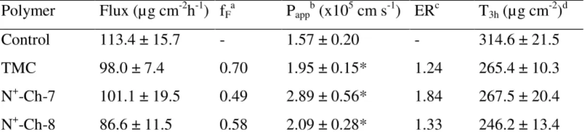
II.3.2.1 Data for NaFlu
Data on NaFlu permeation are listed in Table II.2. As in the case of the Ch derivatives studied in our previous work [55], also the present ones showed a significant binding of the NaFlu molecules, indicated by the relevant fF values. Therefore, the Papp values, calculated by Eq. I.2., were significantly enhanced by the polymers over the control, even though the flux values were not. In fact, with each polymer the mean flux was lower than the control value. These findings support the consideration at the basis of Eq. I.2., i.e., drug–polymer binding counteracted the enhancing effect of polymer on transmembrane flux by limiting the fraction of free drug. On the other hand, the possible hypothesis that the polymer might mediate transepithelial drug transport by concurrently interacting with the drug and with the epithelium is not supported by the flux values, in no case higher than the control value. As appears from Table II.2., the Papp values for N+-Ch-7 and N+-Ch-8 are not significantly different, whereas the value for N+-Ch-7 is significantly higher than that for TMC. No appreciable lag time before attainment of steady-state flux was ever noticed with NaFlu.
II.3.2.2 Data for FD4
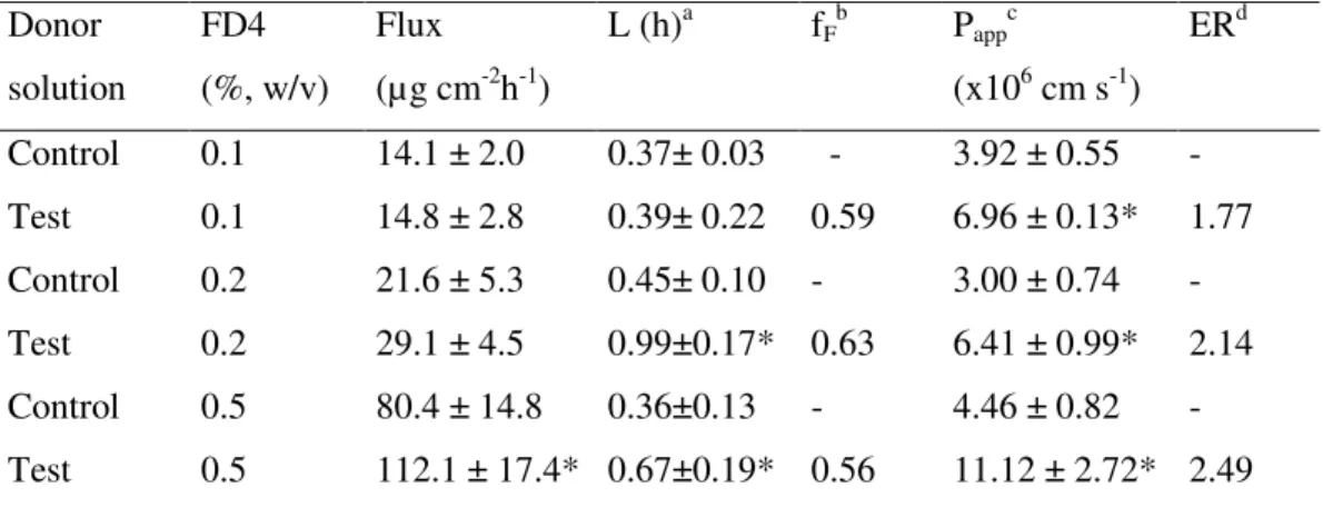
The ability of N+-Ch conjugates to enhance the intestinal epithelium permeability to hydrophilic macromolecules was compared with that of
TMC using FD4 (MW 4400 Da), an extensively used marker of the paracellular route. As appears in Table II.3., the Papp of this macromolecule, in the absence of enhancers, is much lower than that seen in Table II.2. for enhancer-free NaFlu, as expected considering that both were transported across the paracellular route and that the molecular size of FD4 is much larger than that of NaFlu. As in the case of NaFlu, discussed above, the FD4 molecule was significantly bound to the polymers, so the free fraction was used to calculate the relevant Papp values. All polymers resulted effective permeability enhancers, N+-Ch-8 being significantly more effective than N+-Ch-7 or TMC. Because FD4 is supposed to travel the paracellular route, the hypothesis that N+-Ch conjugates act by loosening the epithelial tight junctions is sound. Such an effect is significantly stronger with the conjugate having higher DS and lower n value. In principle, the significant FD4-polymer binding, testified by the fF values in Table II.3., in addition to depressing FD4 transmembrane flux could hamper the polymer action to decrease the epithelium barrier properties. If this were the case, increasing FD4 concentrations at constant N+-Ch concentration would decrease the P
app value. Unexpectedly, the data in Table II.4. indicate that the opposite is true, i.e., the Papp is significantly higher with the highest than with the lowest FD4 concentration. These results could be explained by admitting that the permeant fraction interacting with the polymer is not completely impermeable, possibly due to a parallel penetration mechanism different from passive permeation.
II.3.2.3 Data for Rh-123
In Table II.5., data are listed on Rh-123 permeation across the excised rat jejunum. It is noteworthy that the apparent permeability value for the control is three orders of magnitude higher than that for the respective control value for Rh-123 permeation across excised porcine buccal epithelium, reported previously [31]. N+-Ch-7 was the only conjugate to enhance the Papp significantly with respect to the control,
although its effect on the cumulative transport is not significant, due to a comparatively high lag time. It should be considered, however, that possible enhancing effects on epithelial permeability by the other polymers could be masked by the muscle layer underlying the epithelial membrane. Such a layer, indeed, could control the permeation rate of such a lipophilic molecule as Rh-123. The conjugate N+-Ch-7, having lower DS and higher
n value, exerted a stronger enhancing effect on the permeability of
Rh-123, as can be seen in Table II.5. According to Sakai et al. (1997) [50], Rh-123 crosses the intestinal epithelium by the transcellular, in addition to the paracellular route. This suggests that the effect of N+-Ch-7 on Papp is apparently stronger than that of N+-Ch-8 because of more ability of the former to act on the transcellular route, e.g., by perturbing the order of the plasma membrane lipids. As a hypothesis, such an ability could be due to the higher n value for the pendant chains of N+-Ch-7, which would elicit a stronger interaction with the epithelial cell membrane. TMC was apparently inactive, perhaps because it was not endowed with a similar interactive strength. Considering that the positive charge per unit repeating unit of N+ -Ch-7 and N+-Ch-8 is similar, the hypothesis can be advanced that the pendant chain of adjacent quaternary ammonium groups, whether longer or shorter, determines the ability of N+-Ch conjugates to enhance the transcellular rather than the paracellular route or vice versa. The fF values in Table II.5. testify to an absence of Rh-123 binding to the polymers. The
L value for N+-Ch-7 is significantly higher than the values for the control
and the apparently inactive polymers, indicating that some time was needed for the enhancing effect to be exerted.
II.4 Conclusions
The structural parameters, DS and n, of the N+-Ch conjugates can be controlled by strictly controlling the pH at which the N+-Ch synthesis from Ch is carried out. The pH values of 7 and 8 have reproducibly yielded the more significant derivatives, N+-Ch-7 and N+-Ch-8. The present ex vivo procedure used to determine the Papp of the excised intestinal wall shows
intrinsic limits, e.g., such a membrane comprises both mucosal epithelium and underlying muscle tissue. Therefore, the obtained Papp values can only be used for comparative purposes. In this light, it can be concluded, on the basis of the present results, that the Ch derivatives N+-Ch-7 and N+-Ch-8 are more effective absorption enhancers than the TMC specimen used as a reference in the present study. Of the excised intestinal membrane structures, the mucosal epithelium was rate-controlling enough to discriminate between different polymers as concerns their permeability-enhancing properties. This can be interpreted as a sign of preservation of epithelium integrity, as also demonstrated by the trypan blue test. The permeation data have prompted the hypothesis that the structural parameters, DS and n, for a conjugate may be correlated with the conjugate ability to promote drug penetration across the excised intestinal membrane via the transcellular or the paracellular route. Thus, the penetration of the lipophilic Rh-123 has been enhanced by N+-Ch-7, having lower DS and higher n, more than by N+-Ch-8, which has higher DS and lower n. The opposite is true with the hydrophilic macromolecular FD4, for which N+-Ch-8 has exerted a stronger enhancement than N+ -Ch-7. Only in the case of Rh-123, which showed no appreciable interaction with the polymers, did the Papp enhancement result in a significant enhancement of permeation flux with respect to the control. On the other hand, this was not the case of the negatively charged NaFlu or FD4, because a significant binding of these permeants by the polycationic N+ -Ch conjugates would limit the respective fluxes. Hence, a significant enhancement of paracellular absorption of hydrophilic drugs of either low or high molecular weight by the present chitosan derivatives is expected for drugs that do not interact significantly with the polymer at the absorption site. As for the mode of application of these polymeric enhancers to the intestinal mucosa, it should be considered that they are readily soluble in the fluids of the intestinal lumen, so it would be appropriate, e.g., to introduce them as solid water soluble excipients into
enteric-coated tablets. The viability tests have shown that these excipients are safe on the intestinal mucus and epithelial cells.
TABLES
Table II.1. – Effect of the reaction pH on the structural characteristics of the N+-Ch conjugates
Polymer code Reaction pH DSa (%) nb Notes
N+-Ch-5 5 - - The reaction does not occur
N+-Ch-6 6 - - The reaction is not reproducible
N+-Ch-7 7 40.6 ± 1.3 3.0 ± 0.2 The reaction is reproducible
N+-Ch-8 8 59.2 ± 4.5 1.7 ± 0.1 The reaction is reproducible
N+-Ch-9 9 52.5 ± 8.0 1.4 ± 0.1 The product is not significantly
different from N+-Ch-8
a
Degree of substitution, determined by NMR analysis. Mean ± S.D. of 6 batches.
b
Number of quaternary ammonium groups in pendant chains, determined by NMR analysis.
Table II.2. – Data on NaFlu permeation across excised rat jejunal epithelium from Ringer
buffer pH 6.8 containing 0.2% (w/v) NaFlu and 0.5% (w/v) of different Ch derivatives. Polymer Flux (µg cm-2h-1) fF a Papp b (x105 cm s-1) ERc T3h (µg cm -2 )d Control 113.4 ± 15.7 - 1.57 ± 0.20 - 314.6 ± 21.5 TMC 98.0 ± 7.4 0.70 1.95 ± 0.15* 1.24 265.4 ± 10.3 N+-Ch-7 101.1 ± 19.5 0.49 2.89 ± 0.56* 1.84 267.5 ± 20.4 N+-Ch-8 86.6 ± 11.5 0.58 2.09 ± 0.28* 1.33 246.2 ± 13.4
Means ± S.D. of at least 6 runs. The data marked by (*) are significantly different from the respective controls (P<0.05).
a
Fractionof free permeant. b
Apparent permeability.
c
Enhancement ratio.
d
Table II.3. – Data on FD4 permeation across excised rat jejunal epithelium from Ringer
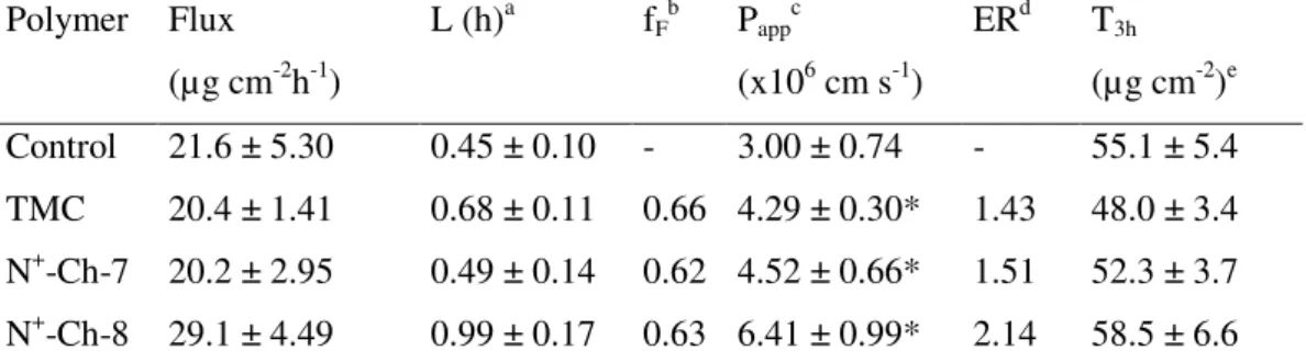
buffer pH 6.8 containing 0.2% (w/v) FD4 and 0.5% (w/v) of different Ch derivatives Polymer Flux (µg cm-2h-1) L (h)a fF b Papp c (x106 cm s-1) ERd T3h (µg cm-2)e Control 21.6 ± 5.30 0.45 ± 0.10 - 3.00 ± 0.74 - 55.1 ± 5.4 TMC 20.4 ± 1.41 0.68 ± 0.11 0.66 4.29 ± 0.30* 1.43 48.0 ± 3.4 N+-Ch-7 20.2 ± 2.95 0.49 ± 0.14 0.62 4.52 ± 0.66* 1.51 52.3 ± 3.7 N+-Ch-8 29.1 ± 4.49 0.99 ± 0.17 0.63 6.41 ± 0.99* 2.14 58.5 ± 6.6
Means ± S.D. of at least 6 runs. The data marked by (*) are significantly different from the respective controls (P<0.05).
a
Lag time.
b
Fractionof free permeant.
c
Apparent permeability.
d
Enhancement ratio.
e