UNIVERSITY OF PISA
BIOS Research Doctorate School in BIOmolecular Science PROGRAM IN EXPERIMENTAL AND MOLECULAR
ONCOLOGY
XXIII CYCLE (2008-2010)
Research Doctorate Thesis
The Pathogenic Role of the Human Mammary
Tumor Virus (HMTV) in Human Breast Cancer
Supervisor Candidate
Prof. Generoso Bevilacqua Dr. Mohammad Al Hamad
Tutor
Dedication
To my father, mother, brothers and sisters for their love and support. To my lovely wife for her support, kindness and patience, and to my
Acknowledgements
I would like to express my deep and sincere gratitude to my supervisor Professor Generoso Bevilacqua for the opportunity to do exciting research in the field of Molecular Oncology. His guidance and support were invaluable.
This dissertation would not be finished without the support, encouragement and help of many people who I appreciate.
My tutor Dr. Chiara Maria Mazzanti whose encouragement, guidance and support from the initial to the final level enabled me to develop an understanding of the subject. Dr. katia Zavaglia for her valuable advice and friendly help. Dr. Giovanni Fanelli for his help in selecting the samples. I would like to thank Ms Maria Pia for her help. I want to thank also my colleagues Mazhar, Francesca, Sara, Alesandro, Claudia, and Evana for the scientific discussions that taught me many things during my research.
I am very grateful to Professor Mauro Pistello for collaboration and the fruitful scientific discussion that significantly influences my thesis.
I would like to express my love and gratitude to my beloved families; for their understanding & endless love, through the duration of my studies.
Table of Contents
ABSTRACT...9
INTRODUCTION: ...11
RISK FACTORS OF BREAST CANCER...11
Age ...11
Reproductive factors ...11
Hormonal status...12
Genetics...13
Virus etiology of mammary tumor ...13
Methods used in the past for viral detection ...14
MOUSE MAMMARY TUMOR VIRUS...19
History: ...19
TAXONOMY ANDCLASSIFICATION:...19
PROPERTIES OF THEVIRION: ...20
VIRAL GENOME...21
Super antigen ...22
MMTV life cycle ...23
Recombination of exogenous and endogenous MMTV...25
MMTV and Tumorigenesis...26
Replication cycle of MMTV ...28
HUMAN ENDOGENOUS RETROVIRUS:...32
The biological function of HERV...34
HERV-K family ...35
MATERIALS AND METHODS ...36
Specimens...36
Histological criteria...38
LASER MICRODISSECTION...41
DNA extraction from microdissected tissue...42
DNAEXTRACTION FROM PARAFFIN EMBEDDED TISSUE...43
RNAEXTRACTION FROM CELL CULTURE...44
cDNA synthesis: ...45
DNAAMPLIFICATION SUITABILITY...46
HMTVSEQUENCE DETECTION...47
SEQUENCING OFHMTVENVPCRFRAGMENTS...49
HMTV Real Time PCR assay...50
Amplification of the probe (2.7 kb env-LTR) ...52
Labeling the probe ...53
CHROMOGEN IN SITU HYBRIDIZATION(CISH) ...54
TISSUE CULTURE...56
Collecting medium ...56
Primary culture medium:...56
Complete culture medium ...56
Antibiotic solution (ABC-PBSA) ...56
Day 1 protocol ...57
Day 2 protocol ...58
Removal of fibroblastic cells from the tissue culture...59
Cryopreservation the cells ...60
STATISTICAL ANALYSIS...61
RESULTS ...62
HMTV frequency in phases of breast cancer progression...62
HMTV viral load with RT-PCR...66
CHROMOGEN IN SITU HYBRIDIZATION(CISH) ...69
The frequency of HMTV in metastatic lymph nodes ...72
The frequency of HMTV in primary and metastatic breast cancer...73
The frequency of HMTV in primary breast cancers and their lymph nodes ...75
TISSUE CULTURE...78
Primary culture of human breast cancer ...78
Presence of HMTV in primary culture of human breast cancer ...80
DISCUSSION ...82
Table of figures and tables
Figure 1 Structure of proviral sequences in human breast cancer. .17
Figure 2 Electron microscopy of MMTV. ...18
Figure 3: The structure of MMTV ... 21
Figure 4: MMTV life cycle... 25
Figure 5: Histological types, and the number of breast cases...37
Figure 6: The cases number of breast cancer and their lymph nodes ...38
Figure 7: Breast cancer progression... 40
Figure 8: Laser capture microdissector... 41
Figure 9: Laser capture microdissection of breast cancer...42
Figure 10: Fluorescent capillary electrophoresis ...49
Figure 11: Rotor gene real time PCR... 52
Figure 12: CISH protocol ... 55
Figure 13: The frequency of HMTV env sequence in normal breast cells and all phases of breast cancer progression...63
Figure 14: Gel electrophoresis of HMTV env sequence ... 66
Figure 15: RT-PCR of HMTV env sequence of 2 cases harbor nor-mal cells, DCIS and IDC ...68
Figure 16: RT-PCR of 7 cases harbor DCIS and IDC ...69
Figure 17: CISH analysis of HMTV env ...71
Figure 18: The frequency of HMTV in primary breast cancers and their metastatic lymph nodes. ...74
Figure 19: The frequency of HMTV in primary breast cancers and their metastatic lymph nodes ...76 Figure 20: The frequency of HMTV in primary breast cancers and their non metastatic lymph nodes. ...77 Figure 21: Primary culture of human breast cancer...79 Figure 22 : Quantification of viral RNA in cell lysates of human breast cells in the presence and absence of dexamethasone ...81
Table 1: Primer sequences... 47 Table 2: Number of cases tested with different histological types and number of HMTV env positive cases found in all phases of breast cancer progression. ……….……….64 Table 3: The presence of HMTV in different breast cancer progression cases………...………. 73
ABSTRACT
A viral etiology of human breast cancer has been postulated for
decades after the identification of MMTV (Murine Mammary
Tu-mor Virus). The detection of HMTV (Human Mammary TuTu-mor
Vi-rus) env exogenous sequences in 30-40% of invasive breast
carci-noma increased the interest towards this hypothesis. To look for
HMTV env exogenous sequences during cancer progression could
contribute to a better understanding of their role in breast cancer.
This work analyzes the presence of HMTV env exogenous
se-quences in the first phases of carcinogenesis, i.e. the pre-invasive,
as well as in metastatic lesions.
Formalin fixed and paraffin embedded samples were utilized: 20
Usual type Ductal Hyperplasia (UDH), 22 Atypical Ductal
Hyper-plasia (AH), 49 Ductal Carcinoma In Situ (DCIS), 20 Infiltrative
Ductal Carcinoma (IDC), 26 Normal Epithelial Cells (NEC)
colla-teral to DCIS or IDC, i.e. present in the same histological section,
22 primary breast cancers and their respective non metastatic lymph
As a negative control we used reductive mammoplasties, thyroid
and colon carcinoma, and blood of healthy donors. All samples
were laser microdissected. Fluorescent nested - PCR was used to
detect the presence of HMTV env exogenous sequences. Generated
fluorescent amplicons were sized on an automatic DNA sequencer.
DNA extracted from tissues of 9 patients was analyzed by
quantita-tive RT-PCR. Moreover, we created primary cell line of human
breast cancer that was positive for HMTV env exogenous
se-quences, and then we treated the cells with 106M Dexamethasone. HMTV env exogenous sequences were found in 19% of NEC
colla-teral to DCIS or IDC, 27% of AH, 80% of DCIS, 35% of IDC,
pri-mary breast cancer cases that do not develop metastasis 50%, their
respective lymphocytes 36%, primary breast cancers that develop
metastasis 69.5%, and their metastatic cancer in lymph nodes
68.4%. Controls results were negative. RT-PCR and CISH
con-firmed these results. The expression of HMTV in primary breast
cancer cell line was started to increase after 16 days of treatment
with Dexamethasone. These data could contribute to understanding
INTRODUCTION:
Risk factors of breast cancer
Breast cancer is the second leading cause of cancer deaths in wom-en today (after lung cancer) and is the most common cancer among women[1]. Despite the fact that it represents the most frequent can-cer in women and that it is largely studied all over the world since many decades, the etiology of breast carcinoma is largely unknown. However, the strongest risk factors for breast cancer include age, familial and reproductive factors, lifestyle and hormonal factors have also been linked to breast cancer risk, as well as the virus but the data is inconsistent and inconclusive.
Age
Aging is considered as one of the greatest risk factors for the devel-opment of new breast cancer. Breast cancer incidence is strongly associated with an increase of age, with estimated 64% of women over the age of 55 at the time of breast cancer diagnosis[2].
Reproductive factors
Breast cancer risk increases in women who experience late age menopause, increasing age at first pregnancy, and low parity[3]. Menopause: Although the risk of breast cancer incidence increases with age, postmenopausal women have a lower risk of developing breast cancer than do premenopausal women of the same age. Each 1 year delay in the onset of menopause is associated with 3% in-crease risk of breast cancer[4].
Age at menarche: later age at menarche (age at 15 or more) is as-sociated with reduction in risk of breast cancer compared to early age (at 12 or less). 1 year delay in the onset of menarche is associ-ated with 5% reduction in the risk of developing breast cancer in later life [5].
Parity: increasing parity is associated with decreased risk for breast cancer compared with nulliparity. Each birth reduces the relative risk of breast cancer by an average of 7%. This reduction in the risk per birth is greater in young women (before age 20 years) compared to older ages[6].
Hormonal status
Breast cancer risk has been extensively reported in women with ex-posure to exogenous sex steroids such as oral contraceptives (OC) and postmenopausal hormone replacement therapy (HRT).
The hormonal effect of OC on breast cancer is complex. They often cause protective anovulatory cycles, but the mixture of progesterone and estrogen may also stimulate the mitotic activity on the breast[7]. One case control study found a relative risk of 1.4 in women who took oral contraceptives for long than 12 years com-pared with non users. These results are consistent with the earlier meta analysis results, which had shown no difference in risk after 10 or more years of discontinuing of oral –contraceptive use [8]. Hormone replacement therapy increase the risk of breast cancer in current users with increasing substantial differences between the effect of estrogen only and estrogen /progesterone preparations[9].
Genetics
Women with a family history of breast cancer are at high risk of developing the disease that estimated to be 5% of breast cancer cases. The number of breast cancer genes is not yet known. How-ever two autosomal dominant genes, BRCA1, BRCA2, which are located on chromosome 17and 13 respectively, have been account for most of the cases of familial breast cancer. The lifetime risk of developing breast cancer for BRCA1 and BRCA2 mutation carriers is 80-85%[10]. According to a combined analysis of 22 studies, the average cumulative risk in BRCA1- mutation carried by age 70 was 65%, and 45% in BRCA2-mutation carriers [11].
Virus etiology of mammary tumor
The etiological role of the Murine Mammary Tumor Virus (MMTV) in the development of tumors of the mammary gland in mice is demonstrated since a long time [12]. It is interesting to note that much of what is known about the pathogenesis of human breast carcinoma was learned by the experimental model of the MMTV-induced mouse mammary tumors [13, 14].
In particular, the concept of cancer progression and the recognition of the so called preinvasive lesions as morphological steps of its development are based on this murine model[15]. Moreover, the promotional role of estrogens was built on the observations con-ducted in mice [16, 17].
These strong similarities between the human and the murine disease represented the reason for the quest of a possible viral etiology of breast carcinoma in women since more than half century: MMTV viral antigens were found in human breast tumors [18] ,MMTV par-ticles were described in human cells and milk [19, 20], MMTV se-quences were found in humans [21].Unfortunately, these data were never considered conclusive mainly because of their scarce repro-ducibility. Moreover, the less sensitive and specific techniques that used at that time are considered the main reason of the variability of results.
Methods used in the past for viral detection
In the '70s, most of the experiments were based on immunohisto-chemical techniques. The envelope protein of MMTV (MMTV gp-52) was studied in human breast tumor, and in human breast cancer cell line T47D [18], but there was conflicting results, that may be due to low sensitive technique used.
The electron microscopy results were also conflicting. Some laboratories report the presence of morphologically related retrovirus particles, called RVLPs (Retrovirus-Like Particles) in samples of human milk [22],and in macrophages of breast cancers tissues as well as in human breast cancer cell line T47D, after stimulation with estradiol, followed by stimulation with progesterone [23]. Other researchers considered those particles as cellular debris and have no correlation with the onset of breast cancer[24].
A subsequent wave of interest came in the early '80s, when some laboratories reported the presence of reverse transcriptase in sam-ples of breast cancer or in the serum of patients. An essential cha-racteristic of retroviruses is to encode a DNA-dependent RNA po-lymerase (called reverse transcriptase) that retrotrascripe the viral RNA into a double helix of DNA. The enzyme reverse transcriptase is therefore considered a marker of retroviruses and can be used as an indicator of their presence[25]. Again, there are conflicting re-sults obtained [20, 26], the variability in the rere-sults obtained by dif-ferent working groups due to the low sensitivity of the techniques used.
The same authors conclude that if the human variant of MMTV ex-ists, it must be present in amount so low be detectable with the techniques used. In addition, the methods discussed do not allow a clear discrimination between the endogenous retroviral sequences and the exogenous virus. In subsequent years it has been shown that certain endogenous sequences (type HERV-K) are capable of pro-ducing complete viral particles, related to MMTV[27, 28].
The molecular biology techniques significantly contributed in de-tecting the MMTV sequences in human breast cancer.
Sequences homologous to MMTV were first shown in human DNA by using hybridization experiments [21, 29], but there a doubt that these sequences due to presence of endogenous retrovirus.
In 1995, Wang et al [30] selected a region of 660 bp of the MMTV envelope gene (MMTV env), with a homology of only 16% to
HERVK10, the prototype human endogenous retrovirus and highly similar to MMTV. Later other authors, by using MMTV-specific primers located in the 660 bp region, identified a MMTV envelope gene-like sequence (MMTV env like) in 38% of a series of human infiltrating breast carcinoma, these sequences were 90–98% homo-logous to the MMTV env,. On the other hand, MMTV env like was found in only 2% of normal human breast samples.
Several other groups were able to confirm these data, [31-33], whe-reas negative results were published too [34, 35].This discrepancy could be consequence of differences in technical procedures, of tis-sue heterogeneity, and of the fact that env sequences are present in few copies.
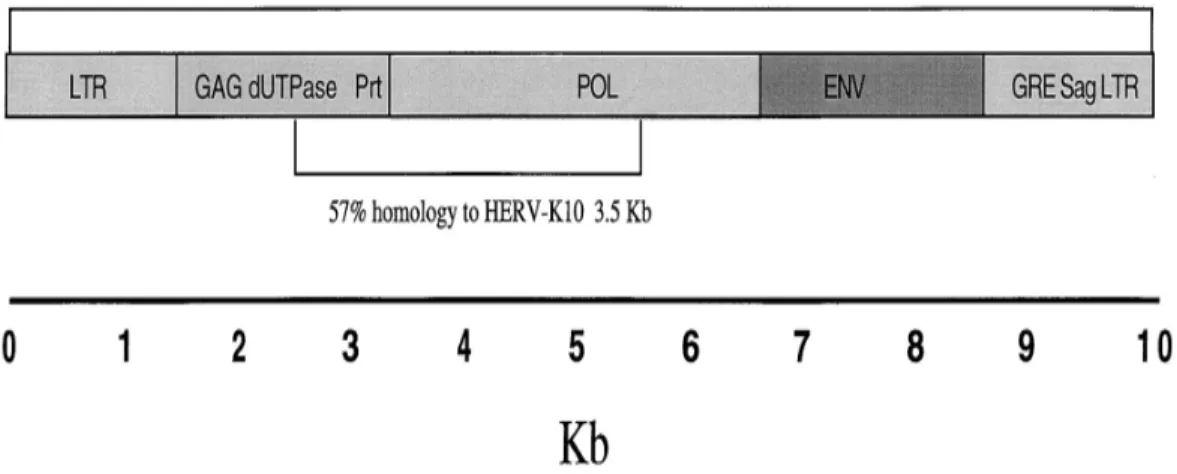
More in favor of the exogenous origin of these env-like sequences relate to the amplification of whole proviral structure from human breast carcinomas (figure: 1) as shown in the total homology from the carcinomas was 95% to MMTV in 9.9 Kb and 57%to HERV-K10 in 3.5Kb. Homology to the endogenous retrovirus was seen primarily in the pol gene, which is known to be conserved among different retroviruses and partially in the gag gene[36].
In 2006 Zammarchi et al [37]from this laboratory designed a rigor-ous methodological approach able to overcome these difficulties, based on the association of a laser microdissection procedure and a highly sensitive fluorescent nested PCR. The MMTV env-like se-quence was detected in 33% of a series of human breast carcinoma, whereas normal breast tissues and other types of human tumors re-sulted negative.
Figure 1 Structure of proviral sequences in human breast cancer showing genes, ORFs, and sequence homology to MMTV and HERV-K10.
Subsequently, in 2007, Pogo et al. isolated the HMTV particles from ascitic and pleural fluid of patient with metastatic breast can-cer whose primary tumor where positive for env sequence and ex-pressing env protein. They demonstrated the presence of retroviral particles electron microscopy technique. The virus particles in cul-ture medium showed retroviruses of type B, with size between 100 and 110 nm, a spherical shape, an eccentric dense core and the
Figure 2 Electron microscopy of MSSM3 cell associates and PF viral particles. A and B, selected examples of cell-associated viral particles. C and D, selected examples of viral particles in PFs. Note that the nucleoids are condensed to varying degrees. Original mag-nification,130,000 (bar, 0.2 Am).
Today the idea that a virus could be involved in the etiology of hu-man breast carcinoma receives much more attention that in the past. Several papers go towards this direction, even if in general they re-strict their research to demonstrate viral particles and proteins in tumor cells [38, 39].
MMTV, if really involved in breast human carcinogenesis, could act as a promoting agent or as an etiological/pathogenetical co-factor linked to some steps of cancer progression, such as infiltra-tion or metastasis. To individuate where during cancer progression MMTV exogenous sequences appear could be useful to unveil their role in human breast cancer.
Mouse mammary tumor virus
History:Mouse mammary tumor virus (MMTV) was first reported in 1933 by the Jackson Memorial Laboratory and by Korteweg in 1934 as an extrachromosomal influence on the incidence of breast cancer in inbred mouse strains[40]. Subsequently, in 1936 Bittner showed that a cancerous agent, which he called “milk factor”, could be transmitted by cancerous mother young mice while nursing [41]. In 1966 it was proven that Bittner’s milk factor was virus that re-mained dormant during the early stage of life in mouse but produce cancer in the middle age when the sexual hormones were in right conditions[42].
Taxonomy and Classification:
MMTV is a prototype species of the genus B retaretrovirus in the family Retroviridae. These viruses previously were referred to as type B retroviruses based on their appearance by electron microsco-py (a characteristic acentric core within particles of c. 100 nm). Multiple double-stranded DNA copies are found in the chromosom-al DNA of most commonly used laboratory strains of mice (cchromosom-alled integrated or endogenous proviruses)[43].
These endogenous proviruses presumably represent viral insertions into chromosomal DNA of germline cells and are referred to as Mtv followed by an Arabic number, for example, Mtv8.
Most endogenous Mtvs have defects in one or more genes and, therefore these proviruses often fail to produce infectious virus [44] . Currently, MMTV is classified with other betaretroviruses includ-ing Mason–Pfizer monkey virus (MPMV), Jaagsiekte sheep retrovi-rus (JSRV), and human endogenous retroviretrovi-rus type-K (HERV-K) [45].
Properties of the Virion:
The mature MMTV virion is100nm in diameter, contain a single-stranded positive-sense RNA, which exists as a dimer, and is encap-sidated as a helical ribonucleoprotein (RNP) by the nucleocapsid (NC) protein; reverse transcriptase (RT) and integrase (IN) are closely associated with the RNP.
The RNP is surrounded by an icosahedral shell composed of capsid (CA) protein.
MMTV capsids are bound via the matrix (MA) protein to the viral envelope, a portion of the cellular plasma membrane that has been modified by the insertion of the surface (SU) and transmembrane (TM) proteins [46] (figure: 3).
Figure 3: The structure of MMTV
Viral genome
The viral RNA is bound at either end by a short direct repeat (R) of 15 bp. The R regions are adjacent to
and 1200 bp, respectively, present at the 5 (U5) or 3 (U3) he structure of MMTV
The viral RNA is bound at either end by a short direct repeat (R) of The R regions are adjacent to a regions of approximately 120 and 1200 bp, respectively, present at the 5 (U5) or 3 (U3). U5: The viral RNA is bound at either end by a short direct repeat (R) of
regions of approximately 120 . U5: A
unique, non-coding region which is the first part of genome to be reversed transcribed, forming 3' of the provirus genome.
U3: A unique, non-coding region which forms the 5' of the provirus after reverse transcription contains the promoter elements responsi-ble for transcription of the provirus.
A cellular tRNA (tRNA3Lys) is bound through 18 bp of comple-mentarity to each copy of the viral RNA at the primer-binding site (PBS) located just downstream of U5.
The first splicing donor (SD) site precedes the group-specific anti-gen (gag) region that encodes a Gag precursor. The virus also en-codes two other precursor polypeptides, gag-pro, gag-pro-pol from genomic RNA. Gag -pro encodes the gag proteins, a dUTPase (DU), and the viral protease (PR). Whereas the gag-pro-pol protein encodes RT, including a ribonuclease H (RNase H) activity and IN. and finally the env region that encodes the envelope protein which compose of surface (SU) and transmembrane protein (TM).
Super antigen
The long terminal repeat (LTR) of MMTV harbor an Open Reading Frame (ORF) that encodes a superantigen (Sag), essential for its life cycle [47]. The superantigens are different from normal anti-gens for their ability to stimulate a greater amount of T cells, this property derives from their ability to bind to all T cells expressing a particular Vβ chain of T-cell receptor (TCR), and not only to the groove formed by the α and β chains, as do antigens conventionally. In this way, all T cells that express the chain
Vβ, have the ability to recognize and be stimulated by superantigens, which are presented as exogenous antigens [48]. Another characteristic of superantigens is to be present only in the context of major histocompatibility complex molecules (MHC) class II [49]. MMTV require functional MMTV sag to establish in-fection of the mammary gland of mice. Laboratory experiments have shown that exogenous virus with impaired function of Sag, are not infectious [50]. MMTV also needs a functional immune system (B cells, T cells) to complete its infectious cycle[51].
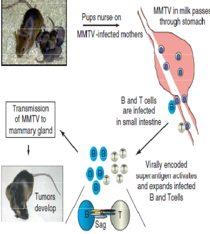
MMTV life cycle
The virus can be transmitted vertically (endogenous virus) when embryonic germline cells are infected [52]but it is usually transmit-ted horizontally through maternal milk (called exogenous virus or milk-borne virus)[12]. MMTV particles ingested by newborn mice in maternal milk that cross the epithelium cells through M cells in small intestine. The virion initially encounters and infects dendritic cells and B cells located in Peyer’s patches of gastrointestinal tract[53]. Following virus expression, superantigen (sag) protein is produced and presented on B cells by MHC II. The Sag binds both MHC II on B cells and the Vβ of the T cell receptor making it a potent T cell stimulus [54]. The stimulated T cell through sag release cyto-kines, which cause proliferation of many B cells that subsequently, also become infected with MMTV [55].
These infected lymphoid cells become a reservoir of MMTV that will preserve MMTV infectivity prior to the onset of puberty in mice
when a source of susceptible and dividing mammary cells becomes available [50].
MMTV infection increases during lactation, where high virion pro-duction ensures that large amounts of viral particles will increase the probability of newborn offspring infection [56].
The chronic infection of mammary cells induces the formation of malignant tumors, mammary adenocarcinomas. Infected T-cells rarely become tumorigenic; though, when T-lymphocytes are trans-formed, T-cell lymphomas do occur [57] (figure: 4 ).
Figure 4: MMTV life cycle
Recombination of exogenous and endogenous MMTV The coexpression of exogenous and endogenous RNA molecules in vivo results in viral recombination. Recombination between ex-ogenous and endex-ogenous genomes occurs during cDNA synthesis and is responsible for the generation of variants virus with new bio-logical characteristics. This recombination occurs because the
RNA-dependent DNA polymerase, reverse transcriptase, can switch templates during replication[58].
When an exogenous MMTV infected cells where the endogenous Mtv are highly expressed, there is a 'high possibility of recombina-tion between two viral forms. This event generates new viruses that have the ability to infect different strains of mice than the parental forms.
Exogenous MMTV infects epithelial cells of mammary gland, en-dogenous Mtv which is highly expressed in this tissue may be co-packaged and result is a recombinant virus. If the hypervariable re-gion of the exogenous MMTV is different from that of the endo-genous Mtv, the recombinant viral particle is capable infecting mu-rine strain different than the original particles of exogenous MMTV [59].
MMTV and Tumorigenesis
MMTV does not encode any oncogene, and mammary tumorigene-sis is therefore takes place after proviral insertion near specific cel-lular proto-oncogene, activating them in a process called insertional mutagenesis [60]. Retroviral integration is not sequence-specific, therefore the greater the amount of virus, the greater probability that the proviral DNA integrates near a proto-oncogene. In fact there has been a considerable correlation between viral load in breast milk and the incidence of breast cancer in a given mouse strain[61].
Once a provirus become integrated into the host genome, expres-sion of proviral DNA is regulated by specific sequences within the
LTR that cause increased viral transcription in response to gluco-corticoid receptor/steroid hormone complexes. The most important steroid hormone that increased the transcription of MMTV during pregnancy is the progesterone [62]. Analysis of mouse mammary tumors induced by MMTV has shown an alteration in six genes[63]. These genes belong to different family members of fibroblast growth factor (FGF) and members of the family Wnt gene. They are: Int-1/Wnt-1, Int-2/Fgf-3, Wnt-3, Hst/k-Fgf/Fgf-4, Wnt-10b and FGF-8.
An interesting feature common to these genes is that they are all involved in the short-range cellular communication, many of them, in fact, encode secreted proteins[64]. The activation of the expression of these genes, caused by MMTV is mainly due to the presence of enhancer sequences in the 5 'LTR (Long Terminal Re-peat) of the genome of MMTV. These regions act on promoters of adjacent genes, located up to 20 kb from the LTR. The MMTV pro-virus integrates generally outside the region coding of the proto-oncogene and only rarely within it[65].
The Wnt genes, members of a family consist of structurally related genes which encode secreted signaling proteins. These proteins have been implicated in oncogenesis and in several developmental processes, including morphology development of the tissue during embryonic and adulthood stage[66].
The other common target for the integration of MMTV is the gene family of fibroblast growth factor (FGF). The proteins en-coded by these genes are potent angiogenic factors in vivo, and
ivolve in cell differentiation [67]. Three of the eight members of the FGF family, namely, Int-2 (FGF-3), FGF-4 and FGF-8, are acti-vated by the insertion of proviral MMTV. As in the case of genes Wnt-1 and Wnt-3, the three FGF genes are not normally expressed in the mammary gland, but are found only or in embryonic life or in adult tissues other than breast [68, 69]. It has been shown that the onset of murine breast cancer is associated with activation of tar-geted genes (int-2 (Fgf-3), hst (Fgf-4), and Fgf-8) by MMTV pro-viral insertion. These observations suggest therefore that different genes cooperate in tumorigenesis [70].
Replication cycle of MMTV
MMTV replication cycle begin when the viral surface glycoprotein ,gp 52 bind to tranferrin receptor (tfr1) that expressed in many ro-dent cells. Human tfr1 is failed to allow viral entry, however use of
MMTV bound to the trf 1 enters the cell via endocytosis of clathrin-coated pits, andreceptors are recycled to the cell surface where they allow entry of additional MMTV particles into previously infected cells.
Following entry and partial uncoating in the cytoplasm, the virally encoded RT is activated. PBS, the primer binding sequence of viral RNA is complementary to the 3’ nucleotides of the host tRNA. The host tRNA, lys-3 acts as the DNA primer for the reverse transcrip-tase to synthesize minus-strand proviral DNA.
The initial product of reverse transcription is an RNA-DNA com-plex. The RNase H digests the RNA strand, and allows the
synthe-sis of double-stranded provirus[71]. However, the product of re-verse transcription is different from the starting template so that the U5 and U3 sequences present uniquely in viral RNA are duplicated to give longer repeats at each end of the provirus (LTRs).
Because nuclear entry of the preintegration complex containing the provirus is thought to require nuclear envelope breakdown during mitosis, it is generally believed that MMTV must infect dividing cells. Once the preintegration complex enters the nucleus the viral integrase randomly inserts the provirus into the host genome[72]. Integrase introduces an asymmetric cut 2 bp (cytosine, and adenine) from the linear ends of the provirus as well as an asymmetric break exactly 6 bp apart on opposite DNA strands of host DNA. Proviral integrations are not site-specific and may occur at transcriptionally active sites. Following the joining reaction, the repair of virus–cell junctions by cellular enzymes generates a 6 bp direct repeat of cell DNA that flanks the viral LTRs[73].
The integrated MMTV LTR is a stretch of DNA (1195 base pairs in length) at the 5’ end of the viral genome that is responsible for the expression of the virus in mammary epithelial cells and for the acti-vation of cellular proto oncogenes.
Transcription is initiated from the standard promoter in the 5' LTR starting at the U3/R junction and terminating at the R/U5 junction. Transcription of the MMTV LTR produces full- length mRNA, spliced mRNAs, as well The spliced mRNAs are the env mRNAs and the sag mRNAs [74, 75] .MMTV also produces a doubly spliced mRNA that encodes the RNA export protein, Rem[76]. In
most cell types, the levels of gag-pol and env mRNAs greatly ex-ceed sag and rem mRNAs.
The viral mRNAs are translated into the viral proteins in the cytop-lasm using the host cellular machinery. The gag mRNA is trans-lated into the precursor protein Pr77, a Gag polyprotein. Proteolytic processing of Pr 77 by the viral protease produces the structural proteins: the matrix protein p10 or MA; the capsid protein p27 or CA; the nucleocapsid protein p14 or NC, and some smaller proteins p21, p3, and p8 that have no known function [77, 78]. Though, p21 is cleaved into a few smaller proteins[79].
Capsid protein forms the structural shell of the core of the virion,
which enclose the ribonucleoprotein comple that contains the genomic RNA. Thecapsid protein appears to be important in the assembly of immature capsid [80].
The nucleocapsid protein has a sequence that includes many basic amino acids and a cysteine-histidine box that is similar to a zinc finger domain seen in DNA-binding proteins, which explain its packaging role of the viral RNA in the cytoplasm[81].
The gag-pro mRNA is translated as a precursor protein Pr 110, a Gag-Pro polyprotein. Within Pr 110, the NC sequence of the gag gene and the first part of the pro gene comprise a dut gene, which encodes DU a dUTPase. DU prevents misincorporation of uracil and mutation of newly synthesized proviruses in nondividing cells[82].
The C terminus end of the pro gene codes for p13 or PR the viral protease. PR is responsible for the cleavage events that produce the
mature virion proteins, MA, p21, p3, p8, CA, and NC, the functions of p21, p3, and p8 are currently unknown[83]. The precursor pro-tein Pr160, a Gag-Pro-Pol polypropro-tein encodes a viral reverse tran-scriptase with RNase H activity and an integrase[84].
The env mRNA is translated into a precursor protein, pr73 env on membrane bounded polyribosomes. This precursor is modified by glycosylation in both the endoplasmic reticulum and the Golgi, where a cellular protease cleaves the Env poly protein to produce gp52, SU a viral surface envelope protein and gp36, TM a viral transmembrane envelope protein. SU and TM remain attached to each other via disulfide bonds[85]. They travel to the host plasma membrane in vesicles and are then incorporated into the lipid envelope of the mature B type virion when the immature A particle buds from the host cell[86].
The sag mRNA is translated into a precursor protein Pr 48, SAg, a superantigen protein. This protein is glycosylated and cleaved in the Golgi by protease to generate SAg protein[87]. Sag is associated with major histocompatibility complex (MHC) class II protein at the surface of antigen-presenting cell. Sag is essential activation of certain T cell subsets containing Vβ TcR [88].
MMTV utilizes a polyprotein strategy for viral assembly. This process insures that none of the individual proteins: matrix, capsid, and nucleocapsid, are individually targeted to the membrane. The order of the proteins in the polyproteins corresponds to their rela-tive location in the virion from the outside of the virus to the inside of the virus.
MMTV polyproteins assemble into a capsid shell or imma-ture A particle in the cytoplasm. The formation of the A particle occurs prior to its transport to the plasma membrane[86],then the immature A particles bud from the tips of long filamentous actin projections as mature virions [89]. The viral envelope proteins are transported to the plasma membrane via the host secretory path-way. The A particles associate with the cell membrane, they ac-quire a viral lipid envelope with viral envelope proteins as the virus buds from the host cell. [90].
The budding process of MMTV seems to involve the host actin cy-toskeleton. The immature viral A particles have been observed at the tips of long filamentous projections [86].In addition, cytochalasin D, which disrupts the actin cytoskeleton, reduced the number of mature viral B particles released into the supernatant by 80% [91]Thus budding of MMTV appears to be an actin-dependent process. Infected human breast cells with MMTV initiated the forma-tion of filamentous projecforma-tions with virus particles at their tips. These filopodial projections emerged from the cell surface of in-fected live cells when viral production was hormonally induced with dexamethasone[92].
Human endogenous retrovirus:
Human endogenous retroviral (HERV) elements comprise 8% of human DNA [93], and are likely to be derived from ancient viral infections during evolution but their biological relevance is largely unknown.
It is thought that exogenous infection allowing HERVs sequences to be inserted into the genome of germ line cells, where they have been replicated along with the host's cellular genes following a Mendelian pattern [94] and[95]. Integration of endogenous retrovi-ruses into the human germ line is thought to have occurred 2 to about 70 million years ago depending on the individual retroviruses and were introduced by mechanisms involving reverse transcrip-tion. Most of human endogenous retroviruses (HERVs) are defec-tive, due to multiple mutations or deletions, and are therefore none of them is capable of encoding complete viral particles and cause infection [96]. Although many HERVs are transcriptional active, they are not able to produce functional proteins. The high number of copies present in the human genome is attributed to repeated cycles of infection and retrotransposition [97].
Classification
The known HERVs families have been grouped into classes. HERV class I shows clustering phylgenetically with Gammaretro-virus, those clustering with the betaretrovirus are class II, and class III HERV clustering with spumaviruses [98].
HERV families that reside in human genome are estimated to be between 30 and 50[99], among them various so-called HERV-K families. The letter K indicates that a primer binding site specific for lysine-tRNA was used to prime reverse transcription. In total, 10 HERV-K families have been defined based on sequence similari-ties, and they have been named K(HML-1) to
HERV-K(HML-10) (for human MMTV [mouse mammary tumor virus]-like) because of some sequence relationship to the mouse mammary tumor virus [100].
The biological function of HERV
The distribution of endogenous retroviral sequences, scattered throughout the genome of mammals, suggests that they have impor-tant biological functions and therefore have been conserved during evolution. Using reverse transcription and subsequent insertion of their genome into the host DNA, these sequences regulate the plas-ticity of genome, accelerating the evolution of new genes and alter-ing the transcription of existalter-ing genes [101].
Their biological function is still unknown and there are many as-sumptions that, to date, are proposed. In analogy with animal mod-el, it has been hypothesized that these endogenous retroviral se-quences are protected in respect of their endogenous counterparts. In the case of MMTV, for example, the expression of endogenous Sag coding sequences during the formation of the immune system, induces clonal deletion of all T cells reactive to that particular Sag and at the same time, makes it immune to the forms of MMTV ex-ogenous coding Sag with the same specificity to T cells [50, 102]. There are several experimental evidences that show the possibility of recombination between exogenous viruses and endogenous re-troviral sequences. It has been identified a variant of MMTV, high-ly tumorigenic, resulting from recombination between sequences
derived from endogenous Mtv, and a strain of exogenous MMTV [59].
HERV-K family
Unlike the majority of HERV, the family of HERV-K has genes an open reading frame (ORF) for essential genes of retroviruses (gag, pol and env) as well as its ability to synthesize all essential retrovir-al proteins [103, 104]. This family was originretrovir-ally identified for its high homology with the Mouse Mammary Tumor Virus (hence the nomenclature further HML, human endogenous MMTV-like). The HERV-K are present in the human genome with about 30-50 pro-viral copies and approximately 10,000 LTRS solitary, arising, prob-ably by homologous recombination between LTRS, followed by excision of the viral genome intermediate[102].
HERV-K families have been classified into six subgroups (HML by HML-1 to-6), based on sequence homology in the conserved region of the pol gene, encoding the enzyme reverse transcriptase [105, 106]. HERV-K10 has been sequenced and showed to have a com-plete provirus of 9 Kb, and contains ORFs for all retroviral genes, but showed to have a stop codon in the env gene and a frameshift in the gene gag[107].
MATERIALS AND METHODS
Specimens
All tissues, formalin fixed and paraffin embedded, were collected and archived (2005-2009) at the Division of Surgical, Molecular and Ultrastructural Pathology, University of Pisa.
We have collected 91 paraffin embedded tissue cases of ductal car-cinoma insitu (DCIS) and invasive ductal carcar-cinoma (IDC), as well as usual ductal hyperplasia (UDH) Atypical ductal hyperplasia (ADH). 49 of samples were DCIS, 20 samples of them have also IDC. It was possible to collect 26 samples of normal epithelial cells (NEC) collateral to DCIS or IDC (c-NEC) (figure: 5).
We have collected 22 samples of primary breast cancer and their respective non metastatic lymph nodes in order to study the HMTV status in the primary tumor and their respective non metastatic lymph nodes. Moreover, we have collected also 23 samples of pri-mary breast cancer and their respective metastatic lymph nodes to compare the HMTV status in non metastatic compared to metastat-ic lymph nodes (figure: 6). As negative control were used: 20 NEC from reduction mammoplasty (nc-NEC); 6 papillary thyroid carci-noma; 6 colon adenocarcinoma, and DNA extracted from 6 healthy blood donors. DNA of a mouse infected by MMTV was the positive control. Of the DCIS, 15 were poorly differentiated, 33 moderately differentiated and 1 well differentiated.
Figure 5: The histological types, and the number of breast cases (benign and malignant) and negative controls.
26 49
20 20 22 20
6 6 6
Normal epithelial cell from cases with DCIS
Ductal carcinoma insitu (DCIS) Invasive breast cancer(DIC) from cases have DICS
usual hyperplasia Atypical hyperplasia
Normal epithelial cells from normal breast
colon adenocarcinoma thyroid papillary carcinoma normal blood samples
Figure 6: The Cases number of nodes.
Histological criteria
The adjective “collateral” is used to indicate: a)
same histological section hosting a preinvasive lesion or ing carcinoma and
logical section hosting an infiltrating carcinoma. Usual ductal hyperplasia (UDH)
proliferative lesion characterized by secondary lumens and strea ing of the central proliferative cells.
(ADH) has cytological and architectural features of low 0 5 10 15 20 25 30 35 40 45 primary breast cancer
Cases number of breast cancer and their lymph
Histological criteria
The adjective “collateral” is used to indicate: a) NEC present in the same histological section hosting a preinvasive lesion or an infiltra ing carcinoma and b) preinvasive lesions present in the same hist logical section hosting an infiltrating carcinoma.
Usual ductal hyperplasia (UDH) indicates a benign ductal epithelial proliferative lesion characterized by secondary lumens and strea ing of the central proliferative cells. A typical ductal hyperplasia
has cytological and architectural features of low-grade metastatic
lymph nodes of metastaticlymphocytes lymphnodes
non metastatic lymphnodes
cancer and their lymph
present in the an infiltrat-
histo-indicates a benign ductal epithelial proliferative lesion characterized by secondary lumens and stream-lasia grade
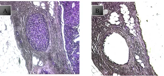
DCIS present in 1 or more ducts or in an aggregate not exceeding 0.2 cm. DCIS can be of high, intermediate, or low grade: a) pleo-morphic large cells with abundant mitoses and with variable archi-tecture, often solid and with a central necrotic debris; b) uniform cells, with small nuclei and frequently with a cribriform or mi-cropapillary configuration; c) the neoplastic nuclei show pleomor-phism of a degree between high and low grade DCIS, (figure: 7).
Figure 7: Breast cancer progression A, normal breast; in B, usual hyperplasia C, Atypical hyperplasia D, ductal carcinoma in situ, E infiltrating ductal carcinoma
E
C D
Laser microdissection
A Leica ASLMD automatic laser microdissector was used to select the epithelial cell population to be studied (Figure: 8). Three µm thick sections were cut from each case using each time a new mi-crotome blade, and applied on microdissecting slide, followed by staining with haematoxylin and eosin, then obtaining a total of 10,000–15,000 cells (figure:9). Stromal and inflammatory cells were carefully excluded. Due to the long experience of the labora-tory with this method, no difficulty was found in selecting areas of interest.
Figure 9: Laser capture microdissection, A) before applying micro-dissection, B) after applying microdissection
DNA extraction from microdissected tissue
Microdissected Cells are suspended in buffer containing 10 mM Tris-HCl, 1 mM ethylenediamine tetraacetic acid (EDTA), 1% Tween 20, and 0.1 mg/ml proteinase K, pH 8.0, and incubated overnight at 56°C. Higher concentrations of proteinase K have been reported to improve the quality of DNA recovered from fixed tissue sections. The sample is subsequently heated at 95°C for 10 minutes to inactivate the proteinase K and the DNA is ready for PCR analy-sis. To avoid cross-contamination, blank DNA samples (lysis buffer with proteinase K) were processed in parallel with the tissue sam-ples. The quality and amount of extracted DNA was evaluated by agarose gel electrophoresis and UV spectrophotometry (NanoDrop, Celbio).
DNA extraction from paraffin embedded tissue
We have used the QIAamp DNA FFPE Tissue Kit for purification of DNA from FFPE tissue sections.
1. Cut 2 sections of 10 µm thick, using sterile forceps and blade for each sample, place it in sterile 1.5ml eppendorf tube
2. Add 180 µl Buffer ATL, and 20 µl proteinase K, and mix by vortexing.
3. Incubate overnight at 56oC
4. Briefly centrifuge the 1.5 ml tube to remove drops from the inside of the lid.
5. Add 200 µl Buffer AL to the sample, and mix thoroughly by vortexing. Then add 200 µl of ethanol (96–100%), and mix again thoroughly by vortexing.
6. Briefly centrifuge the 1.5 ml tube to remove drops from the inside of the lid.
7. Carefully transfer the entire lysate to the QIAamp MinElute column (in a 2 ml collection tube), and centrifuge at 6000 x g (8000 rpm) for 1 min. Place the QIAamp MinElute col-umn in a clean 2 ml collection tube, and discard the collec-tion tube containing the flow-through.
8. Carefully open the QIAamp MinElute column and add 500 µl Buffer AW1 without wetting the rim. Close the lid and centrifuge at 6000 x g (8000 rpm) for 1 min, then discard the collection tube containing the flow-through.
9. Carefully open the QIAamp MinElute column and add 500 µl Buffer AW2 without wetting the rim. Close the lid and centrifuge at 6000 x g (8000 rpm) for 1 min, then discard the collection tube containing the flow-through.
10. Centrifuge at full speed (20,000 x g; 14,000 rpm) for 3 min to dry the membrane completely.
11. Place the QIAamp MinElute column in a clean 1.5 ml mi-crocentrifuge tube and discard the collection tube containing the flow-through. Carefully open the lid of the QIAamp Mi-nElute column and apply 50 µl Buffer ATE to the center of the membrane.
12. Close the lid and incubate at room temperature (15–25°C) for 1 min. Centrifuge at full speed (20,000 x g; 14,000 rpm) for 1 min.
RNA extraction from cell culture
We have obtained cell pellet from primary tissue culture of breast cancer after the first subculture for RNA extraction in order to study the gene expression of the HMTV. RNA extraction was performed using Using Qiagen’s RNA Mini Kit.
1. Harvest cells according to standard tissue culture procedures to obtain a cell pellet, divide the pellet into two separate 15 ml tubes.
2. To the cell pellet in each tube, add 600ul of Buffer RLT containing 6 µl B-Mercaptoethanol.
3. Vortex the pellet to disrupt it as much as possible, then transfer the lysate to QiaShredder columns, and Centrifuge at maximum speed for 2 minutes to fully homogenize the cells.
4. Add 600ul of 70% Ethanol to each tube of the homogenized cells and mix well, Pipet 600ul of the sample in each tube onto RNeasy mini columns.
5. Centrifuge at 11,000rpm for 30 seconds. Discard the flow. 6. Add 700ul of Buffer RW1 to the columns, centrifuge at
11,000rpm for 30 seconds then discard the flow.
7. Add 500ul of buffer RPE to the columns, Centrifuge at 11,000rpm for 30 seconds, and then discard the flow.
8. Add another 500ul of Buffer RPE to the columns, centrifuge at 11,000rpm for 2 minutes then discard the flow.
9. Transfer the RNeasy columns into fresh 1.5mL nucleic acid free tubes.
10. Add 30ul of RNase free water directly onto the columns, centrifuge at 11,000rpm for 1 minute.
11. Quantitate the RNA on the nanodrop, keep it at -70oC. cDNA synthesis:
We have used fermantas cDNA synthesis kit to synthesis cDNA using RNA extracted from cell culture as a template.
1. Add 0.5-1µg of RNA into sterile nuclease-free tube on ice. 2. Add also 1µl hexamer primer
4. Add the following in the indicated order 4µl of 5x reaction buffer
1 µl of Ribolock 10 RNase inhibitor (20u/ µl) 2 µl of mM dNTP mix
1 µl of RevertAID M-MuLV reverse transcriptase (200u/ µl).
5. Mix gently and centrifuge, then incubate for 5min at 25oC. followed by 60 minutes at 42oC.
6. Terminate the reaction by heating at 70oC for 5 minutes. The synthesized cDNA can be used immediately or store at -20oC for less than a week. For longer storage, -70oC is recommended.
DNA amplification suitability
DNA was checked for the absence of PCR inhibitors by amplifying HERV-K10, β actin provirus target templates. Primers used for tar-gete amplifications are in table: 1. Cycle conditions were: 1 cycle, 94 °C, 10 min; 30 cycles, 94 °C, 30 s; 50 °C, 30 s; 72°C, 45 s; final extension, 72 °C, 7 min in 30 μl reaction mixture containing 1× PCR Buffer (500 mM KCl, 150 mM Tris-HCl, pH 8.0), 1.5 mM MgCl2, 200 μM each deoxynucleotidetriphosphates, 0.5 μM of for-ward and reverse primer and 2.5 U AmpliTaq Gold (Applied Bio-systems, Foster City, CA, USA). DNA samples were considered free of PCR inhibitors if HERV-K10, β actin amplicons were clearly visible on a 1.8% agarose gel.
PRIMERS SEQUENCE (3/-5/) HERV-K10 5/ CCACGGGTAAATTTACA HERV-K10 3/ TATTTTTGGTCTTGGGGTAG β-actin 5/ CTTCTGCCGTTTTCCGTAGG β- actin 3/ TGGGATGGGGAGTCTGTTCA ENV 5/ GATGGTATGAAGCAGGATGG ENV 3/ CCTTTTCTCTATATCTATTAG
ENV3/- nested AAGGGTAAGTAACACGGGTCATGTA
ENV5L CCAGATCGCCTTTAAGAAGG
ENVLTR 3 CGAACAGACACAAACACACG
Table 1: Primer sequences
HMTV sequence detection
Fluorescent nested-PCR was used to detect the presence of the HMTV env-like sequence. Generated fluorescent amplicons were sized on an automatic DNA sequencer. The pairs of primer were designed on the bases of the sequence available in GenBank (Ac-cession No. AF243039). Slight changes were introduced in the pro-cedure previously used by ourselves [37] to adapt it to paraffin em-bedded tissues. They consisted mainly in decreasing the length of the amplicons to a maximum of 250bp. The outer primers yield a 248 bp fragment from nucleotide [nt] positions 231–480 of MMTV-like env, the inner primers yield a 202 bp fragment (nt positions 231 and 431). Sequences of the outer primers for the first PCR were: Forward: 5/gatggtatgaagcaggatgg3/ and Reverse: 5/ cctcttttctctatatc-tattag3/ and for the nested PCR the forward primer sequence was
the same as the above while the reverse was Reverse Nested-5/aagggtaagtaacacaggcagatgta3/. Both PCRs were carried out in 50 μl containing 1× standard PCR buffer, 1.5 mm MgCl2, 200 μm each dNTP, 0.5 m unlabelled reverse primer (MWG Biotech), 0.5 μm 6-FAM-labelled forward primer (Applied Biosystems, Milan, Italy) and 2.5 U AmpliTaq Gold (Applied Biosystems). Input target tem-plate was 500 ng genomic DNA in the first round PCR and 2 μl of first round PCR in the second, respectively. Amplification profile was as follows: 1 cycle 94 ◦C, 10 min; 40 (first round) and 30 (sec-ond round) cycles at 94 °C, 45 s; 58 °C, 45 s; 72 °C, 60 s; final ex-tension, 72 °C, 7 min. To prevent PCR contamination, water con-trols and negative DNA samples were included every five samples in each run. Fluorescent amplicons were analysed by capillary elec-trophoresis and appeared as peaks in an electropherogram (figure: 10). Amplicon size was extrapolated from a molecular size ladder re-suspended in PCR buffer and run in parallel. Briefly, 3 μl PCR products from both amplification rounds were mixed with 0.5 μl ROXlabelled size standard (Gene Scan 400 HD ROX; Applied systems) and 11.5 μl formamide (Hi-Di Formamide, Applied Bio-systems). After denaturation at 95 °C for 3 min, samples were loaded onto an ABI PRISM 3100 automatic genetic analyser and analysed using GENESCAN software, version 3.1 (Applied Biosys-tems).
Figure 10: Fluorescent capillary electrophoresis, A: first PCR, B: nested PCR
Sequencing of HMTV env PCR fragments
HMTV env PCR fragments were purified using Multi Screen PCR Plates (Millipore) following this protocol:
Dilute PCR by adding 70 µl of water. Mix gently by pipet-ing up and down 3-5 times.
Transfer diluted reactions into the bottom of the plate wells.
Place the plate on the vacuum manifold. Set the vacuum to 23-25” Hg.
Apply vacuum until the solution has been completely re-moved from the wells.
Shut off the vacuum source and blot the excess liquid from the bottom of the plate.
micro-plate shaker.
Apply vacuum until the solution has been complete-ly removed from the wells.
Shut off the vacuum source and blot the excess liquid from the bottom of the plate.
Resuspend the purified sequencing products in 25 µl of water by shaking for 10 minutes on a microplate shaker.
Transfer the purified sequencing products to an appropriate plate
Then we performed Cycle Sequencing reaction using Big-Dye Terminator kit v3.1 (Applied Biosystems) and 2,5 pmol/µl of each primer, for a final volume of 20 µl. We used these condi-tions: 96 °C using the QIAquick Gel Extraction kit (Qiagen) and sequenced on an ABI PRISM 3130XL (Applied Biosystems). Each sequence was aligned by using BLAST search function
(http://blast.ncbi.nlm.nih.gov) to MMTV sequences deposited in GenBank.
HMTV Real Time PCR assay
Extracted DNA from 9 patients was analysed by quantitative RT-PCR: in 2 patients NEC, DCIS and IDC were available, whereas in the other 7 cases DCIS and IDC only. Briefly, 25μl PCR reaction mixture was prepared using 2X Master Mix (MesaGreeen qPCR, Eurogentec, San Diego, CA), 0.5 μl of HMTV primer (0.3uM) for the ENV region (nested PCR 202bp). The viral load was determined as the mean of triplicate sample values. GAPDH which is a single copy gene was used as an internal reference control. Four ten-fold
dilutions (1X101–104copies/ml) of genomic human DNA (GAPDH gene) and of HMTV env amplicons were included as standard curves on each run. HMTV env-like and corresponding GAPDH. A HMTV positive control DNA extracted from a mouse known to be infected with the virus and a template negative control (PCR mix only) were also included in each PCR run. PCR were run on the Ro-tor-Gene Q from Qiagen (figure 11). The standard curves were plotted by the Rotor-Gene software detector system printing the Ct values against each known concentration of standards (HMTV or GAPDH sequence) and MMTV copy number per DNA sample was calculated by the detector.
Figure 11: Rotor gene real
Amplification of the probe (2.7 kb env We have prepared homemade probe of Amplification of 2.7kb env
kit, the 50 μl reaction contains 3 units
fer, 2 μl of 50 mM mgcl2, 8 μl of 2.5 mM dNTP, 0.2μm of each primer (5L, LTR3)
was incubated at 94 for 2 minutes, 94 for5 seconds 68 for 5minutes for 40 cycles.
Rotor gene real time PCR
Amplification of the probe (2.7 kb env-LTR)
e have prepared homemade probe of HMTV env-LTR 2.7kb. Amplification of 2.7kb env-LTR was achieved using takara EX taq , the 50 μl reaction contains 3 units of enzyme, 5 μl 10xPCR bu fer, 2 μl of 50 mM mgcl2, 8 μl of 2.5 mM dNTP, 0.2μm of each primer (5L, LTR3) [36], 1μg of mouse DNA positive for HMTV at 94 for 2 minutes, 94 for5 seconds 68 for 5minutes LTR 2.7kb. using takara EX taq of enzyme, 5 μl 10xPCR buf-fer, 2 μl of 50 mM mgcl2, 8 μl of 2.5 mM dNTP, 0.2μm of each 1μg of mouse DNA positive for HMTV at 94 for 2 minutes, 94 for5 seconds 68 for 5minutes
Labeling the probe
The band at 2.7kb was cut and purified using centri-sep kit (applied biosystem), then sequenced to make sure it’s the true target.
Labeling probe was done using DIG-NICK translation mix kit (Roche) as the following:
1. Add 1 µg the purified PCR product to sterile, double distill-ed water and end up with a final volume of 16 μl.
2. Add 4 μl DIG-Nick Translation Mix, mix and centrifuge briefly.
3. Incubate for 90 min at 15°C, and then place the reaction on ice.
4. Take a 3 μl aliquot per 20 μl reaction volume from the reac-tion, add gel loading buffer, denature it at 95°C for 3 min, put it on ice for 3 min and run the sample on an agarose mi-nigel along with a DNA molecular weight marker. The probe should range between 200 and 500 nucleotides in length.
5. If necessary reincubate the reaction at 15°C and check the fragment size again.
6. When correct probe length is achieved stop the reaction by adding 1 μl 0.5 M EDTA (pH 8.0) per 20 μl reaction volume and heating to 65°C for 10 min.
Chromogen in situ hybridization (CISH)
Sections were cut at 4 μm thicknesses, mounted onto positively charged microscope slides, and left to dry overnight at 37°C. Sec-tions were then deparaffinized and rehydrated. Antigen retrieval was achieved by heat retrieval using water bath (DAKO). The slides were placed in Coplin jars containing enough 0.01 M sodium citrate solution (pH 6.0) to cover the sections, and then incubate in water bath at 80°C for 30 minutes. In order to expose the nuclear components, proteinase K was applied on the sections for 10 mi-nutes at 37°C. The hybridization solution is prepared just before applying, for each slide make up to 10 µl of the solution of the probe (50 ng) in hybridization solution (2x SSC, 50% formamide, 10% dextran sulfate). The section then dehydrated using absolute ethanol, followed by adding up to 10µl of probe with hybridization solution on tissue, then apply cover slip over the probe solution and avoid bubbles formation, after that seal the edge of cover slip with cement material. Denaturation and hybridization were achieved by incubate the slide for 5minutes at 80°C and 16 hours at 37 °C re-spectively using thermobrite (ABBOTT hybridizer). The day after get the slide out of the hybridizer, remove the cover slip and wash with 0.5 x SSC at 75oC for 30 seconds, followed by incubation in 2x SSC at room temperature. the results were detected by applying anti-digoxigenin-AP conjugate for 30 minutes at 37 °C, that pro-duce a characteristic nuclear black stain after 5-20 minutes of ap-plying NBT/BCIP substrate (figure: 12).
also prepared. The positive control slides were prepared from a case known to be positive for HMTV env in PCR study. The negative control slides were prepared from the same tissue block, but incu-bated with hybridization buffer instead of the HMTV env probe.
4-5µm
Stringent Wash Hybrit Tissue preparation
Block Step Mouse Anti-DIG
NBT-CIP Color formation
Tissue culture
We have prepared the media needed for primary breast cancer tis-sue cultures as the following:
Collecting medium
DMEM F12 with 200U/ml penicillin, 200 µg/ml streptomycin, 5 µg/ml fungizone.
Primary culture medium: DMEM F12 supplemented with
1. 0.5µg/ml hydrocortisone 2. 5µg/ml insulin
3. 20 ng/ml EGFR
4. 200 µg/ml cholera toxin
5. 0.004M/ml bovine pituitary extract 6. B27 2%
Complete culture medium
DMEM F 12 containing 10% heat-inactivated fetal bovine serum albumin (HIFBS).
Antibiotic solution (ABC-PBSA) 1. Phosphate buffered saline (PBSA) 2. 200 U/ml penicillin
3. 200 µg/ml streptomycin 4. 5µg/ml fungizone
Day 1 protocol
1. Preparing the hood by ensuring that it’s clear and swabbing it by 70% alcohol.
2. Brings the reagents and materials necessary for the proce-dure, swab bottles with 70% alcohol and place items re-quired immediately in the hood.
3. Obtain breast tumor biopsies from pathology and transport to the laboratory in colleting medium. If the tissue cannot be processed immediately (recommended),it may be stored re-frigerated for up to 24 h without significant loss of viability 4. Wash tissue extensively with phosphate buffered saline
lacking Ca2+ and Mg2+ (PBSA) supplemented with 200U/ml penicillin, 200 µg/ml streptomycin, 5 µg/ml fungizone (ABS-PBSA)
5. Minced finely by using crossed scalpels in a sterile Petri dish.
6. Wash twice in ABS-PBSA.
7. Disaggregate for 18-20 h in 200 U/ml collagenase A (made up in complete culture medium) in a 37C incubator (either in multiwell plate, or Petri dish)
8. Remove tissue from incubator, and shake vigorously by hand. This will break up any remaining large clumps of par-tially digested tissue
9. If there still undigested tissue re incubate the tissue with fresh medium and collagenase 200U/ml.
Day 2 protocol
1. Transfer the Petri dish contents to 50 ml sterile centrifuge tube, close the cap and wrap it with parafilm.
2. Spin at1200g for 5 minutes.
3. Culture the pellet in the primary cultured medium using primary culture flask (blue cap); incubate the flask at 37oC and 5% Co2.
4. The day after change the medium to get rid of unattached cells in case of attached conditions, after that change the medium every 2 days.
5. For floating cell culture, incubate the cells in
Subculture
The need to subculture implies that cells in the primary culture have increased to become 70-80% confluent.
1. Take the culture flask to the sterile working area under the hood, then remove and discard the medium
2. Add PBSA/EDTA prewash (0.2ml/cm2) to the side of the flask opposite the cells to avoid dislodging cells.
3. Add trypsin (0.1ml/cm2) to the side of the side of the flasks opposites the cells. Turn the flask over and lay them down.
Ensure that the monolayer is completely covered.
4. Incubate at room temperature (5-10 min), with the flasks
monolayer should slide down the surface. Don’t leave the
flask longer than necessary.
5. Add complete culture medium (0.1-0.2ml/ cm2), and dis-persed the cells by repeated pipetting over the surface
bear-ing the monolayer.
6. Finally, pipette the cell suspension up and down for a few
times, with the tip of the pipette resting on the bottom corner
of the flask, taking care not to create foam.
7. Count the cells with hemocytometer; dilute the cell
suspen-sions to appropriate seeding concentration, by adding
ap-propriate volume of cell suspension to a premeasured
vol-ume of medium in a culture flask, and incubate the flasks at
37oC and 5% Co2.
Removal of fibroblastic cells from the tissue culture
We have used Anti-fibroblast microbeads form MACS to get rid of the fibroblast from tissue culture.
1. Trypsinized the cells as mentioned above, and determine the cell number.
2. Centrifuge cells at 300xg for 10 minutes. Aspirate super-natant completely.
4. Add 20 µl of Anti- fibroblast Microbeads per 107 total cells.
5. Mix well and incubate for 10 minutes in the refrigerator (2-8oC).
6. Washing cells by adding 1-2 ml of buffer 107 cells and centrifuge cells at 300xg for 10 minutes. Aspirate super-natant completely.
7. Resuspend in 500 µl buffer.
8. Prepare the magnetic field and the adaptor, then apply the column and prime it with PBS.
9. Apply a sterile collecting tube under the applied column, then add the labelled cells to be separated to the column and collect the negatively selected cells ( fibroblast con-fined in the column)
10. The negatively selected cells (no fibroblast) then sub cultured.
Cryopreservation the cells
Grow the culture to late log phase; prepare a high concentration of cell suspension by:
1. Trypsinize monolayer and resuspend cells in medium at 1-10x1010/ml
2. Add cryoprotectant, dimethyl sulfoxide (DMSO) to 10% v/v 3. Divide cells in a prelabelled polypropylene plastic ampoules 4. Clip the ampoules on to an aluminium cane, insert in a
Statistical analysis
One way Anova test was used to statistically evaluate the differ-ences in viral copy numbers between the early and later stages of tumour progression in the same patient.
RESULTS
HMTV frequency in phases of breast cancer progression The presence and the frequency of HMTV env sequence in normal breast and in all phases of breast cancer progression are described below and reported in Table 2 and in Figure 13.
Normal epithelial cells: a) exogenous HMTV env sequences were found in 19, 2% (5 out of 26) of NEC collateral to DCIS or IDC. b) the 20 cases of normal breast samples obtained by reductive mam-moplasty were result all negative (c and nc stand for collateral and non collateral respectively, Figure.13)
Ductal epithelial hyperplasia: a) the 20 cases of UDH resulted all negative. b) exogenous HMTV env sequences were found in 27% (6 out of 22) samples of AH.
Breast carcinoma: a) exogenous HMTV env sequences were found in 79.6% (39 out of 49) of samples of DCIS. The presence of the virus was not related to the presence of the preinvasive lesion in the same histological section hosting an infiltrating carcinoma. b) 35% (7 out of 20) of samples of IDC resulted positive.
Controls: all the 6 cases of infiltrating papillary thyroid carcinoma, the 6 cases of colon adenocarcinoma and the 6 cases of DNA from blood of healthy donors were negative.
Figure 13: The frequency of HMTV env sequence in normal breast cells and all phases of breast cancer progression.
0% 10% 20% 30% 40% 50% 60% 70% 80% 90% Normal ducts from cases with DCIS Ductal carcinoma In Situ Invasive breast cancer from cases with DCIS Usual Type Hyperplasia (UDH) Atypical
hyperplasia epithelialNormal cells from normal breast (plastic reduction) Thyroid
cancer cancerColon HealthyBlood Donors
Tissue Cases HMTV env positivity
%
Ductal Carcinoma In Situ 49 39 79.6
Invasive Ductal Carcinoma from cases with DCIS
20 7 35
Normal ducts from cases with DCIS 26 5 19
Usual Type Hyperplasia 20 0 0
Atypical Hyperplasia 22 6 27
Normal ducts from plastic breast reductions 20 0 0
Papillary Thyroid carcinoma 6 0 0
Colon Adenocarcinoma 6 0 0
Healthy blood donors 6 0 0
Table 2: Number of cases tested with different histological types and number of HMTV env positive cases found in all phases of breast cancer progression.
Different histological structures obtained from the same patient: in 14 patients with DCIS positive for HMTV env-like se-quences, normal breast and IDC were also available; in one case all the three samples resulted positive (case 1) , in 4 cases both DCIS and IDC were positive (cases 6-9), whereas the normal structures negative; in 3 cases normal structures and DCIS were positive, and IDC negative (cases 2-4); in 6 cases only DCIS were positive (cases 12-17). In the 6 patients with DCIS negative for HMTV env-like sequences in which normal structures and/or IDC were also avail-able also the last two resulted negative (cases 41-46). Of four cases of positive DCIS in which IDC were available, the last one was positive in 2 and negative in 2 (cases 10-11 and 26-27).
In 9 cases of positive DCIS in which only normal gland structures were also available, 8 of the last ones resulted negative and 1 posi-tive (cases 18-25 and 5). In the 16 cases with only DCIS available, 13 were found positive and 3 negative (cases 28-40 and 47-49). Sequence detection
To determine whether the amplified fragments were indeed homo-logous to HMTV, the 248 bp amplicons that showed clear band on agarose gel (Figure: 14) were sequenced, aligned to the correspond-ing region of the prototype MMTV sequence and MMTV (Gen-Bank Accession Nos AY152 721 and AF243039, respectively). Multiple nucleotide alignment showed 97% homology to both MMTV and HTMV (Human MTV) env sequences. Finally, no sig-nificant hits were found when the 2 sequences were compared to the human genome sequences available in GenBank, indicating that
these amplicons were not of human genomic or endogenous retrovi-rus origin.
248 bp
Figure 14: Amplification of 248 bp HMTV env gene by PCR, 2% agarose gel electrophoresis.
HMTV viral load with RT-PCR
Real time PCR was conducted on DNA extracted from DCIS and IDC from 7 different patients. GAPDH was used as the internal gene reference control, so each bar represents the ratio between HMTV DNA copy number and GAPDH DNA copy number (Figure 15). Figure 16 shows also the average of the HMTV/GAPDH DNA copy number ratio (A) for DCIS and IDC of 7 samples. There was a