CASE REPORT
The Canadian Journal of Urology; 12(6); December 2005
Accepted for publication June 2005
*Robert (Bob) L. Stephen leaves us with the memory of an inspiring teacher, scientist, colleague and friend. We sorely miss him.
Address correspondence to Dr. Savino M. Di Stasi, Department of Surgery/Urology, “Tor Vergata” University, Via Torrice 4, 00189 Rome, Italy
Percutaneous sequential bacillus
Calmette-Guèrin and mitomycin C for panurothelial
carcinomatosis
Savino M. Di Stasi, MD, Antonella Giannantoni, MD, *Robert L. Stephen, MD,
Luigi Storti, MD, Francesco Attisani, MD, Andrea De Carolis, MD,
Guido Virgili, MD
Department of Surgery/Urology, “Tor Vergata” University, Rome; Department Of Urology, University of Perugia, Perugia; Physion Laboratories, Medolla; Italy
DI STASI SM, GIANNANTONI A, STEPHEN RL, STORTI L, ATTISANI F, DE CAROLIS A, VIRGILI G. Percutaneous sequential bacillus Calmette-Guèrin and mitomycin C for panurothelial carcinomatosis. The Canadian Journal of Urology. 2005;12(6):2895-2898.
A 59 year old male presented with a 4 month history of lower urinary tract symptoms.
Exhaustive urological investigations revealed papillary tumors and carcinoma in situ extending from the prostatic urethra, throughout the bladder, up both ureters and into the renal pelves.
Tumors were resected where possible and then bacillus Calmette-Guèrin (BCG) and mitomycin C (MMC)
were infused sequentially through bilateral nephrostomy tubes for a total of six BCG and three MMC instillations.
Follow up 1 month post treatment demonstrated a complete response which persisted for 2 years. Then there appeared a solitary papillomatous recurrence in the bladder which was successfully resected. Side effects were the occasional fever and BCG induced granulomatis prostatitis which slowly resolved.
In conclusion, sequential BCG/MMC instillations were effective treatment for widespread panurothelial carcinomatosis.
Key Words: panurothelial, carcinomatosis, immunotherapy, chemotherapy
numbers of patients with varying combinations involving bladder, upper urinary tract (UUT) and prostatic urethra.2 Most reports describe these
combinations as metachronous3 with varying time
intervals between detection of carcinoma at the different sites.4 Synchronous presentation with
carcinoma extending throughout the whole urinary tract is rare. French investigators have described one such probable case but, as the full extent of the disease was defined over a time span of 3 years, the authors themselves were uncertain as to the sequence of events.5
In this report we describe a patient who presented with panurothelial carcinomatosis extending from the prostatic urethra to the renal
2895
Introduction
Although transitional cell carcinoma (TCC) confined to the bladder is the most common site for urothelial cancers,1 panurothelial TCC occurs in significant
The Canadian Journal of Urology; 12(6); December 2005
2896
Percutaneous sequential bacillus Calmette-Guèrin and mitomycin C for panurothelial carcinomatosis
pelves and treatment of this condition with sequential bacillus Calmette-Guèrin (BCG) and mitomycin C (MMC).
Case report
Initial findings
A 59 year old man presented with a history of hematuria, urinary frequency and dysuria of 4 months duration. Further questioning elucidated progressive reduction of urinary flow, nocturia (1-2 times) and incomplete bladder emptying that had been present for 2 years.
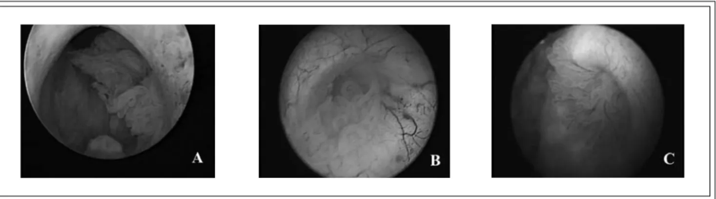
The patient had always worked as a post office employee with no exposure to occupational carcinogens but had smoked 30 cigarettes daily since age 14 (45 years). There was nothing relevant in his past medical history, he was taking no medications, had no allergies and the family history was unhelpful. Physical examination revealed chronic bronchitis, emphysema and slight prostatic enlargement. A urine sample contained malignant cells. An abdominal ultrasound and intravenous pyelogram with micturating cystourethrogram showed one irregular defect in the bladder neck, trigone and posterior bladder wall and another in the prostatic urethra at the verum montanum. Urethrocystoscopy revealed multifocal papillary tumors in the prostatic urethra, Figure 1 panel A, and in the bladder corresponding to the regions of the filling defects. Papillary lesions surrounded and also emerged from both ureteric orifices, Figure 1 panels B and C. All visible tumors were resected (TURBT) and their predominant pathology was transitional cell carcinoma grade G2 penetrating the lamina propria (T1). Obviously there was a need (a) to define the full extent of the carcinomatosis and (b) to initiate appropriate therapy.
Further investigations
A spiral CT scan of the upper urinary tracts (UUT) and ureteropyelography were uninformative. Endoscopic evaluation involving lavage of both UUT for cytology, bilateral ureteropyeloscopy and multiple biopsies of the UUT, the bladder and prostatic urethra delineated the clinical situation. Both ureters contained multiple, flat, hyperemic plaques and some small papillary tumors extending from the bladder up into the renal pelves. Pathology of the tumors was Ta G2 and that of the other biopsies was carcinoma-in-situ (Tis) extending from the prostatic urethra, throughout the bladder and up into the renal pelves: panurothelial Tis with multiple papillary tumors. There was no evidence of cancerous progression into muscle layers and beyond.
Treatment plan
Alternatives: The patient resolved the contentious
issue of surgical extirpation – combined bilateral nephro-uretectomy and cystoprostatectomy – by refusing this option. A proposed experimental treatment with Pasteur bacillus Calmette Guèrin (BCG) and mitomycin C (MMC) was approved by the institutional review board and the patient supplied written informed consent.
Procedures: With ultrasound and fluoroscopy
control bilateral percutaneous nephrostomy tubes were inserted into the superior renal calyces under local anesthesia. Unobstructed flow from the renal pelves to the bladder was assured and pyelovenous/ pyelolymphatic backflow were excluded with fluoroscopy. The bladder was catheterized, with thorough drainage achieved under ultrasound control, then the catheter was removed. The drug solutions prepared for instillations were BCG 54 mg in 100 ml 0.9% NaCl solution and MMC 40 mg in 100 ml water,
Figure 1. Endoscopic pictures of the prostatic urethra (panel A), right (panel B) and left (panel C) ureteric orifices were taken during the initial transurethral resection.
The Canadian Journal of Urology; 12(6); December 2005
DI STASI ET AL.
2897
and were divided into two 50 ml aliquots attached to the two nephrostomy tubes. An immediate bilateral instillation of 10 ml-15 ml theoretically filling the UUT was followed by slow infusion of the remaining drug solution over 1 hour. All symptoms during treatment and the patient’s subsequent overnight stay were recorded. Antibiotic prophylaxis was not employed.
Treatment cycles: The three sequential BCG-MMC
cycles totaled six BCG and three MMC instillations as shown in Figure 2.
Follow-up
Response to therapy was assessed 1 month after the last treatment cycle employing: urinary cytology, saline lavage of both UUT and the bladder with cytology, urethrocystoscopy, bilateral ureteropyeloscopy, and selected biopsies of the renal pelves, ureters, bladder and prostatic urethra. Urine cytology, urethrocystoscopy, bladder biopsies and saline lavage of UUT with cytology were repeated at 3 monthly intervals. An excretory urogram and ureteropyeloscopy were performed yearly.
Results
Response to treatment
The investigations undertaken 1 month post treatment indicated a complete response. We found no evidence of carcinoma along the whole length of the urothelium. Both nephrostomy tubes were removed and the response persisted for 2 years. Symptoms indicative of minor bladder outlet obstruction were relieved by Tamsulosin 0.4 mg daily and no other problems appeared.
Recurrence
Two years after completion of the anti cancer treatment, suspect tumor cells were detected in the
urine. Cystoscopy revealed a papillary tumor 5 mm in diameter on the posterior bladder wall. Generous resection (TURBT) and pathological examination showed transitional cell carcinoma grade G2 stage T1. The exhaustive investigations of the first follow up examination were repeated but there was no evidence of additional Tis or papillary tumors anywhere in the urinary tract. We have commenced an intravesical treatment course of BCG and MMC.
Side effects
Discomfort, transient frequency, urgency and occasional chills all occurred and always resolved within 36 hours. Visible hematuria and fever exceeding 38° C followed instillations # 5 and # 8 forcing postponement of the subsequent instillations for 1 week. Six weeks after cessation of therapy the patient developed urgency, frequency and pelvic pain. Urinalysis showed marked leucocyturia, slight hematuria, and routine culture was negative as was staining for mycobacteria. The prostate specific antigen (PSA) level increased from 2.2 ng/ml to 7.2 ng/ml. Digital rectal examination and transrectal ultrasound demonstrated a nodular region in the right prostatic lobe. Biopsies of the suspicious area showed granulomatous prostatitis, which slowly resolved with a 4 month course of isoniazid 300 mg daily and rifampin 600 mg daily, the PSA level falling to 2.8 ng/ml.
Discussion
It is quite possible that the patient’s initial symptoms of bladder outlet obstruction were caused by the irritative effects of multifocal Tis of the bladder. However, when he presented at our clinic, 2 years later, hematuria had been present for only 4 months. The full extent of the carcinomatosis then induced debates Figure 2. Treatment regimen. Sequential therapy with BCG (bacillus Calmette-Guerin) and MMC (Mitomycin C) instilled at weekly intervals with two “Rest” periods dividing the treatment course into three cycles totalling nine instillations.
The Canadian Journal of Urology; 12(6); December 2005
2898
Percutaneous sequential bacillus Calmette-Guèrin and mitomycin C for panurothelial carcinomatosis
on different modes of treatment. Diffuse Tis of the bladder alone has considerable invasive potential and the prognosis worsens with associated T1 papillary tumors. Obviously the risks are further increased with concomitant Tis and Ta papillary tumors extending into the prostatic urethra and UUT. The patient’s rejection of extensive surgery, lifelong chronic dialysis and no guarantee of a cure reduced the options to localized immunotherapy or chemotherapy, or both.
BCG is generally accepted as the most effective of the localized instillations for Tis whereas chemotherapeutic agents such as MMC are equally as effective in the treatment of Ta papillary tumors and have less side effects.6 BCG instillations into the UUT
have been employed by numerous investigators each with small numbers of patients.7 Some patients have
been treated with ureteric instillations of MMC, usually for papillary tumors and initial results were promising.8 Combined treatment with BCG and MMC
for active urothelial carcinoma has theoretical appeal. BCG is an immuno-modulator and several induced immunological changes result in destruction of tumor cells. MMC causes synthesis inhibition and strand breakage of DNA. These two distinct mechanisms could be additive and a Finnish group used this combination in patients with bladder Tis, whose results were superior to those treated with MMC alone.9
Most side effects were self limiting and associated with BCG instillations. However, the appearance of a nodule in the prostate associated with a rising PSA level was worrisome, although granulomatis prostatitis following intravesical BCG therapy is well recognized.10
It is difficult to attribute this patient’s extensive disease to his cigarette smoking alone, prolonged and intensive though it was. It is likely that some form of genetic polymorphism made him unduly susceptible to carcinogenic exposure. Unquestionably, apparent elimination of the carcinomatosis with three treatment cycles has not eliminated the cancerous diathesis. This was evident 2 years later with intravesical recurrence and the patient is subject to lifelong surveillance.
3. Solsona E, Iborra I, Ricos JV, Monros JL, Rubio J, Almenar S. Clinical panurothelial disease in patients with superficial bladder tumors: therapeutic implications. J Urol 2002;167:2007-2011. 4. Herr HW. Extravesical tumor relapse in patients with superficial
bladder tumors. J Clin Oncol 1998;16:1099-1102.
5. Bellocq JP, Lassabe-Roth C, Viville C, Camey M, Batzenschlager A. Carcinome urothélial in situ étendu à tout l’arbre urinaire: A propos d’un cas. J d’Urol 1982;88:255-260.
6. Duque JLF, Loughlin KR. An overview of the treatment of superficial bladder cancer. Urol Clin North Am 2000;27:125-135. 7. Bassi P, Iafrate M, Longo F, Iannello A, Mostaccio G, Ingrassia A,
Repele M, Tavolini IM. Intracavitary therapy of noninvasive transitional cell carcinomas of the upper urinary tract. Urol Int 2001;67:189-194.
8. Keeley FX Jr, Bagley DH. Adjuvant mitomycin C following endoscopic treatment of upper tract transitional cell carcinoma.
J Urol 1997;158:2074-2077.
9. Rintala E, Jauhiainen K, Rajala P, Ruutu M, Kaasinen E, Alfthan O. Alternating mitomycin C and bacillus Calmette-Guèrin instillation therapy for carcinoma in situ of the bladder: The Finnbladder Group. J Urol 1995;154:2050-2053.
10. Lamm DL, Stogdill VD, Stogdill J, Crispen RG. Complications of bacillus Calmette-Guèrin immunotherapy in 1,278 patients with bladder cancer. J Urol 1986;135:272-274.
References
1. Lynch CF, Cohen MB. Urinary system. Cancer 1995;75 (Suppl.1):316-329.
2. Yousem DM, Gatewood OM, Goldman SM, Marshall FF. Synchronous and metachronous transitional cell carcinoma of the urinary tract: prevalence, incidence, and radiographic detection. Radiology 1988;167:613-618.