or
ig
in
al
r
es
ea
rc
h
ar
tic
le
Introduction
Endodontically treated teeth usually require a post for the die reconstruction and the applica-tion of a fixed dental prosthesis (FDP) in order to handle tooth structure loss due to the en-dodontic treatment itself (1). In fact such teeth
are exposed to a higher fracture risk compared to vital teeth (2).
The difference between the metallic custom-cast post and the dentin elastic modulus has been proved to be responsible for a stress concentra-tion in the cement layer with a consequent pos-sible restoration failure or even root fracture (3). For this reason, different prefabricated post
ma-I
N VITRO EVALUATION OF THE POST
-
SPACE
DEPTH READING WITH AN INTRAORAL
SCANNER
(IOS)
COMPARED TO
A TRADITIONAL SILICON IMPRESSION
A. PINTO
1, L. ARCURI
1, P. CAROSI
2, R. NARDI
2, A. LIBONATI
3,
L. OTTRIA
2, V. CAMPANELLA
21 PhD student, Materials for Health, Environment and Energy, University of Rome “Tor Vergata”, Italy 2Department of Clinical Sciences and Translational Medicine, University of Rome “Tor Vergata”, Italy 3Department of Surgical Sciences, Catholic University of Our Lady of Good Counsel of Tirane, Tirana, Albania
SUMMARY
Objectives. The aim of the study was to assess the depth and quality of the post-space reading, using an IOS without
scan-post, compared to a traditional silicon technique.
Methods. Six extracted bicuspids were decoronated and endodontically treated. After having prepared the space for the
posts, a structure in pink acrylic resin was created with two resin elements. At the center of the structure one sample was put at a time. Digital and traditional impressions were taken for each sample.
Digital impressions were developed through the Computer-aided design (CAD) software in order to integrate the scan-ner results into a three-dimensional grid to make the measurements. A K-file was used to measure the length of the post-space of each sample obtained through the traditional silicon impression and subsequently the measurement results were reported on a millimeter gauge. Furthermore, an assessment of the width of the entrances of the post-spaces was car-ried out.
Results. The mean reading depth discrepancy expressed in percentages (19.58%) indicates that the digital impression
with current technologies fails to impress clearly the post-space. Standard deviation of the data expressed in percentage is 13.89, suggesting that the values were not similar to each other. In two cases the digital technique has achieved less than 10% difference compared to the traditional technique, but there have been also cases in which the variation in depth has reached almost 40%.
The samples that showed the minor discrepancy between the two techniques expressed the widest post-space entrance.
Conclusions. In this in vitro study, the application of the IOS for the post-space reading in order to deliver an anatomic
post has been proven to be still not reliable, as there are still depth reading limitations for the narrow root channels. In fact, in this type of channels it is difficult to reach with the light beam of the IOS the deepest areas of the post-space, with a consequent incomplete post-space reading.
original research article
terials have been introduced, as well as newce-mentation system to increase the longevity of the restoration (3, 4). The ideal post should have mechanical properties similar to the ones of the dentin, and should be cemented with a thin, uni-form, and bubbles free layer of cement to in-crease the survival of the post-endo restoration (1, 5-8). A cement layer between 250 and 500 μm as to be considered acceptable (9). Anyway, one of the main issue is the discrepancy between the prefabricated post shape and the post space shape (10, 11). In fact, root channels can express different anomalies influencing the restoration such as an oval shape, cavities, previous restora-tions with excessive prepararestora-tions, overstrumen-tation, incomplete root formation, internal re-sorption or development anomalies (11-13). The gap between the prefabricated post and the dentin lead to a thick and non-uniform layer of cement, with a consequent higher risk for struc-tural discontinuity. Besides, the increased poly-merization contraction creates internal stresses responsible for fractures and post debonding (8, 11, 14). Polimerization contraction stress has been identified as the main reason for post based restoration failures (15, 16). Several techniques have been proposed to reduce the discrepancy between the prefabricated fiber post and the root channel anatomy such as the composite filling of the root channel (11, 17), the anatomic shaping of the post through a direct resin application (11, 18-21) and the use of an extra post (11, 18, 22). Anyway, it has been proved that anatomic posts are the gold standard for wide root channels for an adhesion reason, better fracture resistance, polymerization contraction stress limitation and reduction in bubbles and gap production during the cementation (11).
The development of the Cad-Cam (Computer aided design-Computer aided manufacturing) technology and its application in the field of dentistry has brought to innovative treatment so-lutions (23) like the production of tailored posts made in zirconia or glass-fiber (24-26).
Furthermore, the new generation of intraoral scanner (IOS) has been validated as an efficient alternative to the traditional impression in the
field of prosthodontics (27, 28).
Consequently, the application of IOS for the post-space impression and the use of Cad-Cam technology for the anatomic post production should be tested.
The aim of the present study was to evaluate in
vitro the depth reading and impression quality of
the post space through the use of an IOS com-pared to a traditional silicon impression.
Materials and methods
Six bicuspids, free of caries and fractures, were extracted for periodontal reasons and used for the present study. X-rays with a mesio-distal and vestibular-lingual projection were executed prior the root canal treatment, and just with a vestibu-lar-lingual projection after the root canal treat-ment and the post space preparation. The ele-ments were stored in distilled water throughout the sampling period. The elements were decoro-nated using a separator disk mounted on a straight handpiece, 1 mm coronally to the ce-ment-enamel junction and perpendicular to the long axis of the tooth. Each channel was pre-pared with Ni-Ti Mtwo rotating instruments (Sweden & Martina) 10/04; 15/05; 20/06; 25/06 mounted on an endodontic motor. Five percent sodium hypochlorite (Niclor 5, Ogna) was used as root irrigant. The channels, so shaped and cleansed, were closed with Microseal technique (Sweden & Martina) and endodontic cement (Pulp canal Sealer, Kerr).Each post-space was created with Mtwo post file (Sweden & Martina). It is a Mtwo 55/06 instru-ment, designed to remove gutta-percha and to create space for the post without changing the shape of the previously prepared channel. It does not alter the shape and does not work on the walls, if not to create the minimum space for the cement. The post-spaces were prepared with a depth between 8.5 and 9.8 mm. After having pre-pared the space for the posts, a structure in pink acrylic resin (Jet Kit, Lang dental manufactur-ing) was created with two resin elements, placed
or
ig
in
al
r
es
ea
rc
h
ar
tic
le
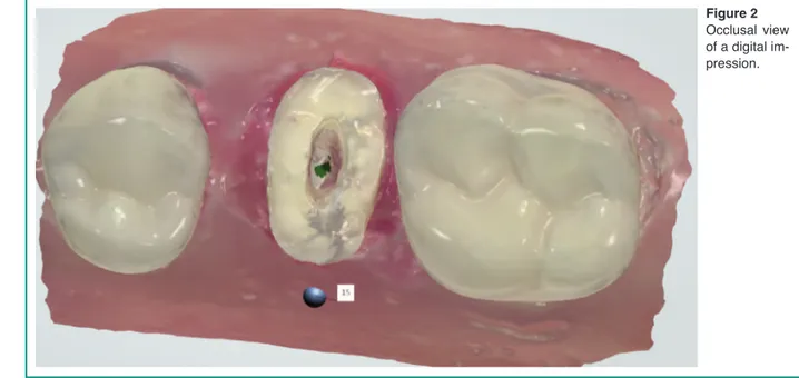
one mesially and one distally to the space for samples in order to simulate a clinical real situa-tion. At the center of the structure one sample was put at a time (Figure 1). Digital and tradi-tional impressions were taken for each sample in order to compare the reading depth of the digital impression of the post-space with the reading depth of the traditional impression. The first group of impressions of the post-spaces was made with the digital technique. The IOS used was the 3shape TRIOS (3hape) (Figures 2, 3). The digital files, produced through the IOS,
were sent to the laboratory (Figure 4). Through the CAD program, the scanned spaces were de-veloped in order to integrate the scanner results into a three-dimensional grid to make the meas-urements. For each sample a light-cured resin in-dividual tray was prepared, taking care to elimi-nate any undercuts with red wax (Figure 5). It was carried out a two-component single-phase impression with the use of a calcinable plastic post inside the post-space to give higher stiff-ness to the light silicon (Aquasil, Dentsply). It was thus obtained the traditional post-space silicon impression of the samples (Figure 6). A digital grid was used to measure the depth of the post-space scanned through the digital technique of each sample from the coronal margin to the most apical part of the preparation (Figure 7). A K-file was used to measure the length of the post-space of each sample obtained through the traditional silicon impression and subsequently the measurement results were reported on a mil-limeter gauge (Figure 8).
Furthermore, an assessment of the width of the entrances of the post-spaces was carried out. Di-ameters and areas of the post-space entrances were measured with a dedicated software (Image Tool 3.0) (Figure 9).
Figure 1
Pink acrylic resin model with one of the samples in the cen-ter and two resin elements on the sides.
Figure 2
Occlusal view of a digital im-pression.
original research article
Results
The data expressed in Table 1 highlight the length discrepancy of the post-space reading be-tween the digital and traditional technique. A shorter reading depth was noticed in all the sam-ples for the digital technique compared to the traditional one. The mean depth discrepancy ex-pressed in percentages (19.58%) indicates that the digital impression with current technologies
Figure 5
Pink acrylic resin model with a positioned sample, the indi-vidual tray and the traditional silicon impression.
Figure 3
Digital impression internal view.
Figure 4
or
ig
in
al
r
es
ea
rc
h
ar
tic
le
fails to impress clearly the post-space. Standard deviation of the data expressed in percentage of Table 1 is 13.89. This data suggests that the val-ues were not similar to each other, but this is probably due to the low number of samples. In two cases the digital technique has achieved less than 10% difference compared to the tradi-tional technique, but there have been also cases
in which the variation in depth has reached al-most 40%.
From the results of the analysis of the root canal entrances (Table 2), it is possible to hypothesize
Figure 6
Detail of the traditional silicon impression with calcinable plastic post-space.
Figure 7
Measurement with a digital grid of the post-space scanned in digital technique.
Figure 8
Measurement with a gauge of the post-space length ob-tained through the traditional technique.
Figure 9
Measurement of the diameter and area of the post-space en-trance.
original research article
that the depth in digital technique, besides being influenced by the IOS hardware and software, is influenced by the amplitude of the surface of the post-spaces entrances.
The samples that showed the minor discrepancy between the two techniques expressed the widest post-space entrance.
Discussion
Several articles have proposed in the literature the use of the IOS and CAD-CAM technology to produce endodontic posts (24). Dedicated tools, called scan-posts, are used to detect the
post-Table 1- Comparison between the depths of the post-spaces obtained through the traditional and digital technique with the
re-spective discrepancies expressed in percentages.
Table 2- Larger and smaller diameter measurement for each sample with the calculation of the post-space entrance areas
or
ig
in
al
r
es
ea
rc
h
ar
tic
le
space through the use of an IOS. Anyway this kind of technique, even though makes it easier to detect the depth of the post-space, is not able to record properly the root channel anatomy. Considering the better features of an anatomic endodontic post, such as the root dentin preser-vation (8, 20, 29-33), reduced cement layer (34-37), minor bubbles formation (38), increased post retention (7, 8, 39, 40), and fracture resist-ance (41, 42), in this study the possibility of pro-ducing an anatomic endodontic post through the use of an IOS has been investigated. To the best of our knowledge no studies have yet investigat-ed the IOS capability to detect the post-space for the production of anatomic posts.In the present paper, the digital impression showed lower capability to read the post-space compared to the traditional impression. Anyway, similar results between the two techniques have been obtained for post-spaces expressing a wide entrance. That is probably due to the increased amount of IOS light able to get into the post-space when a greater entrance is expressed. One major limitation of the present study was the low sample number. Besides an in vitro study design does not take into consideration all the variables of the mouth environment such as oral fluids and the IOS motion limitations. Further studies on a greater sample and with different IOS systems should analyze the possibility of recording the post-space through a digital impression in order to produce an anatomic post.
Conclusions
In this in vitro study, the application of the IOS for the post-space reading in order to deliver an anatomic post has been proven to be still not re-liable, as there are still depth reading limitations for the narrow root channels. In fact, in this type of channels it is difficult to reach with the light beam of the IOS the deepest areas of the post-space, with a consequent incomplete post-space reading. Further improvements in the IOS
hard-ware and softhard-ware are necessary to make the digital impression able to read properly the post-space independently from the root channel anatomy.
References
1. Cheung W. A review of the management of endodon-tically treated teeth. Post, core and the final restoration. J Am Dent Assoc. 2005;136(5):611-9.
2. Dietschi D, Duc O, Krejci I, Sadan A. Biomechanical considerations for the restoration of endodontically treated teeth: a systematic review of the literature, Part II (Evaluation of fatigue behavior, interfaces, and in vivo studies). Quintessence Int. 2008;39(2):117-29. 3. Choudhary S, Choudhary P, Tripathi S, Begum Z.
Com-parative evaluation of retention of prefabricated and conventional cast post: An in vitro study. J Int Soc Prevent Communit Dent. 2014;4(2):87-91.
4. Cohen BI, Pagnillo M, Musikant BL, Deutsch AS. Comparison of the retentive and photoelastic properties of two prefabricated endodontic post systems. J Oral Rehabil. 1999;26(6):488-94.
5. Boschian Pest L, Cavalli G, Bertani P, Gagliani M. Ad-hesive post-endodontic restorations with fiber posts: push-out tests and SEM observations. Dental Materials. 2002;18(8):596-602.
6. Butz F, Lennon AM, Heydecke G, Strub JR. Survival rate and fracture strength of endodontically treated maxillary incisors with moderate defects restored with different post-and-core systems: an in vitro study. The International journal of prosthodontics. 2001;14(1):58-64.
7. Ferrari M, Vichi A, Mannocci F, Mason PN. Retro-spective study of the clinical performance of fiber posts. Am J Dent. 2000;13(Spec No):9B-13B. 8. Grandini S, Goracci C, Monticelli F, Borracchini A,
Ferrari M. SEM evaluation of the cement layer thick-ness after luting two different posts. J Adhes Dent. 2005;7(3):235-40.
9. Assif D, Gorfil C. Biomechanical considerations in restoring endodontically treated teeth. The Journal of Prosthetic Dentistry. 1994;71(6):565-7.
10. Perdigão J, Gomes G, Augusto V. The effect of dowel space on the bond strengths of fiber posts. J Prostho-dont. Blackwell Publishing Inc;2007;16(3):154-64. 11. Gomes GM, Gomes OM, Gomes JC, Loguercio AD,
Calixto AL, Reis A. Evaluation of different restorative techniques for filling flared root canals: fracture re-sistance and bond strength after mechanical fatigue. J Adhes Dent. 2014;16(3):267-76.
original research article
12. Baba NZ, Goodacre CJ, Daher T. Restoration of en-dodontically treated teeth: the seven keys to success. Gen Dent. 2009;57(6):596-603–quiz604–5–595–679. 13. Morgano SM, Rodrigues AHC, Sabrosa CE.
Restora-tion of endodontically treated teeth. Dent Clin North Am. 2004;48(2):vi-397-416.
14. Schmage P, Pfeiffer P, Pinto E, Platzer U, Nergiz I. In-fluence of oversized dowel space preparation on the bond strengths of FRC posts. Operative Dentistry. 2009;34(1):93-101.
15. Davidson CL, Feilzer AJ. Polymerization shrinkage and polymerization shrinkage stress in polymer-based restoratives. Journal of Dentistry. 1997;25(6):435-40. 16. Ferracane JL, Mitchem JC. Relationship between
com-posite contraction stress and leakage in Class V cavi-ties. Am J Dent. 2003;16(4):239-43.
17. Zogheib LV, Pereira JR, do Valle AL, de Oliveira JA, Pegoraro LF. Fracture resistance of weakened roots restored with composite resin and glass fiber post. Braz Dent J. 2008;19(4):329-33.
18. Clavijo VGR, Reis JMDSN, Kabbach W, Silva ALFE, Oliveira Junior OB de, Andrade MF de. Fracture strength of flared bovine roots restored with different intraradicular posts. J Appl Oral Sci. Bauru School of Dentistry;2009;17(6):574-8.
19. Faria-e-Silva AL, Pedrosa-Filho C de F, Menezes M de S, Silveira DMD, Martins LRM. Effect of relining on fiber post retention to root canal. J Appl Oral Sci. Bauru School of Dentistry;2009;17(6):600-4. 20. Grandini S, Sapio S, Simonetti M. Use of anatomic post
and core for reconstructing an endodontically treated tooth: a case report. J Adhes Dent. 2003;5(3):243-7. 21. DDS VCMM, DDS ALFESPM, DDS LRMMPM.
Ef-fect of Cement Type, Relining Procedure, and Length of Cementation on Pull-out Bond Strength of Fiber Posts. Journal of Endodontics. Elsevier Ltd;2010;36 (9):1543-6.
22. Martelli H Jr, Pellizzer EP, Rosa BT, Lopes MB, Go-nini A Jr. Fracture resistance of structurally compro-mised root filled bovine teeth restored with accessory glass fibre posts. Int Endod J. 2008;41(8):685-92. 23. De Vico G, Ferraris F, Arcuri L, Guzzo F, Spinelli D.
A novel workflow for computer guided implant surgery matching digital dental casts and CBCT scan. Oral Im-plantol (Rome). 2016 Jan;9(1):33-48.
24. Awad MA, Marghalani TY. Fabrication of a custom-made ceramic post and core using CAD-CAM tech-nology. The Journal of Prosthetic Dentistry. 2007;98 (2):161-2.
25. Liu P, Deng X-L, Wang X-Z. Use of a CAD/CAM-fab-ricated glass fiber post and core to restore fractured an-terior teeth: A clinical report. The Journal of Prosthetic Dentistry. The Editorial Council of the Journal of Pros-thetic Dentistry. 2010;103(6):330-3.
26. Lee JH, Sohn DS, Lee CH. Fabricating a
fiber-rein-forced post and zirconia core with CAD/CAM tech-nology. The Journal of Prosthetic Dentistry. Editorial Council for the Journal of Prosthetic Dentistry. 2014; 112(3):683-5.
27. Ender A, Mehl A. Full arch scans: conventional versus digital impressions-an in-vitro study. Int J Comput Dent. 2011;14(1):11-21.
28. Arcuri L, Lorenzi C, Cecchetti F, Germano F, Spuntarelli M, Barlattani A. Full digital workflow for implant-prosthetic rehabilitations: a case report. Oral Implantol (Rome). 2015;8(4):114-21.
29. Tilk MA, Lommel TJ, Gerstein H. A study of mandibu-lar and maxilmandibu-lary root widths to determine dowel size. Journal of Endodontics. 1979;5(3):79-82.
30. Raiden G, Costa L, Koss S, Hernández JL, Aceñolaza V. Residual thickness of root in first maxillary premo-lars with post space preparation. Journal of Endodon-tics. 1999;25(7):502-5.
31. Raiden G, Koss S, Costa L, Hernández JL. Radi-ographic measurement of residual root thickness in premolars with post preparation. Journal of Endodon-tics. 2001;27(4):296-8.
32. Pilo R, Tamse A. Residual dentin thickness in mandibu-lar premomandibu-lars prepared with gates glidden and ParaPost drills. The Journal of Prosthetic Dentistry. 2000;83 (6):617-23.
33. Kuttler S, McLean A, Dorn S, Fischzang A. The impact of post space preparation with Gates-Glidden drills on residual dentin thickness in distal roots of mandibular molars. The Journal of the American Dental Associa-tion. 2004;135(7):903-9.
34. Braga RR, Ferracane JL. Alternatives in polymerization contraction stress management. Crit Rev Oral Biol Med. 2004;15(3):176-84.
35. Ferracane JL. Developing a more complete under-standing of stresses produced in dental composites dur-ing polymerization. Dental Materials. 2005;21(1):36-42.
36. Eick JD, Gwinnett AJ, Pashley DH, Robinson SJ. Cur-rent concepts on adhesion to dentin. Crit Rev Oral Biol Med. 1997;8(3):306-35.
37. Miguel A, la Macorra de JC. A predictive formula of the contraction stress in restorative and luting materials at-tending to free and adhered surfaces, volume and de-formation. Dental Materials. 2001;17(3):241-6. 38. Gluskin AH, Ahmed I, Herrero DB. The aesthetic post
and core: unifying radicular form and structure. Pract Proced Aesthet Dent. 2002;14(4):313-21–quiz322. 39. Pirani C, Chersoni S, Foschi F, Piana G, Loushine RJ,
Tay FR, et al. Does hybridization of intraradicular dentin really improve fiber post retention in endodon-tically treated teeth? Journal of Endodontics. 2005; 31(12):891-4.
40. Ferrari M, Vichi A, García-Godoy F. Clinical evalua-tion of fiber-reinforced epoxy resin posts and cast post
or
ig
in
al
r
es
ea
rc
h
ar
tic
le
and cores. Am J Dent. 2000;13(Spec No):15B-18B.41. Lassila LVJ, Tanner J, Le Bell A-M, Narva K, Vallittu PK. Flexural properties of fiber reinforced root canal posts. Dental Materials. 2004;20(1):29-36.
42. Bell A-ML, Lassila LVJ, Kangasniemi I, Vallittu PK. Bonding of fibre-reinforced composite post to root canal dentin. Journal of Dentistry. 2005;33(7):533-9.
Correspondence to:
Dr. Alessandro Pinto
Materials for Health, Environment and Energy University of Rome “Tor Vergata”, Italy E-mail: [email protected]