.
Impact of residual pulmonary obstruction
on the long-term outcome of patients
with pulmonary embolism
Raffaele Pesavento1, Lucia Filippi1, Antonio Palla2, Adriana Visonà3, Carlo Bova4, Marco Marzolo5, Fernando Porro6, Sabina Villalta7, Maurizio Ciammaichella8, Eugenio Bucherini9, Giovanni Nante1, Sandra Battistelli10, Maria Lorenza Muiesan11, Giampietro Beltramello12, Domenico Prisco13, Franco Casazza14, Walter Ageno15, Gualtiero Palareti16, Roberto Quintavalla17, Simonetta Monti18, Nicola Mumoli 19, Nello Zanatta20, Roberto Cappelli21, Marco Cattaneo22, Valentino Moretti23, Francesco Corà24, Mario Bazzan25, Angelo Ghirarduzzi26, Anna Chiara Frigo27, Massimo Miniati13 and Paolo Prandoni27for the SCOPE Investigators28
Affiliations:1Dept of Internal Medicine, University of Padua, Padua, Italy.2Cardio-thoracic Dept, Cisanello Hospital, University of Pisa, Pisa, Italy.3Dept of Internal and Vascular Medicine, Angiology Unit, Civic Hospital, Castelfranco Veneto, Italy.4Dept of Internal Medicine, University Hospital of Cosenza, Cosenza, Italy. 5UOC Medicina Interna, UOS Angiologia Medica, Civic Hospital, Rovigo, Italy. 6UOC Pronto Soccorso e Medicina d’Urgenza, Fondazione IRCCS Ca’ Granda Ospedale Maggiore Policlinico, Milan, Italy.7UOC Medicina Interna I, Ca’ Foncello Hospital, Treviso, Italy.8UOC Medicina d’Urgenza, San Giovanni Addolorata Hospital, Rome, Italy. 9UOS di Angiologia e Medicina Vascolare, Civic Hospital of Faenza, Faenza, Italy. 10Department of Medicine, Surgery and Neuroscience, University Hospital of Siena, Siena, Italy. 11Dept of Clinical and Experimental Sciences, University of Brescia, Brescia, Italy.12Division of Internal Medicine, San Bassiano Hospital, Bassano del Grappa, Italy.13Dept of Experimental and Clinical Medicine, University of Florence, Florence, Italy.14UO Cardiologia Clinica, San Carlo Borromeo Hospital, Milan, Italy.15Dept of Clinical and Experimental Medicine, University of Insubria, Varese, Italy. 16Angiology and Blood Coagulation, University Hospital of Bologna, Bologna, Italy. 17UOC Medicina Interna ad indirizzo Angiologico e Coagulativo, University Hospital of Parma, Parma, Italy.18Istituto di Fisiologia Clinica del CNR e Fondazione Toscana Gabriele Monasterio, Pisa, Italy.19Dept of Internal Medicine, Livorno Hospital, Livorno, Italy. 20Dept of Internal Medicine, Civic Hospital of Conegliano, Conegliano, Italy.21Thrombosis Center, University of Siena, Siena, Italy.22Medicina III, Ospedale San Paolo, ASST Santi Paolo e Carlo, Dipartimento di Scienze della Salute, University of Milan, Milan, Italy.23AAS3 Alto Friuli Collinare, Medio Friuli, San Daniele del Friuli, Italy.24UOS Emergenza Medica, Pronto Soccorso Generale, Civic Hospital of Vicenza, Vicenza, Italy.25UOSD di Ematologia e Malattie Trombotiche CMID, Ospedale San Giovanni Bosco, Torino Emergenza Nord, Turin, Italy.26Dipartimento Medicina Interna e Specialità Mediche, Medicina II, Angiologia, Arcispedale S. Maria Nuova, IRCCS, Reggio Emilia, Italy.27Dept of Cardiac Thoracic and Vascular Sciences, University of Padua, Padua, Italy.28A complete list of the SCOPE investigators can be found in the Acknowledgements section.
Correspondence: Raffaele Pesavento, Dept of Internal Medicine, University of Padua, Via Giustiniani 2, 35128 Padua, Italy. E-mail: [email protected]
@ERSpublications
Residual pulmonary obstruction after PE is an independent predictive factor of VTE recurrences and/or CTEPH http://ow.ly/XK49308SDTd
Cite this article as: Pesavento R, Filippi L, Palla A, et al. Impact of residual pulmonary obstruction on the long-term outcome of patients with pulmonary embolism. Eur Respir J 2017; 49: 1601980 [https://doi.org/ 10.1183/13993003.01980-2016].
ABSTRACT The impact of residual pulmonary obstruction on the outcome of patients with pulmonary embolism is uncertain.
We recruited 647 consecutive symptomatic patients with a first episode of pulmonary embolism, with or without concomitant deep venous thrombosis. They received conventional anticoagulation, were assessed for residual pulmonary obstruction through perfusion lung scanning after 6 months and then were followed up for up to 3 years. Recurrent venous thromboembolism and chronic thromboembolic pulmonary hypertension were assessed according to widely accepted criteria.
Residual pulmonary obstruction was detected in 324 patients (50.1%, 95% CI 46.2–54.0%). Patients with residual pulmonary obstruction were more likely to be older and to have an unprovoked episode. After a 3-year follow-up, recurrent venous thromboembolism and/or chronic thromboembolic pulmonary hypertension developed in 34 out of the 324 patients (10.5%) with residual pulmonary obstruction and in 15 out of the 323 patients (4.6%) without residual pulmonary obstruction, leading to an adjusted hazard ratio of 2.26 (95% CI 1.23–4.16).
Residual pulmonary obstruction, as detected with perfusion lung scanning at 6 months after a first episode of pulmonary embolism, is an independent predictor of recurrent venous thromboembolism and/or chronic thromboembolic pulmonary hypertension.
Introduction
The risk of recurrent venous thromboembolic (VTE) events in patients after a first episode of acute pulmonary embolism is high in many patients, especially in those in whom the episode is unprovoked. In addition, up to 4% of them will develop chronic thromboembolic pulmonary hypertension (CTEPH) [1–4]. In the case of recurrent VTE episodes after an acute pulmonary embolism episode, the probability of experiencing another pulmonary embolism episode is three-fold higher than in patients whose initial acute event is a deep vein thrombosis (DVT) [3]. Moreover, it was observed that recurrent embolism increases the risk of developing CTEPH [4].
Currently, there is virtually no way to identify patients with pulmonary embolism in whom the risk of late complications is high enough to justify indefinite anticoagulation. Although the latest international guidelines suggest the adoption of indefinite anticoagulation in most patients with a first episode of unprovoked pulmonary embolism [5, 6], most physicians often disregard this .recommendation [7]. In an attempt to investigate whether residual pulmonary obstruction (RPO) can identify patients at a higher risk of late complications, computed tomography (CT) angiography was repeated in patients with pulmonary embolism 6 months after the index episode [8–10]. The rate of RPO was found to be much lower than expected (<20%) and was not related to subsequent complications [9–11]. In contrast, the potential for perfusion lung scanning is higher. When assessed prior to hospital discharge, perfusion defects were found to predict a long-term unfavourable outcome [12]. In addition, when assessed 3–6 months after the acute episode, the rate of RPO was consistently found to be∼50% of the investigated patients [13–15]. While it would be of interest to know whether and to what extent the persistence of RPO, as shown by perfusion lung scanning at 6 months after the acute episode, can be of help in identifying patients at a higher risk of developing recurrent VTE and/or CTEPH, the available information is limited and contradictory. In a prospective cohort study by MINIATIet al. [16] addressing the long-term follow-up of
320 patients with pulmonary embolism, three out of the four patients who developed CTEPH had a persistent scintigraphic RPO. By contrast, in a more recent prospective cohort study by POLIet al. [17],
RPO, which was evaluated in 236 patients with pulmonary embolism at variable timing during the follow-up, failed to show any predictive value of VTE recurrences.
In order to assess the impact of RPO on the long-term outcome of patients with pulmonary embolism, we performed a prospective cohort study with central adjudication of the study procedures and outcomes. For this purpose, we recruited a large number of consecutive patients after a first episode of symptomatic pulmonary embolism, with or without concomitant DVT, gave them conventional anticoagulation, assessed the persistence of RPO by perfusion lung scanning after 6 months and followed them up for up to 3 years.
Methods
The SCOPE (Study on the Clinical Course of Pulmonary Embolism) clinical investigation was a multicentre, nationwide, prospective cohort study conducted in 34 university or hospital institutions in Italy over a 7-year period (2008–2014). The study protocol was approved by the institutional review board of each participating centre and all patients provided written informed consent. The study was registered at ClinicalTrials.gov (NCT01781858).
The main end-point of the study was to assess the impact of RPO, as shown by perfusion lung scanning 6 months after a first episode of acute symptomatic pulmonary embolism, on the incidence of recurrent VTE and/or CTEPH.
Patients and risk factors for pulmonary embolism
Consecutive in- and outpatients referred with a first episode of acute symptomatic pulmonary embolism, with or without concomitant DVT, were potentially eligible for the study. Patients were excluded on account of the following: symptoms occurring for >2 weeks, life expectancy <2 years, concomitant severe cardiac or pulmonary diseases accounting for the risk of nonthromboembolic pulmonary hypertension, previous episodes of VTE, unavailability for long-term follow-up, need for anticoagulation for reasons
This article has supplementary material available from erj.ersjournals.com
Earn CME accreditation by answering questions about this article. You will find these at erj.ersjournals.com/journal/cme Received: Oct 09 2016 | Accepted after revision: Feb 07 2017
Clinical trials: This study was registered at Clinicaltrials.gov with identifier NCT01781858. Conflict of interest: None declared.
other than VTE, age <18 years, pregnancy, or refusal to give written informed consent. In addition, patients who developed recurrent VTE prior to completion of the first 6 months of observation were subsequently excluded from any further follow-up or analysis, as were those in whom perfusion lung scanning at 6 months could not be performed.
Recruited patients were categorised as having secondary pulmonary embolism in the presence of active cancer, immobilisation for a medical illness lasting >1 week, known anti-phospholipid syndrome, recent (<1 month) trauma, surgery or puerperium, or ongoing hormonal therapy. All other patients were labelled as having unprovoked pulmonary embolism.
Diagnosis and treatment of pulmonary embolism
Diagnosis of pulmonary embolism was confirmed by multidetector row CT angiography or ventilation/ perfusion (V/Q) lung scanning, and was accepted in the presence of at least one intraluminal filling defect or a (sub)segmental V/Q mismatch, respectively. The anatomic severity of pulmonary embolism was quantified according to the Qanadli and Meyer score, respectively, while the clinical severity of pulmonary embolism was evaluated by means of the original Pulmonary Embolism Severity Index (PESI) [18–20]. Details concerning the Qanadli, Meyer and PESI scores are available in the supplementary material. In patients with concomitant symptoms of DVT, whole-leg ultrasonography was performed to confirm or rule out the clinical suspicion using the criterion of vein compressibility [21].
Patients were treated with full-dose unfractionated heparin, low-molecular-weight heparin or fondaparinux, overlapping with and followed by at least 6 months of vitamin K antagonists (International Normalised Ratio 2.0–3.0). Most patients with active cancer received low-molecular-weight heparin alone. In patients with haemodynamic instability, anticoagulant therapy was preceded by reperfusion therapy at the discretion of the attending physicians. The duration of anticoagulant treatment followed international guidelines with individual adaptation based on patients’ preferences and risk profile [5, 6].
Assessment of residual pulmonary obstruction
At 6 months after the index event, patients underwent perfusion lung scanning, which was evaluated using the Meyer score [19]. Patients were labelled as having RPO in the presence of a positive Meyer score, whatever the degree of vascular defect.
Assessment of the study outcomes
Patients who developed signs or symptoms suggestive of recurrent VTE following the 6-month perfusion lung scanning underwent objective investigation. Recurrent VTE was defined as a composite of (nonfatal or fatal) pulmonary embolism and/or symptomatic DVT in the lower or upper extremities. Nonfatal pulmonary embolism was defined as the presence of a new (sub)segmental V/Q mismatch or of a new intraluminal filling defect on CT angiography. Fatal pulmonary embolism was diagnosed if it was confirmed at autopsy or if was preceded by objectively confirmed VTE events. The ultrasound criterion for (recurrent) DVT was incompressibility of a vein segment that was either initially free from thrombi or had later recanalised. All patients who developed clinical signs or symptoms suggestive of CTEPH (e.g. unexplained exertional dyspnoea, progressive limitation of exercise capacity, syncope, angina or right ventricular failure) underwent transthoracic echocardiography and V/Q lung scanning. Whenever this complication could not be excluded, multidetector CT angiography, pulmonary angiography and/or heart catheterisation were performed according to local availabilities. CTEPH was confirmed in the presence of multiple perfusion defects as shown by V/Q lung scanning, mean pulmonary artery pressure >25 mmHg, pulmonary capillary wedge pressure <15 mmHg, pulmonary vascular resistance >2 Woods Units and angiographic or tomographic evidence of pouching, webs or bands with or without post-stenotic dilation, intimal irregularities, abrupt narrowing or total occlusion.
The scintigraphic images obtained at 6 months and the study outcomes were centrally adjudicated by an independent committee.
Statistical analysis
Details concerning the sample size calculation are provided in the supplementary material. For comparison of baseline characteristics between patients with and without RPO, the Chi-squared test was used for categorical variables and the Wilcoxon’s rank sum test was used for quantitative variables, since their distribution was not normal. Persistence of RPO was estimated with 95% confidence intervals calculated with the binomial exact method. Potential predictors for the development of RPO, found to be statistically significant at the 10% level in the univariate analysis, were included in a multivariate logistic regression model with backward selection. The linearity of the model when considering quantitative covariates was checked with the Hosmer–Lemeshow goodness-of-fit test. Results are presented as p-values and odds
ratios with 95% confidence intervals. The cumulative probability of recurrent VTE and/or CTEPH in patients with and without RPO was estimated with the Kaplan–Meier method. Patients who refused to further participate, moved, died or were lost to follow-up were censored at the time of their last follow-up. Predictors of recurrent VTE and/or CTEPH were tested in a univariate Cox proportional hazard regression model and those found to be statistically significant at the 10% level were included in a multivariate Cox regression model with backward selection. Ties were handled as discrete and the supremum test for proportional hazard assumption for quantitative covariates was performed. Results are presented as p-values and hazard ratios with 95% confidence intervals. Multicollinearity of predictors was excluded since the variance inflation factor was close to 1. Finally, the effect of the degree of RPO on the study outcomes was tested in a univariate logistic regression model and the results expressed as p-values, odds ratios and 95% confidence intervals.
All the statistical tests were two-tailed and conducted at a significance level of 5% if not otherwise stated and the analyses were performed with SAS for Windows version 9.4 (SAS Institute, Cary, NC, USA).
Results
Patients
Out of 887 potentially eligible patients referred from January 2008 to July 2011, 240 were excluded because of an expected survival <2 years (n=39), concomitant severe cardiac or pulmonary diseases (n=35), previous VTE (n=17), need for anticoagulation for reasons other than VTE (n=17), geographical inaccessibility (n=6) or pregnancy (n=1). In addition, 22 patients refused to participate, 96 died or developed recurrent VTE (78 and 18, respectively) before completion of the first 6-month treatment and seven additional patients could not receive the scheduled RPO assessment. Hence, 647 subjects with a first episode of acute symptomatic pulmonary embolism who had completed an uneventful 6-month period of anticoagulation were recruited for the current investigation. The main baseline characteristics of the patients are shown in
TABLE 1Main baseline characteristics of the study patients by presence of residual pulmonary obstruction (RPO) All patients Patients with RPO Patients without RPO p-value Patients 647 324 323 Age years 67 (52–75) 70 (59.5–77) 63 (47–73) <0.0001 Male 318 (49.1) 156 (48.2) 162 (50.2) 0.61 PESI ⩽85 337 (52.1) 148 (45.7) 189 (58.5) 0.005 86–124 233 (36.0) 131 (40.4) 102 (31.6) ⩾125 77 (11.9) 45 (13.9) 32 (9.9)
Pulmonary embolism with concomitant DVT 296 (45.8) 152 (46.9) 144 (44.6) 0.55 Unprovoked pulmonary embolism 404 (62.4) 218 (67.3) 186 (57.6) 0.01 Provoked pulmonary embolism#
Prolonged immobility 24 (9.9) 13 (12.3) 11 (8.0) 0.66
Recent trauma or surgery 92 (37.9) 41 (38.7) 51 (37.2)
Active cancer 57 (23.4) 24 (22.6) 33 (24.1)
Acute medical disease 23 (9.5) 11 (10.4) 12 (8.8)
Others 47 (19.3) 17 (16.0) 30 (21.9)
Pulmonary embolism initial extension %
<33 292 (45.1) 131 (40.4) 161 (49.9) 0.06 33–66 285 (44.1) 155 (47.9) 130 (40.2) >66 70 (10.8) 38 (11.7) 32 (9.9) Therapy Thrombolysis 50 (7.7) 25 (7.7) 25 (7.7) 0.99 OAT withdrawal 410 (63.4) 191 (59.0) 219 (67.8) 0.02
OAT duration months
⩽6 135 (20.9) 57 (17.6) 78 (24.1) 0.02
7–12 201 (31.1) 92 (28.4) 109 (33.8)
13–24 63 (9.7) 33 (10.2) 30 (9.3)
25–36 248 (38.3) 142 (43.8) 106 (32.8)
Data are presented as n, median (interquartile range) or n (%), unless otherwise stated. PESI: Pulmonary Embolism Severity Index; OAT: oral anticoagulant therapy.#: n=243.
table 1. The diagnosis of pulmonary embolism was confirmed by CT scan in 482 patients (74.5%), by lung scan in 156 patients (24.1%) and by pulmonary angiography in nine patients (1.4%). Anticoagulation could be discontinued in 410 patients (63.4%).
Residual pulmonary obstruction
The 6-month perfusion lung scanning showed the persistence of RPO in 324 patients (50.1%, 95% CI 46.2–54.0%). Older age, an unprovoked clinical presentation, a higher clinical severity and a more extensive acute pulmonary embolism episode were associated with the persistence of pulmonary obstruction at the univariate level (table 1). Moreover, a significantly higher prevalence of RPO was found in patients treated for >2 years than in those treated for up to 6 months. At the multivariate regression model, only age (OR 1.03, 95% CI 1.02–1.04; p<0.0001) and the unprovoked nature of pulmonary embolism (OR 1.40, 95% CI 1.01–1.95; p=0.04) were associated with the persistence of RPO.
Among the 156 patients with scintigraphic diagnosis of pulmonary embolism, the persistence of RPO was found in 86 patients (55%) (table 2). The corresponding figures in those with CT or angiographic diagnosis were 233 patients (48.3%) and five patients (55%), respectively ( p=0.17).
Follow-up and study outcomes
During the 3-year follow-up period, two out of 647 patients died because of pulmonary embolism and 26 because of other causes, including cancer (n=19), ischaemic stroke (n=2), pneumonia (n=1), acute heart failure (n=1), traumatic accident (n=1), acute myocardial infarction (n=1) and retroperitoneal bleeding (n=1). In addition, four patients (0.62%) were lost to follow-up after 8, 13, 24 and 30 months, respectively (figure 1). Objectively proven recurrent VTE developed in 40 patients (6.2%, 95% CI 4.5–8.3%) and CTEPH in 11 patients (1.7%, 95% CI 0.9–3.0%). The distribution of the events according to the persistence of RPO is described in table 3.
The combined end-point of recurrent VTE and/or CTEPH occurred in 34 out of the 324 patients (10.5%) with RPO compared with 15 out of the 323 patient (4.6%) without RPO (table 4).
The patients with recurrent VTE and/or CTEPH received oral anticoagulants for up to 6 months in 11 events (22.5%), 12 months in 30 events (61.2%), 2 years in six events (12.2%) and 3 years in two events (4.1%). The corresponding figures in patients free from events were 124 (20.7%), 171 (28.7%), 57 (9.5%) and 246 (41.1%), respectively. Of the 49 events, seven (14.3%) occurred while on anticoagulation: five in the group with RPO and two in the group without RPO. The distribution of the study events according to the degree of RPO showed that the higher the RPO index, the greater the risk of developing the outcomes, and this was true especially for the chance of developing CTEPH (table 5).
In patients with RPO, the cumulative incidence of recurrent VTE and/or CTEPH was 3.4% (95% CI 1.8–5.8%) in the first month of observation, 6.2% (95% CI 3.9–9.2%) after 12 months, 8.8% (95% CI 6.1–1.3%) after 2 years and 11.0% (95% CI 7.8–14.8%) after 3 years. The corresponding figures in patients without RPO were 0% (95% CI 0–1.1%), 0.9% (95% CI 0.3–2.5%), 3.8% (95% CI 2.1–6.4%) and 4.9% (95% CI 2.9–7.8%), respectively (figure 2). Table 6 shows the cumulative incidence of recurrent VTE and of CTEPH, considered separately.
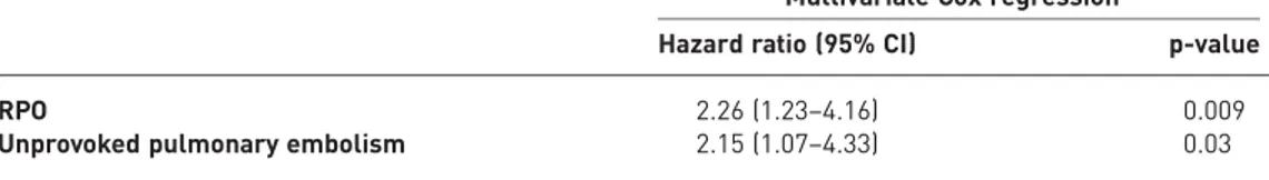
In the multivariate proportional hazards regression model, RPO (hazard ratio 2.26, 95% CI 1.23–4.16; p=0.009) and unprovoked pulmonary embolism (hazard ratio 2.15, 95% CI 1.07–4.33; p=0.03) were independent predictors of VTE recurrences and/or CTEPH (table 7).
TABLE 2Distribution of residual pulmonary obstruction (RPO) index according to the initial pulmonary obstruction index in the 156 patients who received pulmonary embolism diagnosis by means of a lung scan
Pulmonary perfusion index at time of diagnosis % RPO index % p-value <1 1–25 >25
1–25 33 (47.1) 33 (47.1) 4 (5.7) 0.07
>25 37 (43.0) 34 (39.5) 15 (17.4)
Additional observations
CTEPH developed in two out of the 40 (5.0%) patients who had experienced recurrent VTE and in nine out of the 607 (1.5%) patients who had not experienced recurrent VTE ( p=0.14).
Discussion
The results of this prospective, multicentre study conducted in a large cohort of consecutive patients with a first episode of pulmonary embolism suggest that the persistence of RPO is an independent predictor of subsequent late complications, including recurrent VTE and CTEPH, and that the impact of RPO increased
Patients with acute pulmonary embolism n=887 Patients enrolled n=647 VTE recurrences n=40 (6.2%, 95% CI 4.5–8.3%) CTEPH n=11 (1.7%, 95% CI 0.9–3.0%) Patients excluded n=240 Expected survival <2 years n=39 Severe cardiac/pulmonary diseases n=35 Previous VTE n=17
Need for anticoagulant for other reasons n=17 Geographical inaccessibility n=6
Pregnancy n=1 Refusal of consent n=22
Deaths/recurrent VTE within 6 months n=96 Unable to perform 6-month lung scan n=7
Deaths n=28 Cancer n=19 Ischaemic stroke n=2 Pneumonia n=1 Acute heart failure n=1 Pulmonary embolism n=2 Trauma n=1
AMI n=1
Retroperitoneal bleeding n=1 Lost to follow-up n=4
FIGURE 1 Flow diagram of the study. VTE: venous thromboembolism; AMI: acute myocardial infarction; CTEPH: chronic thromboembolic pulmonary hypertension.
TABLE 3Frequency of study outcomes according to the persistence of residual pulmonary obstruction (RPO)
Patients with RPO Patients without RPO
Patients 324 323
Recurrent VTE 25 (7.7) 15 (4.6)
DVT 10 (3.1) 10 (3.1)
Nonfatal pulmonary embolism 14 (4.3) 4 (1.2)
Fatal pulmonary embolism 1 (0.3) 1 (0.3)
CTEPH 11 (3.4) 0
Data are presented as n or n (%). VTE: venous thromboembolism; DVT: deep vein thrombosis; CTEPH: chronic thromboembolic pulmonary hypertension.
according to the degree of the perfusion defects. Our study results have the potential to help assist decisions on the optimal duration of anticoagulation in patients with pulmonary embolism beyond the first months. Our results are robust as they come from the prospective follow-up of a large number of consecutive patients with a first episode of symptomatic and objectively proven pulmonary embolism, irrespective of its clinical and radiological severity, in whom rigorous criteria were adopted for adjudication of the study outcomes. RPO was predefined according to the Meyer score [19], and the diagnostic work-up for recurrent VTE and CTEPH was performed according to international guidelines [5, 6]. A central independent committee unaware of patients’ details adjudicated the presence of RPO and the study outcomes. Finally, loss to follow-up was low and most patients who refused to further participate or moved had a follow-up of at least 8 months. Hence, we believe that the results of our observations are generalisable and applicable to the vast majority of patients with a first episode of clinically symptomatic pulmonary embolism. Our findings are consistent with those found in a single-centre cohort study by PLANQUETTEet al. [22], who
arrived at similar conclusions after following prospectively a smaller sample of patients with pulmonary embolism in whom the lung scan had been performed at variable times after the index episode. In this study, only patients with a perfusion defect index >10% were labelled as having RPO.
Our study results are in apparent contrast to those recently obtained by us [8] and others [9, 10] with the use of multidetector CT angiography as a tool for detecting the persistence of RPO. Indeed, the rate of RPO shown in all three articles [8–10] was unexpectedly much lower than that identified in the current as well as in previous studies with the use of perfusion lung scanning [15], and was unrelated to the development of
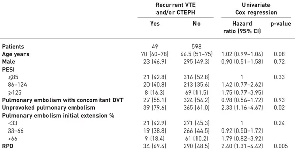
TABLE 4Main characteristics of the study patients by late complications and results of the univariate Cox regression analysis
Recurrent VTE and/or CTEPH Univariate Cox regression Yes No Hazard ratio (95% CI) p-value Patients 49 598 Age years 70 (60–78) 66.5 (51–75) 1.02 (0.99–1.04) 0.08 Male 23 (46.9) 295 (49.3) 0.90 (0.51–1.58) 0.72 PESI ⩽85 21 (42.8) 316 (52.8) 1 0.33 86–124 20 (40.8) 213 (35.6) 1.42 (0.77–2.62) ⩾125 8 (16.3) 69 (11.5) 1.75 (0.77–3.95)
Pulmonary embolism with concomitant DVT 27 (55.1) 324 (54.2) 0.98 (0.56–1.72) 0.93 Unprovoked pulmonary embolism 39 (79.6) 365 (61.0) 2.33 (1.16–4.67) 0.02 Pulmonary embolism initial extension %
<33 21 (42.9) 271 (45.3) 1 0.24
33–66 19 (38.8) 266 (44.5) 0.92 (0.50–1.72)
>66 9 (18.4) 61 (10.2) 1.79 (0.82–3.92)
RPO 34 (69.4) 290 (48.5) 2.40 (1.31–4.42) 0.005
Data are presented as n, median (interquartile range) or n (%), unless otherwise stated. VTE: venous thromboembolism; CTEPH: chronic thromboembolic pulmonary hypertension; PESI: Pulmonary Embolism Severity Index; DVT: deep vein thrombosis; RPO: residual pulmonary obstruction.
TABLE 5Venous thromboembolism (VTE) recurrence and chronic thromboembolic pulmonary hypertension (CTEPH) according to the degree of pulmonary embolism and results of the logistic regression analysis
6-month RPO index % Patients n VTE recurrence and/or CTEPH n (%) OR (95% CI)
<1.00 323 15 (4.6)# 1
1.00–25.00 268 25 (9.3)¶ 2.11 (1.09–4.10)
25.01–72.00 56 9 (16.1)+ 3.93 (1.63–9.50)
72.01–100.00 0
RPO: residual pulmonary obstruction.#: recurrent VTE in all;¶: recurrent VTE in n=20, CTEPH in n=4, both in n=1;+: recurrent VTE in n=3, CTEPH in n=5, both in n=1.
either recurrent VTE or CTEPH [9–11]. The reason for this discrepancy is unclear. Intriguingly, we can speculate that chronic scintigraphic perfusion defects do not simply reflect the presence of mere, persistent thromboembolic material. They are likely to intercept more complex, functional vascular abnormalities. It should be specified, however, that even with the use of the current high-technology scanners, CT angiography is unlikely to be as sensitive as perfusion lung scanning in detecting small peripheral perfusion abnormalities [23–25] and CTEPH can develop in patients with completely recanalised pulmonary arteries, as shown by CT angiography [11]. While its higher specificity makes CT angiography the test of reference for detecting pulmonary embolism in clinically symptomatic patients, lung scintigraphy at 6 months is likely to become the procedure of choice for identification of patients at a higher risk of later serious complications. Of interest, our results were achieved by perfusion lung scanning alone, thus obviating the inconveniences and the economic burden posed by the ventilation scan.
The incidence of recurrent VTE (6.2%) we found in our patients was lower than that reported elsewhere [1, 26, 27], as was that of CTEPH (1.7%) [4, 28–32]. In addition, unexpectedly enough, in only two out of the 11 patients who developed CTEPH was this threatening complication preceded by an episode of recurrent pulmonary embolism. It should be considered, however, that in 237 out of the 647 patients (36.6%) of our cohort, including 162 patients with unprovoked pulmonary embolism, anticoagulation was continued throughout the whole length of follow-up. This decision was taken in agreement with the latest international guidelines [5, 6]. Not surprisingly, therefore, most late complications (42 out of 49) developed in the 410 patients (10.2%) in whom anticoagulation was discontinued, although a substantial proportion of these patients had a transient risk factor for VTE.
A few methodological aspects deserve comment. 1) As the attending physicians were aware of the scintigraphic results, they may have impacted on the decision to prolong the anticoagulant therapy, in such a way confounding the interpretation of our study results. Indeed, the proportion of patients with RPO who received a 3-year therapy was higher than that of patients without it (41% versus 32%) and a higher
0.10 0.11 0.12 0.09 0.07 0.08 0.06 0.04 0.05 0.03 0.02 0.01 0.00 Cumulativ e incidenc e
Time from acute pulmonary embolism months 9 6 12 15 18 21 24 27 30 33 36 307 324 RPO At risk n 299 286 282 273 269 261 255 249 241 322 323 No RPO 318 310 305 299 293 281 274 261 252 RPO No RPO
FIGURE 2 Cumulative incidence of recurrent venous thromboembolism and/or chronic thromboembolic pulmonary hypertension in patients with and without residual pulmonary obstruction (RPO).
TABLE 6Cumulative incidence of recurrent venous thromboembolism (VTE) and chronic thromboembolic pulmonary hypertension (CTEPH) at 1, 12, 24 and 36 months follow-up
Patients with RPO# Patients without RPO
1 month 12 months 24 months 36 months 1 month 12 months 24 months 36 months Recurrent VTE 2.1 (1.0–4.2) 3.7 (2.0–6.2) 6.2 (3.9–9.2) 7.4 (4.9–10.6) 0 (0.0–0.7) 0.9 (0.3–2.5) 3.7 (2.1–6.4) 4.3 (2.5–7.0) CTEPH 1.54 (0.6–3.3) 2.5 (1.2–4.6) 2.5 (1.2–4.6) 3.4 (1.8–5.8) 0 0 0 0 Data are presented as % (95% CI). RPO: residual pulmonary obstruction.#: n=324;¶: n=323.
prevalence of RPO was found in patients treated for >2 years. However, RPO was eventually found to be a powerful and independent risk factor of late complications irrespective of the duration of anticoagulation. Therefore, this potential bias points to the direction of strengthening our conclusions. 2) As we did not collect information on the quality of anticoagulation we are not aware of the impact of this information on the development of recurrent VTE. 3) As we did not mandate the search for thrombophilia or measure D-dimer after stopping anticoagulation, we could not assess their potential role on the development of late complications. 4) As the study was designed when direct oral anticoagulants were not yet available, we can only report on the outcome of patients on conventional anticoagulants. 5) We decided to adopt as the main study end-point a combined outcome (i.e. recurrent VTE and/or CTEPH) rather than assessing each component separately because they have comparable severity and prognostic implications. 6) Patients with previous severe cardiac or pulmonary diseases accounting for the risk of nonthromboembolic pulmonary hypertension were excluded, as were those with previous VTE, those who needed anticoagulation for reasons other than VTE and those who developed recurrent VTE prior to completion of the first 6 months of treatment. Thus, our study conclusions cannot apply to these categories.
In summary, our results suggest that a single assessment of RPO at 6 months can help risk-stratify patients with pulmonary embolism and guide treatment decisions. Elderly patients and those with unprovoked pulmonary embolism are most likely to manifest residual obstruction. Studies addressing the safety of withholding anticoagulation from patients with pulmonary embolism with a negative scintigraphic pattern after 6 months are required, as are studies addressing the impact of the direct oral anticoagulants on the occurrence of RPO and on the development of the long-term complications of pulmonary embolism.
Acknowledgements
The SCOPE investigators are as follows. Executive Committee: Raffaele Pesavento (Padua, Chair), Antonio Pagnan (Padua), Gualtiero Palareti (Bologna), Antonio Palla (Pisa), Vittorio Pengo (Padua), Franco Piovella (Pavia), Paolo Prandoni (Padua). Adjudication Committee: Massimo Miniati (Florence, Chair), Chiara Arcangeli (Pisa), Giorgio De Conti (Padua). Data Handling and Quality Control Committee: Lucia Filippi (Padua, Chair), Davide Ceccato (Padua), Isabella Minotto (Padua). Biostatistical Committee: Anna Chiara Frigo (Padua, Chair), Lucia Filippi (Padua), Raffaele Pesavento (Padua). Participating Centres and Investigators (number of enrolled patients in brackets): Pisa (n=70): Antonio Palla, Letizia Marconi; Padua 1 (n=58): Raffaele Pesavento, Paolo Prandoni, Lucia Filippi; Castelfranco Veneto (n=32): Adriana Visonà, Beniamino Zalunardo, Diego Tonello; Cosenza (n=29): Carlo Bova, Antonio Lanzillotta, Alfonso Noto; Rovigo (n=29): Stefano Cuppini, Marco Marzolo, Domenico Rubello; Milan (n=24): Fernando Porro, Gabriella Gazzano, Giovanni Pagnozzi; Treviso (n=24): Sabina Villalta, Elena Callegari, Matteo Rugolotto; Rome (n=23): Maurizio Ciamaichella, Rosa Maida, Alessandra De Angelis; Faenza (n=22): Eugenio Bucherini, Enrico Carioli, Laura Laghi; Padua 2 (n=22): Giovanni Nante, Giulia Bertozzo; Siena (n=21): Sandra Battistelli, Claudia Baiocchi, Paolo Bertelli; Brescia (n=21): Maria Lorenza Muiesan, Massimo Salvetti, Francesco Marino; Bassano del Grappa (n=20): Giampietro Beltramello, Nadia Xamin, Mario Ernesto De Antoni; Florence (n=20): Domenico Prisco, Daniela Poli; Milan 2 (n=19): Franco Casazza, Maria Garagiola; Varese (n=18): Walter Ageno, Marco Donadini; Bologna (n=18): Gualtiero Palareti, Stefania Cavazza; Parma (n=18): Roberto Quintavalla, Gino degli Antoni; Pisa 2 (n=17): Simonetta Monti; Livorno (n=17): Nicola Mumoli, Jose Vitale; Conegliano Veneto (n=16): Nello Zanatta, Annamaria De Pellegrin; Siena (n=14): Roberto Cappelli, Michele Voglino; Milan 3 (n=14): Marco Cattaneo, Daniela Lambertenghi; San Daniele del Friuli (n=12): Valentino Moretti, Teodora Celino; Vicenza (n=11): Francesco Corà, Katia Mattiello; Turin (n=10): Mario Bazzan, Antonella Vaccarino; Reggio Emilia (n=10): Angelo Ghirarduzzi; Ferrara (n=9): Roberto Manfredini; Mirano (n=6): Ornella Barbato; Perugia (n=6): Leonella Pasqualini; Reggio Emilia 2 (n=5): Mauro Silingardi; Perugia 2 (n=4): Cecilia Becattini; Pavia (n=4): Franco Piovella; Genoa (n=4): Renzo Poggio.
Author contributions: study concept and design: R. Pesavento and P. Prandoni; acquisition, analysis or interpretation of data: all authors; drafting of the manuscript: P. Prandoni, R. Pesavento and L. Filippi; critical revision of the manuscript for important intellectual content: all authors; statistical analysis: A.C. Frigo, R. Pesavento and L. Filippi; administrative, technical or material support: L. Filippi, R. Pesavento and P. Prandoni; study supervision: R. Pesavento, P. Prandoni, A. Palla, V. Pengo, G. Palareti, A. Pagnan and F. Piovella; R. Pesavento had full access to all of the data in the study and takes responsibility for the integrity of the data and the accuracy of the data analysis.
TABLE 7The characteristics of the study patients by late complications and results of the multivariate Cox regression analysis
Multivariate Cox regression#
Hazard ratio (95% CI) p-value
RPO 2.26 (1.23–4.16) 0.009
Unprovoked pulmonary embolism 2.15 (1.07–4.33) 0.03
RPO: residual pulmonary obstruction.#: age, unprovoked pulmonary embolism and RPO were included in the multivariate Cox regression model.
References
1 Prandoni P, Noventa F, Ghirarduzzi A, et al. The risk of recurrent venous thromboembolism after discontinuing anticoagulation in patients with acute proximal deep vein thrombosis or pulmonary embolism. A prospective cohort study in 1626 patients. Haematologica 2007; 92: 199–205.
2 Klok FA, Zondag W, van Kralingen KV, et al. Patient outcomes after acute pulmonary embolism. A pooled survival analysis of different adverse events. Am J Respir Crit Care Med 2010; 181: 501–506.
3 Baglin T, Douketis J, Tosetto A, et al. Does the clinical presentation and extent of venous thrombosis predict likelihood and type of recurrence? A patient level meta-analysis. J Thromb Haemost 2010; 8: 2436–2442.
4 Pengo V, Lensing AW, Prins MH, et al. Incidence of chronic thromboembolic pulmonary hypertension after pulmonary embolism. N Engl J Med 2004; 350: 2257–2264.
5 Kearon C, Akl EA, Ornelas J, et al. Antithrombotic therapy for VTE disease: CHEST guideline and expert panel report. Chest 2016; 149: 315–352.
6 Konstantinides SV, Torbicki A, Agnelli G, et al. 2014 ESC guidelines on the diagnosis and management of acute pulmonary embolism. Eur Heart J 2014; 35: 3033–3069.
7 Ageno W, Samperiz A, Caballero R, et al. Duration of anticoagulation after venous thromboembolism in real world clinical practice. Thromb Res 2015; 135: 666–672.
8 Pesavento R, Filippi L, Pagnan A, et al. Unexpectedly high recanalization rate in patients with pulmonary embolism treated with anticoagulants alone. Am J Respir Crit Care Med 2014; 189: 1277–1279.
9 den Exter PL, van Es J, Kroft LJ, et al. Thromboembolic resolution assessed by CT pulmonary angiography after treatment for acute pulmonary embolism. Thromb Haemost 2015; 114: 26–34.
10 Golpe R, de Llano LA, Castro-Añón O, et al. Long-term outcome of patients with persistent vascular obstruction on computed tomography pulmonary angiography 6 months after acute pulmonary embolism. Acta Radiol 2012; 53: 728–731.
11 Pesavento R, Visonà A, Villalta S, et al. Residual pulmonary obstruction and the risk of late complications in patients with pulmonary embolism. Thromb Res 2016; 137: 228–230.
12 Meneveau N, Ider O, Seronde MF, et al. Long-term prognostic value of residual pulmonary vascular obstruction at discharge in patients with intermediate- to high-risk pulmonary embolism. Eur Heart J 2013; 34: 693–701. 13 Hvid-Jacobsen K, Fogh J, Nielsen SL, et al. Scintigraphic control of pulmonary embolism. Eur J Nucl Med 1988;
14: 71–72.
14 Wartski M, Collignon MA. Incomplete recovery of lung perfusion after 3 months in patients with acute pulmonary embolism treated with antithrombotic agents. J Nucl Med 2000; 41: 1043–1048.
15 Nijkeuter M, Hovens MM, Davidson BL, et al. Resolution of thromboemboli in patients with acute pulmonary embolism: a systematic review. Chest 2006; 129: 192–197.
16 Miniati M, Monti S, Bottai M, et al. Survival and restoration of pulmonary perfusion in a long-term follow-up of patients after acute pulmonary embolism. Medicine 2006; 85: 253–262.
17 Poli D, Cenci C, Antonucci E, et al. Risk of recurrence in patients with pulmonary embolism: predictive role of D-dimer and of residual perfusion defects on lung scintigraphy. Thromb Haemost 2013; 109: 181–186.
18 Qanadli SD, El Hajjam M, Vieillard-Baron A, et al. New CT index to quantify arterial obstruction in pulmonary embolism: comparison with angiographic index and echocardiography. Am J Roentgenol 2001; 176: 1415–1420. 19 Meyer G, Collignon MA, Guinet F, et al. Comparison of perfusion lung scanning and angiography in the
estimation of vascular obstruction in acute pulmonary embolism. Eur J Nucl Med 1990; 17: 315–331.
20 Aujesky D, Roy PM, Le Manach CP, et al. Validation of a model to predict adverse outcomes in patients with pulmonary embolism. Eur Heart J 2006; 27: 476–481.
21 Bernardi E, Camporese G, Büller HR, et al. Serial 2-point ultrasonography plus D-dimer vs whole-leg color-coded Doppler ultrasonography for diagnosing suspected symptomatic deep vein thrombosis: a randomized controlled trial. JAMA 2008; 300: 1653–1659.
22 Planquette B, Ferré A, Peron J, et al. Residual pulmonary vascular obstruction and recurrence after acute pulmonary embolism. A single center cohort study. Thromb Res 2016; 148: 70–75.
23 Tunariu N, Gibbs SJ, Win Z, et al. Ventilation–perfusion scintigraphy is more sensitive than multidetector CTPA in detecting chronic thromboembolic pulmonary disease as a treatable cause of pulmonary hypertension. J Nucl Med 2007; 48: 680–684.
24 Cosmi B, Nijkeuter M, Valentino M, et al. Residual emboli on lung perfusion scan or multidetector computed tomography after a first episode of acute pulmonary embolism. Intern Emerg Med 2011; 6: 521–528.
25 van Es J, Douma RA, Kamphuisen PW, et al. Clot resolution after 3 weeks of anticoagulant treatment of pulmonary embolism: comparison of computed tomography and perfusion scintigraphy. J Thromb Haemost 2013; 11: 679–685.
26 Agnelli G, Prandoni P, Becattini C, et al. Extended oral anticoagulant therapy after a first episode of pulmonary embolism. Ann Intern Med 2003; 139: 19–25.
27 Couturaud F, Sanchez O, Pernod G, et al. Six months vs extended oral anticoagulation after a first episode of pulmonary embolism: the PADIS-PE randomized clinical trial. JAMA 2015; 314: 31–40.
28 Ribeiro A, Lindmarker P, Johnsson H, et al. Pulmonary embolism: one-year follow-up with echocardiography Doppler and five-year survival analysis. Circulation 1999; 99: 1325–1330.
29 Barros A, Baptista R, Nogueira A, et al. Predictors of pulmonary hypertension after intermediate-to-high risk pulmonary embolism. Rev Port Cardiol 2013; 32: 857–864.
30 Guerin L, Couturaud F, Parent F, et al. Prevalence of chronic thromboembolic pulmonary hypertension after acute pulmonary embolism. Prevalence of CTEPH after pulmonary embolism. Thromb Haemost 2014; 112: 598–605. 31 Kayaalp I, Varol Y, Cimen P, et al. The incidence of chronic thromboembolic pulmonary hypertension secondary
to acute pulmonary thromboembolism. Tuberk Toraks 2014; 62: 199–206.
32 Yang S, Yang Y, Zhai Z, et al. Incidence and risk factors of chronic thromboembolic pulmonary hypertension in patients after acute pulmonary embolism. J Thorac Dis 2015; 7: 1927–1938.