2319
—
4
16
with Crohn’s Disease: A Description of 2 Cases
Studied with a Novel Magnetic Resonance
Enterography (MRE) Procedure
ADE 1
Giuseppe Cicero
BE 1
Tommaso D’Angelo
DF 1
Antonio Bottari
BD 2
Giuseppe Costantino
F 1
Carmela Visalli
A 1Sergio Racchiusa
B 1Maria Adele Marino
D 1Marco Cavallaro
F 1
Luciano Frosina
ADE 1
Alfredo Blandino
ABE 1
Silvio Mazziotti
Corresponding Author: Giuseppe Cicero, e-mail: [email protected]
Conflict of interest: None declared
Case series
Patients: Female, 23 • Female, 27
Final Diagnosis: SMA syndrome
Symptoms: Abdominal pain • vomiting
Medication: —
Clinical Procedure: —
Specialty: Radiology
Objective: Rare co-existance of disease or pathology
Background: Superior mesenteric artery syndrome is caused by vascular compression of the third portion of the duodenum between the aorta and the superior mesenteric artery. It may occur with acute or chronic symptomatology, such as vomiting or postprandial abdominal pain, and it is usually caused by a lack of mesenteric fat pad un-der conditions of severe weight loss. Crohn’s disease can be one of them.
Case Reports: We report 2 cases of Crohn’s disease patients with clinical suspicion of jejunal stricture who underwent MR-enterography with a novel approach. In fact, the examinations were performed including prone position of the patients inside the scanner, drinking of contrast medium during the examination, and prompt acquisition of fluoroscopic sequences. Both the exams showed an abrupt termination of the duodenum on its third portion and a decreased aortomesenteric distance, allowing the diagnosis of superior mesenteric artery syndrome.
Conclusions: A correlation between Crohn’s disease and superior mesenteric artery syndrome has never before been report-ed in the literature. The present study provides some practical steps that may be useful in order to improve MRE standard protocol in recognizing this condition while evaluating Crohn’s disease bowel lesions.
MeSH Keywords: Crohn Disease • Magnetic Resonance Imaging • Superior Mesenteric Artery Syndrome
Abbreviations: MRE – magnetic resonance enterography; SMA – Superior mesenteric artery; CD – Crohn’s disease; CDAI – Crohn’s disease activity index; CECT – Contrast-enhanced computed tomography
Full-text PDF: https://www.amjcaserep.com/abstract/index/idArt/908273 Authors’ Contribution: Study Design A Data Collection B Statistical Analysis C Data Interpretation D Manuscript Preparation E Literature Search F Funds Collection G
1 Section of Radiological Sciences – Department of Biomedical Sciences and Morphological and Functional Imaging, University of Messina, Policlinico “G. Martino”, Messina, Italy
2 Clinical Unit for Chronic Bowel Disorders – Department of Clinical and Experimental Medicine, University of Messina Policlinico “G. Martino”, Messina, Italy
Background
Superior mesenteric artery (SMA) syndrome, also called Wilkie’s syndrome, is an uncommon but well recognized clinical entity characterized by compression of the third portion of the duo-denum between the aorta and SMA, causing a chronic, inter-mittent or acute duodenal obstruction that can be complete or partial. SMA syndrome has mainly been described in 2 pa-tient populations: papa-tients who have undergone surgery, re-sulting in loss or distortion of the normal retroperitoneal fat (i.e., scoliosis surgery, aortic aneurysm repair, or bowel resec-tion surgery) and patients who have had severe, rapid weight loss for any number of reasons [1]. Even if both these condi-tions are often concomitant in patients affected by Crohn’s disease (CD), a correlation between CD and SMA syndrome has never been reported.
In fact, according to randomized controlled trials, up to one-third of patients with CD currently undergo major abdominal surgery within 5 years [2]. In addition, weight loss is one of the most common signs of CD, representing a severe problem in patients who have undergone extensive surgical resections (e.g., short bowel syndrome) or with severe Crohn’s Disease Activity Index (CDAI).
Although CT-scan still represent the criterion standard imag-ing modality in diagnosimag-ing SMA syndrome, MR-enterography (MRE), which is a radiation-free imaging modality already in-cluded in the standard diagnostic algorithm of inflammatory bowel disease, could be helpful in recognizing the onset of SMA syndrome while evaluating intestinal Crohn’s disease lesions. We report on 2 CD patients who underwent MRE for clinical suspicion of jejunal stricture. MRE, performed with a novel tech-nical approach, demonstrated the finding of SMA syndrome, never reported in the Crohn’s literature up to now.
MRE – technique
Patients who undergo MRE of the small bowel can be exam-ined by drinking 1500–2000 ml of a polyethylene glycol solu-tion administered within 45 min before the beginning of the scan. On the other hand, intestinal distension can be obtained by injecting the contrast medium through a nasojejunal cath-eter (MR-enteroclysis). This latter procedure is more invasive and is often associated with patient discomfort, and it is ac-tually considered optimal in case of jejunal stricture as it al-lows a better distension of the proximal intestinal segments. In our institution, we developed a novel and “hybrid” proce-dure of MRE that we have been performing in patients with clinical suspicion of duodenal or jejunal stricture. In this pro-cedure, the protocol starts with MR-fluoroscopic sequences.
The patient is asked to lie in prone position inside the scanner and to drink the oral contrast agent through a drinking straw for the whole duration of MR-fluoroscopy (Figure 1).
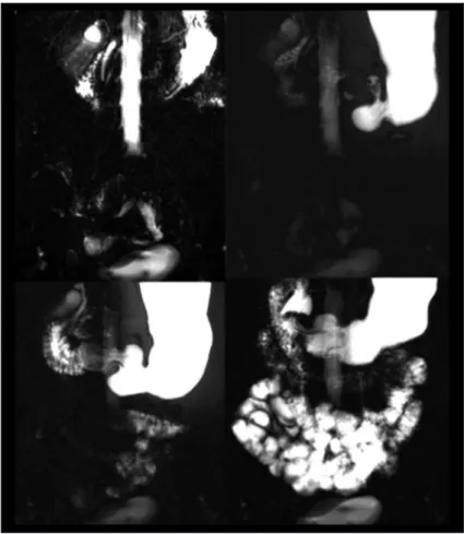
MR-fluoroscopy is performed with several T2-weighted thick-slab turbo spin-echo sequences (TR/TE: ∞/900 ms), usually em-ployed in MR-cholangiopancreatography and MR-pyelography. Images are acquired on the coronal plane (section thickness: 100–180 mm) in order to include the gut and the stomach, and are obtained during and after the oral administration of contrast material, every 1 or 2 min (Figure 2).
When no strictured bowel tract is detected, the fluoroscopic-like images are then used as a guide for assessing the most adequate proximal bowel distension prior to beginning the standard MRE protocol. On the other hand, in case of abnor-mally and persistently dilated bowel segments followed by nar-rowed intestinal tracts, MR-fluoroscopic images are used as landmarks for targeting the following standard MRE sequences. The standard protocol includes various T2-weighted pulse se-quences along axial and coronal planes. High-resolution ultra-fast sequences, such as true-ultra-fast imaging with steady-state (True-Fisp; TR/TE: 4.20/2.10 ms, FA: 60°) and half-Fourier ac-quisition single-shot turbo-spin-echo (HASTE; TR/TE: ∞/80 ms) with and without fat-suppression, are usually performed, to-gether with diffusion-weighted imaging (DWI) sequences, us-ing a diffusion factor b fixed at 0, 400, and 800 s/mm2. DWI
helps to identify segments of bowel affected by CD and to as-sess disease activity [3].
When CD activity is challenging to assess, fat-suppressed three-dimensional gradient-echo images (THRIVE; TR/TE: minimum/ minimum, FA: 10–15°) are added to the protocol either before and after intravenous gadolinium-based contrast material injection.
Figure 1. Picture showing the patient, in prone position inside the MR scanner, drinking oral contrast agent from a drinking straw during MR-fluoroscopy.
Case Report
Case 1
A 23-year-old female, with a diagnosis of CD A2L4bB2G0 ac-cording to the Paris classification [4], presented at our insti-tution with 8-month history of diarrhea and abdominal pain, associated with recurrent postprandial stabbing epigastric pain, nausea, and vomiting. She also stated a weight loss of 8 kg since the symptoms had begun, and she had been treat-ed with adalimumab and azathioprine for the last 6 months but without relief of epigastric pain and nausea.
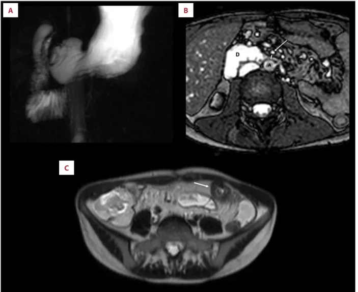
A MRE was performed with the clinical suspicion of proximal bowel obstruction. MR-fluoroscopy showed a persistently dilat-ed proximal duodenum with an abrupt termination at its third portion and mild gastric distention (Figure 3A). Conventional MRE sequences confirmed the presence of a grossly dilated duodenum abruptly narrowed under SMA and revealed an aor-tomesenteric distance of 4 mm (normal values: 10–28 mm) (Figure 3B, 3C). In addition, on delayed MRE sequence acquisi-tion, obtained when the small bowel was sufficiently distend-ed, wall-thickening of several bowel segments was also pres-ent (Figure 3D). Subsequpres-ently, a gastroduodenal endoscopy, performed to exclude the presence of strictures or ulcerations,
revealed only a pulsatile compression of the third portion of the duodenum, further corroborating the diagnosis of SMA syndrome. The patient was managed with adalimumab and high-caloric enteral nutrition and gained 5 kg in 2 months, re-porting a partial relief of symptoms.
Case 2
A 27-year-old female was first diagnosed with CD at age 16 years. Her phenotype was A1bL4bB2B3G1p according to the Paris classification [4]. She presented at our institution with a 10-month history of fatigue associated with an intermit-tent postprandial dull epigastric pain, accompanied by inter-mittent non-bloody vomiting, early satiety, post-prandial full-ness, and severe malnutrition (body mass index: 14.02 Kg/m2).
She had a history of multiple bowel resections, with an esti-mated residual small-bowel length of 170 cm. She was origi-nally on treatment with adalimumab and, after the worsening of symptoms, she had been managed with infliximab for the prior 12 months, without any clinical improvement. Physical examination revealed fullness in the epigastric region. After a gastroenterological consultation, she was suggested to un-dergo MRE to evaluate the presence of possible intestinal seg-ments of narrowing.
Figure 2. MR-Fluoroscopy shows normal progressive filling of the stomach, duodenum and small-bowel.
MR-fluoroscopy showed dilated proximal duodenum with mark-edly slow passage of contrast beyond the third portion, with a cut-off in adjacency of SMA (Figure 4A). MRE also confirmed a persistently dilated duodenum prior to its passage under SMA. The aorto-mesenteric distance measured at the level of the third portion of the duodenum was markedly reduced (4 mm; normal values: 10–28 mm) (Figure 4B). In addition, on delayed
MRE sequence acquisition, wall-thickening of some residual il-eal segments was also present (Figure 4C).
The patient began treatment with metronidazole and predni-sone and was managed with parenteral nutrition and gastric decompression via a nasogastric tube, followed by placement of a Dobbhoff catheter for post-pyloric feeding with Modulen-B
A
C
B
D
Figure 3. MRE images in a 23-year-old CD patient with symptoms of abdominal distention and vomiting. MR-Fluoroscopy image (A) obtained along the coronal plane shows abrupt vertical compression of the third portion of duodenum (arrowheads) and proximal dilatation (asterisk). T2-weighted axial HASTE image (B) from the same study shows a dilated proximal duodenum (D) that abruptly narrows as it travels between the aorta (A) and SMA (arrow); aortomesenteric distance measures 4 mm (line). Sagittal True-Fisp image shows a marked reduction of the angle between aorta (a) and SMA (arrows) (C). Delayed T2-weighted coronal HASTE image shows multiple inflamed bowel segments (arrows) (D). S – stomach; G – gallbladder.
1000 kcal/die (Nestlé, Lausanne, Switzerland). She tolerat-ed oral diet 5 weeks later and starttolerat-ed to gain weight (BMI: 15.62 Kg/m2), with partial relief of symptoms.
Discussion
SMA syndrome was first described comprehensively by Wilkie. It is caused by the vascular compression of the third portion of the duodenum between the aorta and SMA [5].
Typically, patients present postprandial epigastric pain, nau-sea, and gastric distension. These symptoms are characteris-tically described as being partially relieved by lying prone or
in the left lateral decubitus position, because these manoeu-vres release tension on the small-bowel mesentery and thus on the aortomesenteric angle [6]. Identification can be a di-agnostic dilemma and it is frequently made by exclusion, as SMA syndrome is rarely taken into account at first presentation of symptoms because of its low prevalence (0.013–0.3%) [7]. There is a slight female preponderance (64–66%), with 75% of cases occurring in individuals who range in age from 10 to 39 years [8, 9]. The incidence is higher in patients with severe weight loss (e.g., chronic debilitating disease or acute catabolic state) or history of surgery (e.g., scoliosis surgery, aortic aneu-rysm repair, or bowel resection surgery), as both these condi-tions may be responsible for a possible consequent distortion
A
C
B
Figure 4. MRE images in a 27-year-old CD patient with history of multiple bowel resections and intermittent postprandial epigastric pain, vomiting, and severe malnutrition. MR-Fluoroscopy image obtained along the coronal plane shows abrupt vertical compression of the third portion of duodenum and proximal dilatation (A). Axial True-Fisp image from the same study depicts a grossly dilated proximal duodenum (D) that abruptly narrows as it travels between the aorta (A) and SMA (arrow). Aortomesenteric distance measures 4 mm (line) (B). Delayed T2-weighted axial HASTE image shows an inflamed bowel segment (arrow) (C).
of the normal retroperitoneal fat with an increased mesenter-ic tension and caudal pull of SMA [7]. In fact, the duodenum is normally separated from the SMA and abdominal aorta by adipose tissue, which serves as a natural cushion to prevent extrinsic compression [8].
Although a direct association between SMA syndrome and CD has never been reported up to now, CD is a well-known condi-tion that may cause abdominal pain, malabsorpcondi-tion of nutri-ents with weight loss, and intestinal obstruction, potentially occurring in any part of the digestive tract. In addition, ac-cording to Bougen and Peyrin-Biroulet, the rate of surgery at 5 years in patients affected by CD ranges between 25% and 33% [2]. Given the high risk of multiple bowel resection surger-ies, it is not uncommon for these patients to experience short-bowel syndrome and severe weight loss. For this reason, close monitoring of CD patients is mandatory, as it can help identi-fy patients at risk for disease relapses or complications [10]. Since the last decade, CD patients have benefitted from the advances of several imaging techniques, especially MRE. This technique provides data regarding the location, extent of bow-el disease, and disease phenotype (i.e., fibrostenosing, in-flammatory, or perforating) and is accurate for the detection of strictures, extra-luminal complications, and associated dis-eases. However, among complications of CD, SMA syndrome has never been described so far [11–13]. On the other hand, MRE has shown the advantage not only to depict anatomical abnormalities of the small-bowel but also to detect its func-tional disorders. In fact, the use of rapid dynamic thick-slab sequences (e.g., 70–180 mm), a technique also known as MR-fluoroscopy, allows instilling the oral contrast agent with a si-multaneous MR-guide, until good distention of jejunal loops is obtained. Real-time MR-fluoroscopic studies of the small bow-el have been adopted only in MR-enteroclysis so far. In this procedure, intestinal distention is achieved via administration of contrast material through a prepositioned nasojejunal bal-loon-tipped catheter, with serious procedure-related patient discomfort [14]. For this reason, the use of MR-enteroclysis has been limited in routine clinical practice, as opposed to the rel-atively discomfort-free MRE.
In our institution, we have recently introduced a novel proce-dure for performing MRE, which has mainly been employed in the clinical suspicion of proximal bowel stricture, with the aim to further improve the visualization of jejunal loops. This novel procedure requires the prone position of the patient in-side the scanner and the drinking of the oral contrast medium through a drinking straw while acquiring the fluoroscopic se-quences. This allows monitoring not only the small-bowel filling, but also the gastroduodenal emptying, providing information about the peristalsis efficiency and better depicting the prox-imal jejunal tract. In case of delayed emptying of an intestinal
segment that is followed by a narrowed tract, the convention-al MRE sequences are then focused on the region of interest. Up to now, the radiological diagnosis of duodenal obstruction in SMA syndrome has been achieved by means of convention-al contrast fluoroscopy, contrast-enhanced computed tomog-raphy (CECT), and angiogtomog-raphy.
Conventional fluoroscopy usually demonstrates the dilatation of the proximal duodenum followed by a sharp narrowing as the duodenum travels underneath the SMA. On the other hand, CECT imaging has demonstrated that the 2 key signs of SMA syndrome are represented by an aortomesenteric distance of less than 8 mm and by an aortomesenteric angle of less than 22°. The former sign has a sensitivity and specificity of 100% for SMA syndrome, whereas the sensitivity and specificity of the latter are 42.8% and 100%, respectively [15].
In both our cases, MR-fluoroscopy showed a persistently di-lated proximal duodenum with an abrupt narrowing at its third portion and with a mild gastric distention, findings con-sistent with what is seen on conventional contrast fluorosco-py in the SMA syndrome [15]. In addition, MRE revealed also the reduction of the aortomesenteric distance, the major sign of SMA syndrome.
However, MRE is a radiation-free exam whose role is well-es-tablished in the assessment and management of CD patients but not in evaluating SMA syndrome.
In fact, even if it allows an overall view of the abdominal cav-ity and the addition of fluoroscopy sequences can further im-prove its usefulness in highlighting the presence of an upper intestinal tract obstruction, measurement in the prone posi-tion may underestimate the aortomesenteric angle.
Nevertheless, consideration should be given to the fact that MRE could yield additional important information, with a high degree of accuracy, to confirm or dismiss the suspicion of SMA syndrome. Of course, the final diagnosis should be achieved through other imaging modalities (e.g., gastroscopy or CT-scan), which therefore would be performed in a wiser and more advisable way.
To the best of our knowledge, this is the first report in which the SMA syndrome has been detected by means of MR in a CD patient, and it is noteworthy to highlight the role of real-time MR-fluoroscopy in detecting this syndrome prior to per-forming MRE. In fact, it would have hardly been demonstra-ble either by means of conventional MRE (because distention of the duodenum and proximal jejunum is usually poor at the time of scanning) and MR-enteroclysis (because the nasojejunal catheter might go through the duodenal stricture). Moreover,
a clear distinction between an extrinsic compression and the presence of a duodenal or jejunal stricture is crucial, as this latter phenotype is strongly associated with an increased risk of relapse and complicated disease [16].
Both these latter points might represent important pitfalls to avoid when performing MR of the small bowel in CD patients, especially in those with clinically suspected proximal small-bowel disease.
Conclusions
In conclusion, either gastroenterologists and radiologists should be aware of the possible association of SMA syndrome with CD in order to reduce the symptoms and to prevent inappro-priate or aggressive treatments. The prone position and the dynamic evaluation allowed by fluoroscopic sequences dur-ing simultaneous drinkdur-ing of the oral contrast medium can be useful tools to add to the standard MRE protocol in order to recognize the presence of this pathological entity while eval-uating CD small-bowel lesions.
Conflict of interest
None.
References:
1. Raman SP, Neyman EG, Horton KM et al: Superior mesenteric artery syn-drome: Spectrum of CT findings with multiplanar reconstructions and 3-D imaging. Abdom Imaging, 2012; 37: 1079–88
2. Bouguen G, Peyrin-Biroulet L: Surgery for adult Crohn’s disease: What is the actual risk? Gut, 2011; 60: 1178–81
3. Morani AC, Smith EA, Ganeshan D, Dillman JR: Diffusion-weighted MRI in pe-diatric inflammatory bowel disease. Am J Roentgenol, 2015; 204: 1269–77 4. Levine A, Griffiths A, Markowitz J et al: Pediatric modification of the Montreal
classification for inflammatory bowel disease: The Paris classification. Inflamm Bowel Dis, 2011; 17(6): 1314–21
5. Wilkie D: Chronic duodenal ileus. Am J Med Sci, 1927; 173: 643–49 6. Merrett ND, Wilson RB, Cosman, P, Biankin AV: Superior mesenteric artery
syndrome: Diagnosis and treatment strategies. J Gastrointest Surg, 2009; 13: 287–92
7. Welsch T, Büchler MW, Kienle P: Recalling superior mesenteric artery syn-drome. Dig Surg, 2007; 24: 149–56
8. Lee TH, Lee JS, Jo Y et al: Superior mesenteric artery syndrome: Where do we stand today? J Gastrointest Surg, 2012; 16: 2203–11
9. Biank V, Werlin S: Superior mesenteric artery syndrome in children: A 20-year experience. J Pediat Gastroenterol Nutr, 2006; 42: 522–25
10. Sauter B, Beglinger C, Girardin M et al: Monitoring disease activity and pro-gression in Crohn’s sisease. A swiss perspective on the IBD ahead ‘opti-mised monitoring’ recommendations. Digestion, 2014;89: 299–309 11. Horsthuis K, Bipat S, Bennink RJ, Stoker J: Inflammatory bowel disease
di-agnosed with US, MR, scintigraphy, and CT: Meta-analysis of prospective studies. Radiology, 2008; 247: 64–79
12. Mazziotti S, Blandino A, Scribano E et al: MR enterography findings in ab-dominopelvic extraintestinal complications of Crohn’s disease. J Magn Reson Imaging, 2013; 37: 1055–63
13. Mazziotti S, D’Angelo T, Racchiusa S et al: Peritoneal inclusion cysts in pa-tients affected by Crohn’s disease: Magnetic resonance enterography find-ings in a case series. Clin Imaging, 2016; 40: 152–55
14. Siddiki H, Fidler J: MR imaging of the small bowel in Crohn’s disease. Eur J Radiol, 2009; 69: 409–17
15. Fong JK, Poh AC, Tan AG, Taneja R: Imaging findings and clinical features of abdominal vascular compression syndromes. Am J Roentgenol, 2014; 203: 29–36
16. Flamant M, Trang C, Maillard O et al: The prevalence and outcome of je-junal lesions visualized by small bowel capsule endoscopy in Crohn’s dis-ease. Inflamm Bowel Dis, 2013; 19: 1390–96