R E S E A R C H
Open Access
Clinical evaluation of outdoor cats exposed
to ectoparasites and associated risk for
vector-borne infections in southern Italy
Maria Flaminia Persichetti
1, Maria Grazia Pennisi
2*, Angela Vullo
1, Marisa Masucci
2, Antonella Migliazzo
1and Laia Solano-Gallego
3Abstract
Background: Cats can be carriers of infected arthropods and be infected with several vector-borne pathogens (VBP) but there is limited knowledge about their pathogenic role in cats.
Results: A cross-sectional controlled study investigated the clinical status and antibody (Bartonella henselae, Rickettsia conorii, Ehrlichia canis, Anaplasma phagocytophilum, Babesia microti and Leishmania infantum) and/or blood PCR (Mycoplasma spp., Bartonella spp., Rickettsia spp., Ehrlichia/Anaplasma spp., piroplasmids, L. infantum, Hepatozoon felis) prevalence in 197 cats. Outdoor cats lacking ectoparasiticide treatment or hosting ectoparasites (study group [SG], n = 134) and indoor cats treated against ectoparasites (control group [CG], n = 63) were enrolled. Clinical data and retroviral co-infections were compared between the two groups. Multivariable analysis tested associations between variables and VBP exposure. Lymphadenia, stomatitis, and various haematological abnormalities were statistically more frequent in SG. Antibodies against R. conorii, B. henselae, A. phagocytophylum, B. microti, E. canis and L. infantum were detected. Bartonella henselae, Bartonella clarridgeiae, Mycoplasma haemofelis,“Candidatus Mycoplasma haemominutum” and “Candidatus Mycoplasma turicensis” DNA were identified. Very high antibody (87.8%) and PCR (40.1%) positivity to at least one pathogen were detected and were significantly higher in SG. Co-infections were confirmed in about one-third of the cats and were more frequent in SG cats. Molecular and overall (antibody and PCR) positivity to Bartonella and antibody positivity to R. conorii were higher in SG. Multivariable analysis found significant associations of Bartonella spp. infection with Feline Immunodeficiency Virus (FIV) infection and increased globulins, and of Mycoplasma spp. infection with adult age, FIV infection, anaemia, and increased creatinine.
Conclusions: A very high prevalence of exposure to zoonotic VBP was found in cats, with Rickettsia and Bartonella infections being most prevalent. Some risk factors were documented namely for Mycoplasma spp. and Bartonella spp. The lifestyle of cats is clinically relevant and requires specific preventative measures to protect their health.
Keywords: Cat, Vector-borne pathogens, Zoonosis, Risk factor, Ectoparasite, Outdoor lifestyle, Indoor lifestyle Background
Vector-borne infections (VBI) are caused by parasites, bac-teria or viruses transmitted by hematophagous arthropods, and many of them are of zoonotic concern [1–6]. Cats have a high likelihood of ectoparasite exposures when living an outdoor lifestyle and there is a lack of preventive treatment with acaricides. Consequently, these animals can be carriers
of infected arthropods and be infected with several vector-borne pathogens (VBP), as observed in dogs [1,3–5,7–11]. The lack of knowledge on the pathogenic role of most of these VBP in cats may limit the diagnosis of vector-borne diseases (VBD). Also, clinical signs and laboratory abnor-malities associated with VBD are widely variable and non-specific [1, 3, 4]. Moreover, concurrent VBI or retroviral infections can be found, which may influence the clinical course and outcome of VBD in cats [3,12].
Recent literature highlighted some risk factors associ-ated with cat positivity to VBI such as multi-cat * Correspondence:[email protected]
2Dipartimento di Scienze Veterinarie, Università degli Studi di Messina, Polo
Universitario Annunziata, 98168 Messina, Italy
Full list of author information is available at the end of the article
© The Author(s). 2018 Open Access This article is distributed under the terms of the Creative Commons Attribution 4.0 International License (http://creativecommons.org/licenses/by/4.0/), which permits unrestricted use, distribution, and reproduction in any medium, provided you give appropriate credit to the original author(s) and the source, provide a link to the Creative Commons license, and indicate if changes were made. The Creative Commons Public Domain Dedication waiver (http://creativecommons.org/publicdomain/zero/1.0/) applies to the data made available in this article, unless otherwise stated.
household, outdoor access, male gender, FIV positivity,
and abortive FeLV infection [3, 4, 6, 12]. Preventative
control measures against ectoparasites infestation, i.e. regular individual use of ectoparasiticide formulations, seem the most effective tool to prevent infection in cats, and other hosts [3, 4, 12]. The present controlled study evaluated the prevalence and risk factors for some VBP in cats exposed to ectoparasites in southern Italy and assessed the impact of the infections on their health status.
Methods
Study sites, cat enrolment and sampling procedures A total of 197 cats were enrolled from March 2012 to March 2013 at four veterinary clinics located in Sicily (n = 39) (Veterinary Teaching Hospital, Università degli Studi di Messina, Messina and Ambulatorio Veterinario S. Lucia, Lipari-Messina) and Calabria (n = 158) (Clinica Veterinaria Camagna, Reggio Calabria and Ambulatorio Dr Cardone, Gioia Tauro-Reggio Calabria). Cats aged > 6 months and experiencing at least an intere vector-season since birth (April-October) were recruited irrespective of breed and gender. Most cats (n = 144; 73%) were admitted for elective surgery or annual health check. They were enrolled when the following information was available: type of housing and lifestyle and individual application of ectoparasiticides. According to this information and the occurrence of ecto-parasites at physical examination, two groups of cats were considered. The study group (SG, n = 134) included cats with a greater chance of exposure to ectoparasites, i.e. outdoor cats with a lack of regular individual ectoparasiti-cide treatment and having ectoparasites at enrolment. The control group (CG, n = 63) was composed of indoor cats with no evidence of ectoparasites at enrolment, receiving the appropriate ectoparasiticide treatment, and therefore with a low risk for ectoparasites. Cats living in rescue catteries were excluded from this study.
Clinical history and physical examination findings for cats were registered in a clinical form. Also, information on region, age, sex, breed, lifestyle, ectoparasiticide treatments, flea and tick presence was included. Cats were classified as “young” if they had experienced only one vector-season since birth, and“adult” if they had experienced more than one vector-season.
From each cat, blood, conjunctival and oral swabs were obtained. One millilitre of blood was placed into one tube with EDTA and used within 24 hours for complete blood count (CBC) and subsequently stored at -20 °C until further use for molecular investigations. Left-over blood (about 2 ml) was used to perform blood smears (immediately) and to obtain serum after clotting into a dry tube. Blood serum was stored at -20 °C until further use for haematological and serological investigations. Urine samples were obtained by cystocentesis when possible and used for urinalysis
within 2 hours and urine protein and creatinine ratio (UPC) within 24 h from collection. When enlarged lymph nodes were observed, a fine needle aspiration was per-formed. Sealed needles and swabs were stored at -20 °C until further use for molecular tests.
Hematological investigations and urinalysis
Complete blood count was performed using a laser haema-tology analyser (IDEXX ProCyte Dx® Hemahaema-tology Analyser, Idexx Laboratories, Westbrook, Maine, USA). Reference in-tervals of CBC are listed in Additional file 1: Table S1. Blood smears were stained by May Grünwald-Giemsa staining and examined for haematological abnormalities and presence of hemoparasites [13].
A biochemical profile including creatine kinase (CK), aspartate aminotransferase (AST), alanine aminotrans-ferase (ALT), alkaline phosphatase (ALP), gamma gluta-myl transferase (GGT), cholinesterase, total bilirubin, total proteins, albumin, globulin, albumin/globulin ratio,
cholesterol, triglycerides, urea, creatinine, glucose,
calcium, phosphorus, magnesium, sodium, potassium,
sodium/potassium ratio, chloride, correct chloride,
serum iron, unsatured iron-binding capacity (UIBC), total iron-binding capacity (TIBC), transferrin satur-ation, and serum amyloid A (SAA) was performed at the Laboratorio di analisi veterinarie San Marco (Padova, Italy) and reference intervals of the above parameters are shown in Additional file2: Table S2.
Urinalysis was performed using Combur 9 Test strips (Roche Diagnostics, Indianapolis, Indiana, USA), Vet 360 refractometer (Reichert, Seefeld, Germany) and a micro-scopic evaluation of urine sediment was performed using the Kova system (Kova International, Garden Grove, CA, USA). Urine protein and creatinine ratio were eval-uated with Catalyst Dx® Chemistry Analyzer (Idexx La-boratories, Westbrook, Maine, USA), using 0.4 as the cut-off value for proteinuria [14].
Serological investigations
All cats were tested for feline leukaemia virus (FeLV) anti-gen and antibodies to feline immunodeficiency virus (FIV) by a rapid enzyme-linked immunosorbent assay (ELISA) (SNAP Combo Plus FeLV ag/FIV ab test, Idexx Laborator-ies, Westbrook, Maine, USA). Sera from individual cats were also tested for the presence of immunoglobulin G (IgG) antibodies against Bartonella henselae, Rickettsia con-orii, Ehrlichia canis, Babesia microti and Anaplasma
pha-gocytophilum antigens by the immunofluorescence
antibody test (IFAT) using commercial kits (Fuller Labora-tories Fullerton, California, USA). The manufacturer’s protocol was followed for all serological tests using a cut-off dilution of 1:64 for B. henselae, R. conorii and B. microti; and 1:50 for E. canis and A. phagocytophilum. The presence of L. infantum IgG antibodies was investigated by IFAT
according to Persichetti et al. [15] and the cut-off value was established at 1:80 as previously described [15]. Titres of positive samples were determined for all pathogens, with the exception of B. microti, and considered high when they were at least four times over cut-off value.
Molecular investigations
Quantitative real-time PCR (qPCR) technology and sequencing were applied as described elsewhere [10], to detect specific DNA markers for Ehrlichia/Anaplasma spp., piroplasmids (Babesia spp. and Theileria spp.), Hepatozoon felis, hemotropic Mycoplasma spp., Rickettsia spp., Bartonella spp. and L. infantum from feline blood samples, while infections by the latter pathogen were also diagnosed from conjunctival and oral swabs, urine, and lymph node specimens. Species-specific qPCRs were also performed as described by Martinez et al. [16] to discrim-inate among feline hemoplasmas species (Mycoplasma
haemofelis, “Candidatus Mycoplasma haemominutum”
and“Candidatus Mycoplasma turicensis”).
Statistical analysis
The Kolmogorov-Smirnov normality test was used to as-sess the normality of distribution of the continuous vari-able age. Statistical differences between SG and CG cats were tested for significance by Fisher’s exact test for cat-egorical variables and with the Mann-Whitney test for numerical variables using the GraphPad InStat v3.05 for Windows 95 (GraphPad Software Inc., San Diego Cali-fornia, USA, 2000). Differences were considered signifi-cant if the P-value was < 0.05. Independent variables that yielded significant differences between the 2 groups were analysed in the overall 197 cats with multivariable logistic regression analysis using the STATA 9.2 software (StataCorp LP, College Station, Texas) to test for possible risk factors associated with investigated VBP. According to the molecular and antibody detection, outcome vari-ables were as follows: L. infantum (molecular and anti-body tests), Bartonella spp. (molecular and antianti-body tests), Mycoplasma spp. (molecular test), R. conorii (anti-body test), E. canis (anti(anti-body test), B. microti (anti(anti-body test), A. phagocytophilum (antibody test). Dichotomous variables (outcome variable and investigated risk factors) were analysed with multivariable logistic regression ana-lysis, which was applied for each pathogen. Also, an or-dered logistic regression model was applied to the variable response co-infection (i.e. exposure to more than one VBP) and the same potential risk factors were considered for single pathogens. The variable co-infection was measured on an ordinal scale as it considers the number of infections present simultaneously in a cat under investigation (no infection, one infection,≥ 2 in-fections). P-values with an odds ratio (OR) and 95% confidence interval (CI) of multivariable analyses were
obtained. An OR value > 1 implies a positive associ-ation between independent and dependent variables, while an OR < 1 implies an inverse association.
Results
Clinical evaluation
Cats were aged between 5 months and 19 years (median 2 years, 25th percentile 1 year, 75th percentile 5 years); 69 (35%) were males and 128 females (65%); eight pedigree cats were enrolled (five Persians and three Carthusians in the CG). The SG median age (2 years, 25th percentile 0.9 years, 75th percentile 4 years) was significantly lower com-pared to that of the CG (3 years, 25th percentile 1 year, 75th percentile 8 years) (Mann-Whitney U-test: U(195)=
3387, Z = -2.23327, P = 0.0257). Moreover, a significantly higher percentage of SG cats (79%) were admitted for elect-ive surgery or annual health check compared to the CG (60%) (Fisher’s exact test: P = 0.0093, OR = 2.49, 95% CI:1.29–7.79). One or more abnormalities were observed in all cats at a physical examination or laboratory investiga-tions (CBC, biochemical profile and urinalysis); therefore, no cat was considered“healthy”.
Clinical findings observed during physical examination and CBC or biochemical abnormalities in the 2 groups
are described and compared in Tables1,2and3.
Skin lesions observed consisted in crusty dermatitis (n = 22), alopecia (n = 21), excoriations (n = 4), ulcerative dermatitis (n = 4), scaly dermatitis (n = 4), papules (n = 2), erythema (n = 2), abscess (n = 1) and nodule (n = 1). Ocular findings included corneal ulcer (n = 11), purulent conjunctivitis (n = 11), blindness (n =8) and retinal atro-phy (n = 1). Respiratory findings were associated with rhinotracheitis (n = 11) and asthma (n = 1). Reproduct-ive abnormalities included mammary hyperplasia (n = 1) Table 1 Clinical findings observed at physical examination Physical examination SG cats (%) CG cats (%) Total cats (%)
No abnormalities 1 (0.7) 9 (14.3) 10 (5.1)
Lymph node enlargementa 121 (90.3) 38 (60.3) 159 (67.5) Chronic gingivostomatitisa 50 (37.3) 12 (19) 62 (31.5) Hyperthermia 21 (15.7) 13 (20.6) 34 (17.3) Dehydration 17 (12.7) 5 (7.9) 22 (11.2) BCS > 3/5a 1 (0.7) 16 (25.4) 17 (8.6) BCS < 3/5 9 (6.7) 3 (4.8) 12 (6.1) Splenomegaly 4 (3) 0 4 (2) Hepato-splenomegaly 1(< 1) 0 1 (0.5) Skin lesions 40 (29.8) 11 (17.5) 51 (25.8) Ocular findings 18 (13.4) 10 (15.8) 28 (14.2) Respiratory findings 10 (7.5) 2 (3.2) 12 (6.1) Reproductive abnormalities 1 (0.7) 2 (3.2) 3 (1.5) Neurologic signs 1 (0.7) 0 1 (0.5) a
and pyometra (n = 2). Neurological signs consisted in vertical nystagmus observed in one cat.
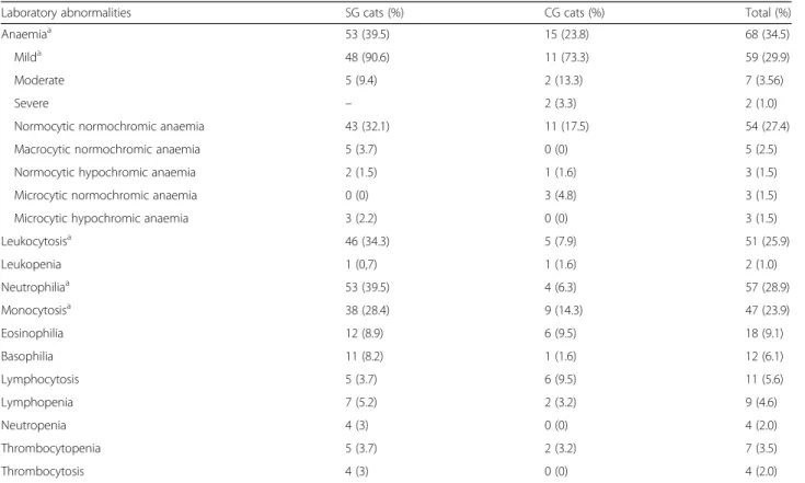
A significant difference was detected between SG and CG concerning the prevalence of three clinical findings. Lymph node enlargement (Fisher’s exact test: P < 0.0001, OR= 7.63, 95% CI: 3.36–17.34), and chronic gingivostoma-titis (Fisher’s exact test: P = 0.0049, OR = 2.90, 95% CI: 1.39–6.08) were more frequent in the SG and a BCS > 3/5 (Fisher’s exact test: P < 0.0001, OR = 0.02, 95% CI: 0.003– 0.17) was more prevalent in the CG. Moreover, anaemia (Fisher’s exact test: P = 0.0367, OR = 2.09, 95% CI: 1.07– 4.11), mild anaemia (Fisher’s exact test: P = 0.0119, OR = 2.64, 95% CI: 1.26–5.53), leukocytosis (Fisher’s exact test: P < 0.0001, OR = 6.06, 95% CI: 2.27–16.17), neutrophilia (Fisher’s exact test: P < 0.0001, OR = 9.65, 95% CI: 3.31– 28.15) and monocytosis (Fisher’s exact test: P = 0.0325, OR= 2.37, 95% CI: 1.07–5.28) were more frequently ob-served in SG compared to CG. Only in one case the an-aemia was regenerative (1 cat of CG). Laboratory abnormalities are reported in Tables2,3.
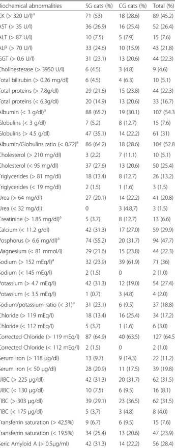
Hemoparasites were not detected at the microscopical evaluation of blood smears. A significant higher preva-lence of increased CK (Fisher’s exact test: P = 0.0013, OR = 2.82, 95% CI: 1.48–5.36), decreased albumin (Fish-er’s exact test: P < 0.0001, OR = 4.43, 95% CI: 2.32– 8.45), decreased A/G ratio (Fisher’s exact test: P < 0.0001, OR = 4.48, 95% CI: 2.34–8.59), increased
phosphorus (Fisher’s exact test: P = 0.0023, OR = 2.65,
95% CI: 1.41–4.98) and decreased sodium/potassium
ra-tio (Fisher’s exact test: P = 0.0303, OR = 2.86, 95% CI: 1.12–7.26) were observed in the SG when compared with CG. Conversely, increased creatinine (Fisher’s exact test: P = 0.0284, OR = 3.75, 95% CI: 1.17–11.99) and so-dium (Fisher’s exact test: P < 0.0001, OR = 5.18, 95% CI: 2.72–9.88) were significantly more prevalent in the CG when compared with SG.
Urinalysis was performed in 127 cats, and 33 (25.98%) showed inappropriate (≤ 1039) urine specific gravity (21 cats in the SG and 12 in the CG). No significant differ-ence was found between the two groups. Only two cats were proteinuric with UPC values of 0.4 in one CG cat and 2.52 in one SG cat, respectively.
Retroviral positivity
Antibodies against FIV (15/197 = 7.6%) were detected in both groups with no significant difference. FeLV antigenemia was detected rarely (only in two cats from SG living in Calabria region). Antibodies against FIV were significantly more prevalent in adult cats compared to young (Fisher’s exact test: P = 0.0105, OR = 9.81, 95% CI: 1.26–76.27), and in males (12.8%) than in females (4.7%) cats (Fisher’s exact test: P = 0.05, OR = 2.97, 95% CI: 1.01–8.74).
Table 2 CBC abnormalities
Laboratory abnormalities SG cats (%) CG cats (%) Total (%)
Anaemiaa 53 (39.5) 15 (23.8) 68 (34.5)
Milda 48 (90.6) 11 (73.3) 59 (29.9)
Moderate 5 (9.4) 2 (13.3) 7 (3.56)
Severe – 2 (3.3) 2 (1.0)
Normocytic normochromic anaemia 43 (32.1) 11 (17.5) 54 (27.4)
Macrocytic normochromic anaemia 5 (3.7) 0 (0) 5 (2.5)
Normocytic hypochromic anaemia 2 (1.5) 1 (1.6) 3 (1.5)
Microcytic normochromic anaemia 0 (0) 3 (4.8) 3 (1.5)
Microcytic hypochromic anaemia 3 (2.2) 0 (0) 3 (1.5)
Leukocytosisa 46 (34.3) 5 (7.9) 51 (25.9) Leukopenia 1 (0,7) 1 (1.6) 2 (1.0) Neutrophiliaa 53 (39.5) 4 (6.3) 57 (28.9) Monocytosisa 38 (28.4) 9 (14.3) 47 (23.9) Eosinophilia 12 (8.9) 6 (9.5) 18 (9.1) Basophilia 11 (8.2) 1 (1.6) 12 (6.1) Lymphocytosis 5 (3.7) 6 (9.5) 11 (5.6) Lymphopenia 7 (5.2) 2 (3.2) 9 (4.6) Neutropenia 4 (3) 0 (0) 4 (2.0) Thrombocytopenia 5 (3.7) 2 (3.2) 7 (3.5) Thrombocytosis 4 (3) 0 (0) 4 (2.0) a
Vector-borne pathogens Serological results
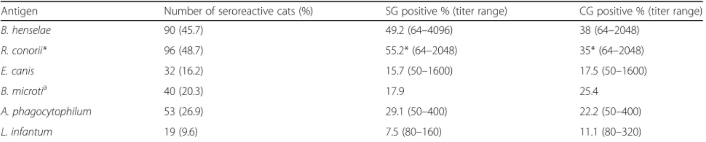
One hundred and seventy-three cats (87.8%) were seroposi-tive at least to one of the tested agents and the difference between SG (91.8%), and CG (79.4%) was significant (Fish-er’s exact test: P = 0.0187, OR = 2.91, 95% CI: 1.22–6.92). One-hundred and thirty-four cats (68%) were seropositive to two or more pathogens with a significant difference be-tween the two groups (75.4% in SG and 60.3% in CG) (Fish-er’s exact test: P = 0.0437, OR = 2.01, 95% CI: 1.06–3.82).
Antibody prevalence concerning the pathogens under consideration is reported in Table4. Prevalence of anti-R. conorii antibodies was significantly higher in SG cats (Fisher’s exact test: P = 0.0094, OR = 2.30, 95% CI: 1.24–4.27). High titers were detected against B. henselae, R. conorii, and E. canis in 10.7, 10.1 and 3.5% of tested cats, respectively, and no significant difference was found between the two groups when comparing the number of cats with high titers. Antibody prevalence to at least one pathogen was 90.8% in young cats (69/76) and 85.9% in adult cats (104/121) with no significant dif-ference between groups.
Moreover, 87.2% of Sicilian cats (100% from SG and 73.7% from CG) and 90.5% of Calabrian cats (93% in SG and 84% in CG) were positive at least to one VBP. Anti-body prevalence for R. conorii (Fisher’s exact test: P = 0.0062, OR = 5.24, 95% CI: 1.45–18.97) and B. microti (Fisher’s exact test: P = 0.0459, OR = 3.36, 95% CI: 1.16, 9.76) were significantly higher in the SG cats from Sicily compared to those from Calabria. No other significant difference was found between the two regions.
Molecular assays
Positive PCR tests were obtained for Bartonella spp. (21.3%), Mycoplasma spp. (18.3%) and L. infantum
(6.6%) (Table 5) but they were negative for Ehrlichia/
Anaplasma spp., piroplasmids (Babesia spp. and
Thei-leriaspp.), Rickettsia spp. and Hepatozoon felis. The fol-lowing species were sequenced (Table 5): B. henselae, B.
clarridgeiae, M. haemofelis,“Ca. Mycoplasma
haemomi-nutum,” “Ca. Mycoplasma turicensis” and L. infantum. Bartonella spp. prevalence was significantly higher in SG compared to CG (Fisher’s exact test: P < 0.0001, OR= 8.21, 95% CI: 2.43–27.76). Mycoplasma spp. infec-tion was significantly more frequent in FIV positive cats compared to FIV negative cats (Fisher’s exact test: P = 0.0002, OR = 8.61, 95% CI: 2.83–26.16). Overall, 79 cats (40.1%) were PCR positive at least to one of the tested agents and this category was significantly more prevalent in the SG (47%) compared to the CG (25.4%) (Fisher’s exact test: P = 0.0049, OR = 2.61, 95% CI: 1.35–5.05). There was no significant difference for molecular posi-tivity rate to at least one pathogen according to gender, age and region.
Table 3 Biochemical abnormalities
Biochemical abnormalities SG cats (%) CG cats (%) Total (%)
CK (> 320 U/l)a 71 (53) 18 (28.6) 89 (45.2) AST (> 35 U/l) 36 (26.9) 16 (25.4) 52 (26.4) ALT (> 87 U/l) 10 (7.5) 5 (7.9) 15 (7.6) ALP (> 70 U/l) 33 (24.6) 10 (15.9) 43 (21.8) GGT (> 0.6 U/l) 31 (23.1) 13 (20.6) 44 (22.3) Cholinesterase (> 3950 U/l) 6 (4.5) 3 (4.8) 9 (4.6) Total bilirubin (> 0.26 mg/dl) 6 (4.5) 4 (6.3) 10 (5.1) Total proteins (> 7.8g/dl) 29 (21.6) 15 (23.8) 44 (22.3) Total proteins (< 6.3g/dl) 20 (14.9) 13 (20.6) 33 (16.7) Albumin (< 3 g/dl)a 88 (65.7) 19 (30.1) 107 (54.3) Globulins (< 3 g/dl) 7 (5.2) 8 (12.7) 15 (7.6) Globulins (> 4.5 g/dl) 47 (35.1) 14 (22.2) 61 (31) Albumin/Globulins ratio (< 0.72)a 86 (64.2) 18 (28.6) 104 (52.8) Cholesterol (> 210 mg/dl) 3 (2.2) 7 (11.1) 10 (5.1) Cholesterol (< 95 mg/dl) 37 (27.6) 13 (20.6) 50 (25.4) Triglycerides (> 81 mg/dl) 18 (13.4) 8 (12.7) 26 (13.2) Triglycerides (< 19 mg/dl) 2 (1.5) 1 (1.6) 3 (1.5) Urea (> 64 mg/dl) 27 (20.1) 14 (22.2) 41 (20.8) Urea (< 32 mg/dl) 0 3 (4.8,7) 3 (1.5) Creatinine (> 1.85 mg/dl)a 5 (3.7) 8 (12.7) 13 (6.6) Calcium (< 11.2 g/dl) 42 (31.3) 17 (27.0) 59 (29.9) Posphorus (> 6.6 mg/dl)a 74 (55.2) 20 (31.7) 94 (47.7) Magnesium (< 81 mmol/l) 29 (21.6) 15 (23.8) 44 (22.3) Sodium (> 152 mEq/l)a 32 (23.9) 39 (61.9) 71 (36) Sodium (< 145 mEq/l) 2 (1.5) 0 2 (1.0) Potassium (> 4.7 mEq/l) 42 (31.3) 12 (19.0) 54 (27.4) Potassium (< 3.5 mEq/l) 1 (0.7) 3 (4.8) 4 (2.0) Sodium/potassium ratio (< 31)a 31 (23.1) 6 (9.5) 37 (18.8) Chloride (> 119 mEq/l) 18 (13.4) 16 (25.4) 34 (17.2) Chloride (< 112 mEq/l) 5 (3.7) 1 (1.6) 6 (3.0) Corrected Chloride (> 119 mEq/l) 87 (64.9) 40 (63.5) 127 (64.5) Corrected Chloride (< 112 mEq/l) 2 (1.5) 0 2 (1.0) Serum iron (> 118μg/dl) 13 (9.7) 9 (14.3) 22 (11.2) Serum iron (< 50μg/dl) 28 (20.9) 11 (17.5) 39 (19.8) UIBC (> 225μg/dl) 42 (31.3) 20 (31.7) 62 (31.5) UIBC (< 130μg/dl) 10 (7.5) 6 (9.5) 16 (8.1) TIBC (> 303μg/dl) 39 (29.1) 23 (36.5) 62 (31.5) TIBC (< 175μg/dl) 5 (3,7) 3 (4.8) 8 (4.0) Transferrin saturation (> 42.5%) 9 (6.7) 6 (9.5) 15 (7.6) Transferrin saturation (< 19.5%) 34 (25.4) 13 (20.6) 47 (23.9) Seric Amyloid A (> 0.5μg/ml) 42 (31.3) 14 (22.2) 56 (28.4) a
Leishmania infantum DNA was amplified in some cases from two specimens/cat as follows: blood and lymph node (n = 2), blood and urine (n = 1) or conjunc-tival and oral swabs (n = 1). Parasitic load for L.
infan-tumranged from 1 to 80,000 Leishmania/ml detected in
blood EDTA, from 1 to 11,000 Leishmania/specimen in lymph nodes, from 7 to 120 Leishmania/specimen in conjunctival swabs, from 16 to 92 Leishmania/specimen in oral swabs, and from 1 to 30 Leishmania/ml in urine.
Co-infections with at least two pathogens were found in 10.1% of cats with 13.4% of SG cats (n = 18) and 3.2% of CG cat (n = 2). Only 2 cats were co-infected with three pathogens: one (1.6%) from CG (L. infantum, M.
haemofelis and “Ca. Mycoplasma haemominutum”) and
one (0.7%) form SG (B. henselae, M. haemofelis and“Ca.
Mycoplasma haemominutum”). The most common co-infection detected was between Bartonella spp. and Mycoplasmaspp. in six cats. Co-infections with L.
infan-tumand other pathogens included two with“Ca.
Myco-plasma haemominutum”, two with B. henselae and one
with B. clarridgeiae. Co-infections with different
Myco-plasma species included“Ca. Mycoplasma
haemominu-tum” and “Ca. Mycoplasma turicensis” (5 cats), M.
haemofelis and “Ca. Mycoplasma haemominutum” (3
cats), and “Ca. Mycoplasma haemominutum”, “Ca.
Mycoplasma turicensis” and M. haemofelis (2 cats). Overall exposure prevalence
An overall prevalence of exposure was calculated for pathogens investigated by both serological and molecu-lar methods. Overall, Bartonella spp. exposure was 48.7% (96/197) and a higher prevalence was observed in FIV positive cats (Fisher’s exact test: P = 0.0002, OR = 17.07, 95% CI: 2.20–132.65). Overall, L. infantum posi-tivity rate was 14.7% (29/197), but no significant differ-ence was observed between SG and CG or according to age, gender or FIV positivity.
One-hundred-eighty-two cats (92.4%) were found positive (by IFAT and PCR) to at least one VBP. The SG cats had higher (96.3%) exposure prevalence to at least Table 4 Overall antibody prevalence and titer range in the study (SG) and control (CG) groups
Antigen Number of seroreactive cats (%) SG positive % (titer range) CG positive % (titer range)
B. henselae 90 (45.7) 49.2 (64–4096) 38 (64–2048) R. conorii* 96 (48.7) 55.2* (64–2048) 35* (64–2048) E. canis 32 (16.2) 15.7 (50–1600) 17.5 (50–1600) B. microtia 40 (20.3) 17.9 25.4 A. phagocytophilum 53 (26.9) 29.1 (50–400) 22.2 (50–400) L. infantum 19 (9.6) 7.5 (80–160) 11.1 (80–320) *
Significant difference of prevalence between SG and CG
a
Titer not determined
Table 5 Positive PCR and sequencing results
Pathogen Number of positive cats (%)
Total cats (n = 197) SG cats (n = 134) CG cats (n = 63) Bartonella spp.a 42 (21.3) 39 (29.1) 3 (4.8) B. henselae 30 (15.2) 28 (20.9) 2 (3.2) B. clarridgeiae 12 (6.1) 11 (8.2) 1 (1.6) Mycoplasma spp. 36 (18.3) 27 (20.2) 9 (14.3) Mycoplasma haemofelis 12 (6.1) 11 (8.2) 1 (1.6)
“Candidatus Mycoplasma haemominutum” 26 (13.2) 18 (13.4) 8 (12.7)
“Candidatus Mycoplasma turicensis” 10 (5.1) 8 (6.0) 2 (3.2)
Leishmania infantum 13 (6.6) 8 (6.0) 5 (7.9) Conjunctival swabs (n = 394) 3 2 1 Oral swabs (n = 197) 3 0 3 Blood EDTA (n = 197) 4 4 0 Lymph nodes (n = 181) 3 2 1 Urine samples (n = 143) 3 2 1 Total 79 (35) 63 (47) 16 (25.4) a
one VBP compared to CG (84.1%) (Fisher’s exact test: P = 0.0068, OR = 4.87, 95% CI: 1.59–14.93).
Two or more co-infections were detected in 62 cats (31.5%). In SG, there was a higher prevalence (37.3%) of multiple positivity compared to CG (19%) (Fisher’s exact test: P = 0.0132, OR = 2.53, 95% CI: 1.23–5.20]). Con-versely, no difference was found in FIV positive cats compared to FIV negative cats.
Multivariable logistic regression analysis
Seventeen variables that showed a significative difference in prevalence of one or more VBP between SG and CG with univariate analysis entered the multivariable logistic regression analysis performed on the 197 cats. Signifi-cant associations were found for exposure to L. infan-tum, Bartonella spp., Mycoplasma spp., B. microti, and
A. phagocytophilum (Table 6) and co-infections
(Additional file3: Table S3). The significant associations concerned adult age and hemoplasma infection, FIV positivity and Bartonella spp. or hemoplasma infection, anaemia and hemoplasma infection, elevated ALP serum activity or low albumin concentration and A.
phagocyto-philum antibody positivity, high globulin concentration
and Bartonella spp. infection, and high creatinine concentration with hemoplasma infection. Moreover, adults had a lower risk of B. microti antibody positivity compared to young cats; elevated serum CK activity was less likely in cats with L. infantum, hemoplasma, co-infections, or B. microti antibody positivity; cats with Bartonella spp infection were less likely to display ele-vated ALP serum activity.
Discussion
This controlled field study examined the prevalence of selected VBP and the clinical and clinicopathological ab-normalities in cats exposed to ectoparasites. Moreover, some risk factors for VBP exposure were identified using
significant associations detected by multivariable logistic regression analysis between some of the investigated pathogens and independent variables.
The study was based on both antibody and molecular detection of feline VBPs under consideration to increase the possibility of evaluating exposure of cats to tested pathogens. We found that cats were extremely exposed to VBP because of the high antibody (88.3%), molecular (40%), and overall (antibody and PCR) positivity (92.4%) to at least one pathogen. Moreover, cats exposed to ectoparasites, because of their outdoor lifestyle and the lack of regular ectoparasiticide treatments (SG), showed a significantly higher molecular and overall (antibody and PCR) positivity compared to indoor cats with no ec-toparasites and subjected to regular application of ecto-parasiticides (CG). Exposure to multiple VBP was also very frequent, as about two-thirds of tested cats were antibody positive, 10% PCR positive and 30.1% antibody and PCR positive to more than one VBP at the time of sampling; the difference between SG and CG was signifi-cant for antibody or antibody and molecular positivity. Epidemiological similarities shared by some VBP can obviously be responsible for vector-borne co-infections but also other factors, such as pathogenic interactions between them, can delay or prevent clearance of VBI and concur to co-infections. Other studies detected a high overall blood PCR positivity to at least one VBP, i.e.
25–29.9% in Portugal [4, 17], 48.9% in northern Italy
[18] and 45.4% in Cyprus [3]. Prevalence of co-infections was lower (2.2–9.8%) in those studies [3,4,17,18].
A small percentage of enrolled cats (12%) were anti-body negative to all of VBP, and this percentage was sig-nificantly higher in the CG. However, we found a significant percentage of CG cats positive to the same VBP detected in the SG, and this may be explained by lack of compliance to ectoparasiticide treatment, low efficacy of the used ectoparasiticide, and/or non-Table 6 Multivariable logistic regression analysis of VBP
Variables with significant ORs
Leishmania infantuma Bartonella spp.a Mycoplasma spp.b Babesia microtic Anaplasma phagocytophilumc
OR (95% Cl) P OR (95% Cl) P OR (95% Cl) P OR (95% Cl) P OR (95% Cl) P Adult age ns ns 4.53 (1.23–16.74) 0.02 0.28 (0.11–0.75) < 0.01 ns FIV positivity ns 11.79 (1.38–100.97) 0.02 5.07 (1.33–19.33) 0.02 ns ns Anaemia ns ns 2.76 (1.05–7.29) 0.04 ns ns High CK 0.28 [0.10; 0.78] 0.01 ns 0.35 (0.12–0.98) 0.04 0.31 (0.12–0.69) 0.01 ns High ALP ns 0.26 (0.09–0.71) 0.01 ns ns 4.40 (1.54–12.57) < 0.001 Low Albumin ns ns ns ns 2.42 (1.05–5.59) 0.04 High Globulins ns 2.99 (1.38–6.48) 0.00 ns ns ns High Creatinine ns ns 7.68 (1.43–41.01) 0.02 ns ns
Abbreviation: ns not significant
a PCR and/or IFAT b PCR c IFAT
vectorial transmission of the pathogen. For instance, in the case of L. infantum infection, pyrethroids are used in dogs for sand fly bite prevention, but almost all of these compounds are toxic to cats, and only a collar contain-ing flumethrin and imidacloprid was able to reduce the incidence of L. infantum infection in cats [19–21]. Blood transfusion is a main non-vectorial transmission route of feline VBI but, at least for hemoplasmas, other routes are strongly suspected [22–24]. Moreover, our model of multivariable analysis did not find significant associa-tions between exposure to any individual or multiple VBP and outdoor lifestyle or individual ectoparasiticide treatment. Attipa et al. [3] also used multivariable logis-tic regression to investigate risk factors of some VBPs detected in cats from Cyprus and, similarly, they did not find any association between positivity to any tested VBP and the lack of ectoparasiticide use while they found an association of outdoor lifestyle only with hemoplasma positivity [3].
Based on antibody detection, R. conorii (or other cross-reacting Rickettsia species) was the most frequent agent circulating among tested cats (48.7%), and it was significantly more common in cats exposed to ectopara-sites (SG). Rickettsia conorii is historically the most im-portant zoonotic species of the Rickettsia genus in the Mediterranean area, and was recently confirmed as a possible causative agent of acute febrile illness in dogs showing a transient positive blood PCR and seroconver-sion [25]. Studies about the infection of cats with
Rick-ettsia spp. of the Mediterranean spotted fever group
reported the collection of infected ticks on cats, and ob-tained positive blood PCR and a high antibody preva-lence [10, 17, 26–28]. These data support the need for prospective investigations about the pathogenic role of spotted fever group Rickettsia spp. in cats.
Antibody prevalence for B. henselae was high (45.7%), as was the seroreactivity for A. phagocytophilum (26.9%), B. microti(20.3%) and E. canis (16.2%) antigens. In many previous serological studies performed by IFAT, antibody titer against tested VBP was not reported, or they were low [6, 10,29, 30]. Interestingly, we detected high titers against B. henselae (10.7%), R. conorii (10.1%) and E.
canis (3.5%) antigens; however, we cannot exclude the
possibility of serological cross-reactions with other spe-cies of the same genus. This is most important for R.
conoriiand E. canis because we documented the
expos-ure of cats by serology, but we were not able to find the pathogen DNA in blood as shown in other studies [31, 32]. Similarly, we did not detect DNA of Anaplasma,
Hepatozoon and piroplasmids such as Babesia or
Cytauxzoon. This might be a consequence of the lack of exposure, clearance or sequestration of the organisms in other tissues, or technical limitations. However, in a study performed on blood collected from outdoor cats
from a confined area of Sicily, only Hepatozoon felis DNA was detected in one cat (0.3%). Therefore, it is likely that some VBPs are not common in Sicily and the Calabria re-gion [33]. Anaplasma/Ehrlichia DNA detection is rare in cats, and more frequently A. phagocytophilum is amplified [4, 17, 31, 34], but A. platys or A. platys-like organisms were also occasionally characterised in cats in southern Europe and the seropositivity detected in this study could be due to different species of Anaplasma [3,35]. However,
Ehrlichia spp. and Anaplasma spp. were not found in
other studies in Greece and Spain [36,37].
Hepatozoon felisdistribution seems to be quite variable in southern Europe. In fact, it is very rare in Italy, but a focus was recently detected in Matera, where single cases of Hepatozoon canis and Hepatozoon silvestris infections were also seen [10,33,38]. Hepatozoon felis is reported in
the Iberian Peninsula with a prevalence range of 1.6–
13.8% [4,17,31,39] and the highest molecular prevalence (37.9%) was recently found in Cyprus [3].
Feline piroplasmid infection is uncommon in Europe. Piroplasmid infection is caused by Cytauxzoon spp. [39– 43], Babesia vogeli, or Babesia canis [4,17]. In Italy, DNA of B. microti was sequenced in cats from Sicily and Milan but, despite 20.3% antibody prevalence for B. microti ob-tained in this study, we were not able to detect piroplas-mid DNA, and we cannot exclude that other Babesia spp. elicited antibody production in cats [44,45]. Interestingly, adult cats were less likely to have antibodies to B. microti compared to those < 1 year of age. Young cats might therefore be more susceptible to Babesia spp. infection than adults but the antibody response would not persist possibly because of clearance of the infection.
Bartonella spp. was VBP with the highest overall
(48.7%) and molecular (21.3%) prevalence in this study, and molecular prevalence was significantly higher in SG. A significant association between Bartonella spp. expos-ure and FIV infections or increased globulin values were found by multivariate analysis as previously reported by univariate analysis only for globulins [46] but not for
FIV [3, 5]. A very high B. henselae antibody prevalence
(45.7%) was also found. Two species were sequenced, and B. henselae was more prevalent (15.2%) than B.
clar-ridgeiae (6.1%). These results confirm data obtained in
42 cats from the same area carrying ticks or fleas at the time of examination, where blood PCR positivity for
Bartonella spp. was 38.1%, and B. henselae (21.4%) was
more frequently detected than B. clarridgeiae (16.6%) [10]. A higher molecular prevalence was previously found in Sicily in a study using a nested-PCR where positivity was 70.6% in blood, 72.9% in lymph node aspi-rates, and 60.0% in oral swabs and B. henselae was the only sequenced species [8]. In other European countries,
lower blood PCR prevalences were reported ranging 1–
investigated cats to fleas or the assays used [3–5,28,31, 36,37]. However, also in these latter studies, B. henselae was usually more frequent than B. clarridgeiae and only in one cat was B. kholerae DNA was sequenced [37].
As in other studies performed in Italy [10,18], Cyprus [3], and Portugal [16, 47], we frequently detected
hemo-plasma DNA in cat blood (18.3%) and “Ca. Mycoplasma
haemominutum” was more commonly sequenced,
com-pared to“Ca. Mycoplasma turicensis” and M. haemofelis.
Other studies reported lower prevalence (range 7.8– 14.9%) but similar species were sequenced and repre-sented possible co-infections [12,36,48,49]. We obtained significant associations by multivariate analysis between hemoplasma positivity and anaemia (OR= 2.76), adult age (OR= 4.53), FIV positivity (OR= 5.07), and increased cre-atinine values (OR=7.68). Interestingly, hemoplasma posi-tive cats have seven times higher risk for an increased creatinine concentration and this association was never found before by multivariate analysis. We performed a cross-sectional study and therefore did not have the possi-bility to confirm chronic kidney disease (CKD) in cats with high creatinine; however, in two-thirds of cats urin-alysis showed inappropriate urine specific gravity that is suggestive of CKD. A causative role for variables cannot be assessed by cross-sectional investigations, and pro-spective surveys on hemoplasma long carrier cats should be considered. Older age of hemoplasma-positive cats was also found in two other studies using multivariable logistic analysis, and could contribute to the association between hemoplasma positivity and high creatinine concentrations [3, 49]. Hemolytic anaemia is the main pathogenic effect of M. haemofelis and less frequently of other hemoplas-mas, but subclinical carriers can be found, and this may explain when in other studies multivariate analysis did not find associations between hemoplasmas and anaemia [3, 49]. Conversely, significant association with FIV was pre-viously reported by other authors using multivariable
ana-lysis, and this comorbidity could be due to
epidemiological factors (sharing of the way of transmis-sion) or facilitation of long-term hemoplasma infections in FIV positive cats [3,49,50].
Feline L. infantum infection can be considered an emergent VBI in endemic areas of canine leishmaniosis [20]. Many studies evaluated antibody and or molecular prevalence in southern Italy, and a wide prevalence range was found by both antibody detection (2.4–59%)
and blood PCR (7.1–61%) [10, 33, 51–53].
Epidemio-logical (endemicity, characteristics of the studied
popula-tion) and technical (serological cut-off, molecular
technique) differences may account for this variability. Antibody prevalence obtained in this study was within the above range (9.6%), but blood DNA detection was as low as 2.0%. Low molecular prevalence was also found by non-invasive samplings as conjunctival (1.5%) and
oral (1.5%) swabs, or by lymph node aspirate (1.7%) or urine (2%). Interestingly, high parasite loads were ob-tained only from blood (up to 80,000 Leishmania/ml), and the clinical relevance of this finding lies in the risk of iatrogenic transmission of L. infantum by blood trans-fusion as reported in dogs [54,55].
Co-infection of L. infantum with “Ca. Mycoplasma
haemominutum”, B. henselae or B. clarridgeiae was
simi-lar to that reported in other studies [3, 10]. However,
other L. infantum co-infections are known including
Anaplasma/Ehrlichia spp., Babesia spp., Hepatozoon
spp., and Borrelia burgdorferi [3,4,17].
Finally, the univariate analysis pointed out some clinical and clinicopathological abnormalities that were significantly more frequent in outdoor cats exposed to ectoparasites than in indoor cats protected from ecto-parasites despite the former being significantly younger and less frequently admitted for health problems. Other infectious and parasite agents in addition to VBP fre-quently affect outdoor cats and may be responsible for
this occurrence [28, 36, 56–58]. The only biochemical
abnormality more frequent in indoor cats was increased creatinine, but these cats were significantly older than those of SG, and this bias could influence the result. Indoor cats were also significantly more overweight compared to outdoor cats and therefore are predisposed to metabolic or urinary problems [59–61]. The clinical relevance of these findings is that lifestyle significantly influences the health of cats and adequate preventative measures have to be tailored accordingly.
Conclusions
A very high prevalence of zoonotic VBP exposure was found in cats, with Rickettsia spp. and Bartonella spp. being the most prevalent. Overall, cats exposed to ecto-parasites for the lack of preventative measure and/or an outdoor lifestyle had a higher risk for VBI and co-infections and for some clinical and clinicopathological abnormalities. Some risk factors were documented by multivariable logistic regression analysis providing a better understanding of the epidemiology for selected feline pathogens, namely for Mycoplasma spp. and Bartonella spp. infections that were both found associated with FIV.
Also, the lifestyle of cats appeared clinically relevant for diseases other than those associated with VBP expos-ure and requires specific preventative measexpos-ures to pro-tect their health.
Additional files
Additional file 1:Table S1. Feline CBC reference intervals. (DOCX 15 kb)
Additional file 2:Table S2. Serum biochemistry reference intervals. (DOCX 15 kb)
Additional file 3:Table S3. Ordered logistic regression analysis of VBP. (DOCX 14 kb)
Abbreviations
ALP:alkaline phosphatase; ALT: alanine aminotransferase; AST: aspartate aminotransferase; BCS: body condition score; CBC: complete blood count; CG: control group; CI: confidence interval; CK: creatine kinase; CKD: chronic kidney disease; GGT: gamma glutamyl transferase; OR: odds ratio; SAA: serum amyloid A; SG: study group; TIBC: total iron-binding capacity; UIBC: unsatured iron-binding capacity; VBI: vector-borne infections; VBP: vector-borne pathogen
Acknowledgements
The authors are grateful to Angela Burrascano for technical assistance; Dr Laura Altet for technical support with molecular investigations; Dr Tommaso Furlanello and Dr Marco Caldin for technical support with biochemical profiles. The publication of this paper has been sponsored by Bayer Animal Health within the framework of the 13th CVBD World Forum Symposium. Funding
This clinical study was funded by Bayer Animal Health Care - Animal Health Division (Monheim, Germany).
Availability of data and materials
The datasets supporting the conclusions of this article are included within the article and its additional files. All analysed data are available from the corresponding author upon reasonable request.
Authors’ contributions
MGP and LSG conceived the research study. MFP worked in the field and performed laboratory techniques. MGP and MFP contributed to data analysis and interpretation, wrote the first draft and revised the manuscript. LS and MM supervised the performance of laboratory techniques, contributed to data analysis and interpretation and revised the manuscript. AV contributed to data analysis, wrote and revised the manuscript. AM contributed to Leishmania tests. All authors read and approved the final manuscript. Ethics approval
This clinical study was conducted from March 2012 to March 2013 in accordance with FVE European code of conduct and under the ethical requirements of the Department of Veterinary Science of University of Messina. Informed consent was obtained from the owners of enrolled cats. Consent for publication
Not applicable. Competing interests
The authors declare that they have no competing interests.
Publisher’s Note
Springer Nature remains neutral with regard to jurisdictional claims in published maps and institutional affiliations.
Author details
1
Istituto Zooprofilattico Sperimentale della Sicilia, A. Mirri, Via G. Marinuzzi 3, 90129 Palermo, Italy.2Dipartimento di Scienze Veterinarie, Università degli
Studi di Messina, Polo Universitario Annunziata, 98168 Messina, Italy.
3Departament de Medicina i Cirurgia Animals, Facultat de Veterinària,
Universitat Autònoma de Barcelona, Bellaterra, Cerdanyola, 08193 Barcelona, Spain.
Received: 18 November 2017 Accepted: 16 February 2018 References
1. Otranto D, Dantas-Torres F. Canine and feline vector-borne diseases in Italy: current situation and perspectives. Parasit Vectors. 2010;3:2.
2. Scorza AV, Lappin MR. Prevalence of selected zoonotic and vector-borne agents in dogs and cats on the Pine Ridge Reservation. Vet Sci. 2017;4
3. Attipa C, Papasouliotis K, Solano-Gallego L, Baneth G, Nachum-Biala Y, Sarvani E, et al. Prevalence study and risk factor analysis of selected bacterial, protozoal and viral, including vector-borne, pathogens in cats from Cyprus. Parasit Vectors. 2017;10:130.
4. Maia C, Ramos C, Coimbra M, Bastos F, Martins A, Pinto P, et al. Bacterial and protozoal agents of feline vector-borne diseases in domestic and stray cats from southern Portugal. Parasit Vectors. 2014;7:115.
5. Bergmann M, Hartmann K. Vector-borne diseases in cats in Germany. Tierarztl Prax Ausg K Klientiere Heimtiere. 2017;45:329–35.
6. Ayllón T, Diniz PPVP, Breitschwerdt EB, Villaescusa A, Rodríguez-Franco F, Sainz A. Vector-borne diseases in client-owned and stray cats from Madrid, Spain. Vector Borne Zoonotic Dis. 2012;12:143–50.
7. Solano-Gallego L, Rossi L, Scroccaro AM, Montarsi F, Caldin M, Furlanello T, et al. Detection of Leishmania infantum DNA mainly in Rhipicephalus sanguineus male ticks removed from dogs living in endemic areas of canine leishmaniosis. Parasit Vectors. 2012;5:98.
8. Pennisi MG, La Camera E, Giacobbe L, Orlandella BM, Lentini V, Zummo S, et al. Molecular detection of Bartonella henselae and Bartonella clarridgeiae in clinical samples of pet cats from Southern Italy. Res Vet Sci. 2010;88:379–84. 9. Pennisi MG. La leishmaniosis felina dalla A alla Z. In: Proceedings 2nd
International Congress on Canine Leishmaniasis; 2010. p. 71–6.https://cms. scivac.it/PDF/SCIVAC/Proceedings/2_Leishmaniosi.pdf. Accessed 22 Dec 2017. 10. Persichetti MF, Solano-Gallego L, Serrano L, Altet L, Reale S, Masucci M, et al.
Detection of vector-borne pathogens in cats and their ectoparasites in southern Italy. Parasit Vectors. 2016;9:247.
11. Pennisi MG, Persichetti MF, Serrano L, Altet L, Reale S, Gulotta L, et al. Ticks and associated pathogens collected from cats in Sicily and Calabria (Italy). Parasit Vectors. 2015;8:512.
12. Bergmann M, Englert T, Stuetzer B, Hawley JR, Lappin MR, Hartmann K. Risk factors of different hemoplasma species infections in cats. BMC Vet Res. 2017;13:52.
13. Piaton E, Fabre M, Goubin-Versini I, Bretz-Grenier M-F, Courtade-Saïdi M, Vincent S, et al. Guidelines for May-Grünwald-Giemsa staining in haematology and non-gynaecological cytopathology: recommendations of the French Society of Clinical Cytology (SFCC) and of the French Association for Quality Assurance in Anatomic and Cytologic Pathology (AFAQAP). Cytopathol Off J Br Soc Clin Cytol. 2016;27:359–68. 14. International Renal Interest Society. IRIS Staging of CKD.www.iris-kidney.
com/pdf/003-5559.001-iris-website-staging-of-ckd-pdf_220116-final. pdf#page=7. Accessed 22 Dec 2017.
15. Persichetti MF, Solano-Gallego L, Vullo A, Masucci M, Marty P, Delaunay P, et al. Diagnostic performance of ELISA, IFAT and Western blot for the detection of anti-Leishmania infantum antibodies in cats using a Bayesian analysis without a gold standard. Parasit Vectors. 2017;10:119.
16. Martínez-Díaz VL, Silvestre-Ferreira AC, Vilhena H, Pastor J, Francino O, Altet L. Prevalence and co-infection of haemotropic mycoplasmas in Portuguese cats by real-time polymerase chain reaction. J Feline Med Surg. 2013;15:879–85.
17. Vilhena H, Martinez-Díaz VL, Cardoso L, Vieira L, Altet L, Francino O, et al. Feline vector-borne pathogens in the north and centre of Portugal. Parasit Vectors. 2013;6:99.
18. Spada E, Proverbio D, Galluzzo P, Della Pepa A, Perego R, Bagnagatti De Giorgi G, et al. Molecular study on selected vector-borne infections in urban stray colony cats in northern Italy. J Feline Med Surg. 2014;16:684–8. 19. Solano-Gallego L, Miró G, Koutinas A, Cardoso L, Pennisi MG, Ferrer L, et al.
LeishVet guidelines for the practical management of canine leishmaniosis. Parasit Vectors. 2011;4:86.
20. Pennisi MG, Cardoso L, Baneth G, Bourdeau P, Koutinas A, Miró G, et al. LeishVet update and recommendations on feline leishmaniosis. Parasit Vectors. 2015;8:302.
21. Brianti E, Falsone L, Napoli E, Gaglio G, Giannetto S, Pennisi MG, et al. Prevention of feline leishmaniosis with an imidacloprid 10%/flumethrin 4.5% polymer matrix collar. Parasit Vectors. 2017;10:334.
22. Museux K, Boretti FS, Willi B, Riond B, Hoelzle K, Hoelzle LE, et al. In vivo transmission studies of“Candidatus Mycoplasma turicensis” in the domestic cat. Vet Res. 2009;40:45.
23. Pennisi MG, Hartmann K, Addie DD, Lutz H, Gruffydd-Jones T, Boucraut-Baralon C, et al. Blood transfusion in cats: ABCD guidelines for minimising risks of infectious iatrogenic complications. J Feline Med Surg. 2015;17:588–93. 24. Lappin MR. Feline haemoplasmas are not transmitted by Ctenocephalides
25. Solano-Gallego L, Caprì A, Pennisi MG, Caldin M, Furlanello T, Trotta M. Acute febrile illness is associated with Rickettsia spp infection in dogs. Parasit Vectors. 2015;8:216.
26. Solano-Gallego L, Hegarty B, Espada Y, Llull J, Breitschwerdt E. Serological and molecular evidence of exposure to arthropod-borne organisms in cats from northeastern Spain. Vet Microbiol. 2006;118:274–7.
27. Segura F, Pons I, Miret J, Pla J, Ortuño A, Nogueras M-M. The role of cats in the eco-epidemiology of spotted fever group diseases. Parasit Vectors. 2014;7:353. 28. Diakou A, Di Cesare A, Accettura PM, Barros L, Iorio R, Paoletti B, et al. Intestinal
parasites and vector-borne pathogens in stray and free-roaming cats living in continental and insular Greece. PLoS Negl Trop Dis. 2017;11:e0005335. 29. Ayllón T, Villaescusa A, Tesouro MA, Sainz A. Serology, PCR and culture of
Ehrlichia/Anaplasma species in asymptomatic and symptomatic cats from central Spain. Clin Microbiol Infect. 2009;15(Suppl. 2):4-5.
30. Ebani VV, Bertelloni F, Fratini F. Occurrence of Bartonella henselae types I and II in Central Italian domestic cats. Res Vet Sci. 2012;93:63–6.
31. Tabar M-D, Altet L, Francino O, Sánchez A, Ferrer L, Roura X. Vector-borne infections in cats: molecular study in Barcelona area (Spain). Vet Parasitol. 2008;151:332–6. 32. Bayliss DB, Morris AK, Horta MC, Labruna MB, Radecki SV, Hawley JR, et al.
Prevalence of Rickettsia species antibodies and Rickettsia species DNA in the blood of cats with and without fever. J Feline Med Surg. 2009;11:266–70. 33. Otranto D, Napoli E, Latrofa MS, Annoscia G, Tarallo VD, Greco G, et al.
Feline and canine leishmaniosis and other vector-borne diseases in the Aeolian Islands: pathogen and vector circulation in a confined environment. Vet Parasitol. 2017;236:144–51.
34. Bergmann M, Englert T, Stuetzer B, Hawley JR, Lappin MR, Hartmann K. Prevalence of selected rickettsial infections in cats in southern Germany. Comp Immunol Microbiol Infect Dis. 2015;42:33–6.
35. Zobba R, Anfossi AG, Visco S, Sotgiu F, Dedola C, Pinna Parpaglia ML, et al. Cell tropism and molecular epidemiology of Anaplasma platys-like strains in cats. Ticks Tick-Borne Dis. 2015;6:272–80.
36. Ravicini S, Pastor J, Hawley J, Brewer M, Castro-López J, Beall M, et al. Prevalence of selected infectious disease agents in stray cats in Catalonia, Spain. JFMS Open Rep. 2016;2:2055116916634109.
37. Mylonakis ME, Schreeg M, Chatzis MK, Pearce J, Marr HS, Saridomichelakis MN, et al. Molecular detection of vector-borne pathogens in Greek cats. Ticks Tick-Borne Dis. 2017;
38. Giannelli A, Latrofa MS, Nachum-Biala Y, Hodžić A, Greco G, Attanasi A, et al. Three different Hepatozoon species in domestic cats from southern Italy. Ticks Tick-Borne Dis. 2017;8:721–4.
39. Díaz-Regañón D, Villaescusa A, Ayllón T, Rodríguez-Franco F, Baneth G, Calleja-Bueno L, et al. Molecular detection of Hepatozoon spp. and Cytauxzoon sp. in domestic and stray cats from Madrid, Spain. Parasit Vectors. 2017;10:112. 40. Carli E, Trotta M, Chinelli R, Drigo M, Sinigoi L, Tosolini P, et al. Cytauxzoon
sp. infection in the first endemic focus described in domestic cats in Europe. Vet Parasitol. 2012;183:343–52.
41. Carli E, Trotta M, Bianchi E, Furlanello T, Caldin M, Pietrobelli M, et al. Cytauxzoon sp. infection in two free-ranging young cats: clinicopathological findings, therapy and follow up. Turk Parazitolojii Derg. 2014;38:185–9. 42. Alho AM, Silva J, Fonseca MJ, Santos F, Nunes C, de Carvalho LM, et al. First report of
Cytauxzoon sp. infection in a domestic cat from Portugal. Parasit Vectors. 2016;9:220. 43. Legroux J-P, Halos L, René-Martellet M, Servonnet M, Pingret J-L,
Bourdoiseau G, et al. First clinical case report of Cytauxzoon sp. infection in a domestic cat in France. BMC Vet Res. 2017;13:81.
44. Pennisi MG, Alongi A, Agnone A, Vitale F, Reale S, Torina A. Cats as reservoir of Babesia microti. Parassitologia. 2007;49:100.
45. Spada E, Proverbio D, Galluzzo P, Perego R, Bagnagatti De Giorgi G, Roggero N, et al. Frequency of piroplasms Babesia microti and Cytauxzoon felis in stray cats from northern Italy. BioMed Res Int. 2014;2014:943754. 46. Whittemore JC, Hawley JR, Radecki SV, Steinberg JD, Lappin MR. Bartonella
species antibodies and hyperglobulinemia in privately owned cats. J Vet Intern Med. 2012;26:639–44.
47. Duarte A, Marques V, Correia JHD, Neto I, Bráz BS, Rodrigues C, et al. Molecular detection of haemotropic Mycoplasma species in urban and rural cats from Portugal. J Feline Med Surg. 2015;17:516–22.
48. Rosenqvist MB, Meilstrup A-KH, Larsen J, Olsen JE, Jensen AL, Thomsen LE. Prevalence of feline haemoplasma in cats in Denmark. Acta Vet Scand. 2016;58:78. 49. Ravagnan S, Carli E, Piseddu E, Da Rold G, Porcellato E, Zanardello C, et al.
Prevalence and molecular characterization of canine and feline hemotropic mycoplasmas (hemoplasmas) in northern Italy. Parasit Vectors. 2017;10:132.
50. Jenkins KS, Dittmer KE, Marshall JC, Tasker S. Prevalence and risk factor analysis of feline haemoplasma infection in New Zealand domestic cats using a real-time PCR assay. J Feline Med Surg. 2013;15:1063–9.
51. Pennisi MG, Masucci M, Catarsini O. Presenza di anticorpi anti-Leishmania in gatti FIV+ che vivono in zona endemica. Società Italiana delle Scienze Veterinarie - Atti 52° Convegno; 1998. p. 265–6.
52. Pennisi MG, Maxia L, Vitale F, Masucci M, Borruto G, Caracappa S. Studio sull’infezione da Leishmania mediante PCR in gatti che vivono in zona endemica. Società Italiana delle Scienze Veterinarie - Atti 54° Convegno; 2000. p. 215–6. 53. Pennisi MG, Lupo T, Malara D, Masucci M, Migliazzo A, Lombardo G.
Serological and molecular prevalence of Leishmania infantum infection in cats from southern Italy. J Feline Med Surg. 2012;14:656–7.
54. de Freitas E, Melo MN, da Costa-Val AP, Michalick MSM. Transmission of Leishmania infantum via blood transfusion in dogs: potential for infection and importance of clinical factors. Vet Parasitol. 2006;137:159–67. 55. Owens SD, Oakley DA, Marryott K, Hatchett W, Walton R, Nolan TJ, et al.
Transmission of visceral leishmaniasis through blood transfusions from infected English foxhounds to anemic dogs. J Am Vet Med Assoc. 2001;219: 1076–83.
56. Diakou A, Di Cesare A, Barros LA, Morelli S, Halos L, Beugnet F, et al. Occurrence of Aelurostrongylus abstrusus and Troglostrongylus brevior in domestic cats in Greece. Parasit Vectors. 2015;8:590.
57. Piyarungsri K, Pusoonthornthum R. Risk and protective factors for cats with naturally occurring chronic kidney disease. J Feline Med Surg. 2017;19:358–63. 58. Hwang J, Gottdenker N, Min M-S, Lee H, Chun M-S. Evaluation of
biochemical and haematological parameters and prevalence of selected pathogens in feral cats from urban and rural habitats in South Korea. J Feline Med Surg. 2016;18:443–51.
59. Lund HS, Sævik BK, Finstad ØW, Grøntvedt ET, Vatne T, Eggertsdóttir AV. Risk factors for idiopathic cystitis in Norwegian cats: a matched case-control study. J Feline Med Surg. 2016;18:483–91.
60. Rowe E, Browne W, Casey R, Gruffydd-Jones T, Murray J. Risk factors identified for owner-reported feline obesity at around one year of age: dry diet and indoor lifestyle. Prev Vet Med. 2015;121:273–81.
61. Chandler M, Cunningham S, Lund EM, Khanna C, Naramore R, Patel A, et al. Obesity and associated comorbidities in people and companion animals: a One Health perspective. J Comp Pathol. 2017;156:296–309.
• We accept pre-submission inquiries
• Our selector tool helps you to find the most relevant journal
• We provide round the clock customer support
• Convenient online submission
• Thorough peer review
• Inclusion in PubMed and all major indexing services
• Maximum visibility for your research Submit your manuscript at
www.biomedcentral.com/submit