DOTTORATO INTERNAZIONALE DI RICERCA IN
NEUROBIOLOGIA
XXV CICLO
UNIVERSITA’ DEGLI STUDI DI CATANIA
SEDI CONSORZIATE: UNIVERSITA’ DI CATANIA, ROMA E PAVIA SEDE AMMINISTRATIVA: UNIVERSITA’ DI CATANIA
Dott.ssa Manuela Pennisi
______________
REDOX PROTEOMICS, THIOL HOMEOSTASIS AND NEUROPHYSIOLOGICAL CORRELATIONS IN AGING AND
NEURODEGENERATION ______________
TESI DI DOTTORATO
_______________
Tutor
Chiar.mo Prof. Vittorio Calabrese
Coordinatore
Chiar.mo. Prof. Roberto Avola
CONTENTS
1. Introduction 3
2. Aim of the research 28
3. Materials and methods 31
4. Results 46
5. Discussion 55
6. References 72
1.
INTRODUCTIONThe terms "aging" and "neurodegeneration" are often used in a broad and generalized manner. Actually, they are particularly complex and multifaceted processes, involving different biochemical systems [1].
Increasing evidence supports the notion that reduction of cellular expression and activity of antioxidant proteins and the resulting increase of oxidative stress are fundamental causes in the aging processes and neurodegenerative diseases [2]. Within the frame of free radical hypothesis of aging, several lines of evidence suggest that accumulation of oxidative molecular damage is a causal factor in senescence. It is also increasingly evident that the mitochondrial genome may play a key role in aging and neurodegenerative diseases. Mitochondrial dysfunction is characteristic of several neurodegenerative disorders, and evidence for mitochondria being a site of damage in neurodegenerative disorders is partially based on decreases in respiratory chain complex activities in Parkinson’s disease (PD), Alzheimer’s disease (AD), and Huntington’s disease (HD) [3]. Such defects in respiratory complex activities, possibly associated with oxidant/antioxidant balance perturbation, are thought to underlie defects in energy metabolism and induce cellular degeneration [4]. Efficient functioning of mantainance and repair process seems to be crucial for both survival and physical quality of life. This is accomplished by a complex network of the so-called "longevity assurance processes", which are composed of several genes, termed vitagenes [5]. Among these, heat shock proteins, also known as stress proteins and molecular chaperones, are highly conserved proteins for the preservation and repair of the correct conformation of cellular macromolecules, such as proteins, RNAs and
contribute to the onset of multigenic diseases, such as age-related disorders, atherosclerosis and cancer [6,7]. Recent studies have shown that the heat-shock response contributes to establishing a cytoprotective state in a wide variety of human diseases, including ischemia and reperfusion damage, inflammation, metabolic disorders, cancer, infection, trauma, and aging [7]. The major neurodegenerative diseases, Alzheimer’s disease (AD), Parkinson’s disease (PD), amyotrophic lateral sclerosis (ALS), Huntington’s disease (HD), and Friedreich’s ataxia (FA), are all associated with the presence of abnormal [3]. Given the broad cytoprotective properties of the heat-shock response, there is now strong interest in discovering and developing pharmacological agents capable of inducing the heat-shock response. These findings have opened up new perspectives in medicine and pharmacology, as molecules inducing this defense mechanism appear to be possible candidates for novel cytoprotective strategies [8]. Particularly, modulation of endogenous cellular defense mechanisms such as the heat-shock response, and the proteasomal system, through nutritional antioxidants or pharmacological compounds may represent an innovative approach to therapeutic intervention in diseases causing tissue damage, such as neurodegeneration. Moreover, by maintaining or recovering the activity of vitagenes, it would be possible to delay the aging process and decrease the occurrence of age-related diseases with resulting prolongation of a healthy life span.
1.1 Oxidative Stress
The brain has a large potential oxidative capacity but a limited ability to counteract oxidative stress [9,10,11]. Within the cell, reactive oxygen species (ROS) are physiologically present at minimal concentration as by-products of aerobic metabolism as well as second messengers in many signal transduction pathways and, in normal conditions, there is a steady-state balance between
pro-oxidants and antipro-oxidants which is necessary to ensure optimal efficiency of antioxidant defenses [2,12,13,14].
However, when the rate of free radical generation exceeds the capacity of antioxidant defenses, oxidative stress ensues with consequential severe damage to DNA, proteins and lipids and plays a pivotal role in leading an irreversible cellular damage [15,16,17].
Numerous experimental data shows the involvement of oxidative stress in the mechanism of aging and neurodegeneration [18].
Oxidative stress is therefore characterized by an imbalance of the redox state of oxidants/antioxidants that may lead to altered cellular function and oxidative damage of fundamental biological macromolecules like protein (protein carbonyls, nitration of tyrosine), lipids (products of lipid peroxidation) and nucleic acids [19,20].
Oxidative stress is induced by both exogenous and endogenous sources The first include drugs and toxic chemicals that change the balance of oxidants/antioxidants in favour of the oxidation; the latter includes overproduction of reactive oxygen intermediates by the mitochondrial electron transport chain. One of the main causes of oxidative stress is therefore the excessive release of reactive oxygen species (ROS) [18].
1.2 Reactive oxygen species (ROS)
ROS are defined as molecular entities that react with cellular components, causing harmful effects on their functions. ROS include both free radicals (containing highly reactive unpaired electrons) such a superoxide anion (O2-•), nitric oxide (NO•) and hydroxyl radical (OH•) and other molecular species, such as hydrogen peroxide (H2O2) and peroxynitrite (ONOO-).
reduction of oxygen to water (see figure below).
The oxygen molecule is a biological paradox in which one side is an essential molecule for aerobic life, the other a biological hazard due to its high toxicity. In fact, the oxygen that is taken from the external environment through breathing, is required in mitochondrial respiration for the production of energy in the form of ATP using a complex process called "oxidative phosphorylation”; oxygen, acting as a final acceptor of electrons subtracted to molecules and combining with the protons subtracted under the same, allows the complete oxidation in water and carbon dioxide of various molecules (glycidol, fatty acids, amino acids, etc.), with release of all the energy that they contain.
At the same time, an amount equal to 2-4% of oxygen uptake by cells is converted into free radicals, highly reactive molecules with an unpaired electron, which subtract the electron they need to restore the even number of electrons in their orbital, from the molecules they are in contact with [21]. The activation of molecular oxygen can usually occur in two ways: one using electron and a means of energy [3]. The free radicals which can be formed during the sequential
(HOOH) and hydroxyl radical (OH˙).
In the energetic activation pathway, 22 kcal of energy are sufficient for transitions electronic in the orbital molecular oxygen that lead to formation of singlet oxygen (O2) that is not a radical, as it does not there is an unpaired electron, but has a strong oxidizing ability, and, through the degradation, generates superoxide anion.
Ionizing radiation, photosensitizing agents, heat, death of a cell entropic phenomena all thermodynamically favor the release of the amount of energy above this threshold and, therefore, sufficient for these electronic transitions take place [21].
Excessive formation of NO·, a physiologically important molecule for the regulation of vascular tone and immunomodulatory processes, can generate radical forms when associated with a concomitant overproduction of superoxide anion [22].
The endothelium seems to continuously produce small amounts of superoxide that can react with nitric oxide (both free radicals) to form nitrate ions, a product which is not radical. For this reason, variations in the production of nitric oxide and superoxide by the endothelium may represent a mechanism of regulation of vascular tone. The peroxynitrite anion, degrading, form the hydroxyl radical [22,23].
and triggers a chain reaction until it forms a stable compound. It then passes from the stage of "generation" to that of "propagation" of the free radical [21].
The formation of oxygen radical species is thus an occurrence which cannot be eliminated in the cellular environment.
1.3 ROS Toxicity
Certain clinical situations or the intensification of external factors such as environmental pollution, smoking, a high-fat diet, alcohol abuse, solar radiation, the use of certain drugs, physical and mental stress, are conditions generally associated with the overproduction of free radicals.
When the generation of free radicals exceeds the capacity of detoxifying antioxidant defenses, it establishes a condition of "oxidative stress". This represents a risk to the structural and functional integrity of important molecules such as DNA, proteins and lipids [20].
Free radicals, and in particular the OH˙, can react with various molecules, dramatically altering both their chemical states and their functions. The proteins can be oxidized at the level of sulfhydryl groups through a process involving the deactivation of channel proteins, receptor or important enzyme activities [24,25]; for example, enzymes such as phosphofructokinase, complex I and complex IV of mitochondrial respiratory chain are inactivated with severe deterioration of the cell's ability to supply energy. The calcium pump is inactivated with a consequent tendency to maintain high levels of calcium citosoluble.
Nucleic acids are sensitive to free radical attack at both of the bases of the pentose resulting in rupture of the propellers with the formation of modified bases, such as 8-hydroxy-guanine, and alteration of the genetic code [21].
leads to the formation of lipid peroxides and hydroperoxides from the oxidation of a methylene bridge at the level of a polyunsaturated fatty acid of membrane lipids such as arachidonic acid and linolenic acid. The lipoidroperoxides tend to move from the hydrophobic membrane to the surface, leading to a disorganization of the structure of the membrane itself. Consequently it causes irreversibile damage to the morfofunctionality of intracellular and cellular membranes or lipoproteins [26].
The extended oxidative damage against important molecules like DNA, proteins and lipids, elicited by activated oxygen species and of NO, is considered, in light of current experimental and clinical evidence, the most important cause of fisiopathogenetic and biochemical changes observed during aging of the CNS including neurodegenerative disorders [22].
Several lines of evidence suggest that accumulation of oxidative molecular damage is a causal factor in senescence.
Among the correlative evidence supporting the involvement of oxidative stress are the following: (a) oxidative molecular damage to DNA and proteins increases exponentially with age, and concomitantly, the rates of mitochondrial O2¯· and H2O2 generation as well as the susceptibility of tissues to experimentally induced oxide. Among the correlative evidence supporting the involvement of oxidative stress are the following: (b) experimental regimens that extend the lifespan, such as caloric restriction in mammals and reduction of metabolic rate in insects, decrease the accumulation rates of oxidative damage; (c) mitochondria make two rather contradictory contributions to cell survival. The classically recognized function is the synthesis of ATP for energizing endergonic reactions, the other is generation of reactive oxygen species which may compromise the long-term survival of cells and constitute a major underlying cause of the aging process.
mitochondrial respiration.
The resulting alteration in the redox and mitochondrial dysfunction is involved in the pathogenesis of various diseases including neurodegenerative disorders such as multiple sclerosis (MS), Parkinson's disease (PD), Alzheimer's disease, (AD) and aging [27].
CNS has a large potential oxidative capacity due to the high level of tissue oxygen consumption [28]. However, the ability of the brain to withstand oxidative stress is limited because of: (a) a high content of easily oxidizable substrates, such as polyunsaturated fatty acids and catecholamines; (b) relatively low levels of antioxidants such as glutathione and vitamin E and antioxidant enzymes (such as glutathione peroxidase, catalase and superoxide dismutase); (c) the endogenous generation of reactive oxygen free radicals through several specific reactions; (d) the elevated content of iron in specific areas of the human brain, such as globus pallidus and substantia nigra (SN), while cerebrospinal fluid has very little iron-binding capacity owing to its low content of transferrin; (e) CNS contains non-replicating neuronal cells which, once damaged, may be permanently dysfunctional or committed to programmed cell death (apoptosis).
Numerous experimental evidence lead to the conclusion that the dysfunction of the cellular energy metabolism is an important factor in the neurotoxicity mediated by NO and that the cellular content of thiols is crucial in determining the sensitivity of cells to oxidative and nitrosative stress [29].
1.4 Mechanisms of Antioxidant Defence
In normal conditions, there is a steady-state balance between prooxidants and antioxidants, which is necessary to ensure optimal efficiency of antioxidant defenses during normal cellular metabolism [30].
until now have been considered only due to aging, is necessary condition for optimal cell function since it leads to greater efficiency in defense systems and increased cell survival [29].
In the cell, on the front of the insult oxidative level of the cytoplasm, mitochondria and also in the extracellular fluid exist efficient enzymatic and non enzymatic mechanisms of antioxidant defense.
The enzymatic mechanisms are represented by cytoprotective enzymes (Superoxide dismutase, catalase, glutathione peroxidase) that act as "scavengers" towards free radicals removing them just formed or preventing their formation. The non-enzymatic antioxidants mechanisms work through substrates capable of reacting with free radicals, abducting and neutralizing them, blocking the reactions of lipid peroxidation, thus preventing their detrimental action and the propagation of free radicals. These include: molecules able to bind the singlet O2 (β-carotenes, retinoids), inhibitors of xanthine oxidase (allopurinol), low molecular weight molecules, both water-soluble (ascorbic acid and glutathione, operating in blood plasma and in the cytosol) both fat-soluble, such as α-tocopherol (vitamin E), the bilirubin, uric acid, estrogen, which instead exert their action in the hydrophobic core of cell membrane or plasma lipoproteins [21]. In recent years, since oxidative stress has been considered the basis of some aspects of neurodegeneration, numerous experimental investigations have been conducted in order to reduce the effects of oxidative stress through the use of scavengers of free radicals.
There are two general classes of antioxidants, endogenous and exogenous. Among the former there are the tripeptide glutathione (GSH), various vitamins, and products of reactions catalyzed by enzymes that are upregulated in response to oxidative stress, e.g., bilirubin from heme oxygenase and products of antioxidant
antioxidants, there are different classes of molecules: some that increase endogenous GSH levels and others that have reactive SH functionalities, vitamins, and phenolic and polyphenolic compounds [31,32]. The major regulator of intracellular redox state is glutathione (γ-glutamyl-cysteinyl-glycine),a cysteine-containing tripeptide with reducing and nucleophilic properties. Glutathione (GSH) is required for mantaining the thiol redox status of cell, particularly in the brain, protecting against oxidative damage, detoxificating of endogenous and exogenous reactive metals and electrophiles, storage and transport of cysteine, as well as for protein and DNA synthesis, cell cycle regulation and cell differentiation [33].
Glutathione and glutathione-related enzymes play a key role in protecting the cell against the effects of reactive oxygen species.
The key functional element of glutathione is the cysteine moiety, which provides the reactive thiol group. Glutathione is the predominant defense against reactive oxygen species (ROS), which are reduced by GSH in the presence of GSH peroxidase. As a result, GSH is oxidized to GSSG, which, in turn, is rapidly reduced back to GSH by GSSG reductase at the expense of NADPH. The thiol-disulfide redox cycle also aids in maintaining reduced protein and enzyme thiols. Lacking a process to reduce protein disulfides, vulnerable cysteinyl residues of essential enzymes might remain oxidized, leading to changes in catalytic activity. Glutathione also aids in the storage and transfer of cysteine as well. Cysteine self-oxidizes rapidly into cystine, producing toxic oxygen radicals. To avoid the toxicity of cystine, most of the nonprotein cysteine is stored in glutathione. In addition toprotection against ROS, glutathione is an excellent scavenger of lipid peroxidation products such as HNE and acrolein, both of which have been found to bind proteins inhibiting their activities. Glutathione also reacts with saturated carbon atoms (epoxides), unsaturated carbon atoms (quinones, esters), and
aromatic carbon atoms (arylnitro compounds). This detoxification involves nucleophilic attack by GSH on an electrophilic carbon. This reaction can occur spontaneously, but most often is catalyzed by glutathione S-transferase. Glutathione also forms metal complexes via nonenzymatic reactions.
GSH functions in the storage, mobilization and delivery of metal ions between ligands, in the transport of metal across cell membranes, as a source of cysteine for metal binding, and as a reductant in redox reactions involving metals [33]. The sulfhydryl group of the cysteine moiety of GSH has a high affinity for metal ions such as mercury, silver, cadmium, arsenic, lead, gold, zinc, and copper, forming a thermodynamically stable complex that can be eliminated from the body.
Recent data demonstrate that, besides intracellular functions, GSH has also important extracellular functions in brain. In this respect astrocytes appear to play a key role in the GSH metabolism of the brain, since astroglial GSH export is essential for providing GSH precursors to neurons. Of the different brain cell types studied in vitro only astrocytes release substantial amounts of GSH. In addition, during oxidative stress astrocytes efficiently export glutathione disulfide (GSSG) [34].
Vitamin E, a phenolic compound, acts as an antioxidant by scavenging free
radicals via the phenolic H-atom. The reactions of vitamin E, vitamin C, and glutathione may be linked by various recycling pathways, thereby increasing efficiency of these moieties against oxidative stress.
Polyphenols are natural substances ubiquitously present in fruits and vegetables,
as well as, beverages obtained from plants such as tea, red wine and olive oil. Flavonoids compose the largest group of polyphenols. Their skeletal structure consists of an aromatic ring condensed to a heterocyclic ring, attached to a second
derivative of anthocyanidin, present in colorful flowers and fruits, and anthoxantins, colorless compounds further divided in several categories including flavones, flavans, flavonols, flavanols, and isoflavones. The remarkable antioxidant activity of these compounds is conferred by the numerous phenolic hydroxyl groups on the aromatic ring.
The rapid donation of a hydrogen atom to lipid peroxyl radical results in the formation of the polyphenol phenoxyl radical (PP•) according to the reaction
ROO• + PPH→ROOH → PP•
that can be stabilized by further donation of another hydrogen or by reacting with another radical. In addition, flavonoids present efficient iron chelating activity, for which the 3-OH is important [35]. The physiological effects of flavonoids are particularly significant in those pathologies where the oxidative stress hypothesis is accepted and supported by experimental data, such as AD. In vitro, flavonoids are capable of scavenging superoxide anions and hydroxyl radicals [36,37]. Once ingested, these compounds are capable of elevating the redox and antioxidant level [38]. In red blood cells, polyphenols enhance cell resistance to oxidative insult, as well as inhibit LDL oxidation in plasma [39, 40]. The importance of these molecules in protecting cells from oxidative stress goes beyond the simple radical oxygen species (ROS) scavenging properties. In a recent study on neuronal cells, three different mechanisms of protection have been identified: flavonoids can prevent cell death after glutamate injury by scavenging radicals, maintaining the correct glutathione levels and inhibiting Ca2+ influx, which represents the last step in the cell death cascade [41]. These properties, together with anti-inflammatory properties attributed to some polyphenols renders this class of compounds suitable for application where oxidative stress, together with
inflammation and antioxidant defense depletion take place, such as AD [42]. 1.5 Defense mechanism "Heat shock response”
Increasing evidence demonstrates that oxidative stress alters the expression of antioxidant enzymes and enhances expression and/or DNA binding of numerous transcription factors, including AP-1, fos, jun, myc, erg-1, SAPK and NfkB(61) [43].
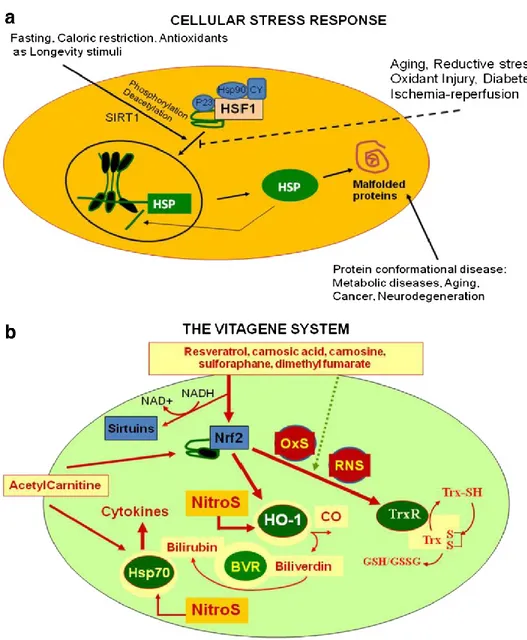
Cellular stress response is the ability of a cell to counteract stressful conditions (Figure 1a, 1b). This phenomenon, which includes heat shock response (HSR), represents an ancient and highly conserved cytoprotective mechanism [44-52]. Production of heat shock proteins, including protein chaperones, is essential for the folding and repair of damaged proteins, serving thus to promote cell survival conditions that would otherwise result in apoptosis [53-57]. The term ‘molecular chaperone’ denotes a large family of ubiquitous proteins that function as part of an ancient defense system in our cells. Chaperones promote cell survival by sequestering damaged proteins and preventing their aggregation. During stressful conditions, such as elevated temperature they prevent protein aggregation by facilitating the refolding or elimination of misfolded proteins. The stress-induced response to damaged proteins is helped by a sophisticated regulatory system, which shuts down most cellular functions and, in parallel, induces the synthesis of several chaperones and other survival-promoting proteins. Therefore, many of the chaperones are also called stress or ‘heat shock’ proteins in reference to the archetype of cellular stress, heat shock.
Besides their role during stress, chaperones have multiple roles under normal conditions, as such they promote the transport of macromolecules (e.g. proteins or RNA) and participate in remodeling events involving larger protein complexes,
stress response requires the activation of pro-survival pathways which, under control of protective genes called vitagenes [58] produce molecules (heat shock proteins, glutathione, bilirubin) endowed with anti-oxidant and anti-apoptotic activities.
Generally, molecular chaperones help a multitude of signaling molecules to keep their activation-competent state, and regulate various signaling processes ranging from signaling at the plasma membrane to transcription. In addition to these specific regulatory roles, recent studies have revealed that chaperones act as genetic buffers stabilizing the phenotypes of various cells and organisms. Among the cellular pathways conferring protection against oxidative stress, a key role is played by the products of vitagenes [59-61]. These include members of the heat shock protein (Hsp) family, such as heme oxygenase-1 and Hsp72, sirtuins and the thioredoxin/thioredoxin reductase system [62]. Recent studies have shown that the heat shock response contributes to establishing a cytoprotective state in a wide variety of human diseases, including inflammation, cancer, aging and neurodegenerative disorders [63]. Given the broad cytoprotective properties of the heat shock response there is now strong interest in discovering and developing pharmacological agents capable of inducing the heat shock response [64]. Molecular chaperones are known to disrupt aggregates but also to promote active aggregation when the concentration of the aggregating protein is high. Consistent with this notion, although protein aggregation is hazardous under certain circumstances, the creation of apparently less-toxic large aggregates is protective. This hypothesis is the basis of the therapeutic potential of heat shock proteins (HSPs), which prevent protein misfolding and aggregation [65].
Cellular stress response is regulated at the transcriptional, translational and post-translational levels by a family of heat shock transcription factors (HSFs) that are expressed and maintained in an inactive state under nonstress conditions [66].
Hsps consist of both stress inducible and constitutive family members. The constitutive form performs basic physiological functions. However, some of them are up-regulated by stress. The inducible form prevents the denaturation of proteins and assembly of abnormal polypeptides during exposure to stressful conditions. Denaturated proteins induced stress protein. The 70 kDa family of stress proteins is one of the most extensively studied. Included in this family are HSC70 (heat shock cognate, the constitutive form), HSP70 (the inducible form, also referred to as HSP72).
Another important family is HSP 32, or heme oxygenase (HO). There are three isoforms of heme oxygenase: HO-1, or inducible isoform; HO-2, or constitutive isoform; and the recently discovered HO-3.
Heme oxygenase-1 exerts protective role, by degrading the intracellular levels of prooxidant heme and by producing biliverdin, the precursor of bilirubin, this latter being an endogenous molecule with potent antioxidant and antinitrosative features and also produces carbon monoxide, a molecule involved in regulating vessel active pathway of NO [67].
Increasing evidence suggests that the HO-1 gene is redox regulated and contains in its promoter region the antioxidant responsive element (ARE), similarly to other antioxidants that bind specific transcription factors Nrf2 as sensitive to the alteration or NFkB redox balance; in fact, nitrosative stress and depletion of GSH up-regulate the protein It was seen that the cells overexpressing the Hsps are resistant to several types of oxidizing agents and to the heat shock; the Hsps play a protective role against oxidative damage to DNA [68]. The experimental evidence that: a) inhibition of antioxidant defenses increases the susceptibility to the heat shock, b) the Hsps confers resistance to oxidative stress [69]; c) the induction of Hsp-70, suggests, inhibited by antioxidant compounds, a correlation between
role in induction of Hsp70 [70].
The thioredoxin (Trx) system (Trx and Trx reductase), has received a considerable attention in the last years, as a stress responsive gene, redox in light of new experimental evidence, is a leading member of the ubiquitous multifunctional redox regulation of cell redox [71].
Trx is a ubiquitous thiol oxidoreductase system that regulates cellular redox balance and constitute a family of proteins all of which have a conserved catalytic site (Cys-Gly-Pro-Cys) which undergoes reversible oxidation of the cysteine pair while reducing disulfide bridges of various proteins [72]. The thioredoxin has evolved similar to a protein chaperone ensuring the maintenance of the structure dithiol/ disulphydryl biological function of proteins. Indeed, scientific evidence shows that Trx binds to specific proteins, modulating the structural conformation. The thioredoxin system, originally identified in Escherichia coli, in 1964, as a hydrogen donor for ribonucleotide reductase required for DNA synthesis, plays a key role in cell function by limiting oxidative stress directly via antioxidant effects and indirectly by protein–protein interactions [73].
It is well established that, in mammals, cellular redox regulation of many processes is provided by the cooperation between the Trx and glutathione systems [71].
Indeed, Trx and GSH systems are involved in a variety of redoxdependent pathways such as supplying reducing equivalents for ribonucleotide reductase, and peptide methionine sulfoxide reductase, the latter being involved in antioxidant defence and regulation of the cellular redox state [71].
Therefore, Trx and GSH form a powerful system controlling redox regulation of gene expression, signal transduction, cell proliferation, protection against oxidative stress, anti-apoptotic functions, growth factor and co-cytokine effects, as well as regulation of the redox state of the extracellular environment [74]. The
promoter of the Trx gene contains a series of stress responsive elements, various transcription factor binding sites, such as SP1, AP-1, NF-jB, and the antioxidantresponse element (ARE) [75].
GSH is thought to be largely responsible for maintaining a low redox potential and free thiol levels inside cells and organelles due to its high intracellular concentration (1–10 mM). The Trx system, rather may play a critical role in the redox regulation of protein thiols involved in signal transduction and gene regulation [76].
In addition, the thioredoxin (Trx), which essentially acts as a soluble protein after breakup cells, exists in an isoform cytoplasmic (Trx-1) and in a mitochondrial (Trx-2) [77].
Molecular studies show that the cytoplasmic isoform of the mitochondrial Trx as well the mithocondrial one protect against oxidative stress and both are essential for the survival of mammalian cells [78]. Given the large amount of functions performed by Trx redox seems reasonable to say that it is a critical molecule essential for cell survival. Overexpression of Trx system/TrxR is generally associated with activation of cellular mechanisms of tolerance to stress and in general, a resistance to oxidative damage and/or nitrosative mediated a wid variety of stressors, including compounds such as doxorubicin and etoposide [79-81]. The Trx plays a cytoprotective role against different forms of stress in a variety of biological systems. It is considered basically as a stress inducible protein with a typical intracellular cytosolic localization [77].
Many physicochemical stimuli, such as UV irradiation and hydrogen peroxide, have been shown to induce Trx expression and secretion, as a redox-sensitive molecule with cytokine-like and chemokine-like activities in the prevention of cellular damage from oxidative stress. In addition to UV irradiation, treatment of
cisplatin and hemin has been reported to cause the translocation of Trx from the cytoplasm to the nucleus, where it regulates the redox-activation and DNA binding activity of critical transcription factors (Jun, Fos, p53, CREB, PEBP2/CBF, Myb), all involved in fundamental processes, such as gene expression, cell growth and apoptosis.
The Trx-1, the most extensively studied isoform, is primarily a cytosolic soluble protein without a specific localization signal. Several studies indicate that the Trx is expressed constitutively associated with protein sulfhydryl on the surface of the plasma membrane of different cell types [77].
Thioredoxin plasma levels in normal individuals vary between 20 and 30 ng/ml (80, 81) and increase in certain human diseases including HIV infection and cancer [75]. Elevated Trx levels may contribute to increased cancer cell proliferation and resistance to chemotherapy by several mechanisms as the stimulation of DNA synthesis and the activation of redox-modulated transcription factors.
Recent work suggests that Trx-1 is involved in nerve growth factor (NGF) signaling pathways. NGF, a neurotrophic factor regulating development, maintenance and function of the CNS, has been shown to activate Trx-1 expression via cyclic AMP (cAMP)-response elements (AREs) present in the Trx-1 gene promoter, and also to induce nuclear translocation of TrxTrx-1 [75]. Several data suggest that, beyond its ability to regulate the function of proteins through thiol-disulfide exchange reactions, Trx and its substrates may also have beneficial effects during oxidative stress by upregulating HO-1, with important cytoprotective pleiotropic effects deriving from heme degradation and bilirubin formation [82, 83]. Besides the role as a source of reducing equivalents, Trx per
se acts as antioxidant or ROS scavenger. In fact, Trx eliminates singlet oxygen,
Trx has been shown to be involved in the neuroprotection against oxidative stress mediated by estrogens [75]. It has also been reported that some of the neuroprotective effects of GSNO on beta-amyloid- or ferrous citrate-induced toxicity in rat cortical neurons or in rat substantia nigra can be due to the activation of multiple signalling pathways including thioredoxin [84,85;].
The sirtuins are a group of proteins linked to aging, metabolism and stress tolerance in several organisms. In mammalian cells seven sirtuins have been identified. SIRT1, 2, 3, 6 and possibly 5 are NAD-dependent deacetylases, SIRT4 and 6 are ADP-ribosyltransferases, and the activity of SIRT7 has not been defined [86]. The sirtuin family of histone deacetylases (HDACs) was named after their homology to the Saccharomyces cerevisiae gene silent information regulator 2 (Sir2). In the yeast, Sir2 has been shown to mediate the effects of caloric restriction on the extension of life span, with high levels of Sir2 activity promoting longevity [87]. Like their yeast homologs, the mammalian sirtuins (SIRT1-7) are class III HDACs and require NAD+ as a cofactor to deacetylate substrates ranging from histones to transcriptional regulators. Through this activity, sirtuins are shown to regulate important biological processes, such as apoptosis, cell differentiation, energy transduction or glucose homeostasis [88]. In particular, the NAD+/NADH ratio can be considered as a ‘‘biochemical sensor’’ to evaluate the energetic status of the cell; in fact, among the several mechanisms through which dietary antioxidants may be useful for tissues, it is noteworthy to mention the improvement of metabolic conditions secondary to proinflammatory damage [88]. In this light, the interaction between NAD+/NADH and the members of the sirtuins family, puts in a single frame the cytoprotective activity of dietary antioxidants through the regulation of both cellular redox and metabolic state [88]. Since the Sir2 family of proteins exert their enzymatic activity not only
they are involved in many cellular processes, e.g., gene silencing, DNA repair, progression of the cell cycle, whereby controlling the mechanism of cellular ageing [88].
Several experimental evidences have shown the role of SIRT1 protein in human cell survival. SIRT1 specifically associates with the p53 tumor suppressor protein and deacetylates it, resulting in negative regulation of p53- mediated transcriptional activation. Importantly, p53 deacetylation by SIRT1 also prevents cellular senescence and apoptosis induced by DNA damage and stress. SIRT1 regulates important aspects of mitochondrial biology, e.g. it deacetylates the essential cofactor PGC-1a (PPAR-c coactivator-1a) in mitochondrial biogenesis. An up regulation of the mitochondrial activity might be of therapeutic benefit for various diseases related to aging such as metabolic disorders (e.g. diabetes type 2) or mitochondrial disorders.
These studies provide important information on the activity of SIRT1 and offer a promising approach for the treatment of metabolic disorders. In addition, SIRT1 activation significantly decreases neuronal cell death induced by amyloid-beta( Ab) peptides through inhibition of NF-jB signaling [88].
1.6 Aging
Aging is a complex biological process characterized by a gradual decline in biochemical and physiological functions of most organs and is considered one of the most significant risk factors for age-related neurodegenerative diseases, such as Alzheimer’s, Parkinson's disease, ALS, Huntington disease, Fredreich ataxia and multiple sclerosis [89, 90, 47, 52]. In general, aging is associated with changes in physiological characteristics, including muscle weakness and hair decolorization, and many physiological functions. The causes, however, are
multifactorial and several studies have suggested that oxidative stress plays an important role. Age-related changes in the brain include reduction of trophic supports, decreased proteosomal enzyme activities, mitochondrial dysfunction, change in the redox status which promotes a more pro-inflammatory environment associated with increased formation of reactive oxygen species (ROS) [91, 92]. The free radical theory of aging postulates that ROS may produce oxidative damage directly to critical biological molecules including proteins, DNA and lipids [92, 93, 94]. To counteract increasing levels of ROS, the cell has developed a number of antioxidant defense systems such as antioxidant enzymes (superoxide dismutase (SOD), catalase (CAT) and glutathione peroxidase (GPx)) as well as non-enzymatic antioxidant molecules (carotenoids, vitamin E, GSH). The imbalance between the activity of free radicals generation and scavenging systems is known as oxidative stress and is considered one of the most important mediators in the progressive decline of cellular function during aging [90, 95]. In particular, the aging process is accompanied by a general decline of physiological functions in the CNS. The CNS is particularly vulnerable to oxidative injury for its high oxygen consumption per unit weight, consistent with the generation of high levels of ROS, and the little amount of ROS defence systems (lower in the nervous system than other tissues) that allow ROS to remain elevated once formed. Since the brain contains high levels of polyunsaturated fatty acids, which upon oxidation form neurotoxic lipid peroxidation products (MDA, HNE), neuronal tissue is extremely vulnerable to oxidative modification of its cellular components. The oxidative damage to cellular macromolecules such as DNA, proteins, and lipids accumulates with age and has been postulated to be the main, but not the only, type of endogenous damage strongly involved in the aging process [96]. Indeed, when post mitotic neurons are injured by oxidative
over the lifetime of the neuron population. The chemical reactions resulting from attack of ROS/RNS on proteins are complex and lead to a variety of products, many as yet uncharacterized. The oxidative damage to proteins is reflected by increasing levels of protein carbonyls and decreasing levels of protein thiols [97, 98] Protein carbonylation, among different types of post-translational modifications, serve as useful biomarker for the accumulation of oxidatively modified proteins [97]. It appears that such modifications target very specific proteins and can affect the integrity and functioning of the proteome.
A number of studies indicated that the levels of oxidized proteins, exhibiting carbonyl groups, increase progressively with age in brain extracts of rats of different ages [99]. Furthermore, due to its central role in producing energy (ATP), mitochondria were brought to attention in aging biology, in order to understand the decline of basal metabolic rate and, consequently, physiological performances observed in aged mammals. Mitochondria produce the majority of free radicals (49%) and as a powerful source of these toxic oxidants are also their potential victim. In fact, the mitochondrial components (e.g. mtDNA or mt enzymes) are surely more susceptible to oxidative damage than all the other components [13, 100, 101]. This damage increase might have important consequences on mitochondrial structure, on the activity of the respiratory chain complex and on the global functionality of these organelles. Increasing body of evidence also suggests that the decay of mitochondria accompanied by an impairment of cell energy metabolism are important factors in the pathogenesis of most important neurodegenerative disorders [102]. Dysfunctional mitochondria are observed in aging, and also in pathological situations as ischemia-reperfusion and inflammation, moreover studies on senescence-accelerated mice (SAMP8) showed the mitochondria decay as one of the main contributor to the acceleration of aging [103, 104]
1.7 Multiple Sclerosis
Multiple sclerosis (MS) is an autoimmune-mediated neurodegenerative disease with characteristic foci of inflammatory demyelination in the brain, spinal cord, and optic nerves. Recent studies have demonstrated not only that axonal damage and neuronal loss are significant pathologic components of MS and experimental autoimmune encephalomyelitis (EAE), but that this neuronal damage is thought to cause the permanent neurologic disability often seen in MS patients. Current treatments for MS involve immunomodulation, which can reduce the incidence of inflammatory relapses. However, existing therapies are often not fully effective, and limited evidence suggests that these therapies prevent the long-term neuronal damage and physical disability of MS patients [105, 106]. New therapies that prevent neurodegeneration through nonimmunomodulatory mechanisms have a tremendous potential to work synergistically with current MS therapies [107]. MS pathology is characterized by perivenous infiltration of lympho-cytes and macrophage leading to damage of myelin and axons in the brain and spinal cord, which underlie the clinical disease course usually occurring with recurrent and reversible episodes of neurological dysfunction affecting one or several sites, during late adolescence and early adulthood; this form is called relapsing-remitting form (RR) and it is the most prevalent. Usually, approximately 20 years later, this clinical pattern transforms into a secondary progressive phase with continuous and progressive neurological decline [108]. Although evidence indicates that MS is a multifactorial disease caused by a complex interplay between genetic and environmental factors, it is still unclear which are the causes or the factors that contribute to its unpredictable course. It is generally accepted that, virus infections of the CNS, vascular factors and/or disturbed immune
the immune system attacks and destroys myelin and the myelin-forming cell, leading to the pathological hallmarks of MS: the classical actively demyelinating lesions, the cortical demyelination and the diffuse white matter injury, the latter particularly evident in the later stage of the disease [110, 111]. Existing evidence indicates that CNS responds to the attack by immune cells and their secreted products through modulation of its metabolism and gene expression [112]. In addition, cytokines, immunoglobulins, and complement complexes may elicit a survival response in the oligodendrocytes, involving the induction of endogenous heat shock proteins and other protective molecules, which indicates that redox systems and therefore the oxidant/antioxidant balance in these cells are of great importance in MS [113-116]. The adaptation and survival of cells and organisms requires the ability to sense proteotoxic insults and to coordinate protective cellular stress response pathways and chaperone networks related to protein quality control [47]. Despite the abundance and apparent capacity of chaperones and other components of homeostasis to restore folding equilibrium, brain cells appears poorly adapted for chronic proteotoxic stress which increases in neurodegenerative diseases such as MS [50].
1.8 Alzheimer’s disease
Alzheimer’s disease (AD) is a progressive neurodegenerative disorder and represents the most common cause of dementia in the elderly, accounting for 50-60% of all cases in Western world [117, 118]. The prevalence rates for AD rise exponentially with age, increasing markedly after 65 years. AD is characterized by cognitive decline beginning usually with impairment of episodic memory, involving progressively all cognitive functions in the late stage [119]. Although some cases are familial, sporadic AD is more common, affecting more than 15 million people worldwide [120].
The pathological hallmarks of AD are amyloid plaques, containing amyloid-β peptide, derived from the transmembrane amyloid precursor protein, and neurofibrillary tangles, composed of hyperphosphorylated tau protein, in the medial temporal lobe structures and cortical areas of the brain together with neuronal death and synapses loss [121, 122].
Many approaches have been undertaken to understand AD, including Aβ aggregation, but the heterogeneity of the etiologic factors makes it difficult to define the clinically most important factor determining the onset and progression of the disease [123]. However, increasing evidence indicates that factors such as oxidative stress and disturbed protein metabolism and their interaction in a vicious cycle are central to AD pathogenesis and progression [124-127]. Amyloid-β peptide (1–42) has been shown to induce protein oxidation in both in vitro and in
vivo studies [128-131]. As a result, amyloid-β peptide (1–42) has been proposed
to play a central role in the pathogenesis of AD [132]. A previous study has shown that increased protein oxidation and lipid peroxidation are present in the brain from patients with mild cognitive impairment (MCI), as compared to aged-matched control brain [133, 134]. Because many researchers consider MCI to be the transition zone between normal cognition and the dementia of early AD, these findings suggest that oxidative stress is fundamental to the progression of AD and not simply a consequence of AD [44, 135].
2. AIM OF THE RESEARCH
The cell antioxidant defence systems operate very efficiently and there is a balance between pro-oxidant and antioxidant factors, that, during normal cellular metabolism, is able to eliminate all free radicals that are produced [30].
Numerous experimental data show evidence of the involvement of oxidative stress in aging and neurodegenerative disorders [18]. In recent years, since oxidative stress has been considered the basis for some aspects of neurodegeneration, a number of experimental studies have been carried out in order to identify a way to counter the effects of oxidative stress through scavengers of free radicals [8]. From a molecular point of view, the central nervous system (CNS) cells are able to fight oxidative stress with many resources including bioactive molecules (glutathione, thioredoxin, flavonoids), lipoic acid, enzymes (heat shock proteins, superoxide dismutase, catalase, glutathione peroxidase, thioredoxin reductase, etc.) and redox sensitive transcription factor protein [136].
The heat shock proteins (HSP) is one of the most studied active defence systems against oxidative damage. The heat shock response is able to produce a cytoprotective state in a wide variety of human diseases such as inflammation, cancer, aging and neurodegenerative disorders, opening new perspective in medicine and pharmacology about molecules capable of activating these defence mechanisms as potential target for novel cytoprotective strategies [7, 137-140].
2.1 Aging
In this study, we examine the free radical hypothesis of aging employing a redox proteomics technique. More in details, the aims of the study were:
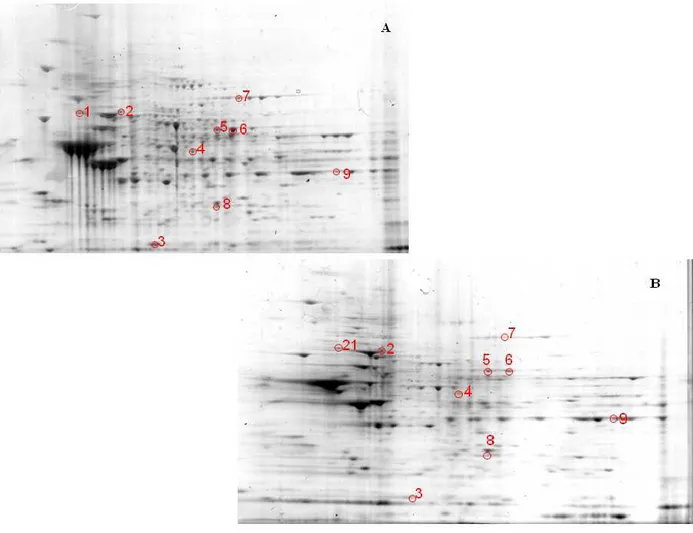
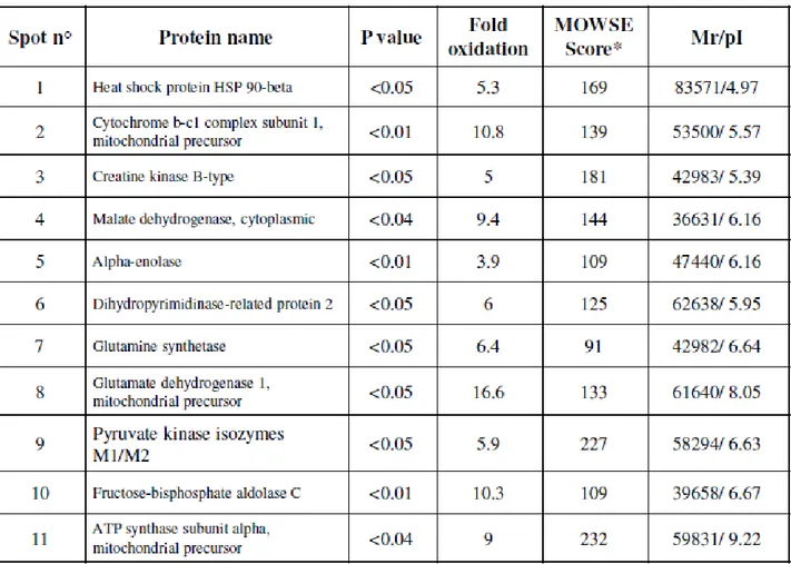
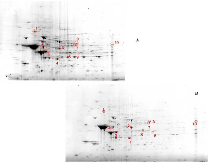
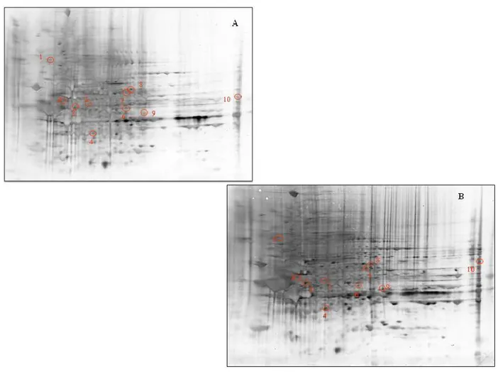
- to dose levels of expression of stress proteins by Western blot analysis, in brain areas of aged rats, using antibodies specific: Hsp-90, Hsp-70, CN1, 4-HNE, DPNH.
- to study the role of free radicals in the aging process, through a redox proteomics approach.
- to investigate the oxidation of specific proteins by measuring the protein carbonyl levels in four different brain regions of rats "(Hippocampus, cerebellum, cortex and striatum) of 28 months (senescent) and 12 months (adults).
- to identify proteins that are specifically oxidized during the process of aging in different brain regions because many of these proteins are related to the functionality of mitochondria, the energy metabolism and activity of chaperones. 2.2 Multiple sclerosis
The present study was undertaken in order to investigate systemic stress response and the associated oxidative stress measured through the determination of markers of protein and lipid oxidation in plasma, lymphocytes and CSF of patients with active MS, as compared to age-matched controls, in order to gain a better insight into the molecular mechanisms regulating the cellular stress response during the progression of the disease and, as such, provide a potential target for novel cytoprotective strategies impacting the clinical settings of this degenerative disease.
The research included the following objectives:
- study, using a redox proteomics approach, the role played by free radicals in multiple sclerosis.
- investigate the oxidation of specific proteins by measuring the protein carbonyl levels in the serum of patients with MS and control subjects.
2.3 Alzheimer’s disease
In the present study we evaluate stress response mechanisms in plasma and lymphocytes of control patients compared to AD patients, in order to provide evidence of an imbalance of oxidant/antioxidant mechanisms and oxidative damage in AD patients and the possible protective role of vitagenes.
The research has been focused on determining the levels of Sirt-1, Sirt-2 and Trx in plasma and in lymphocytes from control subjects and patients with AD by Western blot analysis.
3. MATERIALS AND METHODS 3.1 Aging
3.1.1Chemicals
5,5'-Dithiobis-(2-nitrobenzoic acid) (DTNB), 1,1,3,3 tetraethoxypropane, purified bovine blood SOD, NADH, reduced glutathione (GSH), oxidized glutathione (GSSG), β- NADPH (type 1, tetrasodium salt), glutathione reductase (GR; Type II from Bakers Yeast), SIN-1 (3-Morpholinosydnonimine hydrochloridte) were from Sigma Chemicals Co, St. Louis (USA). All other chemicals were from Merck (Germany) and of the highest grade available.
3.1.2 Animals and samples preparation
All animal protocols were approved by the University of Catania laboratory Animal Care Advisory Committee. Male Wistar rats purchased from Harlan (Udine, Italy) were maintained in a temperature and humidity-controlled room with a 12 h light: dark cycle. Rats (n = 8, per group) of 12 (aged) and 28 (senescent) months, were fed ad libitum a certified diet prepared according to the recommendations of the AIN.
After sacrifice, brains were quickly removed and dissected into the cerebral cortex, cerebellum, and striatum according to a standardized procedure, in a cold anatomical chamber and following a protocol that allows a maximum of 50 s time-variability for each sample across animals. Brain samples from hippocampus, cerebellum, striatum and cerebral cortex were minced and suspended in 10 mM HEPES buffer (pH 7.4) containing 137 mM NaCl, 4.6 mM KCl, 1.1 mM KH2PO4, 0.1 mM EDTA, and 0.6 mM MgSO4 as well as proteinase inhibitors: leupeptin (0.5 mg/ml), pepstatin (0.7 μg/ml), type II S
centrifuged at 14,000 × g for 10 min to remove debris. Protein concentration in the supernatant was determined by the “Coomassie Plus Protein Assay” (Pierce, Rockford, IL, USA).
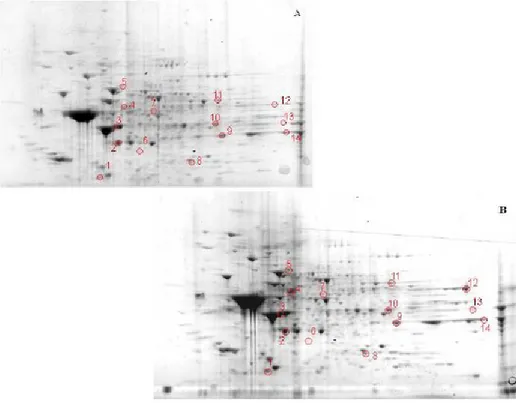
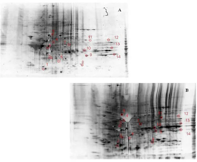
3.1.3. Two-dimensional gel electrophoresis
Samples (200 μg) were incubated at room temperature for 30 min in four volumes of 10 mM 2,4-dinitrophenylhydrazine (DNPH) in either 2 M HCl for protein carbonyl derivatization/oxyblots or 2 M HCl for gel maps and mass spectrometry analysis. This was followed by precipitation of proteins by addition of ice-cold 100% trichloroacetic acid (TCA) to a final concentration of 15% and samples were placed on ice for 10 min.
Precipitates were centrifuged at 15,800 g for 2 min. The pellets were washed with 0.5 ml of 1:1 (v/v) ethanol/ethyl acetate solution. After centrifugation and washing with ethanol/ethyl acetate solution three times, the samples were then dissolved with 185 μl of rehydration buffer (8 M urea, 20 mM dithiothreitol, 2.0% (w/v) CHAPS, 0.2% Biolytes, 2 M thiourea and bromophenol blue). For the first-dimension electrophoresis, 200 μl of sample solution were applied to a ReadyStrip™ IPG strip (Bio-Rad). The strips were soaked in the sample solution for 1 hour to allow uptake of the proteins. The strip was then actively rehydrated in protean IEF cell (Bio-Rad) for 16 hours at 50V. The isoelectric focusing was performed at 300V for 2 hours linearly; 500V for 2 hr linearly; 1000V for 2 hr linearly, 8000V for 8 hr linearly and 8000V for 10 hr rapidly. All the processes above were carried out at 22°C. The focused IEF strip was stored at –80°C until second dimension electrophoresis was performed. For second dimension electrophoresis, thawed IPG® Strips pH 3-10 were equilibrated for 10 min in 50 mM Tris-HCl (pH 6.8) containing 6M urea, 1% (w/v) sodium dodecyl sulfate (SDS), 30% (v/v) glycerol, and 0.5% dithiothreitol, and then re-equilibrated for 15
min in the same buffer containing 4.5% iodacetamide in place of dithiothreitol. Linear Gradient (8-16%) Precast criterion Tris-HCl gels (Bio-Rad) were used to perform second dimension electrophoresis. Precision ProteinTM Standards (Bio-Rad) were run along with the sample at 200V for 65 min. After electrophoresis, the gels were incubated in fixing solution (7% acetic acid, 10% methanol) for 20 min. Approximately 40 ml of Coomassie Safe Gel Stain (Bio-Rad) were used to stain the gels for 1 hour, on a gently continuous rocker. The gels were placed in deionized water overnight for destaining.
3.1.4. Western Blotting
The same amount of protein samples (200 μg) was used for detecting specific protein carbonyl levels and the electrophoresis was carried out in the same way as described above. Proteins (200 μg) were incubated with 4 volumes of 20 mM 2,4- dinitrophenylhydrazine (DNPH) at room temperature (25°C) for 20 min. The gels were prepared in the same manner as 2D-electrophoresis. The proteins from the second dimension electrophoresis gels were transferred to nitrocellulose (Bio-Rad) using a Criterion Blotter Apparatus (Bio-(Bio-Rad) at 15V for 2 h. The 2,4-dinitrophenyl hydrazone (DNP) adducts of the carbonyls of the proteins were detected on the nitrocellulose paper using a primary rabbit antibody (Chemicon) specific for DNP-protein adduct (1:100), followed by a secondary goat anti-rabbit IgG (Sigma, St Louis, MO, USA) antibody. The resultant stain was developed using 5-bromo-4-chloro-3-indolyl phosphate/nitro blue tetrazolium (BCIP/NBT) solution (SigmaFast tablets; Sigma).
3.1.5. Image Analysis
The gels (n=8 aged and n=8 senescent) and nitrocellulose blots were scanned and saved in TIF format using a HP Deskjet H2180 (Hewlett Packard). PDQuest 2-D Analysis Software (Bio-Rad, Inc.) was used for matching and analysis of visualized protein spots among differential gels and membranes to compare protein and DNP immunoreactivity content between senescent and aged rats brain samples. This sophisticated software offers powerful comparative analysis and is specifically designed to analyze many gels or blots at once. Powerful automatching algorithms quickly and accurately match gels or blots and sophisticated statistical analysis tools identify experimentally significant spots. The principles of measuring intensity values by 2-D analysis software were similar to those of densitometric measurement. The average mode of background subtraction was used to normalize intensity values, which represents the amount of protein (total protein on gel and DNP-bound protein on the membrane) per spot. After completion of spot matching, the normalized intensity of each protein spot from individual gels (or membranes) was compared between groups using statistical analysis. Statistical significance was assessed by a two-tailed Student’s t-test. P values <0.05 were considered significant for comparison between aged and senescent rats. This is the method of statistical analysis most appropriate for proteomic analysis of small number of protein spots in contrast to statistical approach used for large analysis common in gene microarray studies (Maurer et al., 2005).
3.1.6. Trypsin digestion
The selected protein spots were excised with a clean blade and transferred into clean microcentrifuge tubes. The protein spots were then washed with 0.1 M
ammonium bicarbonate (NH4HCO3) at room temperature for 15 min. Acetonitrile was added to the gel pieces and incubated at room temperature for 15 min. The solvent was removed, and the gel pieces were dried in a flow hood. The protein spots were incubated with 20 μl of 20 mM DTT in 0.1M NH4HCO3 at 56°C for 45 min. The DTT solution was then removed and replaced with 20 μl of 55 mM iodacetamide in 0.1 M NH4HCO3. The solution was incubated at room temperature in the dark for 30 min. The iodacetamide was removed and replaced with 0.2 ml of 50 mM NH4HCO3 and incubated at room temperature for 15 min 200 μl of acetonitrile was added. After 15 min incubation, the solvent was removed, and the gel spots were dried in a flow hood for 30 min. The gel pieces were rehydrated with 20 ng/μl methylated trypsin (Promega, Madison, WI) in 50 mM NH4HCO3 with the minimal volume to cover the gel pieces. The gel pieces were chopped into smaller pieces and incubated at 37°C overnight in shaking incubator.
3.1.7. Protein identification by mass spectrometry
Selected spots were manually excised from gels and submitted to trypsin proteolysis. Briefly, after three destaining steps using a solution of 50 mM ammonium bicarbonate (5 min), 50% acetonitrile in 50 mM ammonium bicarbonate (15 min) and 100% acetonitrile (15 min), about 100 ng of trypsin (Trypsin Gold, Mass Spectrometry Grade, Promega, Madison, WI, USA), solubilised in 10 μl of a 25 mM ammonium bicarbonate digestion buffer, were added to each vacuum-dried gel spot. Digestion was performed at 37°C overnight. An aliquot (1 μl) of each peptide mixture was mixed with the same volume of α-cyano-4-hydroxy-trans-cinnamic acid matrix solution (10mg/ml) in 70% acetonitrile containing 0.2% TFA (v/v) and spotted onto a MALDI target plate.
MALDI-ToF MS analyses were performed in a Voyager-DE STR instrument (Applied Biosystems, Framingham, MA) equipped with a 337 nm nitrogen laser and operating in reflector mode. Mass data were obtained by accumulating several spectra from laser shots with an accelerating voltage of 20 kV. Two tryptic autolytic peptides were used for the internal calibration (m/z 842.5100 and 2807.3145). Identification by peptide mass fingerprint (PMF), with the monoisotopic mass list obtained from each spot, was performed after exclusion of expected contaminant mass values by Peak Erazor program (http://www.protein.sdu.dk/gpmaw/Help/PeakErazor/peakerazor.html), using the Mascot search engine (v. 2.2) against SwissProt database (v. 55.3, 366226 sequences). Up to one missed cleavage, 50 ppm measurement tolerance, oxidation at methionine (variable modification) and carbamidomethyl cysteine (fixed modification) were considered. Posttraslational modifications were not considered. Identifications were validated when the probability-based Mowse protein score was significant according to Mascot.
3.1.8. Determination of reduced glutathione and oxidized glutathione in the cytosol.
Brain regions were homogenized on ice for 10 s in 100 mM potassium phosphate, pH 7.5, which contained 12 mM disodium EDTA. The homogenate was divided in two aliquot, for total glutathione (GSH+GSSG) assay 0.25 ml of homogenate was added to equal volume of 100mM potassium phosphate buffer pH7.5, containing 17.5mM EDTA and 10 mM 5,5’-dithiobis-(2-nitrobenzoic acid) (DTNB) (Sample SS1). For oxidized glutathione (GSSG) assay 0.25 ml of homogenate was added to100 mM potassium phosphate buffer pH 6.5, containing 17.5 mM EDTA and 10 mM N-ethylmaleimide NEM (Sample SS2). The samples were centrifuged at 800g for 20 min, and the supernatant fractions were then
centrifuged at 10,000 g for 30 min. The supernatant of SS1 and SS2 represented the cytosolic fractions and were used for the spectrofotometric assay of total or oxidized glutathione. Before spectrofotometric determination, 0.25 ml aliquot of SS2 sample was passed through a C18 Sep-Pak cartridge (Waters, Watford, U.K.) to remove the excess of NEM and washed with 0.5 ml of buffer 100 mM potassium phosphate buffer pH 7.5, containing 5 mM EDTA. Spectrofotometric assay of glutathione was performed adding the samples to a cuvette containing 0.5 unit of glutathione reductase, 0.2mM DTNB in a final volume of 1ml of 100 mM potassium phosphate buffer pH 7.5, 5 mM EDTA and the reaction initiated by adding NADPH (220 nmoles). The change in absorbance at 412 nm was recorded over a period of 5 min for SS1 sample or 10 min for SS2 sample using a reference cuvette containing equal concentrations of NADPH, DTNB and enzyme. The GSH and GSSG content, expressed as nmol/mg protein, was determined by comparison with a standard curve obtained with GSH and GSSG solution.
3.1.9. Determination of total glutathione (GSH+GSSG) and oxidized glutathione (GSSG) in mitochondria.
The pellet of SS1 and SS2 samples obtained after centrifugation at 10,000 g was utilized for determination of total and oxidized glutathione in mitochondria. The SS1 pellet was resuspended in 0.32 M sucrose, 1 mM EDTA, 10 mM Tris, pH 7.4, 5 mM DTNB. The SS2 pellet was resuspended in 0.32 M sucrose, 1 mM EDTA, 100 mM potassium phosphate buffer pH6.5, containing 5mM N-ethylmaleimide (NEM). Low and high speed differential centrifugations for mitochondrial isolation were, respectively, 1,300 g for 3 min and 21,200 g for 10 min for all brain regions examined; mitochondrial preparations were washed once with a 100mM potassium phosphate buffer pH 7.5, containing 5mM EDTA. This
from the cytosolic fraction. The mitochondria were then mixed, sonicated and centrifuged at 10,000 g per 11 min., the supernatant was used for spectrofotometric determination assay. Before of spectrophotometric determination 0.25 ml aliquots of SS2 sample were passed through a C18 Sep-Pak cartridge (Waters, Watford, U.K.) to remove the excess of NEM and washing with 0.5 ml of buffer 100 mM potassium phosphate buffer pH 7.5, containing 5 mM EDTA.
Spectrophotometric assay of glutathione was performed adding the samples to a cuvette containing 0.5 unit of glutathione reductase, 0.2 mM DTNB in a final volume of 1 ml of 100 mM potassium phosphate buffer pH 7.5, 5 mM EDTA and the reaction initiated by adding NADPH (220 μM). The change in absorbance at 412 nm was recorded over a period of 5 min for SS1 sample or 10 min for SS2 sample using a reference cuvette containing equal concentrations of NADPH, DTNB and enzyme. Protein concentration was determined in the samples with NEM, according to Bradford method, using BSA as standard. The GSH and GSSG content, expressed as nmol/mg prot, was determined by comparison with a standard curve obtained with known concentrations of GSH and GSSG solution. 3.1.10 Enzyme assays
Pyruvate kinase: The enzyme activity of pyruvate kinase (PK) was determined at 37 °C by lactate dehydrogenase-coupled spectrophotometric assay (Hayashi et al., 1979). The standard reaction mixture contained 100 mM Tris pH 8.0, 100 mM KCl, 10 mM MgCl2, 0.5 mM EDTA, 0.2 mM NADH, 10 μg LDH, 10 mM Phosphoenolpyruvate, and 1.5 mM ADP in a final volume of 1 ml. The reaction was started by adding enzyme solution (0.5–1 μg).
One unit of activity is the amount of enzyme catalyzing the oxidation of 1 μmol NADH/min under the above conditions. The assay was carried out in a microplate reader (Labsystem Multiscan MS).
Glyceraldehyde-3-phosphate dehydrogenase: To determine the activity of GAPDH, 20 μg of protein homogenate was added to an assay mixture (100 mM 3-phosphoglyceric acid, 200 U/ml 3-3-phosphoglyceric phosphokinase, 200 mM cysteine, 100 mM MgSO4, 34 mM ATP, 7.0 mM β-NADH) in a UV-transparent microtiter plate (Corning). The change in absorbance at 340 nm was monitored during a 5-min period with a microplate reader (Labsystem Multiscan MS).
3.1.11. Statistical analysis
Results were expressed as means ± SEM of at least eight separate experiments. Statistical analyses were performed using the software package SYSTAT (Systat Inc., Evanston IL, USA). The significance of the differences, evaluated by two-way ANOVA, followed by Duncan’s new multiple-range test, was considered significant at P<0.05.
3.2 Multiple sclerosis
3.2.1. Ethical permission
The study was conducted according to guidelines of local Ethics Committee, and informed consent was obtained from all patients.
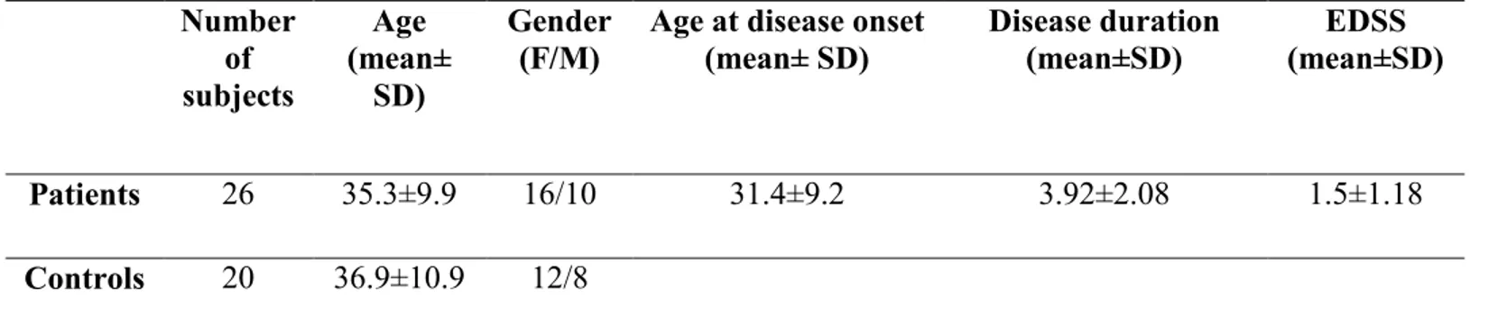
Twenty-six patients with an age range of 20–60 years were used as the ‘‘test’’ group for this study. The mean disease duration was 3.9±2.0 years. All these patients had a confirmed clinical diagnosis of MS according to the diagnostic criteria of McDonald et al. [141]. Furthermore, all the subjects were clinically classified having the relapsing remitting form of MS according to the criteria described in Lublin and Reingold [142]. The patients had not been undergoing corticosteroid or immunosuppressive treatment for at least 2 months before the CSF samples were collected. Twenty control patients, with an age range of 30–60 years, underwent lumbar puncture because of suspected subarachnoid hemorrage, pseudotumor cerebri, oculomotor palsies, or other indications in the usual neurological survey. Laboratory and neuroimaging tests were normal. Therefore the final diagnosis was mainly tension headache or conversion disorder. Clinical and demographical data of patients are shown in Table1.
3.2.3. Sampling
Blood was collected from controls and patients by venipuncture from an antecubital vein into tubes containing EDTA as an anticoagulant. Immediately after sampling, 1 ml the blood was centrifuged at 10,000 g for 10 min at 4 8C to separate serum from red blood cells and 4 mL were utilized for lymphocytes purifica-tion. Lymphocytes from peripheral blood were purified using the Ficoll Paque System following the procedure provided by the manufacturer (GE Healthcare, Piscataway, NJ, USA). CSF was obtained (on ice) from all subjects by lumbar spinal tap. The CSF samples were immediately centrifuged at 10,000 x g for 3 min at 4 °C to remove any contaminating cells and kept on ice until the biochemical assays were performed. Chemical analysis of CSF showed no heme present in the CSF in any of the control or MS CSF samples. CSF lymphocytes